thromboembolic disease - oklahoma academy of · pdf filecan be referred to as venous...
TRANSCRIPT

Thromboembolic Disease Dr. Scott Prater M.D., DABR, RPVI
Financial Disclosures
None
Deep Vein Thrombosis
3
Venous thromboembolism is a major national
health problem, with an overall incidence of
more than 1-3 per 1,000 annually.
Deep Vein Thrombosis (DVT) is a potentially life-
threatening pathology that can lead to pulmonary
embolism (PE), and/or post-thrombotic syndrome
(PTS).
The spectrum of DVT to pulmonary embolism
can be referred to as venous thromboembolic
disease (VTE).
http://www.acep.org/Clinical---Practice-Management/Focus-On--Emergency-Ultrasound-For-Deep-Vein-Thrombosis.
Accessed January 2016.
4
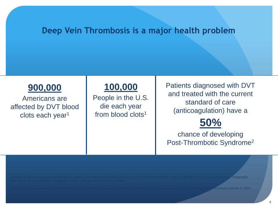
Deep Vein Thrombosis is a major health problem
900,000 Americans are
affected by DVT blood
clots each year1
100,000 People in the U.S.
die each year
from blood clots1
Patients diagnosed with DVT
and treated with the current
standard of care
(anticoagulation) have a
50% chance of developing
Post-Thrombotic Syndrome2
1Centers for Disease Control and Prevention. Venous Thromboembolism (Blood Clots). Venous Thromboembolism: Impact of Blood Clots on the United States – Infographic.
http://www.cdc.gov/ncbddd/dvt/infographic-impact.html. Accessed October 5, 2015.
2Centers for Disease Control and Prevention. Venous Thromboembolism (Blood Clots). Data & Statistics. http://www.cdc.gov/ncbddd/dvt/data.html. Accessed October 5, 2015.

The Challenge - Approximately half of patients
with DVT are asymptomatic.
Symptoms of DVT include swelling, pain,
tenderness, warmth, and prominent superficial
veins on the affected limb.
Patients with suspected DVT frequently present to hospital emergency
departments. Since symptoms and signs of DVT can be non-specific and
found in a wide variety of non-thrombotic disorders, timely diagnostic
testing must be performed to correctly identify patients with VTE.
Diagnosing DVT
5

DVT: What we know
Venous thromboembolism (VTE) is manifested clinically by deep venous thrombosis
(DVT) and pulmonary embolism (PE). DVT, usually of the lower extremity, nearly always
precedes PE. The risk of VTE increases greatly after age 50.
Other Risk Factors
• Age
• Trauma
• Cancer
• Immobilization
• Surgery
• Medications-OCPs
• Inherited Coagulopathies- (Factor V
Leiden, Protein S/C deficiency,
Antiphospholipid, etc.
• Infection/inflammatory Disorders
Occurrence
The disease most often occurs in
hospitalized patients, particularly
those with cancer or following
surgical procedures, but also
occurs sporadically in the
community. In both settings,
multiple risk factors are usually
present.
6

No prophylaxis + routine objective screening for DVT
Risk of DVT in Hospitalized Patients
Patient Group DVT Incidence
Medical patients 10 - 26 %
Major gyne/urol/gen surgery 15 - 40 %
Neurosurgery 15 - 40 %
Stroke 11 - 75 %
Hip/knee surgery 40 - 60 %
Major trauma 40 - 80 %
Spinal cord injury 60 - 80 %
Critical care patients 15 - 80 %
7
Heit – Mayo Clin Proc 2001;76:1102
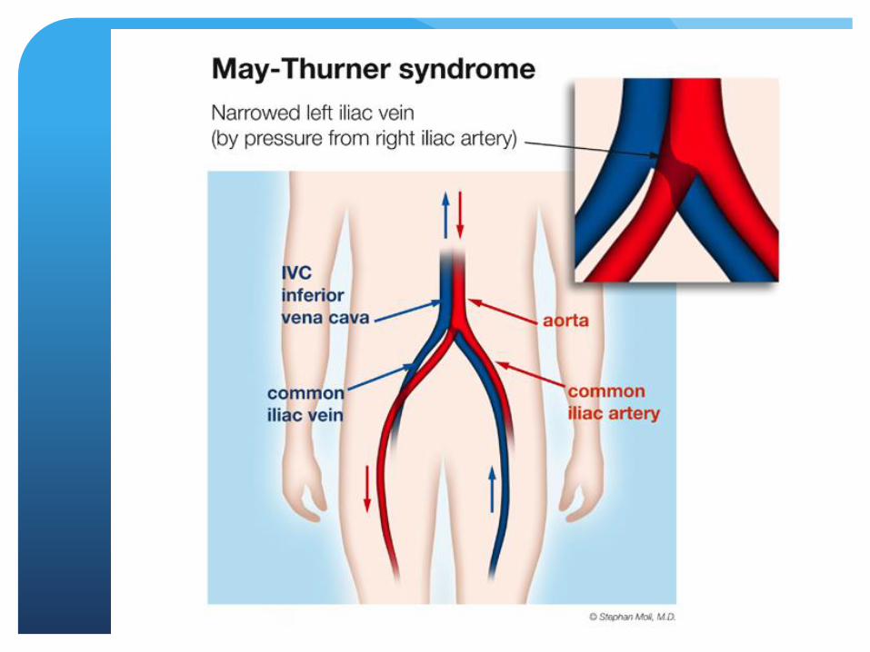
May Thurner Syndrome
An anatomical variant where the right common iliac artery compresses the left common iliac vein
LEFT Thigh swelling common, PE rare
Extensive iliofemoral DVT
Iliac vein compression syndrome & ‘Cockett syndrome’
High incidence of PTS
Excellent 5 yr patency with thrombolysis and stents
Think of it in young women


24 yo female with left leg swelling

Tx with Stent
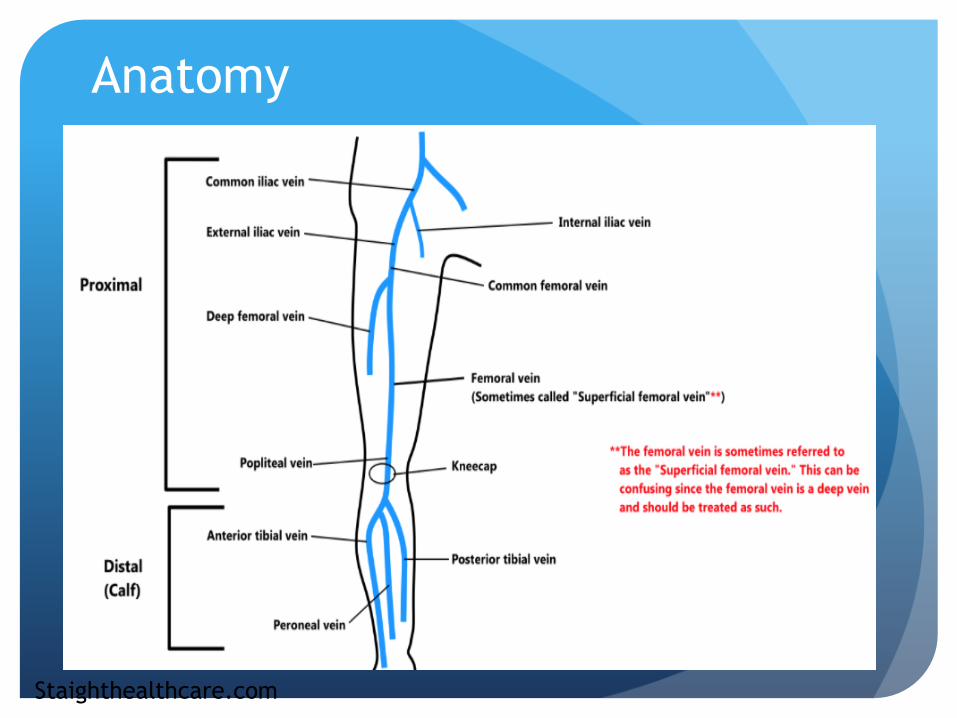
Anatomy
Staighthealthcare.com

U/S
U/S Just checks CF vein to
ankle (Does not see iliac
veins)
Direct signs of DVT=
Noncompressible vein, clot
in the vein, and lack of
compressibility.
Indirect Signs of DVT- Lack
of augmentation and
respiratory waveform.

Respiratory Variance
http://www.criticalecho.com/content/tutorial-10-vascular-ultrasound
www.jultrasoundmed.org

Augmentation
Heather L. Gornik, and Aditya M. Sharma Circulation.
2014;129:917-921

Treatment Options
Anticoaguation
Thrombolysis
Thrombectomy
IVC filters.

Factor Xa Inhibitors
Xarelto (Rivaroxaban)
15 mg x 2 for 21 days PO with food.
20 mg for remaining period of treatment
Avoid in hepatic impairment (child class B and C), pregnancy/nursing.
Can be checked with anit-factor Xa activity assay.
Partially reverse with 4-factor PCC (Kcentra
Eliquis (Apixaban)
10 mg x 2 PO for 7 days
5 mg for remaining period of treatment
Avoid in severe hepatic impairment, pregnancy/nursing.
Can be checked with anit-factor Xa activity assay.
Partially reverse with 4-factor PCC (Kcentra)
1. Xarelto.com
2. Eliquis.com
3. UW Guidelines for Reversal of Anticoagulation

Factor Xa Inhibitors
Savaysa (Edoxaban)
60 mg once daily PO
Avoid in mechanical heart
valves, nursing,
mod/severe hepatic
impairment.
Reverse with 4-factor PCC
(Kcentra) 50 Units/kg up to
5000 Units
Arixtra (Fondaparinux)
Dosed based on body weight-( 5mg <50 kg, 7.5 mg-50kg-100kg, 10mg- >100kg) delivered SQ
Used as bridging agent (avg time administered is 5-9 days)
Contraindicated in severe renal impairment, bacterial endocarditis, and thrombocytopenia with + anti-platelet ab
Reverse with rFVIIa (Novoseven) 90mcg/kg
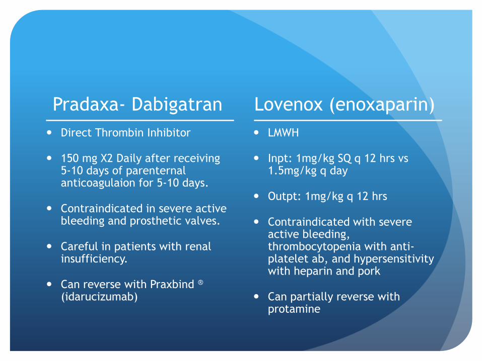
Pradaxa- Dabigatran
Direct Thrombin Inhibitor
150 mg X2 Daily after receiving 5-10 days of parenternal anticoagulaion for 5-10 days.
Contraindicated in severe active bleeding and prosthetic valves.
Careful in patients with renal insufficiency.
Can reverse with Praxbind ® (idarucizumab)
Lovenox (enoxaparin)
LMWH
Inpt: 1mg/kg SQ q 12 hrs vs 1.5mg/kg q day
Outpt: 1mg/kg q 12 hrs
Contraindicated with severe active bleeding, thrombocytopenia with anti-platelet ab, and hypersensitivity with heparin and pork
Can partially reverse with protamine

Warfarin (coumadin)
Vitamin K antagonist
Individualized dosing to maintain INR btwn 2-3 with target of 2.5
Contraindicated pregnancy (unless has mechanical valve), noncompliant pts without sufficient supervision, Malig HTN
Can cause Tissue Necrosis
Reverse with Vit K, FFP, and Kcentra.
Heparin
Antithrombin III activator (blocks thrombin and Factor X)
Inpt: Bolus 5000 U followed by 1300 units an hr infusion.
Outpt: 17,500 Units SQ q 12 hrs
Maintain PTT to 1/5-2.5 x normal control
Avoid in severe thrombocytopenia
Can cause HIT
Reverse with protamine.

Lovenox/Heparin Injections


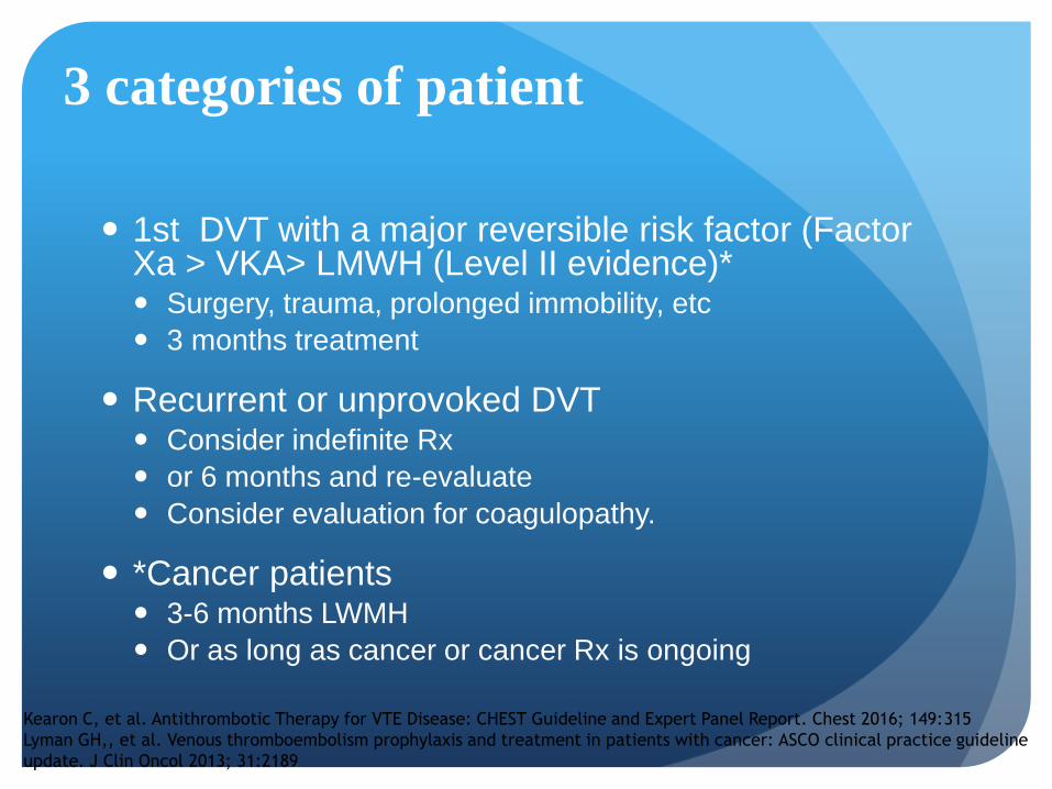
3 categories of patient
1st DVT with a major reversible risk factor (Factor Xa > VKA> LMWH (Level II evidence)* Surgery, trauma, prolonged immobility, etc
3 months treatment
Recurrent or unprovoked DVT Consider indefinite Rx
or 6 months and re-evaluate
Consider evaluation for coagulopathy.
*Cancer patients 3-6 months LWMH
Or as long as cancer or cancer Rx is ongoing
Kearon C, et al. Antithrombotic Therapy for VTE Disease: CHEST Guideline and Expert Panel Report. Chest 2016; 149:315
Lyman GH,, et al. Venous thromboembolism prophylaxis and treatment in patients with cancer: ASCO clinical practice guideline
update. J Clin Oncol 2013; 31:2189
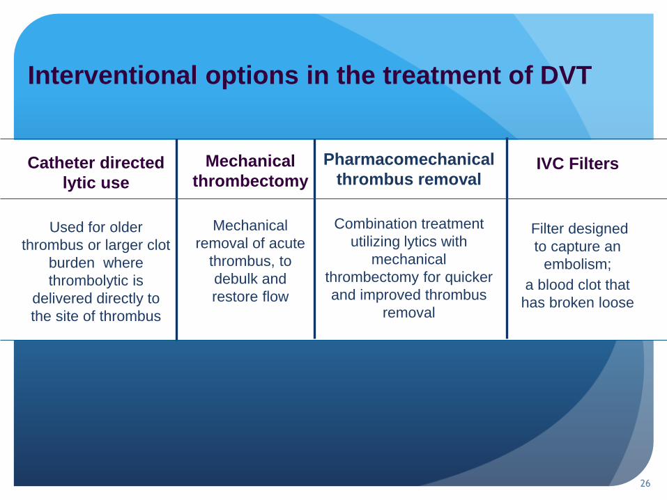
Interventional options in the treatment of DVT
26
Pharmacomechanical
thrombus removal
Combination treatment
utilizing lytics with
mechanical
thrombectomy for quicker
and improved thrombus
removal
Catheter directed
lytic use
Used for older
thrombus or larger clot
burden where
thrombolytic is
delivered directly to
the site of thrombus
Mechanical
thrombectomy
Mechanical
removal of acute
thrombus, to
debulk and
restore flow
IVC Filters
Filter designed
to capture an
embolism;
a blood clot that
has broken loose
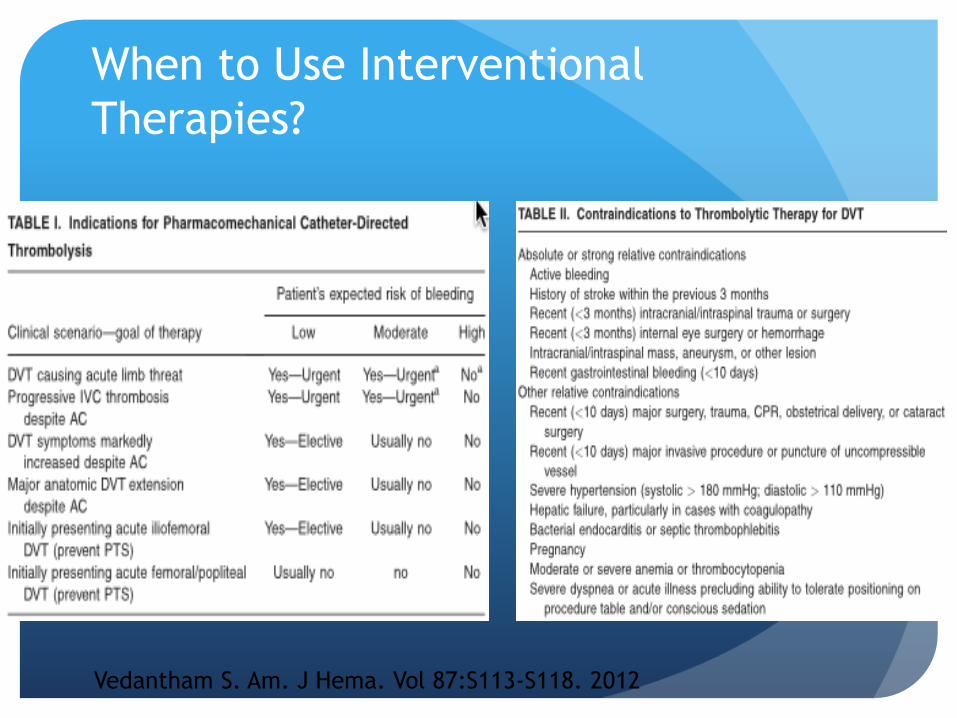
When to Use Interventional
Therapies?
Vedantham S. Am. J Hema. Vol 87:S113-S118. 2012

Acute Threatened Limb/Phlegmasia
Cerulea Dolens

Acute Threatened Limb/Phlegmasia

Post 12 hours tPA @ 1 mg/hr
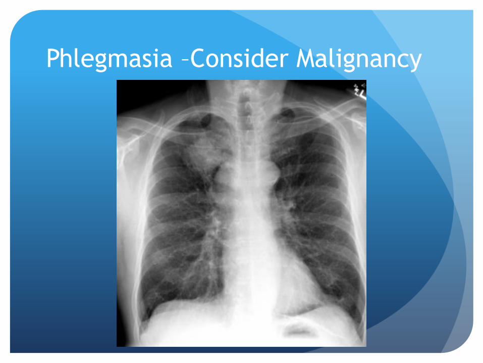
Phlegmasia –Consider Malignancy
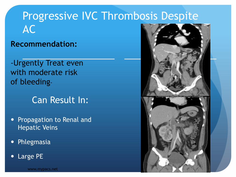
Progressive IVC Thrombosis Despite
AC
Can Result In:
Propagation to Renal and
Hepatic Veins
Phlegmasia
Large PE
Recommendation:
-Urgently Treat even
with moderate risk
of bleeding-
www.mypacs.net
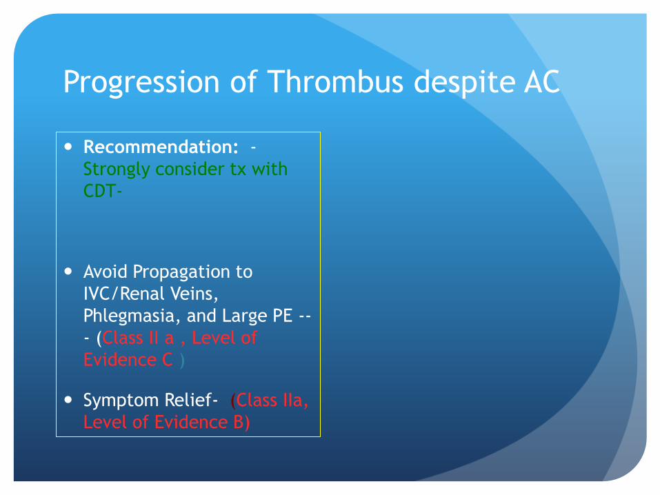
Progression of Thrombus despite AC
Recommendation: -
Strongly consider tx with
CDT-
Avoid Propagation to
IVC/Renal Veins,
Phlegmasia, and Large PE --
- (Class II a , Level of
Evidence C )
Symptom Relief- (Class IIa,
Level of Evidence B)

Initial Treatment of Ileofemoral DVT
Recommendation:
Evidence supports treatment
with CDT would benefit pt
from preventing Post
Thrombotic Syndrome if low
risk of bleeding.

Sara R. Vazquez, and Susan R. Kahn Circulation.
2010;121:e217-e219
Mechanism of Post Thrombotic
Syndrome
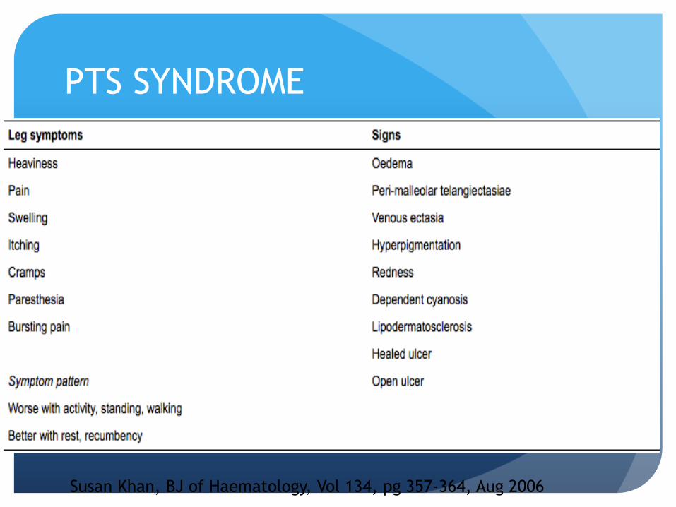
PTS SYNDROME
Susan Khan, BJ of Haematology, Vol 134, pg 357-364, Aug 2006
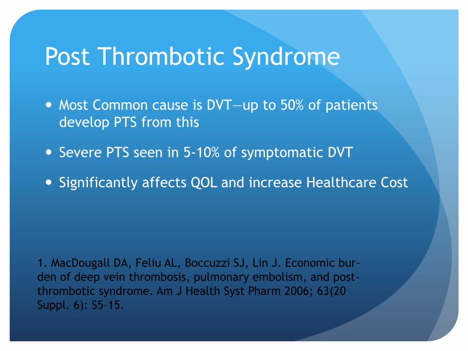
Post Thrombotic Syndrome
Most Common cause is DVT—up to 50% of patients
develop PTS from this
Severe PTS seen in 5-10% of symptomatic DVT
Significantly affects QOL and increase Healthcare Cost
1. MacDougall DA, Feliu AL, Boccuzzi SJ, Lin J. Economic bur-
den of deep vein thrombosis, pulmonary embolism, and post-
thrombotic syndrome. Am J Health Syst Pharm 2006; 63(20
Suppl. 6): S5–15.
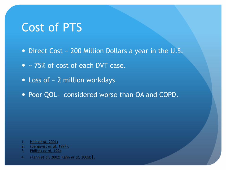
Cost of PTS
Direct Cost ~ 200 Million Dollars a year in the U.S.
~ 75% of cost of each DVT case.
Loss of ~ 2 million workdays
Poor QOL- considered worse than OA and COPD.
1. Heit et al, 2001)
2. (Bergqvist et al, 1997).
3. Phillips et al, 1994
4. (Kahn et al, 2002; Kahn et al, 2005b).
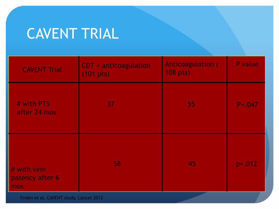
CAVENT TRIAL
P value
# with PTS
after 24 mos
# with vein
patency after 6
mos
CAVENT Trial CDT + anticoagulation
(101 pts)
Anticoagulation (
108 pts)
Enden et al, CAVENT study, Lancet 2012
37 55
58 45 p=.012
P=.047
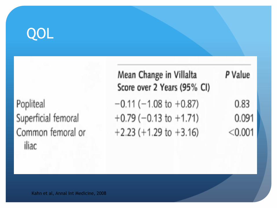
QOL
Kahn et al, Annal Int Medicine, 2008
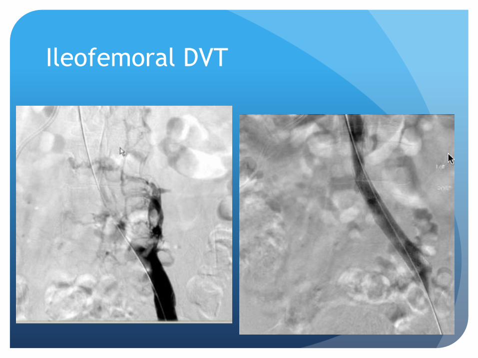
Ileofemoral DVT

What’s Coming?
ATTRACT TRIAL
Randomized control, NIH funded trial
692 pts from 56 hospitals
Goal- Determine if the use of CDT in patients with acute
proximal (DVT) prevents PTS and improves QOL.
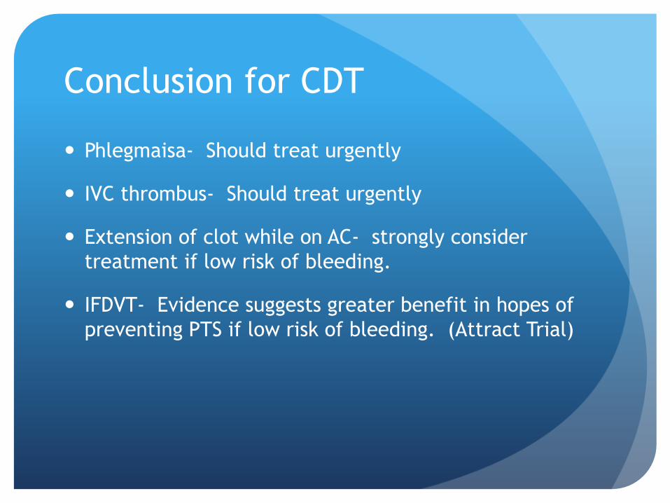
Conclusion for CDT
Phlegmaisa- Should treat urgently
IVC thrombus- Should treat urgently
Extension of clot while on AC- strongly consider
treatment if low risk of bleeding.
IFDVT- Evidence suggests greater benefit in hopes of
preventing PTS if low risk of bleeding. (Attract Trial)
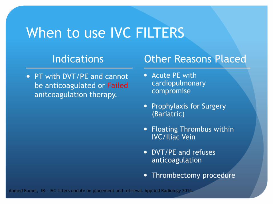
When to use IVC FILTERS
Indications
PT with DVT/PE and cannot
be anticoagulated or Failed
anitcoagulation therapy.
Other Reasons Placed
Acute PE with cardiopulmonary compromise
Prophylaxis for Surgery (Bariatric)
Floating Thrombus within IVC/Iliac Vein
DVT/PE and refuses anticoagulation
Thrombectomy procedure
Ahmed Kamel, IR – IVC filters update on placement and retrieval. Applied Radiology 2014.

IVC filter History/Background
PREPIC--Improved prevention of PE in first 12 days without significant benefit in comparison to anticoagulation over 2 years
8 year Followup—Similar incidence of venous thromboembolic dz with increase in LE DVT in the filter population.
No significant change in post-thrombotic syndrome.
Decousus H, Leizorovicz A, Parent F, et al. A clinical trial of vena caval filters in the prevention of pulmonary embolism in patients with proximal deep-vein thrombosis. Prévention du Risque d'Embolie Pulmonaire par Interruption Cave Study Group. N Engl J Med 1998; 338:409.
PREPIC Study Group. Eight-year follow-up of patients with permanent vena cava filters in the prevention of pulmonary embolism: the PREPIC (Prevention du Risque d'Embolie Pulmonaire par Interruption Cave) randomized study. Circulation 2005; 112:416.

Increase Utilization
http://www.fda.gov/MedicalDevices/Safety/AlertsandNotices/ucm221676.htm
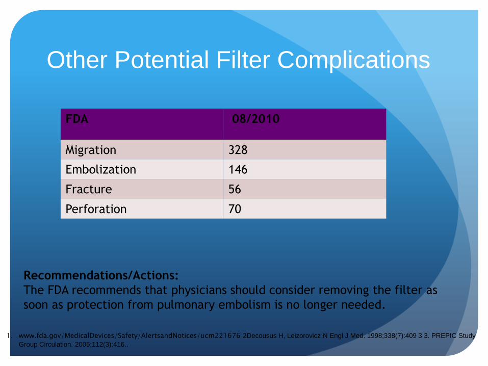
Other Potential Filter Complications
1. www.fda.gov/MedicalDevices/Safety/AlertsandNotices/ucm221676 2Decousus H, Leizorovicz N Engl J Med. 1998;338(7):409 3 3. PREPIC Study
Group Circulation. 2005;112(3):416..
FDA 08/2010
Migration 328
Embolization 146
Fracture 56
Perforation 70
Recommendations/Actions:
The FDA recommends that physicians should consider removing the filter as
soon as protection from pulmonary embolism is no longer needed.

Pulmonary Embolism

Icoper: Cumulative Mortality After Diagnosis of PE M
ort
ality
(%
)
Days From Diagnosis
17.5%
At 90days
7 14 30 60 90
0
5
10
15
20
25
Lancet. 1999;353:1386-1389.
50
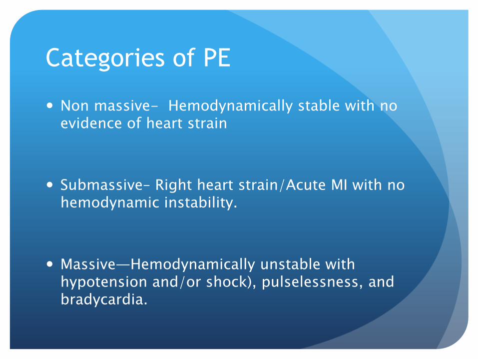
Categories of PE
Non massive- Hemodynamically stable with no evidence of heart strain
Submassive– Right heart strain/Acute MI with no hemodynamic instability.
Massive—Hemodynamically unstable with hypotension and/or shock), pulselessness, and bradycardia.

PE: Indicators of Poor Outcome
ESC criteria (based on consensus; lack of
validation) Criteria mortality
High risk Cardiovascular shock or
persistent hypotension
> 30 %
Intermediate risk Lab (troponin, BNP)
or RV dysfunction
1-30 %
Low risk nl labs (troponin, BNP);
nl RV function
< 1 %
[Torbicki A et al. Eur Heart J 2008;2276-315]

VQ Scan
Ventilation Phase (TcDTPA
vs Xeon)
Perfusion Phase (TcMAA)
Looking for mismatch
perfusion defects.

VQ SCAN
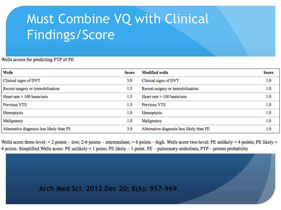
Must Combine VQ with Clinical
Findings/Score
Arch Med Sci. 2012 Dec 20; 8(6): 957–969.
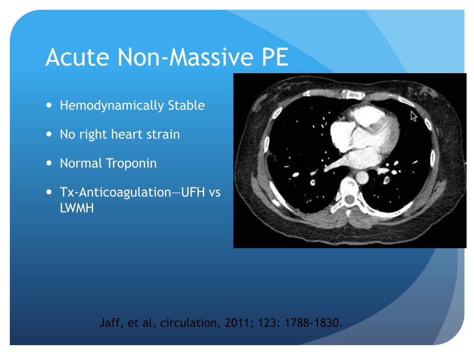
Acute Non-Massive PE
Hemodynamically Stable
No right heart strain
Normal Troponin
Tx-Anticoagulation—UFH vs
LWMH
Jaff, et al, circulation, 2011; 123: 1788-1830.
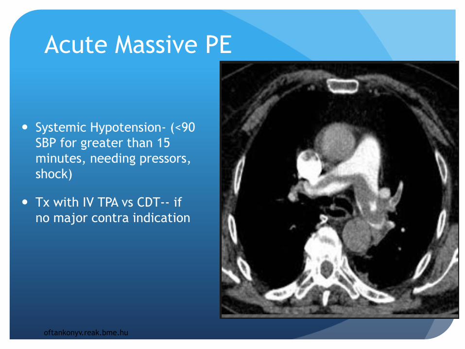
Acute Massive PE
Systemic Hypotension- (<90
SBP for greater than 15
minutes, needing pressors,
shock)
Tx with IV TPA vs CDT-- if
no major contra indication
oftankonyv.reak.bme.hu

Acute Submassive PE ?
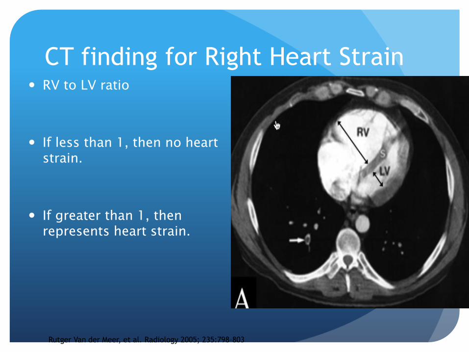
CT finding for Right Heart Strain RV to LV ratio
If less than 1, then no heart strain.
If greater than 1, then represents heart strain.
Rutger Van der Meer, et al. Radiology 2005; 235:798–803
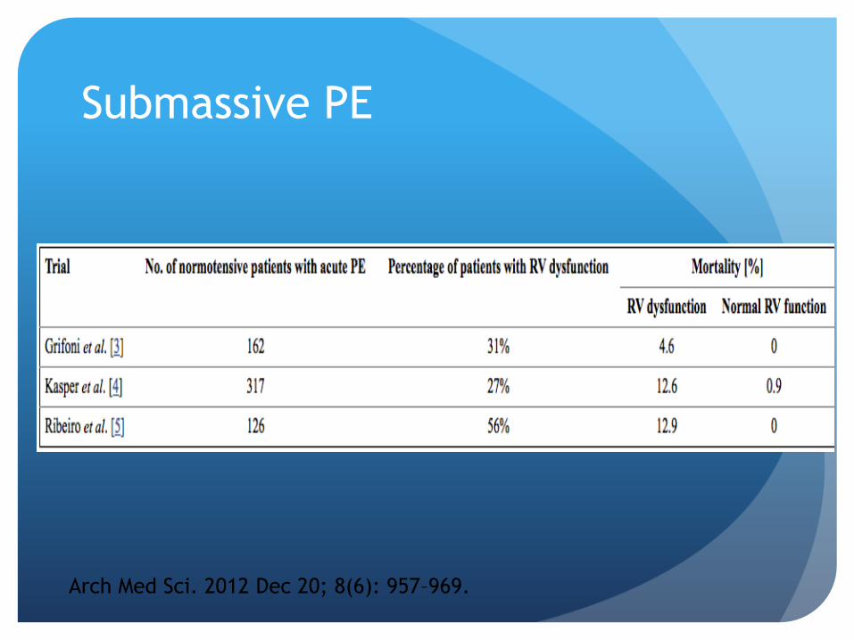
Submassive PE
Arch Med Sci. 2012 Dec 20; 8(6): 957–969.
Goldhaber SZ, Bounameaux H. Lancet. 2012 May 12; 379:1835-46
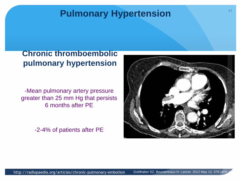
Pulmonary Hypertension
Chronic thromboembolic
pulmonary hypertension
-Mean pulmonary artery pressure
greater than 25 mm Hg that persists
6 months after PE
-2-4% of patients after PE
61
http://radiopaedia.org/articles/chronic-pulmonary-embolism

From: Thrombolysis for Pulmonary Embolism and Risk of All-Cause Mortality, Major Bleeding, and Intracranial Hemorrhage: A
Meta-analysis
JAMA. 2014;311(23):2414-2421. doi:10.1001/jama.2014.5990
Submassive PE

Treatment for Acute PE
Non-Massive Acute PE Massive Acute PE Submassive Acute PE
Anticoagulation UFH vs LMWH IV TPA vs CDT + AC
Jaff, et al, circulation, 2011; 123: 1788-1830.

Questions?