4. coprinus comatus damages nematode cuticles
TRANSCRIPT
8/8/2019 4. Coprinus Comatus Damages Nematode Cuticles
http://slidepdf.com/reader/full/4-coprinus-comatus-damages-nematode-cuticles 1/8
APPLIED AND ENVIRONMENTAL MICROBIOLOGY, June 2007, p. 3916–3923 Vol. 73, No. 120099-2240/07/$08.000 doi:10.1128/AEM.02770-06Copyright © 2007, American Society for Microbiology. All Rights Reserved.
Coprinus comatus Damages Nematode Cuticles Mechanically with Spiny Balls and Produces Potent Toxins
To Immobilize Nematodes
Hong Luo,1† Yajun Liu,1† Lin Fang,2 Xuan Li,3 Ninghua Tang,4 and Keqin Zhang1*
Laboratory for Conservation and Utilization of Bio-resources, Yunnan University, Kunming 650091, Yunnan, People’s Republic of China1 ; Middle School Attached to Yunnan Normal University, Kunming 650106, Yunnan, People’s Republic of China2 ; Faculty of Environmental Science and Engineering, Kunming University of Science and Technology, Kunming 650093,
Yunnan, People’s Republic of China3 ; and State Key Laboratory of Phytochemistry and Plant Resources in West China, Kunming Institute of Botany, Chinese Academy of Sciences, Kunming 650204, People’s Republic of China4
Received 28 November 2006/Accepted 11 April 2007
We reported recently a unique fungal structure, called the spiny ball, on the vegetative hyphae of Coprinus comatus (O. F. Mull.:Fr.) Pers. Although some observations regarding the role of this structure
were presented, its function remained largely unknown. In this study, we showed that purified (isolatedand washed) spiny balls could immobilize and kill the free-living nematode Panagrellus redivivus Goodey
highly efficiently. Scanning electron microscopy studies illustrated that the spiny structure damaged thenematode cuticle, suggesting the presence of a mechanical force during the process of nematode immo-bilization. Severe injuries on nematode cuticles caused the leakage of inner materials of the nematodes.
When these structures were ground in liquid nitrogen, their killing efficacy against nematodes was lost,indicating that the shape and the complete structure of the spiny balls are indispensable for their function.However, extraction with organic solvents never lowered their activity against P. redivivus, and the extractsshowed no obvious effect on the nematode. We also investigated whether C. comatus was able to producetoxins which would aid in the immobilization of nematodes. In total, we identified seven toxins from C.
comatus that showed activity to immobilize the nematodes P. redivivus and Meloidogyne incognita (Kofoidet White) Chitwood. The chemical structures of these toxins were identified with nuclear magneticresonance, mass spectrometry, infrared, and UV spectrum analysis. Two compounds were found to benovel. The toxins found in C. comatus are O-containing heterocyclic compounds.
Nematophagous basidiomycetes refer to those fungi possess-
ing clamp connections on hyphae with the capability of cap-turing, killing, and decomposing nematodes. These fungi aremembers of commonly known mushrooms, and they have de- veloped several mechanisms to utilize nematodes as foodsources, including that of nitrogen (19, 23, 24, 26). Most of these mushrooms use delicate and effective small weaponscalled appendages on either hyphae or conidia to attack nem-atodes. For example, Nematoctonus spp. produce hourglass-shaped adhesive knobs containing a secretory cell and themucilage it produces (7, 8, 9, 10, 11). The knobs are envelopedin a thick mucus sheath and can capture nematodes by firststicking to the nematode cuticle. Subsequently, the knobs pen-etrate and kill the nematodes to start a new cycle of growth.
Hourglass-shaped adhesive knobs are also found on Hohen- buehelia, which has been proved to be the teleomorph of thegenus Nematoctonus (3). Stephanocysts are one- or two-celledstructures on the hyphae of some species of Hyphoderma (5,14). For many years, these structures were merely considered acuriosity, and their function was not known until Tzean andLiou made the exciting discovery that these specialized cells
were coated with fibrous mucilages that can adhere to nema-
todes and eventually invade the nematode hosts (26). Tinysecretory cells on the vegetative hyphae of Pleurotus spp. canalso attack nematodes in a remarkably different fashion. Theseappendages secrete droplets containing potent toxins whichcan paralyze nematodes in seconds but do not kill them (4).The specialized directional hyphae are then attracted by theparalyzed victims. The cuticle is penetrated and the contentsdigested by the hyphae. Secretory appendages similar to thoseof Pleurotus were found on the hyphae of the lawn mushroomConocybe lactea, and its pattern of attacking nematodes is alsosimilar to that of Pleurotus (15). The potent toxin dropletsproduced by the secretory cell can paralyze and kill the nem-atode victim. Finally, an acanthocyte is a spiky structure found
in Stropharia species (12). It was recently discovered that thisstructure can damage the cuticle of nematodes and kill them very efficiently. These observations suggest that acanthocytesmight be a nematode-attacking device (20).
Coprinus comatus, the shaggy mane mushroom, has darkred-brown spores and is commonly seen on newly disturbedgrounds, grassy places, and road sides. This edible species iscultivable and has been commonly seen on the tables of Chi-nese people in recent years. We previously reported a novelfungal structure called spiny ball on vegetative hyphae of C.
comatus. Typical spiny balls consist of a rod-shaped part in thecore and quite a few peripheral projections with contractedends (19). The spiny ball is quite different from other known
* Corresponding author. Mailing address: Laboratory for Conserva-tion and Utilization of Bio-resources, Yunnan University, Kunming650091, Yunnan, People’s Republic of China. Phone: (86) 871 5034878.Fax: (86) 871 5034838. E-mail: [email protected].
† These authors contributed equally to this article. Published ahead of print on 20 April 2007.
3916
8/8/2019 4. Coprinus Comatus Damages Nematode Cuticles
http://slidepdf.com/reader/full/4-coprinus-comatus-damages-nematode-cuticles 2/8
fungal structures on other mushrooms in two aspects. First, itsspiny shape is morphologically distinct and its size is muchsmaller than those of the other known appendages. Secondly,the spiny ball does not possess nuclei and seems to be anincomplete cell, while the others are obviously cells. Someobservations showed that these structures are probably linkedto the ability of C. comatus to attack nematodes (19). However,the function of spiny balls has not been elucidated.
The nematode-immobilizing effect of C. comatus is high. About half of the nematodes could be immobilized within 1 to4 h by C. comatus cells grown on potato dextrose agar (PDA)or cornmeal agar plates. Within 8 h of incubation, about 90%of the worms were immobilized (19). The high efficiency of immobilization suggested that low-molecular-weight toxinsproduced by the fungus might be responsible. Although someearlier studies have isolated and identified nematotoxic com-pounds, the known number of such compounds is still quitelimited. Until now, fewer than 100 metabolites from variousfungi have been proven to possess nematicidal activity (6).Some of them are thought to be specifically produced to attack
nematodes. These compounds included trans-2-decanedioicacid, which was isolated from Pleurotus ostreatus (18), andlinoleic acid, p-anisoylalcohol, p-anisaldehyde, S-coriolic acid,1-(4-methoxyphenyl)-1,2-propanediol, and Z-hydroxy-(4-methoxy)-propiophenone, which were isolated from Pleurotus
pulmonarius (22). The nematicidal abilities of C. comatus
prompted us to investigate whether C. comatus producesnematotoxic compounds.
The aim of this paper is to provide some direct mechanisticevidences for the nematicidal activity by C. comatus. In anearlier study, a simple method to purify spiny balls was devel-oped (19). The purified spiny balls showed limited efficacyagainst nematodes, which were much weaker than live hyphae.
Such observations suggested to us that the nematicidal effects were due to a number of factors. In this paper, we investigated whether the ability to attack nematodes was due to the forma-tion of spiny balls and, if so, what kind of role the spiny ballsplay during the interaction between the fungus and nematodes.In addition, to answer the question of whether C. comatus
could secrete toxins to facilitate the infection process, we car-ried out a study on isolating and identifying nematotoxins.Since many nematodes are parasites on plants and can causesevere damage to world agriculture (1), the identification andstructure elucidation of novel compounds with nematotoxicability are of great interest for developing new nematicides andantihelminthic drugs.
MATERIALS AND METHODS
Strains and cultivation conditions. Two strains of C. comatus were used in thisstudy: strain LHA-7, used in our previous study, and an isolate designated C-1from a basidiocarp found in the field in Yunnan, China. The strains were
deposited in the Laboratory for Conservation and Utilization of Bio-resources(Yunnan University, Kunming, Yunnan, China) as YMF1.00135 and YMF1.02579,respectively. The strains were maintained on PDA slants and transferred every 2
months. The free-living nematode Panagrellus redivivus was grown axenically insemiliquid oat medium (10 g of oat, 6 ml of distilled water) at 28°C for 4 to 6
days, stored at 4°C, and used within 15 days.Regenerating PDA plates. The C. comatus strains LHA-7 and C-1 were cul-
tured on PDA in petri dishes (diameter, 9 cm) for 8 to 10 days. When the agar
surface was fully covered by fungal mycelia, these plates were carefully scraped with a scalpel to completely remove the aerial fungal hyphae under sterile
conditions in a laminar flow cabinet. These plates were then incubated at 20°C
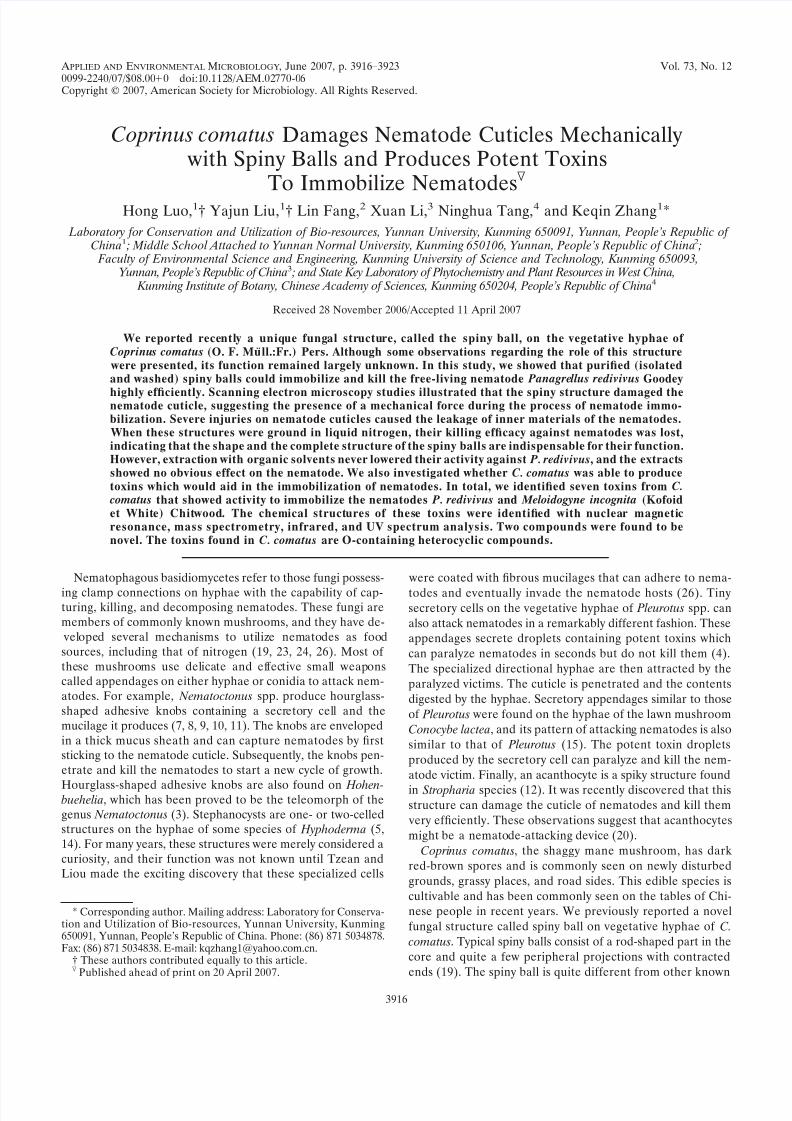
for 18 to 28 h. During this time period, special aerial hyphae grew and produced
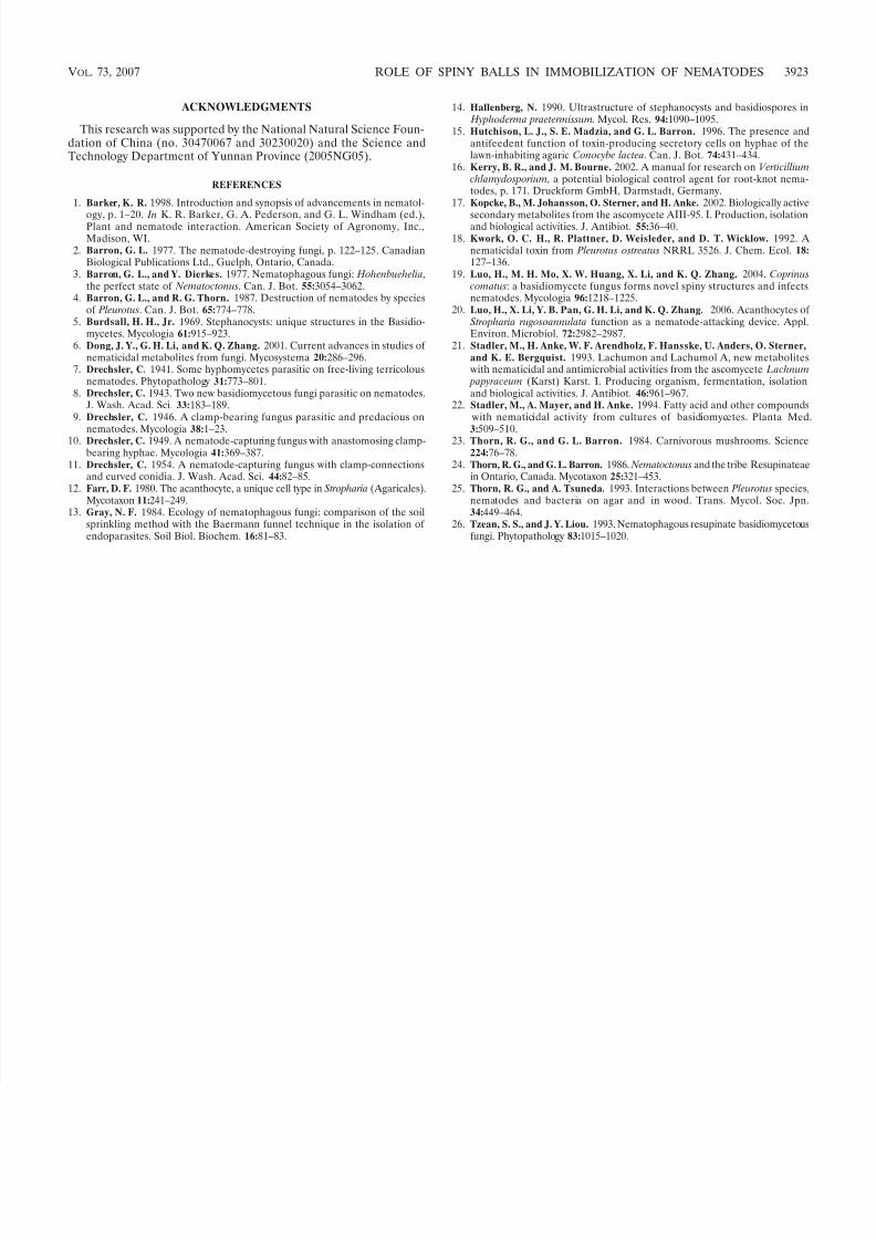
abundant spiny balls. However, normal aerial hyphae formed only very sparsely(Fig. 1). These plates were designated regenerating PDA plates. The purpose of
constructing the regenerating plate was to expose the spiny balls which wouldhave been covered by dense aerial hyphae on normal culture plates of C. coma-
tus. Bioassays with this regenerating plate were compared to those with the
normal culture plate.Bioassay with the regenerating PDA plate. The regenerating PDA plates
prepared as described above were inspected for the production of spiny balls
before being used in bioassays. The P. redivivus nematode was thoroughly rinsedfour times before use. The washed nematodes were picked with an inoculating
loop, and 50 to 100 worms were transferred to a selected site on the regeneratingPDA plate with abundant newly formed spiny balls. Three sites were selected toreceive the nematodes on one plate. The plates were incubated at 24°C, and
mobile and immobile nematodes were counted after 15 and 30 min at three
random sites by using a light microscope (magnifications of
40). Nematodesadded to blank PDA plates served as a control. This experiment was carried out with three replicates and conducted twice.
Bioassay with purified spiny balls and smashed spiny balls. Purified spiny
balls were obtained by following a previously described method (19). The spinyball suspension was adjusted to ca. 1010 per ml. To smash the purified spiny balls,200 l of the spiny ball suspension was thoroughly grounded with liquid nitrogen
by using mortars, and 5 l of the suspension was inspected under a microscopeto confirm that the vast majority of these spiny balls were completely smashed
(like starch paste without or with few complete spiny balls). Then, blank wateragar (WA) plates were prepared for the bioassay. On one plate, 20 l of suspension of the purified spiny balls and 20 l of the ground spiny balls were
transferred to two close sites (ca. 1 cm between each other) in the middle of theplate and the suspension drops formed two circles with diameters of ca. 1 cm.
About 15 P. redivivus worms prepared as described above were handpicked witha fine needle and added to each of the two circles. Immediate observations on thebehavior of the added nematodes were made using a microscope at room tem-
perature. Mobile and immobile nematodes were counted 5 and 10 min after theiraddition. The negative control contained a blank WA plate with about 15 nem-atodes added to the middle of the plate. This experiment was conducted twice,
each with three replicates.Bioassay with organic solvent-extracted spiny balls. According to our previous
hypothesis, the spiny ball could be a reservoir for some potent toxins againstnematodes. To test the hypothesis, 100 l of the purified spiny ball suspension(ca. 1010 per ml) was first extracted in 1.0 ml of ethanol at 28°C for 24 h in a
1.5-ml Eppendorf tube. After centrifugation at 13,000 g for 5 min, the super-natant was transferred to another Eppendorf tube and then dried in Eppendorf
concentrator 5301. Then, 1.0 ml of acetone was added to the tube and held at28°C for 24 h. After another centrifugation, the supernatant was transferred anddried as described above. Finally, 1.0 ml petroleum ether was added to the tube
and was kept at 28°C for another 24 h. After the supernatant (treated as de-scribed above) was removed, the precipitate of the purified spiny balls was washed with 1.0 ml of sterile water for five times, followed by centrifugation at
FIG. 1. A survey view (planform) of a regenerating PDA plate,showing abundantly produced spiny balls with sparse aerial hyphae.Bar, 100 m.
VOL. 73, 2007 ROLE OF SPINY BALLS IN IMMOBILIZATION OF NEMATODES 3917
8/8/2019 4. Coprinus Comatus Damages Nematode Cuticles
http://slidepdf.com/reader/full/4-coprinus-comatus-damages-nematode-cuticles 3/8
13,000 g for 5 min to remove (discard) the supernatant. Then, 20 l of thespiny ball suspension was transferred to a blank WA plate at the central locationof the plate and the suspension drop formed a circle with a diameter of ca. 1 cm.
The plate was held at room temperature for 15 min to allow the excessive waterto be absorbed by the agar. P. redivivus was prepared as described above, and
about 15 nematodes were picked with a fine needle and placed in the circlecontaining spiny balls on the WA plate. The immediate interaction between thespiny balls and the nematodes was studied by microscopy at room temperature.
After 5 and 10 min, mobile and immobile nematodes were counted. This exper-
iment was carried out with three replicates and repeated twice. To construct thesupernatant control, 100 l of distilled water was added to each of the dried
supernatant-containing tubes and the mixture was vortexed for 1 min to dissolvethe extracts. The resulting solutions were pooled and 20 l of the mixture was
transferred to the same WA plate containing the suspension of spiny balls toproduce another circle (ca. 1 cm away from the suspension circle). About 15nematodes were added to the area containing the solution mixtures on the WA
plate to serve as the negative control.Scanning electron microscopy study of immobilized nematodes. Some immo-
bilized nematodes in the bioassay with regenerating PDA as well as some nem-atodes in the bioassay with purified spiny balls as described above were hand-picked and immediately immersed in a 2.5% glutaraldehyde solution at 4°C for
4 h in a 1.5-ml Eppendorf tube. A brief centrifugation was adopted to precipitatethe nematodes, and the glutaraldehyde solution was carefully removed anddiscarded. The fixed nematodes were then rinsed three times in 1 ml of 0.2 M
phosphate buffer (pH 7.2), followed by a brief centrifugation to remove the
buffer with a pipette. Then, 300
l of 1% OsO4 was added to the tube and thesenematodes were postfixed in the same buffer for 1 h at room temperature. Agraded acetone series was used to dehydrate the nematodes, and each step wasfollowed by a brief centrifugation to remove the liquid. Then, the nematodes
were exchanged in an isoamyl acetate series, dried with an HCP-2 critical pointdryer (Hitachi) for 7 h, mounted on aluminum stubs, coated with a gold-palla-
dium mixture by an IB-3 ion coater (Eiko), and viewed with a scanning electronmicroscope (Philips XL30 ESEM) operating at 20 to 30 kV.
Toxin extraction. One of the two strains, C-1, was used to further examine
whether there is toxin production for the fungus. Strain C-1 was cultured on PDA(20 liters) at 25°C for 15 days. The cultures were extracted twice with 15 litersMeOH at room temperature. The crude extracts (160 g) were resuspended in 500
ml distilled water and extracted twice with 500 ml ethyl acetate. The organicphase and residues were separately reduced to solid extracts. The extracts were
tested for nematicidal activity, and only the ethyl acetate extract showed such aproperty. The extracts (10 g) containing active compounds were subjected toliquid chromatography on Silica Gel G (200 to 300 m; column, 55 by 450 mm;
Meijing Co. Ltd.). Stepwise elutions with chloroform-methanol (20:1 [vol/vol],2.5 liters, and 9:1 [vol/vol], 2 liters) were performed. The eluant was collected
with 50-ml flasks and combined for thin-layer chromatography (TLC). TLC wascarried out using precoated Silica Gel G plates (0.2 mm thick; Meijing Co. Ltd.).Each 20-l eluant was developed with chloroform-methanol (9:1; vol/vol) under
saturated conditions. Chromatograms were detected by the color reaction with5% sulfuric acid-ethanol spraying solution after heating at 100°C. Subsequently,the combined eluants were tested for nematicidal activity. Active fractions were
further separated and purified by a silica column and eluted stepwisely withpetroleum ether-acetone (3:1 and 1:1, vol/vol). The eluants were collected with
20-ml flasks. Each eluant was developed with petroleum ether-acetone (3:1, vol/vol) on TLC and was tested for nematicidal activity. We obtained sevencompounds with nematicidal activity from C. comatus. All purified compounds
were subjected to nuclear magnetic resonance (NMR), mass spectrometry (MS),infrared (IR), and UV spectrum analyses for identification and structure eluci-dation.
Spectrum analysis. IR spectra were measured on a Bio-Rad FTS-135 spec-trometer with KBr pellets. UV spectra were obtained on a Shimadzu double-beam 210A spectrometer. Mass spectra were recorded on a VG Autospec-3000
spectrometer. One-dimensional and two-dimensional NMR spectra were run ona Bruker AM-400 and DRX-500 instrument, respectively, with trimethylsilane asan internal standard. TLC was performed on plates precoated with Silica Gel G
(Qingdao Marine Chemical Ltd., China).Nematicidal assay of compounds. Worms of the root-knot nematode Meloi-
dogyne incognita were cultivated on tomato plants in the greenhouse at 25°C, and
second-stage juveniles were extracted and stored according to the method of Kelly and Bourne (16). The nematode P. redivivus was cultured as described above.
The nematodes were separated from the culture medium by the Baermannfunnel technique (2, 13). The assay for nematicidal activity was carried out as
described by Stadler et al. (21).
RESULTS
Immobilization effect on regenerating PDA plate. Regener-ating PDA plates allowed for the exposure of spiny balls withsparse hyphae (Fig. 1). We compared the amount of spiny balls yielded on a normal culture plate with that on a regeneratingPDA plate by purifying the spiny structures. The resultsshowed that the number of spiny balls from a regenerating
PDA plate is less than that from a normal plate (data notshown). However, nematodes were immobilized much fasteron a regenerating PDA plate than on a normal culture plate(19). As shown in Table 1, 73.7 and 98.3% of the P. redivivus
nematodes added were immobilized by the C. comatus strainLHA-7 and 75.7 and 98.9% of the nematodes were immobi-lized by strain C-1 after 15 and 30 min of contact with thestrains, respectively. Highly significant differences were ob-served between the samples treated and the controls ( P
0.005). Most of the immobilized nematodes looked completeunder an optical microscope.
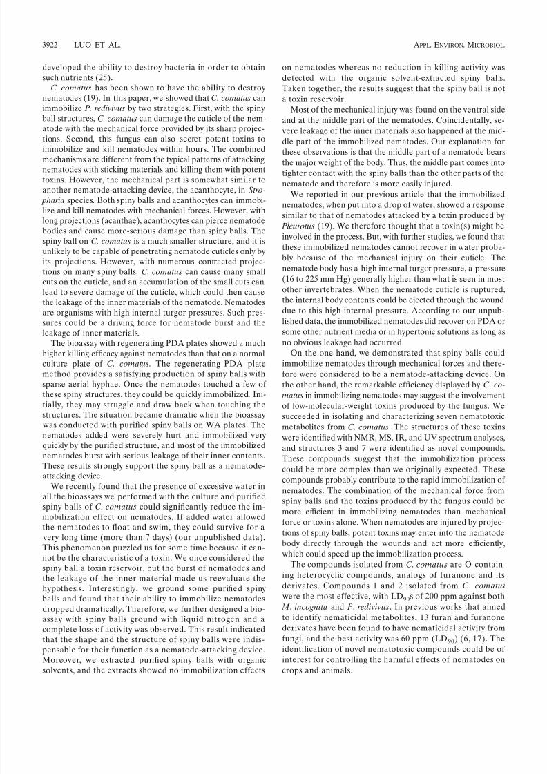
Immobilization of nematodes by purified spiny balls. Intactpurified spiny balls on WA plates exhibited an outstandingcapacity to immobilize P. redivivus. The nematodes added wererestricted to a small area and immobilized the nematodes veryquickly (Fig. 2A). For C. comatus strain LHA-7, 75.0 and93.8% of the nematodes added were immobilized by the non-smashed spiny balls 5 and 10 min after being added, respec-tively. For strain C-1, 76.9 and 92.3% of the nematodes added were immobilized (Table 2). Highly significant differences ex-isted between the samples treated and the negative controls( P 0.005). Grinding spiny balls with liquid nitrogen can finelydestroy the structure of the spiny balls, but no immobilizationeffect was detected with the smashed spiny balls (data notshown).
Immobilization of nematodes by organic solvent-extracted
purified spiny balls. When the spiny balls were in turn ex-
TABLE 1. Immobilization of P. redivivus on regenerating PDA plates by C. comatus strains
StrainIncubationtime (min)
No. of nematodes % Immobilizednematodes
No. of controls2 value a P value
Immobile Mobile Immobile Mobile
LHA-7 15 76 27 73.7 3 96 103.2 0.00530 116 2 98.3 3 102 199.6 0.005
C-1 15 56 18 75.7 1 88 95.5 0.00530 92 1 98.9 2 76 155.3 0.005
a Results of 2 tests (1 df) comparing the frequencies of mobilized and immobilized nematodes in treated versus control samples.
3918 LUO ET AL. APPL. ENVIRON. MICROBIOL.
8/8/2019 4. Coprinus Comatus Damages Nematode Cuticles
http://slidepdf.com/reader/full/4-coprinus-comatus-damages-nematode-cuticles 4/8
tracted with ethanol, acetone, and petroleum ether, the struc-tures maintained their spiny shape with no obvious changes. To
determine whether these organic solvent-extracted spiny ballshave an immobilization effect on nematodes, we performed a
bioassay with WA. As shown in Table 3, the strain LHA-7immobilized 82.4 and 94.1% of the nematodes and C-1 immo-
bilized 71.4 and 92.9% of the nematodes 5 and 10 min after thenematodes were added, respectively. There are highly signifi-
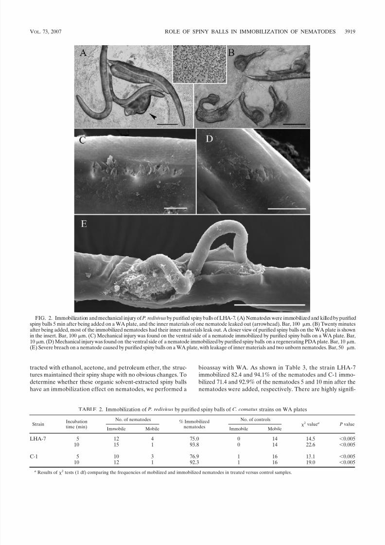
FIG. 2. Immobilization and mechanical injury of P. redivivus by purified spiny balls of LHA-7. (A) Nematodes were immobilized and killed by purifiedspiny balls 5 min after being added on a WA plate, and the inner materials of one nematode leaked out (arrowhead). Bar, 100 m. (B) Twenty minutesafter being added, most of the immobilized nematodes had their inner materials leak out. A closer view of purified spiny balls on the WA plate is shownin the insert. Bar, 100 m. (C) Mechanical injury was found on the ventral side of a nematode immobilized by purified spiny balls on a WA plate. Bar,10m. (D) Mechanical injury was found on the ventral side of a nematode immobilized by purified spiny balls on a regenerating PDA plate. Bar, 10 m.(E) Severe breach on a nematode caused by purified spiny balls on a WA plate, with leakage of inner materials and two unborn nematodes. Bar, 50 m.
TABLE 2. Immobilization of P. redivivus by purified spiny balls of C. comatus strains on WA plates
StrainIncubationtime (min)
No. of nematodes % Immobilizednematodes
No. of controls2 value a P value
Immobile Mobile Immobile Mobile
LHA-7 5 12 4 75.0 0 14 14.5 0.00510 15 1 93.8 0 14 22.6 0.005
C-1 5 10 3 76.9 1 16 13.1 0.00510 12 1 92.3 1 16 19.0 0.005
a Results of 2 tests (1 df) comparing the frequencies of mobilized and immobilized nematodes in treated versus control samples.
VOL. 73, 2007 ROLE OF SPINY BALLS IN IMMOBILIZATION OF NEMATODES 3919

8/8/2019 4. Coprinus Comatus Damages Nematode Cuticles
http://slidepdf.com/reader/full/4-coprinus-comatus-damages-nematode-cuticles 5/8
cant differences between the samples treated and the controls( P 0.005). These results were similar to the immobilizationefficiency produced by normal purified spiny balls (Table 2).Nevertheless, none of the extracts obtained showed any obvi-ous effects on the tested nematodes.
Mechanical injury of nematodes by spiny balls. Under anoptical microscope, most of the immobilized nematodesshowed no obvious changes just after they were immobilized.
However, within two to four more minutes, a few of them hadtheir inner materials leaked (Fig. 2A). Fifteen to 30 minutesafter the nematodes were added, the vast majority of the addednematodes burst, accompanied by the leakage of the innermaterials (Fig. 2B). The burst of nematodes happened fre-quently in the bioassay with purified spiny balls. On regener-ating PDA plates, this burst phenomenon could be observedbut at a much lower frequency (10%). In the bioassays withnormal culture plates of C. comatus, this phenomenon wasseldom observed. We found only a few cases during extensivesearches.
Similar results were found with scanning electron micros-copy studies of the nematodes immobilized on regenerating
PDA plates and those immobilized by purified spiny balls.Figure 2C shows that a nematode was immobilized by purifiedspiny balls and mechanically injured on its ventral side. Nem-atodes immobilized on regenerating PDA plates also illus-trated the presence of mechanical injury on the ventral side of the nematode body (Fig. 2D). Because only purified spiny ballscame into contact with the nematodes in the bioassay, thesephysical damages on nematode cuticles must have been causedby those spiny structures. We previously reported that theprojections on the spiny balls have a contracted end (19), andthe contraction made the projection very sharp, enabling it tocut into the nematode cuticle. Severe damage of nematodecuticles caused the nematodes to breach and their inner ma-terials to leak out (Fig. 2E).
Spectrum data and structure elucidation of nematotoxins
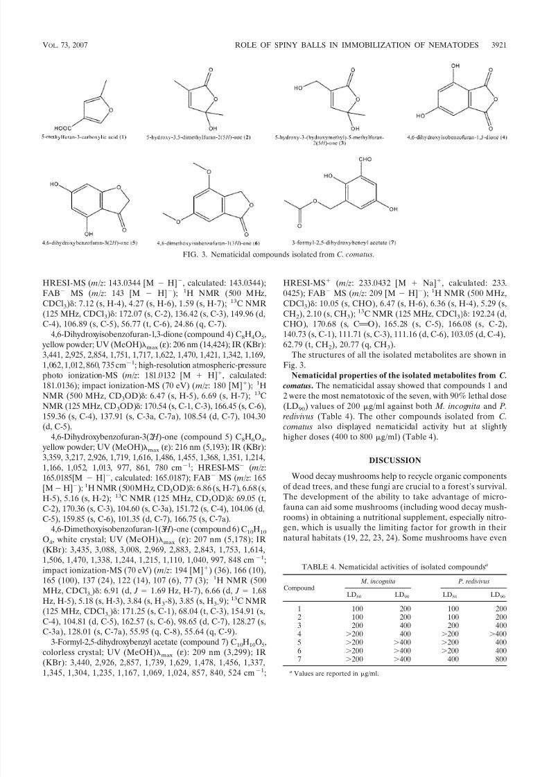
from C. comatus. We successfully isolated and identified sevenpotent compounds against nematodes from strain C-1. Theseven compounds are shown in Fig. 1. The structures of thesecompounds were identified with NMR, IR, UV spectrum, andMS analyses. Among these, compounds 3 and 7 were identifiedas novel natural products never reported previously.
High-resolution MS experiments of compound 3 suggestedthat its molecular formula is C6H8O4, which was confirmed by1H and 13C NMR spectra. The 13C NMR and distortionlessenhancement by polarization transfer spectra of compound 3showed six signals, including one methyl, one methylene, onemethine, and three quaternary carbon atoms (one carbonyl
carbon). In the 1H NMR spectrum, 7.12, 4.27, and 1.59 ppmcorresponded to methine at C-4, methylene at C-6, and methylprotons at C-7, respectively. The methylene signal at 56.77ppm was downfield shifted, but it should have been connectedto hydroxyl. The IR spectrum exhibited absorptions at 3,381and 1,765 cm1 due to the hydroxyls and to the unsaturatedcarbonyl group. Absorptions at 1,631, 1,553, and 1,450 cm1
indicated the presence of a furan ring. Based on the above-
described analysis, the structure of compound 3 was identifiedas 5-hydroxy-3-(hydroxymethyl)-5-methylfuran-2(5 H )-one.
High-resolution MS experiments with compound 7 sug-gested that its molecular formula is C10H10O5 (high-resolutionelectrospray ionization [HRESI]-MS, m/z: 233.0432 [M
Na], calculated: 233.0425). This structure contains six unsat-urated degrees. Four of these could be assigned to a benzenering, and the NMR data suggested the presence of carbonyland formyl carbon. The methylene signals downfield shifted at62.79 ppm should be connected to an oxygen atom. Hetero-nuclear multiple bond correlation experiments showed thatmethylene correlated to methyl and carbonyl and that formylcorrelated to C-3, C-5, and C-6. Also, the single signals of
protons suggested that two methines should be at the metaposition. The IR spectrum exhibited absorption at 3,440 cm1, which confirmed the existence of hydroxyls. The structure of compound 7 was identified as 3-formyl-2,5-dihydroxybenzyl ac-etate.
The spectrum data of the seven isolated compounds from C.
comatus are as follows. 5-Methylfuran-3-carboxylic acid (com-pound 1) C6H6O3, colorless crystal; UV (MeOH)max (ε): 204nm (8,945); IR (KBr): 3,448, 2,919, 2,850, 1,752, 1,710, 1,631,1,552, 1,462, 1,339, 1,111, 1,041 cm1. FAB MS m/z: 125[M H]; 1H NMR (500 MHz, CDCl3): 7.93 (s, H-2), 6.33(s, H-4), 2.29 (s, H-7); 13C NMR (125 MHz, CDCl3): 147.56(d, C-2), 119.72 (s, C-3), 105.55 (d, C-4), 154.06 (s, C-5), 168.41(s, C-6), 13.35 (q, C-7).
5-Hydroxy-3,5-dimethylfuran-2(5 H )-one (compound 2)C6H8O3, colorless crystal; []D
28: 16.67° (c 0.06, MeOH);UV (MeOH)max (ε): 204 nm (8,317); IR (KBr): 3,440, 2,929,1,766, 1,747, 1,629, 1,447, 1,404, 1,374, 1,188, 1,051, 929, 875,762 cm1; FAB MS ( m/z: 127 [M H]); 1H NMR (500MHz, CDCl3): 6.79 (s, H-4), 1.85 (s, H-6), 1.62 (s, H-7); 13CNMR (125 MHz, CDCl3): 171.74 (s, C-2), 131.71 (s, C-3),147.64 (d, C-4), 104.19 (s, C-5), 10.34 (q, C-6), 24.71 (q, C-7).
5-Hydroxy-3-(hydroxymethyl)-5-methylfuran-2(5 H )-one (com-pound 3) C6H8O4, colorless crystal; []D
28: 0.75° (0.00669g/ml); UV (MeOH)max (ε): 226 nm (11,594); IR (KBr): 3,381,2,995, 2,939, 1,765, 1,631, 1,553, 1,450, 1,407, 1,378, 1,348,1,271, 1,223, 1,180, 1,113, 1,066, 1,022, 927, 874, 797, 759,cm1;
TABLE 3. Immobilization of P. redivivus by purified spiny balls extracted by organic solvents
StrainIncubationtime (min)
No. of nematodes % Immobilizednematodes
No. of controls2 value a P value
Immobile Mobile Immobile Mobile
LHA-7 5 14 3 82.4 0 16 19.6 0.00510 16 1 94.1 0 16 25.6 0.005
C-1 5 10 4 71.4 0 15 13.3 0.00510 13 1 92.9 0 15 21.6 0.005
a Results of 2 tests (1 df) comparing the frequencies of mobilized and immobilized nematodes in treated versus control samples.
3920 LUO ET AL. APPL. ENVIRON. MICROBIOL.
8/8/2019 4. Coprinus Comatus Damages Nematode Cuticles
http://slidepdf.com/reader/full/4-coprinus-comatus-damages-nematode-cuticles 6/8
HRESI-MS ( m/z: 143.0344 [M H], calculated: 143.0344);FAB MS ( m/z: 143 [M H]); 1H NMR (500 MHz,CDCl3): 7.12 (s, H-4), 4.27 (s, H-6), 1.59 (s, H-7); 13C NMR(125 MHz, CDCl3): 172.07 (s, C-2), 136.42 (s, C-3), 149.96 (d,C-4), 106.89 (s, C-5), 56.77 (t, C-6), 24.86 (q, C-7).
4,6-Dihydroxyisobenzofuran-1,3-dione (compound 4) C8H4O5, yellow powder; UV (MeOH)max (ε): 206 nm (14,424); IR (KBr):3,441, 2,925, 2,854, 1,751, 1,717, 1,622, 1,470, 1,421, 1,342, 1,169,1,062, 1,012, 860, 735 cm1; high-resolution atmospheric-pressurephoto ionization-MS ( m/z: 181.0132 [M H], calculated:181.0136); impact ionization-MS (70 eV) ( m/z: 180 [M]); 1HNMR (500 MHz, CD3OD): 6.47 (s, H-5), 6.69 (s, H-7); 13C
NMR (125 MHz, CD3OD): 170.54 (s, C-1, C-3), 166.45 (s, C-6),159.36 (s, C-4), 137.91 (s, C-3a, C-7a), 108.54 (d, C-7), 104.30(d, C-5).
4,6-Dihydroxybenzofuran-3(2 H )-one (compound 5) C8H6O4, yellow powder; UV (MeOH)max (ε): 216 nm (5,193); IR (KBr):3,359, 3,217, 2,926, 1,719, 1,616, 1,486, 1,455, 1,368, 1,351, 1,214,1,166, 1,052, 1,013, 977, 861, 780 cm1; HRESI-MS ( m/z:165.0185[M H], calculated: 165.0187); FAB MS ( m/z: 165[MH]); 1H NMR (500MHz, CD3OD): 6.86 (s, H-7), 6.68 (s,H-5), 5.16 (s, H-2); 13C NMR (125 MHz, CD3OD): 69.05 (t,C-2), 170.36 (s, C-3), 104.60 (s, C-3a), 151.72 (s, C-4), 104.06 (d,C-5), 159.85 (s, C-6), 101.35 (d, C-7), 166.75 (s, C-7a).
4,6-Dimethoxyisobenzofuran-1(3 H )-one (compound 6) C10H10
O4
, white crystal; UV (MeOH)max
(ε): 207 nm (5,178); IR(KBr): 3,435, 3,088, 3,008, 2,969, 2,883, 2,843, 1,753, 1,614,1,506, 1,470, 1,338, 1,244, 1,215, 1,110, 1,040, 997, 848 cm1;impact ionization-MS (70 eV) ( m/z: 194 [M]) (36), 166 (10),165 (100), 137 (24), 122 (14), 107 (6), 77 (3); 1H NMR (500MHz, CDCl3,): 6.91 (d, J 1.69 Hz, H-7), 6.66 (d, J 1.68Hz, H-5), 5.18 (s, H-3), 3.84 (s, H3-8), 3.85 (s, H3-9); 13C NMR(125 MHz, CDCl3,): 171.25 (s, C-1), 68.04 (t, C-3), 154.91 (s,C-4), 104.81 (d, C-5), 162.57 (s, C-6), 98.65 (d, C-7), 128.27 (s,C-3a), 128.01 (s, C-7a), 55.95 (q, C-8), 55.64 (q, C-9).
3-Formyl-2,5-dihydroxybenzyl acetate (compound 7) C10H10O5,colorless crystal; UV (MeOH)max (ε): 209 nm (3,299); IR(KBr): 3,440, 2,926, 2,857, 1,739, 1,629, 1,478, 1,456, 1,337,1,345, 1,304, 1,235, 1,167, 1,069, 1,024, 857, 840, 524 cm1;
HRESI-MS ( m/z: 233.0432 [M Na], calculated: 233.0425); FAB MS ( m/z: 209 [M H]); 1H NMR (500 MHz,CDCl3): 10.05 (s, CHO), 6.47 (s, H-6), 6.36 (s, H-4), 5.29 (s,CH2), 2.10 (s, CH3); 13C NMR (125 MHz, CDCl3): 192.24 (d,CHO), 170.68 (s, C A O), 165.28 (s, C-5), 166.08 (s, C-2),140.73 (s, C-1), 111.71 (s, C-3), 111.16 (d, C-6), 103.05 (d, C-4),62.79 (t, CH2), 20.77 (q, CH3).
The structures of all the isolated metabolites are shown inFig. 3.
Nematicidal properties of the isolated metabolites from C.
comatus. The nematicidal assay showed that compounds 1 and2 were the most nematotoxic of the seven, with 90% lethal dose
(LD90) values of 200 g/ml against both M. incognita and P. redivivus (Table 4). The other compounds isolated from C.
comatus also displayed nematicidal activity but at slightlyhigher doses (400 to 800 g/ml) (Table 4).
DISCUSSION
Wood decay mushrooms help to recycle organic componentsof dead trees, and these fungi are crucial to a forest’s survival.The development of the ability to take advantage of micro-fauna can aid some mushrooms (including wood decay mush-rooms) in obtaining a nutritional supplement, especially nitro-gen, which is usually the limiting factor for growth in theirnatural habitats (19, 22, 23, 24). Some mushrooms have even
FIG. 3. Nematicidal compounds isolated from C. comatus.
TABLE 4. Nematicidal activities of isolated compounds a
Compound M . incognita P. redivivus
LD50 LD90 LD50 LD90
1 100 200 100 2002 100 200 100 2003 200 400 200 4004 200 400 200 4005 200 400 200 4006 200 400 200 4007 200 400 400 800
a Values are reported in g/ml.
VOL. 73, 2007 ROLE OF SPINY BALLS IN IMMOBILIZATION OF NEMATODES 3921
8/8/2019 4. Coprinus Comatus Damages Nematode Cuticles
http://slidepdf.com/reader/full/4-coprinus-comatus-damages-nematode-cuticles 7/8
developed the ability to destroy bacteria in order to obtainsuch nutrients (25).
C. comatus has been shown to have the ability to destroynematodes (19). In this paper, we showed that C. comatus canimmobilize P. redivivus by two strategies. First, with the spinyball structures, C. comatus can damage the cuticle of the nem-atode with the mechanical force provided by its sharp projec-tions. Second, this fungus can also secret potent toxins toimmobilize and kill nematodes within hours. The combinedmechanisms are different from the typical patterns of attackingnematodes with sticking materials and killing them with potenttoxins. However, the mechanical part is somewhat similar toanother nematode-attacking device, the acanthocyte, in Stro-
pharia species. Both spiny balls and acanthocytes can immobi-lize and kill nematodes with mechanical forces. However, withlong projections (acanthae), acanthocytes can pierce nematodebodies and cause more-serious damage than spiny balls. Thespiny ball on C. comatus is a much smaller structure, and it isunlikely to be capable of penetrating nematode cuticles only byits projections. However, with numerous contracted projec-
tions on many spiny balls, C. comatus can cause many smallcuts on the cuticle, and an accumulation of the small cuts canlead to severe damage of the cuticle, which could then causethe leakage of the inner materials of the nematode. Nematodesare organisms with high internal turgor pressures. Such pres-sures could be a driving force for nematode burst and theleakage of inner materials.
The bioassay with regenerating PDA plates showed a muchhigher killing efficacy against nematodes than that on a normalculture plate of C. comatus. The regenerating PDA platemethod provides a satisfying production of spiny balls withsparse aerial hyphae. Once the nematodes touched a few of these spiny structures, they could be quickly immobilized. Ini-
tially, they may struggle and draw back when touching thestructures. The situation became dramatic when the bioassay was conducted with purified spiny balls on WA plates. Thenematodes added were severely hurt and immobilized veryquickly by the purified structure, and most of the immobilizednematodes burst with serious leakage of their inner contents.These results strongly support the spiny ball as a nematode-attacking device.
We recently found that the presence of excessive water inall the bioassays we performed with the culture and purifiedspiny balls of C. comatus could significantly reduce the im-mobilization effect on nematodes. If added water allowedthe nematodes to float and swim, they could survive for a very long time (more than 7 days) (our unpublished data).This phenomenon puzzled us for some time because it can-not be the characteristic of a toxin. We once considered thespiny ball a toxin reservoir, but the burst of nematodes andthe leakage of the inner material made us reevaluate thehypothesis. Interestingly, we ground some purified spinyballs and found that their ability to immobilize nematodesdropped dramatically. Therefore, we further designed a bio-assay with spiny balls ground with liquid nitrogen and acomplete loss of activity was observed. This result indicatedthat the shape and the structure of spiny balls were indis-pensable for their function as a nematode-attacking device.Moreover, we extracted purified spiny balls with organicsolvents, and the extracts showed no immobilization effects
on nematodes whereas no reduction in killing activity wasdetected with the organic solvent-extracted spiny balls.Taken together, the results suggest that the spiny ball is nota toxin reservoir.
Most of the mechanical injury was found on the ventral sideand at the middle part of the nematodes. Coincidentally, se-
vere leakage of the inner materials also happened at the mid-dle part of the immobilized nematodes. Our explanation forthese observations is that the middle part of a nematode bearsthe major weight of the body. Thus, the middle part comes intotighter contact with the spiny balls than the other parts of thenematode and therefore is more easily injured.
We reported in our previous article that the immobilizednematodes, when put into a drop of water, showed a responsesimilar to that of nematodes attacked by a toxin produced by
Pleurotus (19). We therefore thought that a toxin(s) might beinvolved in the process. But, with further studies, we found thatthese immobilized nematodes cannot recover in water proba-bly because of the mechanical injury on their cuticle. The
nematode body has a high internal turgor pressure, a pressure(16 to 225 mm Hg) generally higher than what is seen in mostother invertebrates. When the nematode cuticle is ruptured,the internal body contents could be ejected through the wounddue to this high internal pressure. According to our unpub-lished data, the immobilized nematodes did recover on PDA orsome other nutrient media or in hypertonic solutions as long asno obvious leakage had occurred.
On the one hand, we demonstrated that spiny balls couldimmobilize nematodes through mechanical forces and there-fore were considered to be a nematode-attacking device. Onthe other hand, the remarkable efficiency displayed by C. co-
matus in immobilizing nematodes may suggest the involvement
of low-molecular-weight toxins produced by the fungus. Wesucceeded in isolating and characterizing seven nematotoxicmetabolites from C. comatus. The structures of these toxins were identified with NMR, MS, IR, and UV spectrum analyses,and structures 3 and 7 were identified as novel compounds.These compounds suggest that the immobilization processcould be more complex than we originally expected. Thesecompounds probably contribute to the rapid immobilization of nematodes. The combination of the mechanical force fromspiny balls and the toxins produced by the fungus could bemore efficient in immobilizing nematodes than mechanicalforce or toxins alone. When nematodes are injured by projec-tions of spiny balls, potent toxins may enter into the nematode
body directly through the wounds and act more efficiently, which could speed up the immobilization process.
The compounds isolated from C. comatus are O-contain-ing heterocyclic compounds, analogs of furanone and itsderivates. Compounds 1 and 2 isolated from C. comatus
were the most effective, with LD90s of 200 ppm against both
M . incognita and P. redivivus. In previous works that aimedto identify nematicidal metabolites, 13 furan and furanonederivates have been found to have nematicidal activity fromfungi, and the best activity was 60 ppm (LD90) (6, 17). Theidentification of novel nematotoxic compounds could be of interest for controlling the harmful effects of nematodes oncrops and animals.
3922 LUO ET AL. APPL. ENVIRON. MICROBIOL.
8/8/2019 4. Coprinus Comatus Damages Nematode Cuticles
http://slidepdf.com/reader/full/4-coprinus-comatus-damages-nematode-cuticles 8/8
ACKNOWLEDGMENTS
This research was supported by the National Natural Science Foun-dation of China (no. 30470067 and 30230020) and the Science andTechnology Department of Yunnan Province (2005NG05).
REFERENCES
1. Barker, K. R. 1998. Introduction and synopsis of advancements in nematol-
ogy, p. 1–20. In K. R. Barker, G. A. Pederson, and G. L. Windham (ed.),Plant and nematode interaction. American Society of Agronomy, Inc.,Madison, WI.
2. Barron, G. L. 1977. The nematode-destroying fungi, p. 122–125. CanadianBiological Publications Ltd., Guelph, Ontario, Canada.
3. Barron, G. L., and Y. Dierkes. 1977. Nematophagous fungi: Hohenbuehelia,the perfect state of Nematoctonus. Can. J. Bot. 55:3054–3062.
4. Barron, G. L., and R. G. Thorn. 1987. Destruction of nematodes by speciesof Pleurotus. Can. J. Bot. 65:774–778.
5. Burdsall, H. H., Jr. 1969. Stephanocysts: unique structures in the Basidio-mycetes. Mycologia 61:915–923.
6. Dong, J. Y., G. H. Li, and K. Q. Zhang. 2001. Current advances in studies of nematicidal metabolites from fungi. Mycosystema 20:286–296.
7. Drechsler, C. 1941. Some hyphomycetes parasitic on free-living terricolousnematodes. Phytopathology 31:773–801.
8. Drechsler, C. 1943. Two new basidiomycetous fungi parasitic on nematodes.J. Wash. Acad. Sci. 33:183–189.
9. Drechsler, C. 1946. A clamp-bearing fungus parasitic and predacious on
nematodes. Mycologia38:
1–23.10. Drechsler, C. 1949. A nematode-capturing fungus with anastomosing clamp-bearing hyphae. Mycologia 41:369–387.
11. Drechsler, C. 1954. A nematode-capturing fungus with clamp-connectionsand curved conidia. J. Wash. Acad. Sci. 44:82–85.
12. Farr, D. F. 1980. The acanthocyte, a unique cell type in Stropharia (Agaricales).Mycotaxon 11:241–249.
13. Gray, N. F. 1984. Ecology of nematophagous fungi: comparison of the soilsprinkling method with the Baermann funnel technique in the isolation of endoparasites. Soil Biol. Biochem. 16:81–83.
14. Hallenberg, N. 1990. Ultrastructure of stephanocysts and basidiospores in Hyphoderma praetermissum. Mycol. Res. 94:1090–1095.
15. Hutchison, L. J., S. E. Madzia, and G. L. Barron. 1996. The presence andantifeedent function of toxin-producing secretory cells on hyphae of thelawn-inhabiting agaric Conocybe lactea. Can. J. Bot. 74:431–434.
16. Kerry, B. R., and J. M. Bourne. 2002. A manual for research on Verticillium chlamydosporium, a potential biological control agent for root-knot nema-todes, p. 171. Druckform GmbH, Darmstadt, Germany.
17. Kopcke, B., M. Johansson, O. Sterner, and H. Anke. 2002. Biologically active
secondary metabolites from the ascomycete AIII-95. I. Production, isolationand biological activities. J. Antibiot. 55:36–40.18. Kwork, O. C. H., R. Plattner, D. Weisleder, and D. T. Wicklow. 1992. A
nematicidal toxin from Pleurotus ostreatus NRRL 3526. J. Chem. Ecol. 18:
127–136.19. Luo, H., M. H. Mo, X. W. Huang, X. Li, and K. Q. Zhang. 2004. Coprinus
comatus: a basidiomycete fungus forms novel spiny structures and infectsnematodes. Mycologia 96:1218–1225.
20. Luo, H., X. Li, Y. B. Pan, G. H. Li, and K. Q. Zhang. 2006. Acanthocytes of Stropharia rugosoannulata function as a nematode-attacking device. Appl.Environ. Microbiol. 72:2982–2987.
21. Stadler, M., H. Anke, W. F. Arendholz, F. Hansske, U. Anders, O. Sterner,
and K. E. Bergquist. 1993. Lachumon and Lachumol A, new metabolites with nematicidal and antimicrobial activities from the ascomycete Lachnum papyraceum (Karst) Karst. I. Producing organism, fermentation, isolationand biological activities. J. Antibiot. 46:961–967.
22. Stadler, M., A. Mayer, and H. Anke. 1994. Fatty acid and other compounds with nematicidal activity from cultures of basidiomycetes. Planta Med.3:
509–510.23. Thorn, R. G., and G. L. Barron. 1984. Carnivorous mushrooms. Science224:76–78.
24. Thorn, R. G., and G. L. Barron. 1986. Nematoctonus and the tribe Resupinateaein Ontario, Canada. Mycotaxon 25:321–453.
25. Thorn, R. G., and A. Tsuneda. 1993. Interactions between Pleurotus species,nematodes and bacteria on agar and in wood. Trans. Mycol. Soc. Jpn.34:449–464.
26. Tzean, S. S., and J. Y. Liou. 1993. Nematophagous resupinate basidiomycetousfungi. Phytopathology 83:1015–1020.
VOL. 73, 2007 ROLE OF SPINY BALLS IN IMMOBILIZATION OF NEMATODES 3923