3rd faculty of medicine charles university, prague, czech republic · 2010-12-22 · 3rd faculty of...
TRANSCRIPT

1
Primary glomerulonephritides
Ivan Rychlík3rd Faculty of Medicine
Charles University, Prague,Czech Republic

2
Contents of the lecture
• definition of GN• classification• clinical presentation and epidemiology• proliferative PGN (IgAN)• non-proliferative PGN
– MGN– MCD– PSGS
• podocytopaties and slit diaphragm• summary
3
Definition of glomerulonephritis
Glomerulonephritides are supposedlyimmunologically mediatedglomerular diseases, often, but not always, inflammatory in nature
4
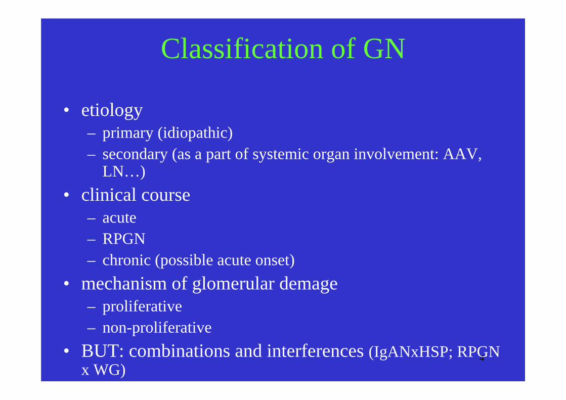
Classification of GN
• etiology– primary (idiopathic)– secondary (as a part of systemic organ involvement: AAV,
LN…)• clinical course
– acute– RPGN– chronic (possible acute onset)
• mechanism of glomerular demage– proliferative– non-proliferative
• BUT: combinations and interferences (IgANxHSP; RPGN x WG)

5
Mechanisms of glomerular damage1. glomerular inflamation – inflamatory pathways2. ultrastructural changes – non-inflamatory pathwas

6
Glomerular inflammation
1. Exsudation of neutrophils and/ormacrophages
2. Proliferation of mesangial and/orendothelial cells

7
Ultrastructural changes in non-proliferative vs. proliferative glomerulonephritides

8
Simplified classification of primary glomerulonephritides
1. Proliferative -mesangiopathies- IgA nephropathy- membranoproliferative GN
2. Nonproliferative - podocytopathies- minimal change disease- focal segmental glomerulosclerosis- membranous nephropathy
3. Diseases with endothelium as primary target- preeclampsia, HUS/TTP- type III and IV LN, - AAV

9
Clinical presentation• very variable• urinary findings
- HU: - glomerular (phase contrast!)- isolated vs. combined with PU; - micro vs. macro
- PU- low range vs. nephrotic- selective (albiminuria) vs. non-selective
- combination of HU + PU- (normal)
• renal function– normal vs. declined (rapid /RPGN/ or slow)
• BP– usually hypertension– normal
• nephritic vs. nephrotic conditions
10
11
Epidemiology – facts:• relatively rare disease (~ incidence biopticaly proven primary
GN 30-40/PMP)
• numerous subtypes
• significant part of GN unrecognised – „clinicaly silent“(incidental dg)
• important role of renal biopsy (cave: different local indicationpolicy)
• national (or large population) registries of RB
• variation: – geographical (e.g. IgAN: Asia – Europe – North America)– ethnicity (e.g. Indians)– population group (e.g. elderly vs. children, gender)
12
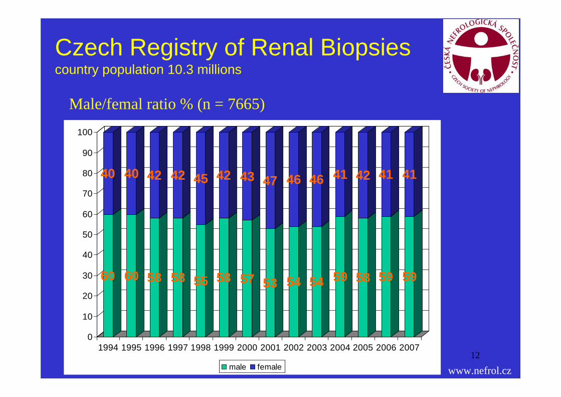
Czech Registry of Renal Biopsiescountry population 10.3 millions
www.nefrol.cz
Male/femal ratio % (n = 7665)
60
40
60
40
58
42
58
42
55
45
58
42
57
43
53
47
54
46
54
46
59
41
58
42
59
41
59
41
0
10
20
30
40
50
60
70
80
90
100
1994 1995 1996 1997 1998 1999 2000 2001 2002 2003 2004 2005 2006 2007
male female

13
Czech Registry of Renal Biopsies
www.nefrol.cz
Presence of erytrocyturia % (n = 7665)
64 67 64 66 66 67 68 63 5666 66 72 68
9 9 10 8 9 10 96
98 10 8 10
24 23 26 24 22 23 23 29 3525 23 18 21
0%
20%
40%
60%
80%
100%
1995 1996 1997 1998 1999 2000 2001 2002 2003 2004 2005 2006 2007
micro macro none not-reported

14
Czech Registry of Renal Biopsies
www.nefrol.cz
Presence of proteinuria % (n = 7665)
24 19 23 22 15 13 18 17 20 19 14
32
40 46 40 4642 40
44 48 46 4447
24
27 27 29 2630
3028 25 22 28 29
31
9 9 7 68 17 9 8 11 8 9 11
0%
10%
20%
30%
40%
50%
60%
70%
80%
90%
100%
1995 1996 1997 1998 1999 2000 2001 2002 2003 2004 2005 2007
none < 3g 4-10g >10g NA
15
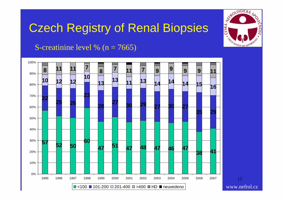
Czech Registry of Renal Biopsies
www.nefrol.cz
S-creatinine level % (n = 7665)
57 52 5060
47 51 47 48 47 46 4738 41
2225 26
23
2827 30 29 27 30 27
35 29
10 12 1210
13 13 11 13 14 14 14 15 16
8 11 11 78 7 11 7 9 9 9 9 11
0%
10%
20%
30%
40%
50%
60%
70%
80%
90%
100%
1995 1996 1997 1998 1999 2000 2001 2002 2003 2004 2005 2006 2007
<100 101-200 201-400 >400 HD neuvedeno

16
Czech Registry of Renal Biopsies
www.nefrol.cz
Presence of arterial hypertension % (n = 7665)
57 54 63 65 5646 63 48 64 47
40 31 32
24 3334 41 31
38 57 45 55 4964 50 67
0%
20%
40%
60%
80%
100%
1995 1996 1997 1998 1999 2000 2001 2002 2003 2004 2005 2006 2007
normo hyper NA
17
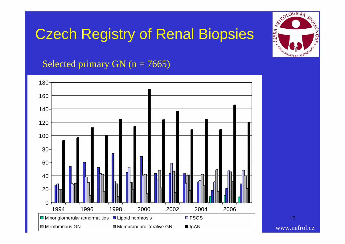
Czech Registry of Renal Biopsies
www.nefrol.cz
Selected primary GN (n = 7665)
0
20
40
60
80
100
120
140
160
180
1994 1996 1998 2000 2002 2004 2006Minor glomerular abnormalities Lipoid nephrosis FSGS
Membranous GN Membranoproliferative GN IgAN
18
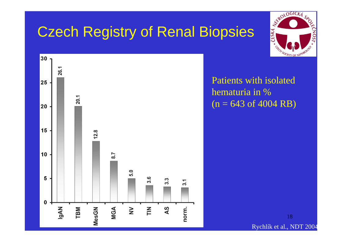
Czech Registry of Renal Biopsies
Rychlík et al., NDT 2004
Patients with isolatedhematuria in %(n = 643 of 4004 RB)
19
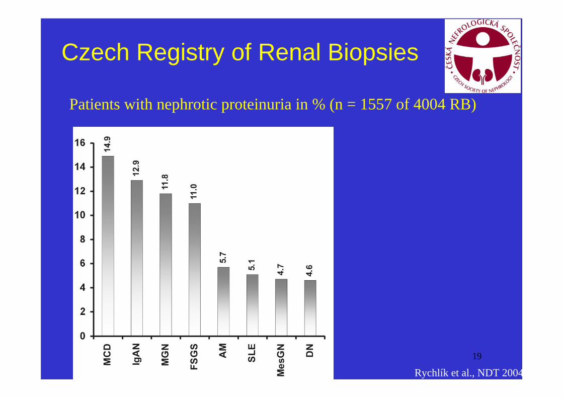
Czech Registry of Renal Biopsies
Rychlík et al., NDT 2004
Patients with nephrotic proteinuria in % (n = 1557 of 4004 RB)
20
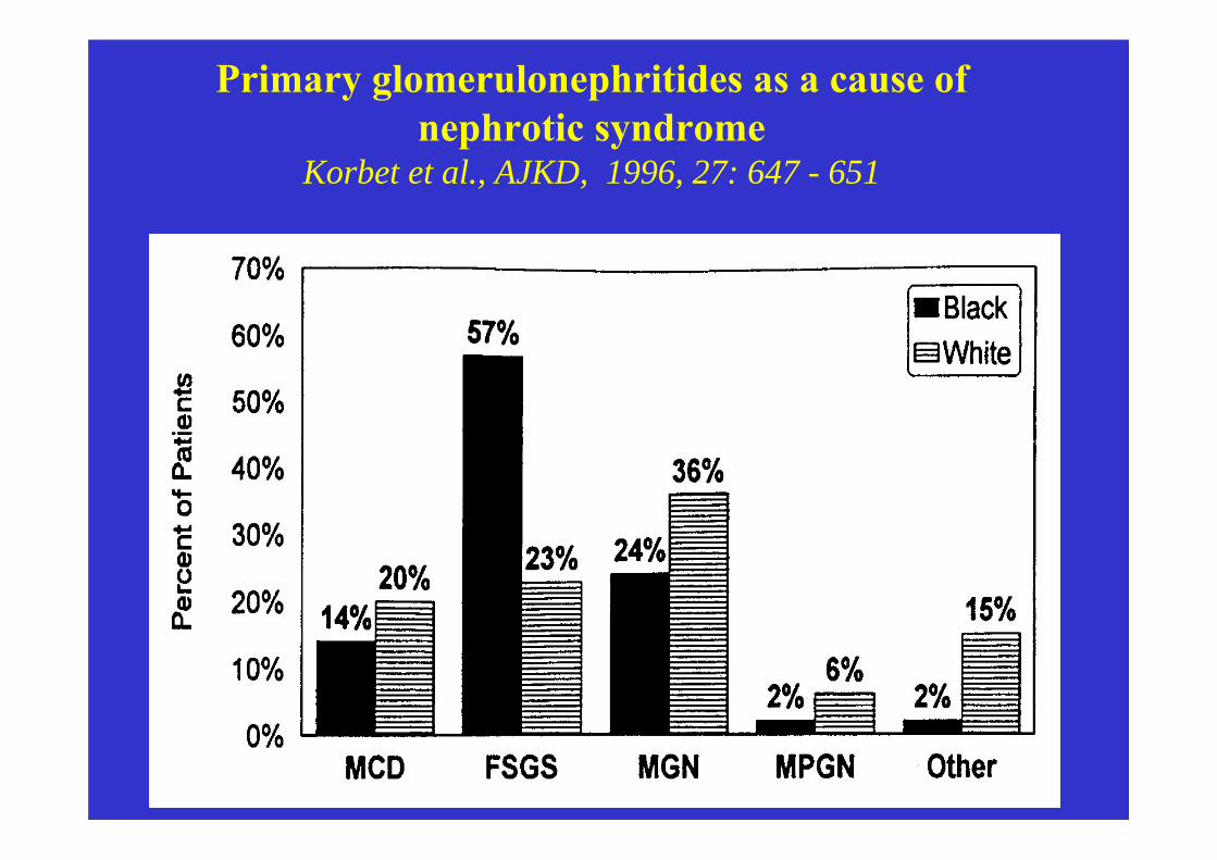
Primary glomerulonephritides as a cause of nephrotic syndrome
Korbet et al., AJKD, 1996, 27: 647 - 651
21
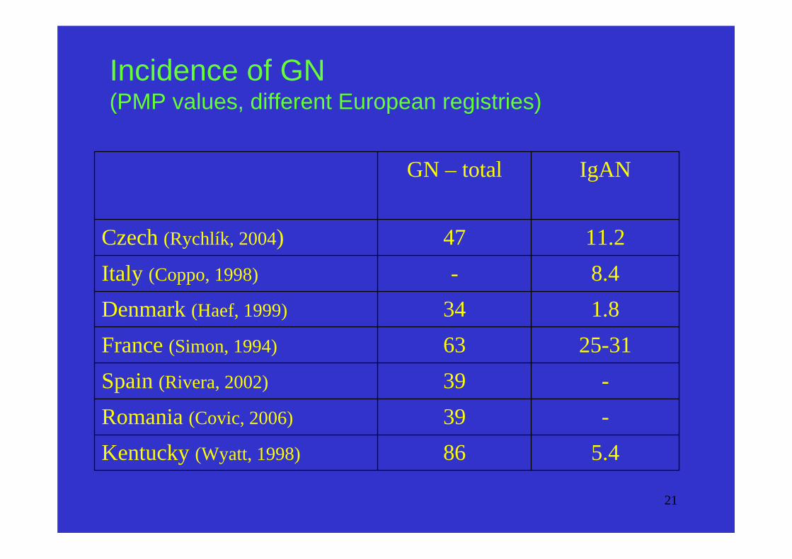
Incidence of GN (PMP values, different European registries)
5.486Kentucky (Wyatt, 1998)
-39Romania (Covic, 2006)
-39Spain (Rivera, 2002)
25-3163France (Simon, 1994)
1.834Denmark (Haef, 1999)
8.4-Italy (Coppo, 1998)
11.247Czech (Rychlík, 2004)
IgANGN – total
22
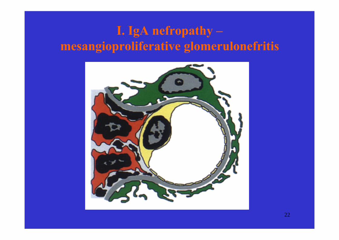
I. IgA nefropathy –mesangioproliferative glomerulonefritis

23
IgA nephropathy

24
IgA nephropathy

25
IgA nephropathy

26
IgA nephropathy – clinical features
1. the most common glomerulonephritis in Europe(20-40% out of all primary GN)
2. typical clinical presentation:• asymptomatic microscopic hematuria or
• episodes of parainfectious macroscopic hematuria
• young males
3. outcome of untreated IgA nephropathy is not benign – at least 20% of pts develop ESRD during20 years

27
IgA nephropathy –negative prognostic factors
a. clinical- hypertension- proteinuria (> 1 g/24 hrs)- decreased GFR at presentation
b. histologic- glomerulosclerosis- interstitial fibrosis- vascular sclerosis
28
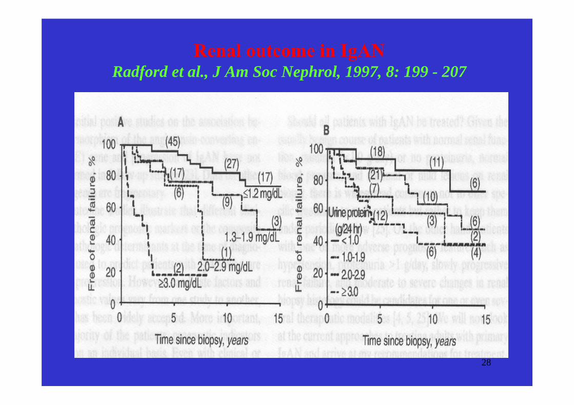
Renal outcome in IgAN Radford et al., J Am Soc Nephrol, 1997, 8: 199 - 207
29
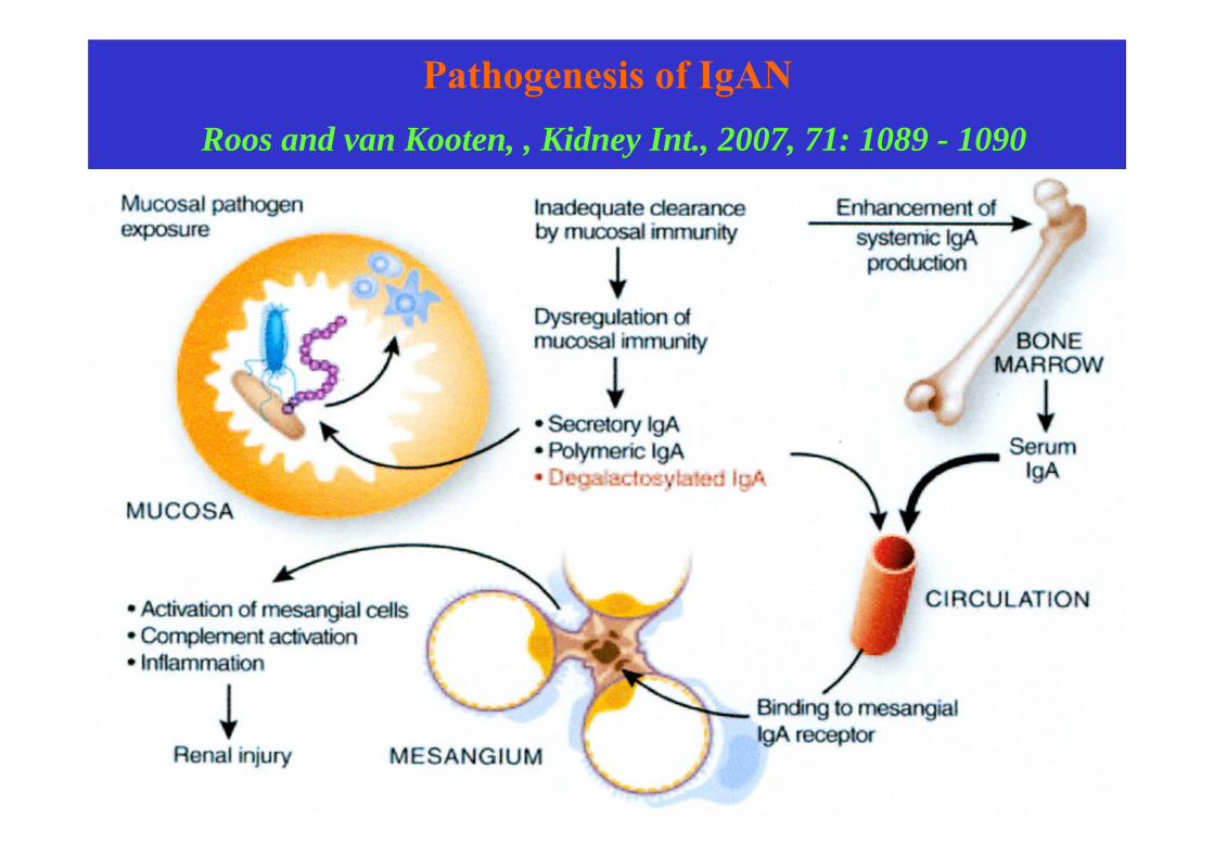
Pathogenesis of IgANRoos and van Kooten, , Kidney Int., 2007, 71: 1089 - 1090

30
Hinge region of human IgA1 and IgA2Mestecky et al., Kidney Blood Press Res, 2008, 31: 29 - 37

31
Pathogenesis of IgANMestecky et al., Kidney Blood Press Res, 2008, 31: 29 - 37

32
IgA nephropathy - treatment1. optimal control of blood pressure using
ACEI and/or AIIA2. fish oil in pts with slowly pregressive renal
insufficiency3. corticosteroids in proteinuric pts with normal
or only slightly decreased RF4. cytotoxics in pts with progressive renal
insufficiency
33
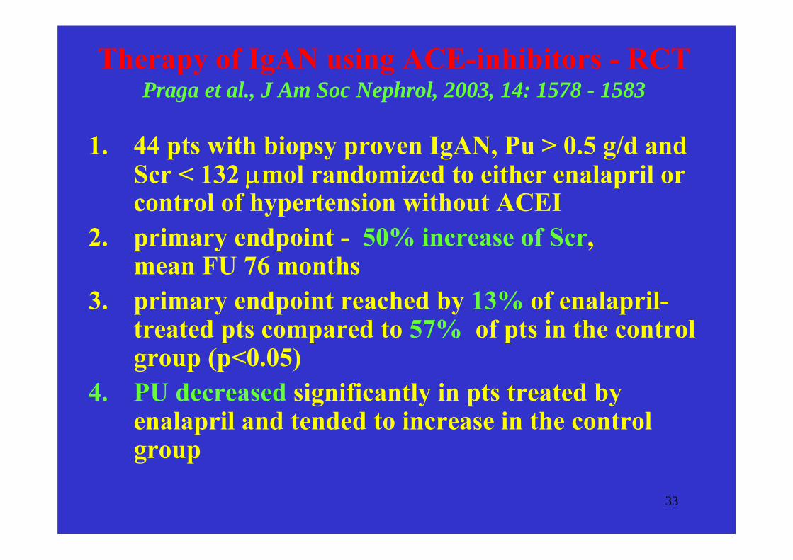
Therapy of IgAN using ACE-inhibitors - RCT Praga et al., J Am Soc Nephrol, 2003, 14: 1578 - 1583
1. 44 pts with biopsy proven IgAN, Pu > 0.5 g/d andScr < 132 μmol randomized to either enalapril orcontrol of hypertension without ACEI
2. primary endpoint - 50% increase of Scr, mean FU 76 months
3. primary endpoint reached by 13% of enalapril-treated pts compared to 57% of pts in the controlgroup (p<0.05)
4. PU decreased significantly in pts treated by enalapril and tended to increase in the controlgroup

34
The effect of fish oil on renal function in IgANStrippoli et al., Am J Kidney Dis, 2003, 41: 1129 - 1139
35
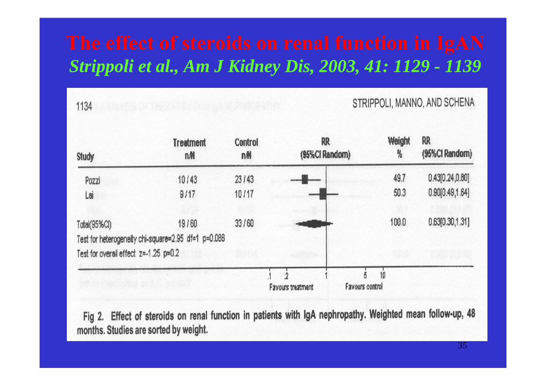
The effect of steroids on renal function in IgANStrippoli et al., Am J Kidney Dis, 2003, 41: 1129 - 1139

36
Corticosteroids in IgAN: long-term outcome
Pozzi et al., J Am Soc Nephrol, 2004, 15: 157 - 163
1. secondary analysis of multicentric RCT -86 pts with IgAN treated 6 m with MP and Pred or only by symptomatic treatment
2. 10-yr renal survival significantly better in pts treated by steroids (97% vs. 53%, p=0.0003)
3. PU decreased in pts who did not reach the doubling of Scr and increased in pts withprogressive renal insufficiency

37
The effect of cytotoxics on renal function in IgANStrippoli et al., Am J Kidney Dis, 2003, 41: 1129 - 1139

38
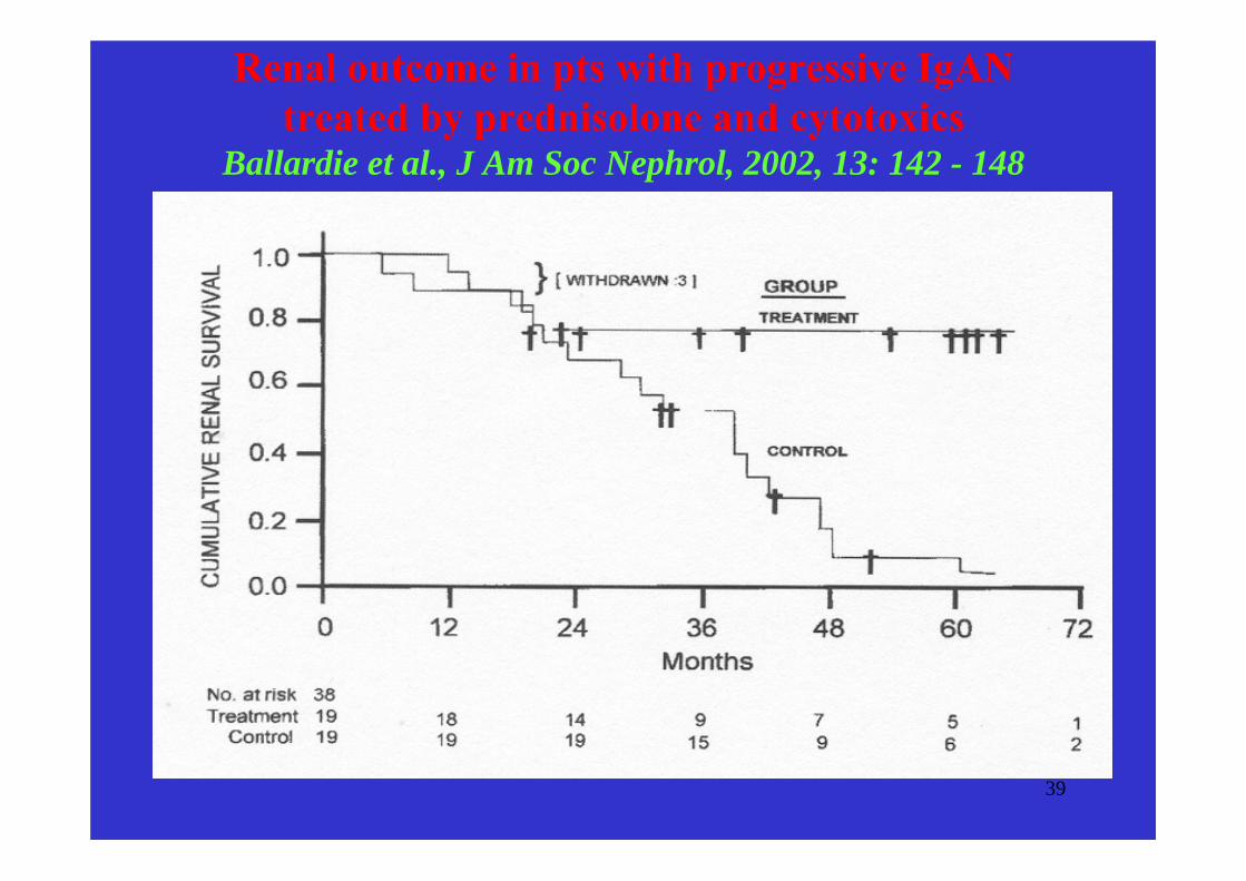
Rate of GFR loss in pts with progressive IgAN treated 2-yrs by CS and cytotoxics (CPH→AZA)
Ballardie et al., J Am Soc Nephrol, 2002, 13: 142 - 148
39
Renal outcome in pts with progressive IgAN treated by prednisolone and cytotoxics
Ballardie et al., J Am Soc Nephrol, 2002, 13: 142 - 148

40
Relative efficacy of IgAN treatment optionsLaville and Alamartine, NDT, 2004, 71: 1947 - 1951

41
Evidence-based recommendations in IgAN Floege, Nephrol Dial Transplant, 2003, 18: 241 - 245
1. in pts with PU < 1.5 g/d and normal GFR steroids may decrease Pu, but their long-term effect remains uncertain
2. in pts with PU 1 – 3.5 g/d and preserved renalfunction - 6-months treatment withcorticosteroids
3. in pts with progressive renal insufficiencyinsuficiencí and Scr < 250 μmol/l - treatmentwith corticosteroids and cytotoxics
42
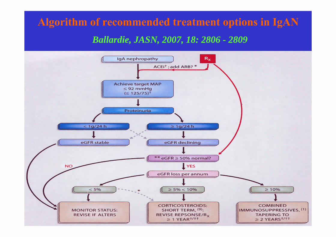
Algorithm of recommended treatment options in IgANBallardie, JASN, 2007, 18: 2806 - 2809

43
II. Podocytopathies
Damage to the glomerular capillary wall resultingin:
1. nephrotic selective proteinuria - minimal change disease
2. nephrotic non-selective proteinuria withmicroscopic hematuria
- focal segmental glomerulosclerosis- idiopathic membranous nephropathy

44
Podocytopathies
Damage to the podocyte caused by:1. antipodocyte antibodies
- membranous nephropathy
2. Genetic, viral, toxic and immunologicmechanisms
- focal segmental glomerulosclerosis- minimal change disease

45
IIa. Membranous nephropathy

46
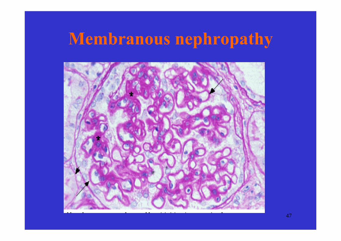
Membranous nephropathy
47
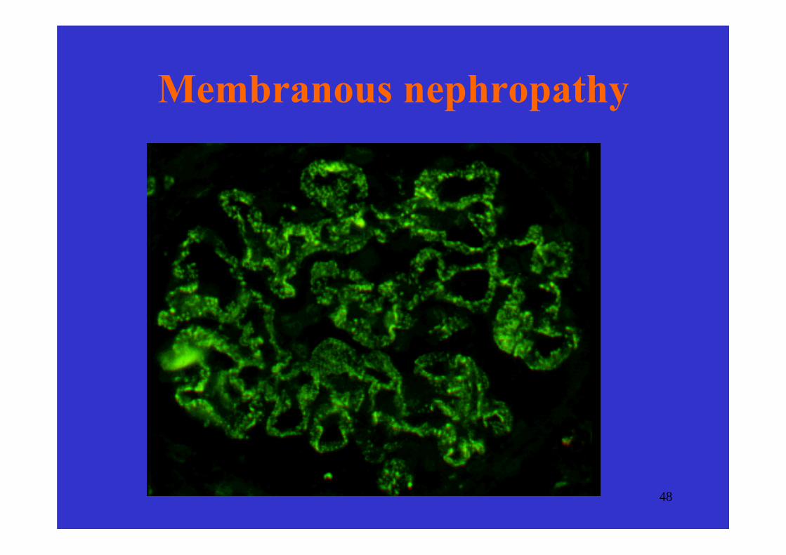
Membranous nephropathy
48
Membranous nephropathy

49
Membranous nephropathy

50
Membranous nephropathy

51
Membranous nephropathy1. Secondary – planted antigens?
- infections(hepatitis B, syphilis, malaria)
- drugs(organic gold, penicillamine, NSAID)
- neoplasms(carcinomas, e.g. Colon, lung, orstomach, and lymphomas)
- systemic lupus erythematosus2. Idiopathic – antibodies directed to podocyte
antigens?

52
Idiopathicmembranous nephropathy
1. very common - 15-25% of adultnephrotic syndrome
2. Nephrotic proteinuria in about 80% of pts
3. Microscopic hematuria common4. Hypertension and chronic renal failure
are uncommon at presentation, but maydevelop during follow-up

53
Pathogenesis of IMN
1. Experimental model of IMN – Heymann’snephritis in rats
- antibodies against megalin(not expressed by human podocytes)
2. Antenatal membranous nephropathy in a child of a woman with truncating mutations of MME(metallomembrane endopeptidase) gene
- alloimmunisation against NEP3. Common IMN – supposedly antibodies against
other podocyte proteins
54
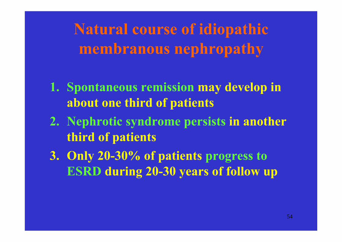
Natural course of idiopathicmembranous nephropathy
1. Spontaneous remission may develop in about one third of patients
2. Nephrotic syndrome persists in anotherthird of patients
3. Only 20-30% of patients progress to ESRD during 20-30 years of follow up

55
High incidence of remission in untreatedmembranous nephropathy
Mosconi et al., NEJM, 1993

56
Outcome of nephrotic vs. non-nephrotic pts with IMN Yoshimoto et al., Kidney Int., 2004, 65: 148 - 153
A- renal survival B-renal and patient death

57
Treatment of IMN

58
Corticosteroids vs. placebo in adults with IMN Muihead, Kidney Int., 1999, 55 (Suppl. 70): S-47 –S-55
Study No of pts Duration Outcome
CSAINS, 1979 72 8-12 w more CR and PR
RF preserved
Cameron,1990 107 8 w no difference
Cattran, 1989 158 6 mo no difference

59
Conservative vs. IS treatment in IMNTorres et al., Kidney Int., 2002, 61: 219 - 227
Retrospective study - 20 pts conservative (CON), 19 pts - CS and CHLB (IST)
At the end of FU:
CON: 65% dialyzed, 10% advanced RF, 25% death
IST: 58% stable renal function, 38% CR or PR
IST - better 4-year (90% vs. 55%) and 7-year renalsurvival (90% vs. 20%)

60
Serum creatinine in progressive IMN on conservative vs. immunosuppresive treatment
Torres et al., Kidney Int., 2002, 61: 219 - 227
61
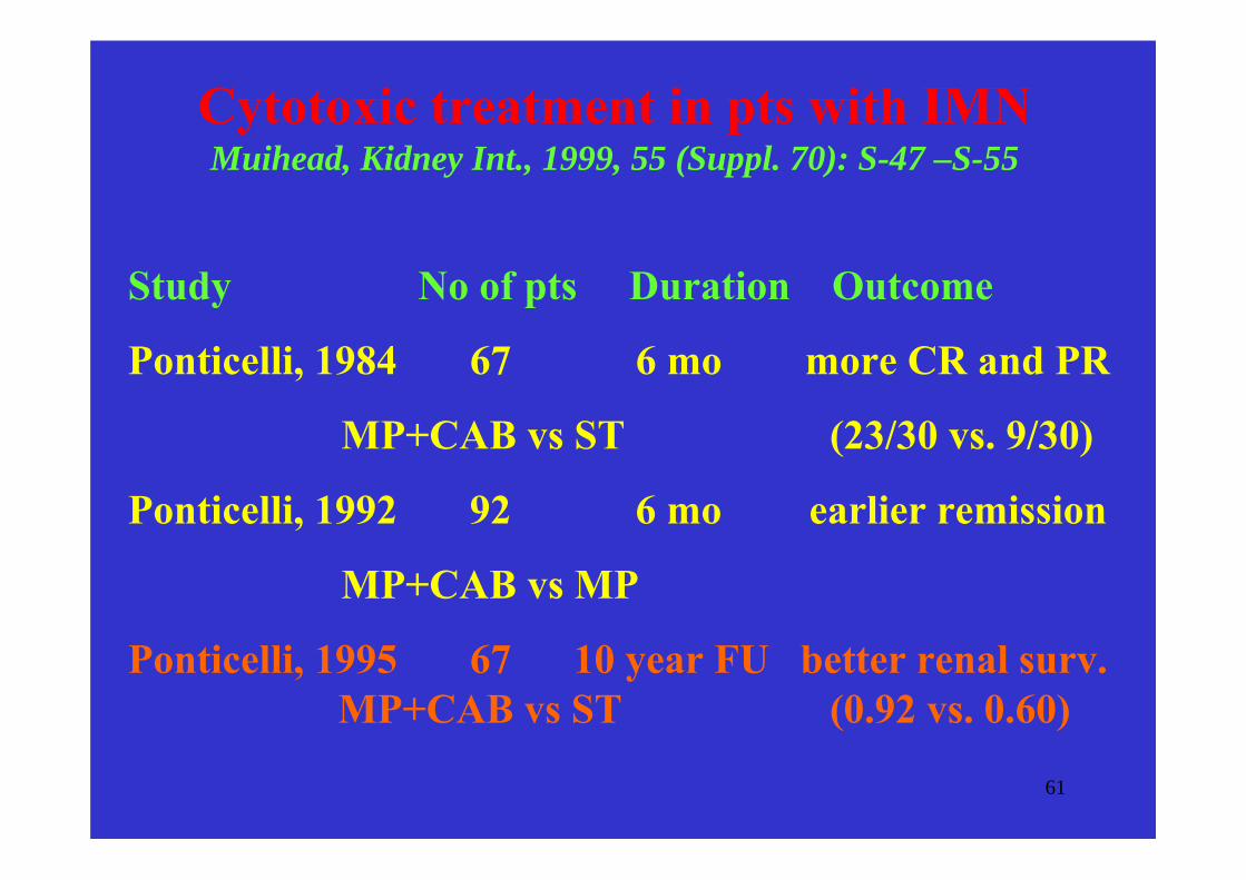
Cytotoxic treatment in pts with IMNMuihead, Kidney Int., 1999, 55 (Suppl. 70): S-47 –S-55
Study No of pts Duration Outcome
Ponticelli, 1984 67 6 mo more CR and PR
MP+CAB vs ST (23/30 vs. 9/30)
Ponticelli, 1992 92 6 mo earlier remission
MP+CAB vs MP
Ponticelli, 1995 67 10 year FU better renal surv. MP+CAB vs ST (0.92 vs. 0.60)

62
Efficacy of different regimens in membranousnephropathy
Ponticelli et al., NEJM, 1992
63
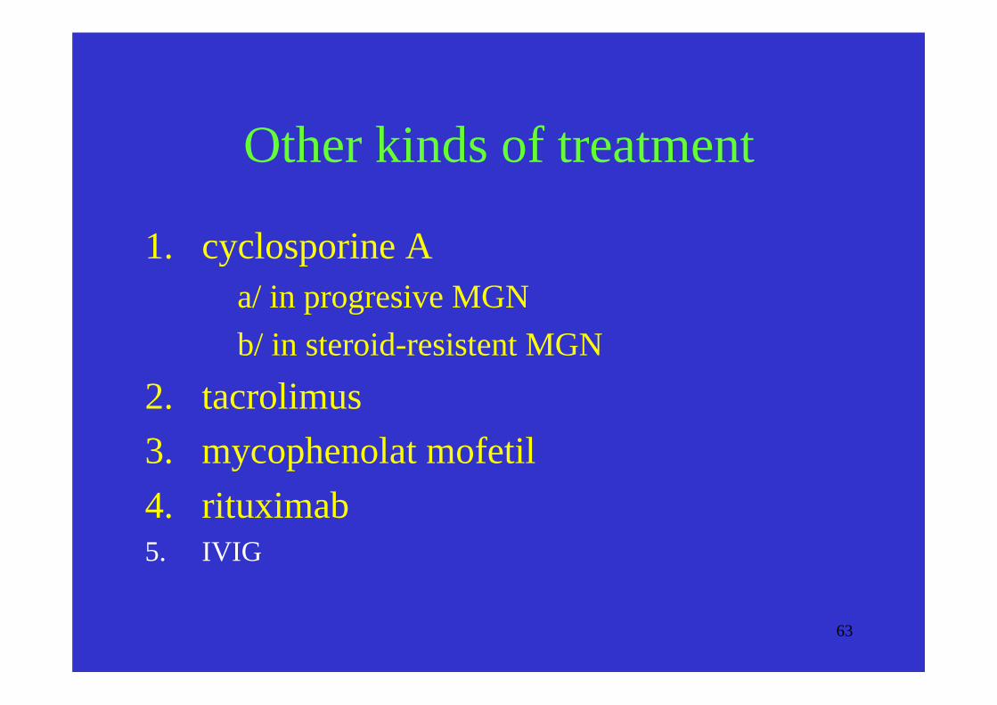
Other kinds of treatment
1. cyclosporine Aa/ in progresive MGNb/ in steroid-resistent MGN
2. tacrolimus3. mycophenolat mofetil4. rituximab5. IVIG

64
1.a/ Cyclosporine in progressive IMNCattran et al., Kidney Int., 1995, 47: 1130 - 1135
- 64 pts followed without treatment for 12 m
- 17 pts - decline of GFR (> 8 ml/min/yr and persistent nephrotic PU)
were randomized to 12-m:
1. CyA (9 pts) or 2. placebo (8 pts)
Proteinuria and rate of decline of GFR decreasedonly in CyA

65
Cyclosporine in progressive IMNCattran et al., Kidney Int., 1995, 47: 1130 - 1135
-2.1-2.2Placebo
-0.7-2.4CyA
AfterbeforeGFR (ml/min/m)
9.212.8Placebo
4.511.5CyA
AfterbeforePU (g/day)

66
1.b/ Cyclosporine in steroid-resistant MNCattran et al., Kidney Int., 2001, 59: 1484 - 1490
51 pts still nephrotic on high-dose CS for 8 weeks
Randomized to 26 w of:
1) CyA and low-dose CS,
2) placebo and low dose CS
Mean follow-up was 78 weeks

67
% Remission of NS in pts with steroid-resistant MN treated by CyA
(26-w treatment)Cattran et al., Kidney Int., 2001, 59: 1484 - 1490
68
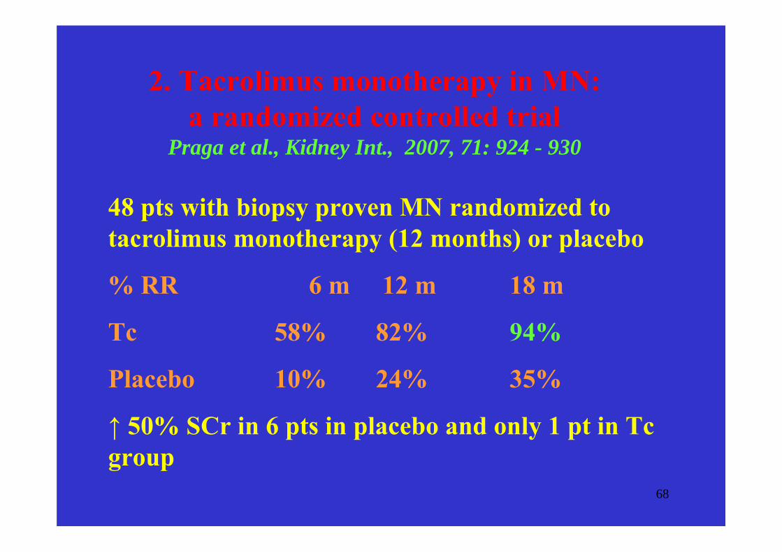
2. Tacrolimus monotherapy in MN: a randomized controlled trial
Praga et al., Kidney Int., 2007, 71: 924 - 930
48 pts with biopsy proven MN randomized to tacrolimus monotherapy (12 months) or placebo
% RR 6 m 12 m 18 m
Tc 58% 82% 94%
Placebo 10% 24% 35%
↑ 50% SCr in 6 pts in placebo and only 1 pt in Tcgroup

69
3. Mycophenolate mofetil in IMN: compared with a historic group treated with CPH
Branten et al., AJKD., 2007, 50: 248 - 256
32 pts with IMN treated 1 yr by MMF (2g/day) compared with 32 historical controls treated also 1 yr by CPH (1.5 mg/kg/day), the steroid regimen the same in both limbs
PU 0 m 12 m Cr 0m 12 m
MMF 8.4 1.4 g/24h 159 124 umol/l
CPH 9.2 1.1 g/24h 159 115 umol/l

70
4.a Rituximab in IMN, who can benefit from the treatment
Remuzzi et al., Lancet, 2002, 360: 923 – 924,Ruggenenti et al., CJASN, 2006, 1: 738 – 748.
8 pts with IMN treated with RTX (375 mg/m2)
- PU decreased during 4 m from 8.6 to 3.8 g/24h
- Therapy was not effective in pts with tubularatrophy and interstitial fibrosis

71
4. Rituximab in IMN, comparison of standard and B-cell titrated regimen
Cravedi et al., CJASN, 2007, 2: 932 - 937
12 new incident IMN pts treated by B-cell driven RTX compared to 24 historical controls (standard RTX protocol – 4x 375 mg/m2)

72
Immunosuppressive treatment for IMN: a systematic review
Perna et al., Am. J. Kidney Dis., 2004, 44: 385 - 401
1. Systematic review of RCT in IMN in adults with NS followed for at leats 6 months
2. Four therapeutic studies identified: steroids alone, alkylating agents, calcineurin inhibitors, antiproliferative agents
3. With the exception of a beneficial effect of alkylating agents on complete remission no positive influence of IST on the outcome of the patients withIMN was documented
73
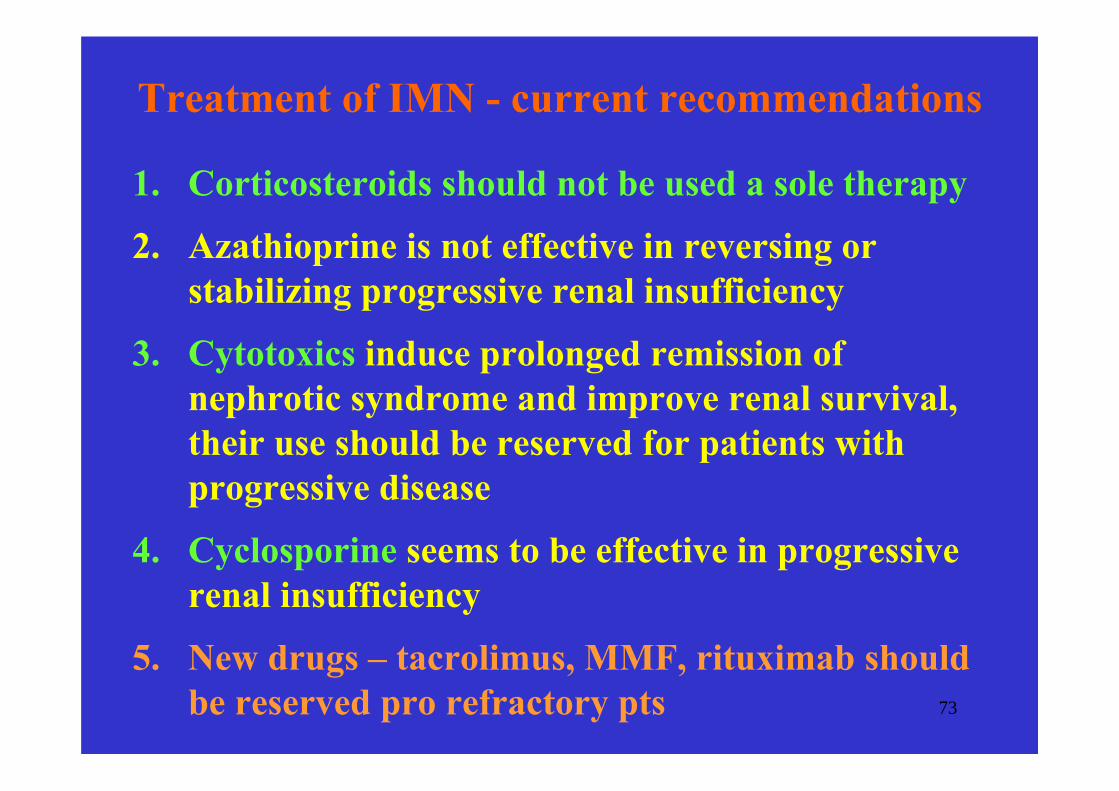
Treatment of IMN - current recommendations
1. Corticosteroids should not be used a sole therapy
2. Azathioprine is not effective in reversing orstabilizing progressive renal insufficiency
3. Cytotoxics induce prolonged remission of nephrotic syndrome and improve renal survival, their use should be reserved for patients withprogressive disease
4. Cyclosporine seems to be effective in progressiverenal insufficiency
5. New drugs – tacrolimus, MMF, rituximab shouldbe reserved pro refractory pts

74
Guidelines for the treatment of IMN Cattran, Kidney Int., 2001, 59: 1983 - 1994

75
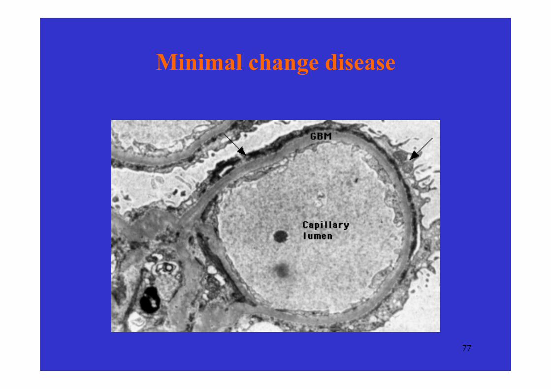
IIb. Minimal change disease

76
Minimal change disease
77
Minimal change disease

78
MCD – clinical presentation
1. nephrotic syndrome with selectiveproteinuria
2. uncommon: hematuria, hypertension and reduced renalfunction

79
Minimal change disease-prevalence among nephrotic patients
Children - 85 – 95%Young adults - 50%Adults > 40 years - 20 – 25%

80
Classification of patients with minimalchange disease based on response to
corticosteroids
1. Steroid responsive (sensitive)develop complete remission of proteinuria within 8 – 12 weeks of treatment(in adults remission should develop within 16 weeks)
2. Steroid dependentdevelop relapse during tapering of steroidsor within 2 weeks after cessation of therapy
3. Steroid resistantfail to respond to steroid treatment at all
81
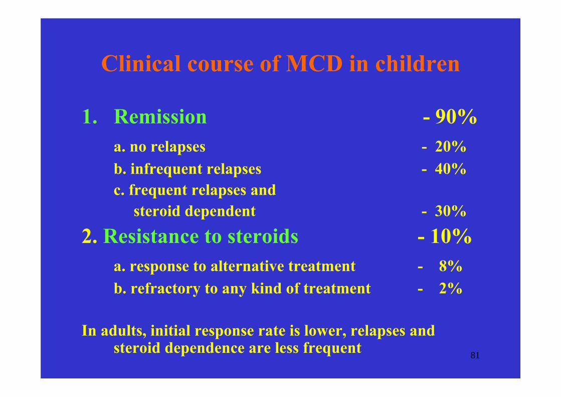
Clinical course of MCD in children
1. Remission - 90%a. no relapses - 20%b. infrequent relapses - 40%c. frequent relapses and
steroid dependent - 30%
2. Resistance to steroids - 10%a. response to alternative treatment - 8% b. refractory to any kind of treatment - 2%
In adults, initial response rate is lower, relapses and steroid dependence are less frequent
82
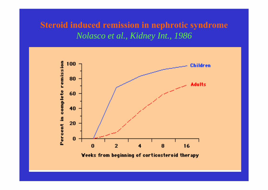
Steroid induced remission in nephrotic syndromeNolasco et al., Kidney Int., 1986

83
Therapy of MCD in children –current recommendations
1. Initially course of prednisone 60 mg/m2 for 4-6 weeks with 40 mg/m2 every alternate day for another 4-6 weeks
2. Relapses treated in a similar way, buttapering of prednisone starts when urine becomes protein free
3. Frequent relapsers and steroid dependentpatients treated either by cyclophosphamide 2 mg/kg/day for 8 weeks or by cyclosporine5 mg/kg/day for 6-12 months
4. Treatment of steroid resistant patients isusually unsatisfactory

84
Therapy of MCD –modifications in adults
1. Initially course of prednisone 1mg/kg for 8-16 weeks or for one week after remission isachieved, then several weeks (one month) 1 mg/kg on alternate days, thereaftercorticosteroids are slowly tapered during severalmonths
2. Relapses treated in a similar way3. Frequent relapsers and steroid dependent
patients treated either by CPH 2 mg/kg/dayfor 8 weeks or by CyA 5 mg/kg/day for 6-12 months
4. Treatment of steroid resistant patients isusually unsatisfactory

85
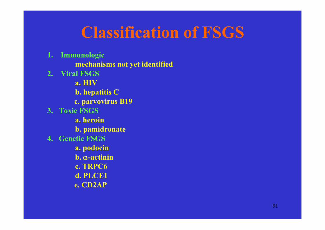
IIc. Focal segmental
glomerulosclerosis

86
Etiology of FSGS1. Primary FSGS
a. classical variantb. glomerular tip lesionc. collapsing glomerulopathy
2. Secondary FSGSa. healing focal lesions (FSGN)b. hyperfiltration in residual nephrons
- agenesis of one kidney- vesicoureteral reflux- morbid obesity
c. damage to epithelial cells- HIV nephropathy- heroin nephropathy
87
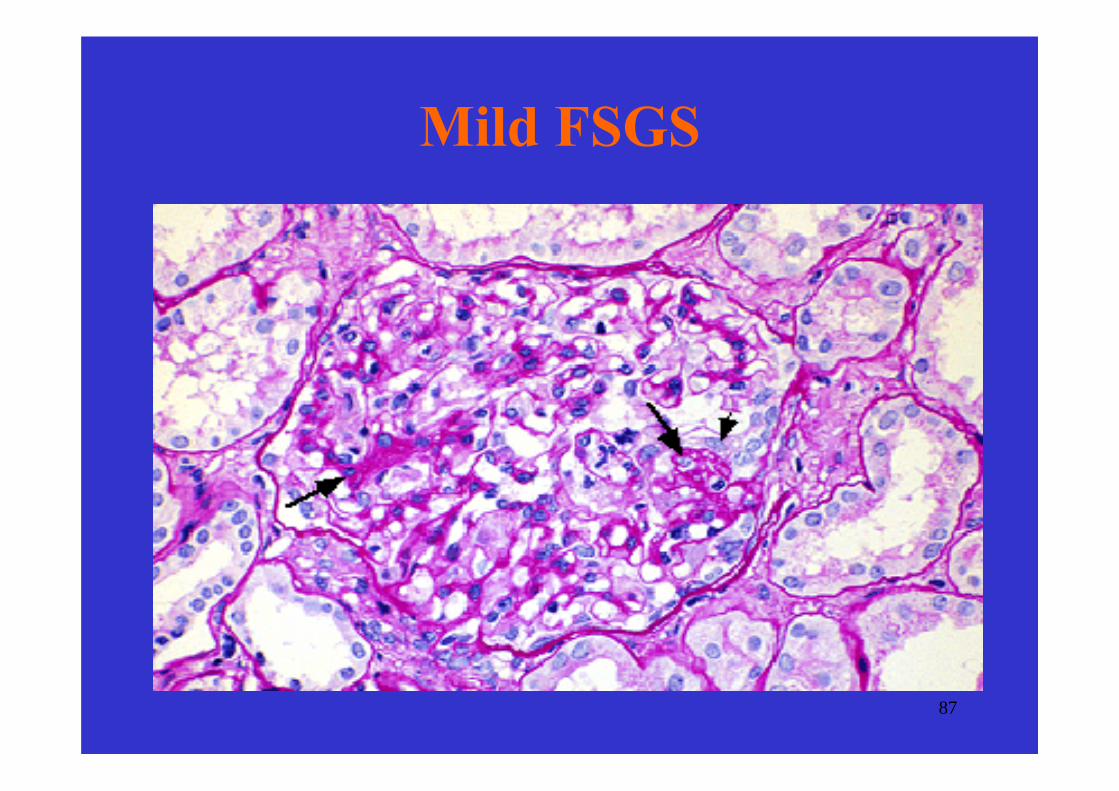
Mild FSGS

88
Moderate FSGS

89
Tip lesion in early FSGS

90
Collapsing FSGS
91
Classification of FSGS1. Immunologic
mechanisms not yet identified2. Viral FSGS
a. HIVb. hepatitis Cc. parvovirus B19
3. Toxic FSGSa. heroinb. pamidronate
4. Genetic FSGSa. podocinb. α-actininc. TRPC6d. PLCE1e. CD2AP

92
Proposed taxonomy of the podocytopathies
Barisoni et al., Clin. J. Am. Sco. Nephrol., 2007, 2: 529 - 542

93
Pathogenesis of primary FSGS1. Circulating permeability factors
a. imunoglobulin, or Ig-like moleculeb. protein of MW about 30-50 kDa c. factor inhibiting inducible NO synthase in mesangial cells
(hemopexin)
2. Deficient inhibitors of permeability factors lost in urineapolipoproteins of HDL complex(e.g. apo J, apo E2 and apo E4)
3. Late onset congenital FSGSdeficiency of podocyte proteins(podocin, α-actinin, CD2AP, et al.)
94
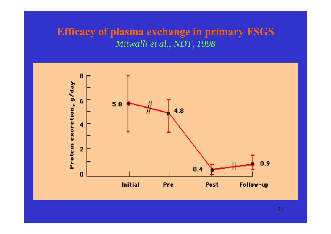
Efficacy of plasma exchange in primary FSGSMitwalli et al., NDT, 1998

95
Identified nonsyndromic FSGS/NS genes
Disease Locus Inherit. Gene Protein
Congenital NS 19q13.1 AR NPHS1 NephrinSRNS 1q25-32 AR NPHS2 PodocinFSGS1 19q13 AD ACTN4 α-actininFSGS2 11q21-22 AD FSGS2 TRPC6FSGS3 6q AD, AR FSGS3 CD2APDMS 10q23.32-24.1 AR NPHS3 PLCE1 SSNS1 2p AR SSNS1 unknown

96
FSGS – clinical presentation
1. Asymptomatic proteinuria or full blownnephrotic syndrome
2. Hypertension, microscopic hematuriaand decreased renal function common
3. Slowly progressive disease – 50% 10-year renal survival
4. Sclerosis of segments of glomerular tuft

97
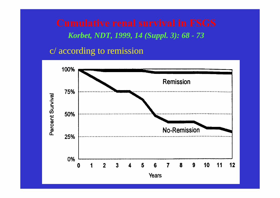
Cumulative renal survival in FSGS Korbet, NDT, 1999, 14 (Suppl. 3): 68 - 73
a/ according to base-line proteinurie

98
Cumulative renal survival in FSGS Korbet, NDT, 1999, 14 (Suppl. 3): 68 - 73
b/ according to base-line renal function
99
Cumulative renal survival in FSGS Korbet, NDT, 1999, 14 (Suppl. 3): 68 - 73
c/ according to remission

100
Immunosupresive treatment of FSGS
1. corticosteroids2. cyclosporin A3. novel drugs
– tacrolimus– mycophenolat mofetil– rituximab

101
1. Corticosteroids in adults with FSGSFranceschini et al., Seminars in Nephrology, 2003, 23: 229 - 233
Study No of pts Duration CR Relapse
Aggarwal, 1993 38 6 mo 31% NA
Rydel, 1995 30 5.5 mo 33% 67%
Cattran, 1998 17 5-6 mo 47% 25%
Ponticelli, 1999 53 6 mo 40% 40%
Chitalia, 1999 28 3 mo 21% NA
102
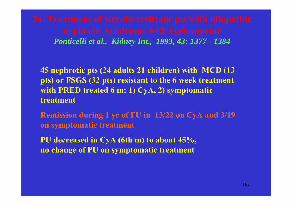
2a. Treatment of steroid-resistant pts with idiopathicnephrotic syndrome with cyclosporine
Ponticelli et al., Kidney Int., 1993, 43: 1377 - 1384
45 nephrotic pts (24 adults 21 children) with MCD (13 pts) or FSGS (32 pts) resistant to the 6 week treatmentwith PRED treated 6 m: 1) CyA, 2) symptomatictreatment
Remission during 1 yr of FU in 13/22 on CyA and 3/19 on symptomatic treatment
PU decreased in CyA (6th m) to about 45%, no change of PU on symptomatic treatment

103
Proteinuria in steroid-resistant pts with INS treatedby CyA or symptomatic treatment
Ponticelli et al., Kidney Int., 1993, 43: 1377 - 1384

104
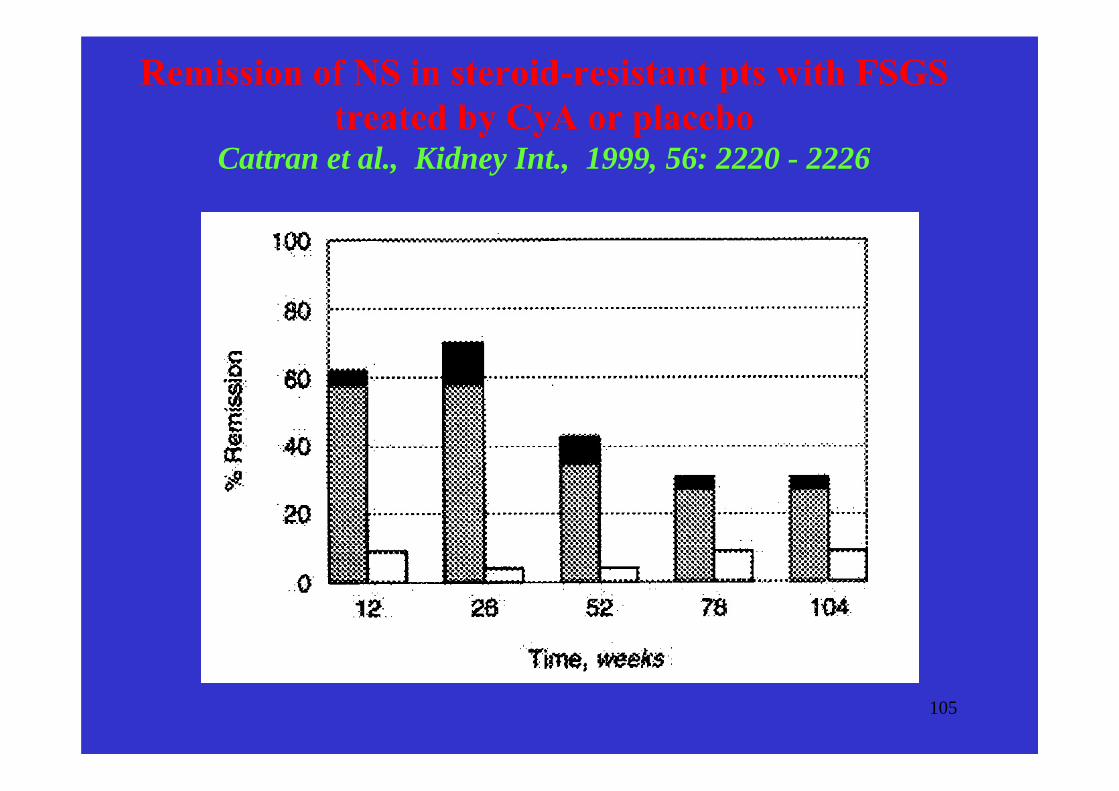
2b.Treatment of steroid-resistant FSGS with CyACattran et al., Kidney Int., 1999, 56: 2220 - 2226
49 adult FSGS resistant to 8 w Pred treated for 26 w: → A. CyA and low-dose CS or→ B. placebo and low-dose CS FU at least 200 w
Results:26 w:
PR and CR - CyA 70%, placebo 4%
78 w:Relapse - 40%, resp. 60% of responding pts50% CCr decreased in 25% CyA pts, in 52%
placebo pts
105
Remission of NS in steroid-resistant pts with FSGS treated by CyA or placebo
Cattran et al., Kidney Int., 1999, 56: 2220 - 2226

106
3a. Treatment of FSGS in adultwith tacrolimus monotherapy
Duncan et al., Nephrol Dial Transplant, 2004, 19: 3062 - 7
6 naive FSGS nephrotic pts treated by tacrolimus - all in remission after 6.5 months- Pu 11.0 vs 2.8 g/day)
5 FSGS pts in remission on CyA with worseningRF switched to tacrolimus - Pu further improved, slightly improved RF

107
3b. Mycophenolate mofetil in FSGSCattran et al., Clin. Nephrol., 2004, 62: 405 - 411
18 FSGS pts resistant to CS (75% also to CPH or CyA) switched to MMF
6 m: substantial improvement of Pu in 44% (8/18)
1 yr post treatment, Improvement sustained in 50% (4/8), no pt CR
No deterioration of RF during treatment, 3 pts progressed to CRF during FU

108
Mycophenolate mofetil in FSGSSegarra et al., NDT, 2007, 22: 1351 - 1360
Among 98 pts with primary GN 22 pts with FSGStreated by MMF
CR in 2 pts, PR in 10 pts, response rate 54%
Median time to response 150 days

109
3c. Rituximab and FSGSAhmed and Wong., NDT, 2008, 23: 11 - 17
Only case reports both in post-transplant FSGS and FSGS in native kidneys(3 children)
Overal success rate in FSGS 8/12 and 7/7 in MCD.

110
Treatment of primary FSGS –current recommendations
1. Response to corticosteroids may increase from only 10-30% up to 60% with longer treatment with higher dose (60 mg/m2 at least 3 months, patients should be consideredsteroid resistant after 6 months)
2. Cyclosporine may reduce proteinuria and lower the risk of progression to ESRD even in steroid resistant patients, treatment should be long (at least 6 months), relapses aftercyclosporine withdrawal are common
3. Cytotoxics remain only second-line therapy, the evidence for their effect in steroid resistant patients is not conclusive
4. Newer drugs – tacrolimus, sirolimus, MMF, rituximabshould be reserved only for refractory pts

111
Podocytes and slit diaphragms

112
Glomerular capillary wallDeen, J.Clin.Invest., 2004, 1412 - 1414
113
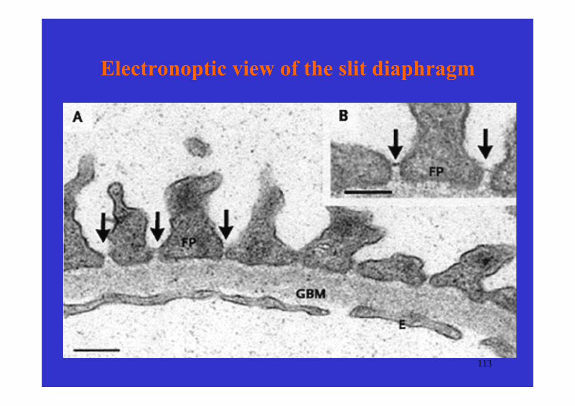
Electronoptic view of the slit diaphragm

114
Slit diaphragm in congenital nephrotic syndrome (D)

115
Nephrin and slit diaphragm
116
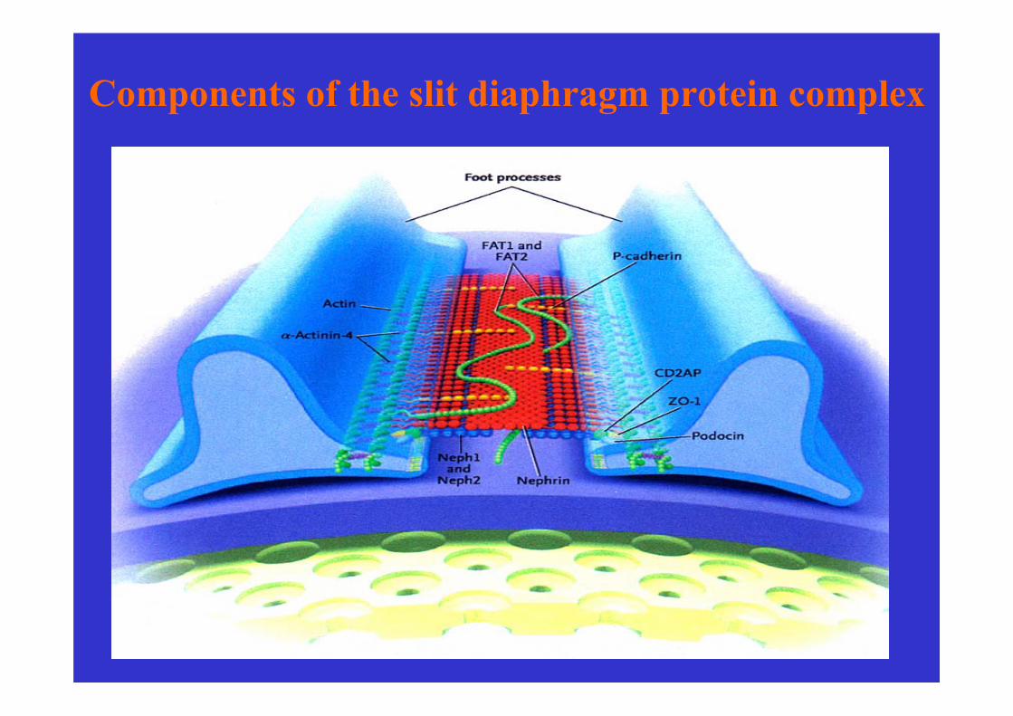
Components of the slit diaphragm protein complex
117
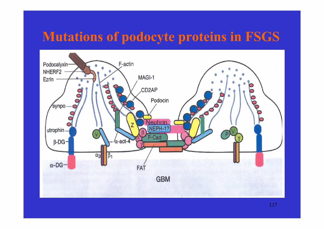
Mutations of podocyte proteins in FSGS

118
Major causes of podocyte effacement
1. Slit diaphragm and its lipid raftnephrin, podocin, TRPC6, CD2AP
2. Podocyte cytoskeletonα-actinin
3. Adhesion of podocyte to GBMβ -dystroglycan, β1-integrins
4. Loss of podocyte electronegative chargepodocalyxin

119
Conclusions - podocytopaties
1. recent progress in the podocyte biology may resultin the elucidation of the pathogenesis of the commonacquired podocytopathies
2. direct effect of different drugs on podocytes maychange the current paradigm that the reduction of proteinuria is exerted either by the immunosuppressive or hemodynamic effect
3. better understanding of the podocyte biology maylead to the discovery of new drugs aimed atpreservation of the glomerular capillary wall
120
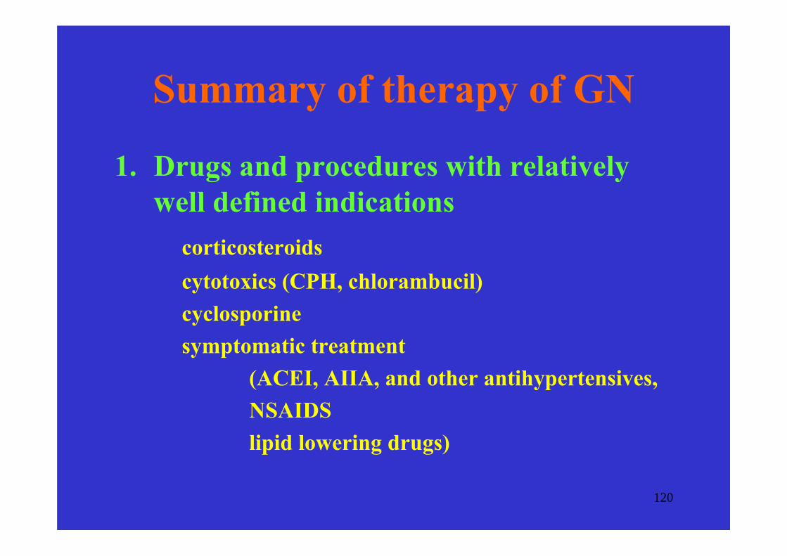
Summary of therapy of GN
1. Drugs and procedures with relativelywell defined indications
corticosteroidscytotoxics (CPH, chlorambucil)cyclosporinesymptomatic treatment
(ACEI, AIIA, and other antihypertensives,NSAIDSlipid lowering drugs)

121
Summary of therapy of GN – cont´2. Drugs and procedures with limited experience
and not well defined indicationsmycophenolate mofetiltacrolimusrapamycin
intravenous immunoglobulinsmonoclonal antibodies (e.g. infliximab, rituximab)soluble cytokine receptors (e.g. etanercept)
plasma exchangeimmunoadsorption

122
Conclusions – primary GN1. Patients with primary GN endangered by:
a. complications of nephrotic syndromeb. progression to ESRF
2. Urinary findings are important, but renalbiopsy remains essential for diagnosis, treatment and assesment of outcome
3. primary GN treatable diseases, pts should be treated according to availableevidence
4. further progress in treatment depends on better understanding of their pathogenesis

123
XLVIII ERA-EDTA Congress
Prague, Czech RepublicJune 23-26, 2011