3d bioprinting for cartilage lillian margolis biomedical engineering october 21, 2015
TRANSCRIPT

3D Bioprinting For Cartilage3D Bioprinting For Cartilage
Lillian MargolisBiomedical EngineeringOctober 21, 2015

IntroductionIntroduction• Tissue Engineering
– Study of growth of connective tissue
– Repair or replace tissue• Cartilage
– Connective tissue– Avascular– Three Types: Hyaline,
Elastic, and Fibrous• 3D Bioprinting: scaffolds and
bio-ink
[2]
[1]

MethodsMethods
• Scaffolds: three dimensional polymer mold that guides the tissue as it cultures and grows– Mesenchymal stem cells– Chemical cues to mimic
the original tissue
[3]
[3]

MethodsMethods
• 3D Bioprinting: directly repair or recreate cartilage and integrates with original cartilage– Sizes of tissues printed: under 400 micrometers– 5 options
• Extrusion• Laser• Inkjet
• Thermal Inkjet• Piezoelectric Inkjet
[5]
[4]

StudiesStudies
• Thermal Inkjet Study (Human)– Layer by layer, articular cartilage, and polyethylene glycol
dimethacrylate– 4mm diameter, thickness of 2mm, nominal 0.23 microliters bioink– ~1140 chondrocytes– Each layer printed and photopolymerized, 18micrometers thick– 2 mins total printing time– Printed cartilage with 3d biopaper had higher levels of
glycosaminoglycan (GAG) content than cartilage printed without– Result: Importance of direct cartilage repair; success in
placement of individual cells, preserving cell viability, maintaining chondrogenic phenotype, and integrating with original tissue tissue
[5]
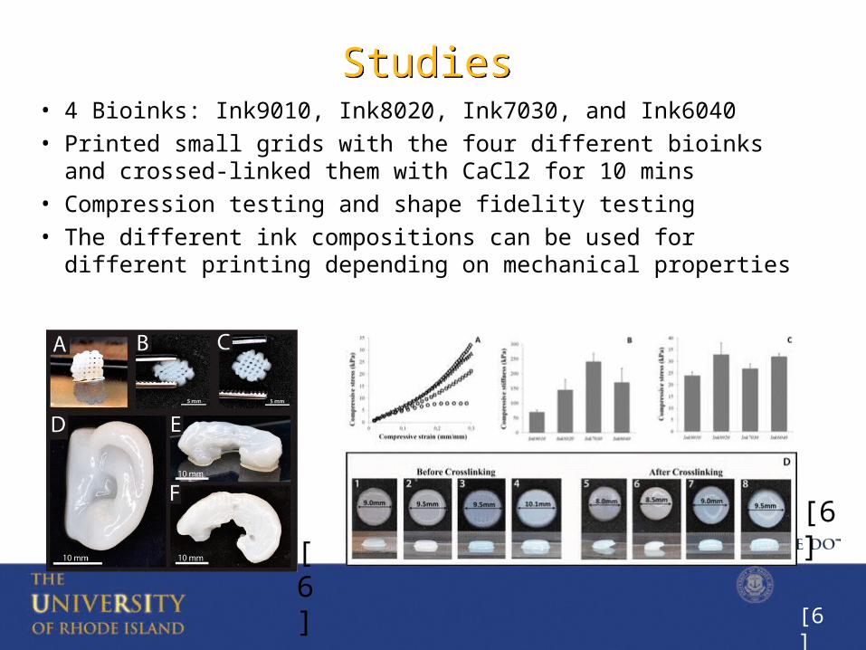
StudiesStudies• 4 Bioinks: Ink9010, Ink8020, Ink7030, and Ink6040• Printed small grids with the four different bioinks and
crossed-linked them with CaCl2 for 10 mins• Compression testing and shape fidelity testing• The different ink compositions can be used for different
printing depending on mechanical properties
[6]
[6]
[6]

ConclusionConclusion• Thermal Inkjet Bioprinting
– Print both soft and hard tissue– Best option for repairing cartilage
• Ink8020 is most suitable bioink for printing• Future
– Optimizing scaffolds– Targeted Drug Therapy– Gene Transfection
[YW]

Questions?Questions?
[1]
Resources Besides the Ones in the AbstractResources Besides the Ones in the Abstract
1. (n.d.). Retrieved October 17, 2015, from http://www.millerplace.k12.ny.us/webpages/lmiller/photos/636532/large23_Cartilage Types.jpg
2. WHAT IS TISSUE ENGINEERING. (N.D.). RETRIEVED OCTOBER 17, 2015, FROM HTTP://WWW.RPI.EDU/DEPT/CHEM-ENG/BIOTECH-ENVIRON/PROJECTS00/TISSUE/WHAT IS TISSUE ENGINEERING.HTM
3. Camarero-Espinosa, S., Rothen-Rutishauser, B., Weder, C., & Foster, E. (2015). Directed cell growth in multi-zonal scaffolds for cartilage tissue engineering. Biomaterials, 42-52. doi:10.1016/j.biomaterials.2015.09.033
4. Murphy, S., & Atala, A. (2014). 3D bioprinting of tissues and organs. Nat Biotechnol Nature Biotechnology, 773-785. doi:10.1038/nbt.2958
5. Gao, G., & Cui, X. (2015). Three-dimensional bioprinting in tissue engineering and regenerative medicine. Biotechnology Letters, 1-9. doi:10.1007/s10529-015-1975-1
6. Markstedt, K., Mantas, A., Tournier, I., Ávila, H., Hägg, D., & Gatenholm, P. (2015). 3D Bioprinting Human Chondrocytes with Nanocellulose–Alginate Bioink for Cartilage Tissue Engineering Applications. BioMacroMolecules, 1489-1496. doi:10.1021/acs.biomac.5b00188
7. Digital image. University of Rhode Island. N.p., n.d. Web. <http://www.uri.edu/news/releases/html/images/rhody.jpg>.