3d anatomical basis for transobturator surgeries prof. paulo palma
Post on 20-Dec-2015
223 views
TRANSCRIPT

3D ANATOMICAL BASIS FOR TRANSOBTURATOR SURGERIES3D ANATOMICAL BASIS FOR TRANSOBTURATOR SURGERIES
Prof. Paulo Palma

Paradigm shifts
1. Integral Theory, 1993 (Peter Petros)– Creating neoligaments using tapes– Reinforcement of pubourethral ligament– Midurethral tape placement.
2. Transobturator Approach, 2001 (Delorme)
- Reinforcement of urethropelvic ligament
- Less visceral and vascular complications
- Less irritative voiding symptoms

Sacro UterineSacro Uterine LigamentLigament
BladderBladder
UterusUterus
PP
Tendineous Tendineous ArcArc
PubourethralPubourethralLigamentLigament
SacruSacrumm
Urethropelvic LigamentUrethropelvic Ligament
VaginaVagina
Anterior Median Posterior
CompartimentsCompartiments

PP
Tendineous ArcTendineous Arc
SacrumSacrum
VaginaVagina
PubourethralPubourethralLigamentLigament
Urethropelvic LigamentUrethropelvic Ligament
Sacro Sacro UterineUterine
LigamentLigament
LIGAMENTSLIGAMENTS

RestRest
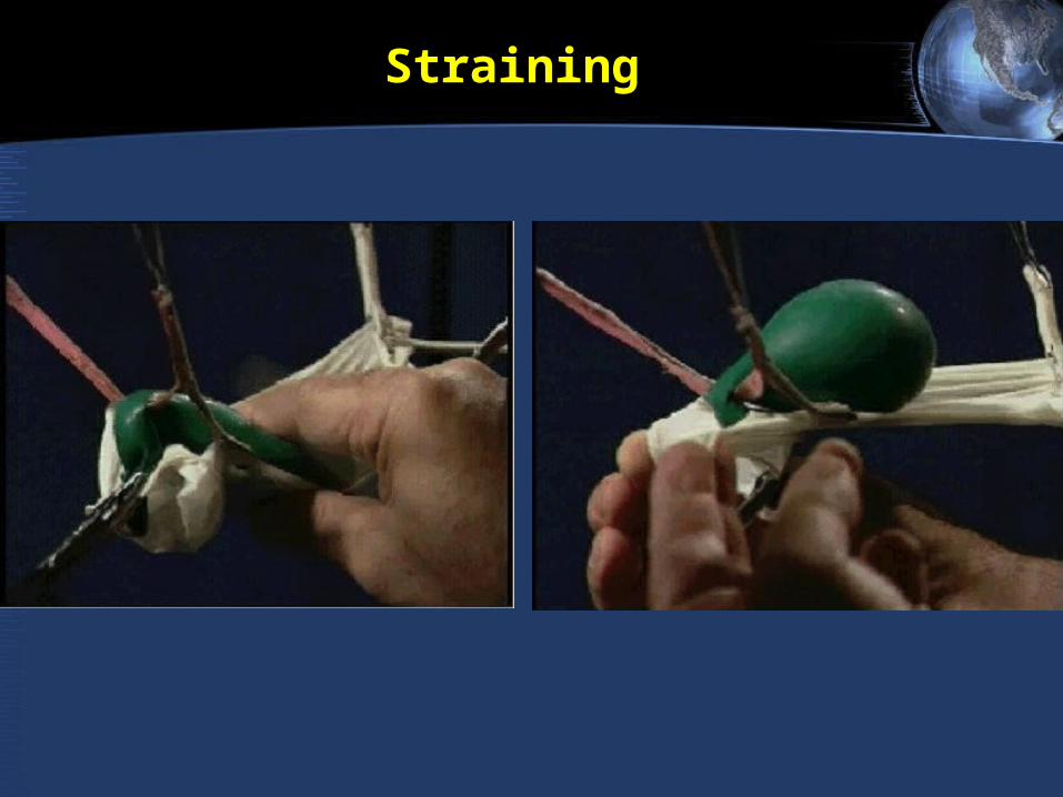
StrainingStraining
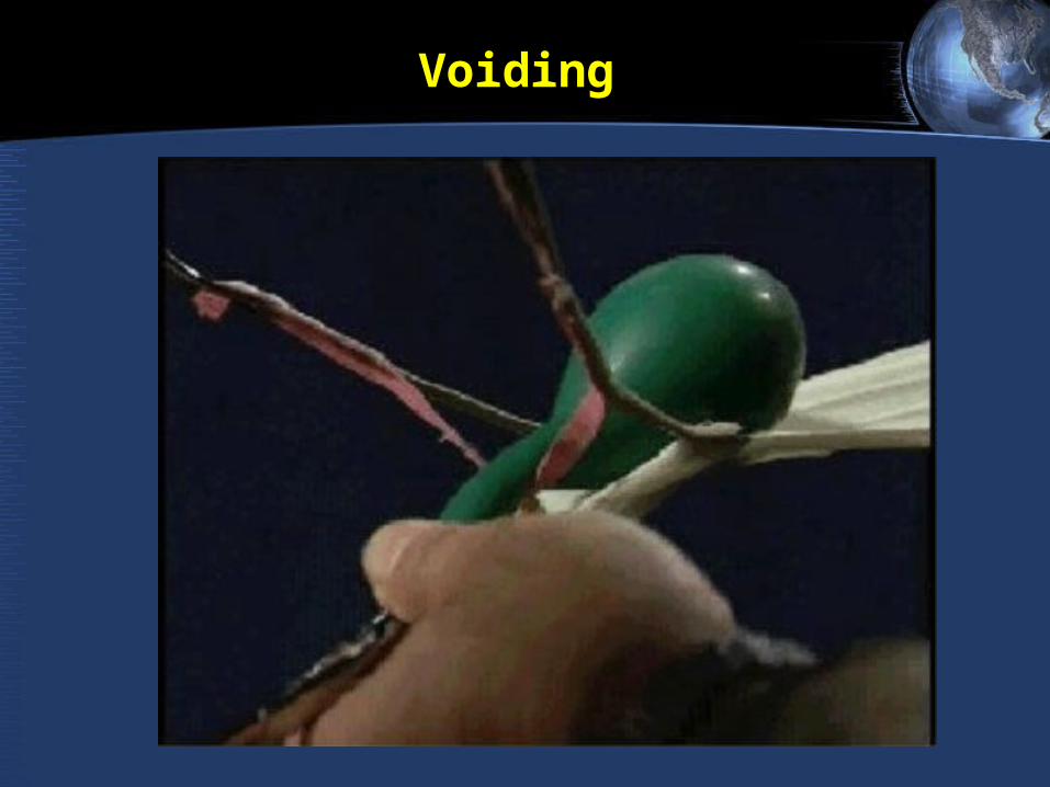
VoidingVoiding

1
2 3
4
1. Superficial Transverse 1. Superficial Transverse Muscle of perineumMuscle of perineum
2. Isquiocavernosous muscle2. Isquiocavernosous muscle
3. Bulbocavernosous Muscle3. Bulbocavernosous Muscle
4. External Anal Esfincter 4. External Anal Esfincter
First muscular layer

5
5.Deep Transversal5.Deep TransversalMuscle of perineum Muscle of perineum
Second Muscular LayerSecond Muscular Layer

6677
886. Pubovaginal6. Pubovaginal7. Puborectal7. Puborectal
8. Ileococcigeous8. Ileococcigeous
White LineWhite Line

RECTOVAGINAL FASCIARECTOVAGINAL FASCIA

VESICOVAGINAL FASCIAVESICOVAGINAL FASCIA

Bony Anatomy
Ischial tuberosity
Pubic Symphysis
Ischiopubic ramus
Obturator Canal
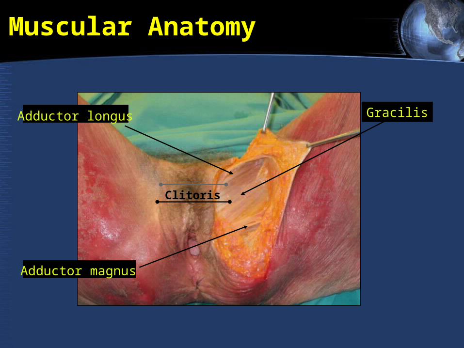
Muscular Anatomy
Clitoris
GracilisGracilisAdductor longus
Adductor magnus

Muscular Anatomy
Muscle Function Innervation
Adductor longusAdducts & flexes
thighObturator (L2,3)
Adductor brevisAdducts & flexes
thighObturator (L2,
3)
Adductor magnus
Adducts, extends, flexes & rotates
thigh
Obturator (L2, 3) & Tibial (L4,
5)
GracilisAdducts thigh,
flexes kneeObturator (L2,
3)
Obturator internus
Lateral rotator of thigh
Obturator (L5, S1)
Obturator externus
Lateral rotator of thigh
Obturator (L3, 4)
PectineusFlexes thigh, Adducts thigh
Femoral (L2, 3)
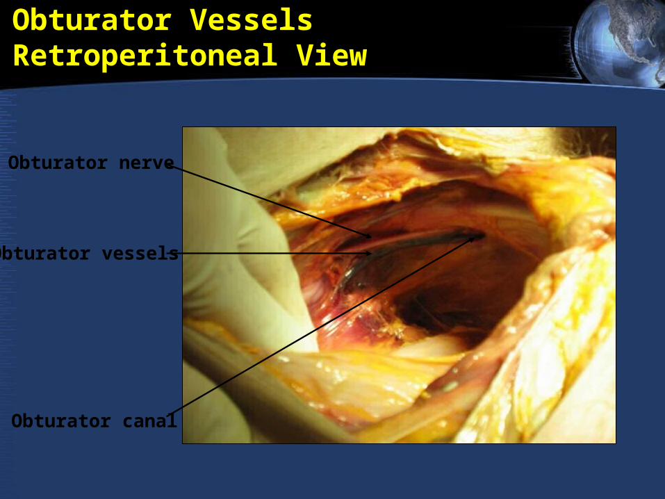
Obturator VesselsRetroperitoneal View
Obturator vessels
Obturator nerve
Obturator canal

Obturator Vascular Anatomy

Obturator - Neuroanatomy
• From anterior division of L2-4• Innervates all muscles of the obturator region,
except pectineus• Passes through obturator canal and splits to
innervate the muscles that adduct the thigh and help with external rotation
• Small sensory area on the medial thigh

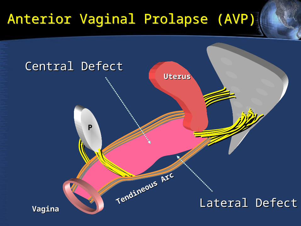
PPTendineous Arc
Tendineous Arc
UterusUterus
VaginaVagina
Anterior aginal Wall Defects

PPTendineous Arc
Tendineous Arc
UterusUterus
VaginaVagina
LateralLateralDefectDefect
Anterior aginal Wall Defects
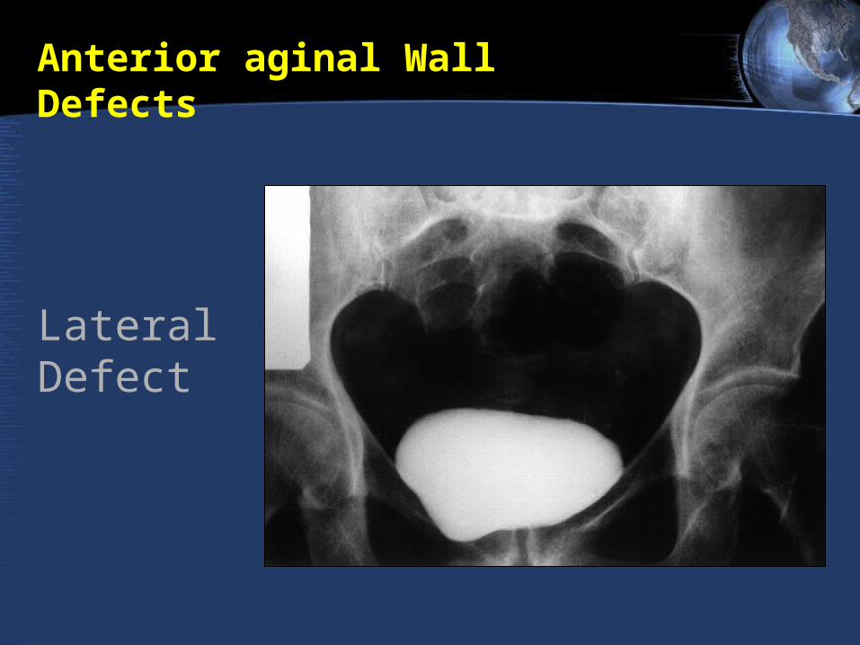
LateralDefect
Anterior aginal Wall Defects
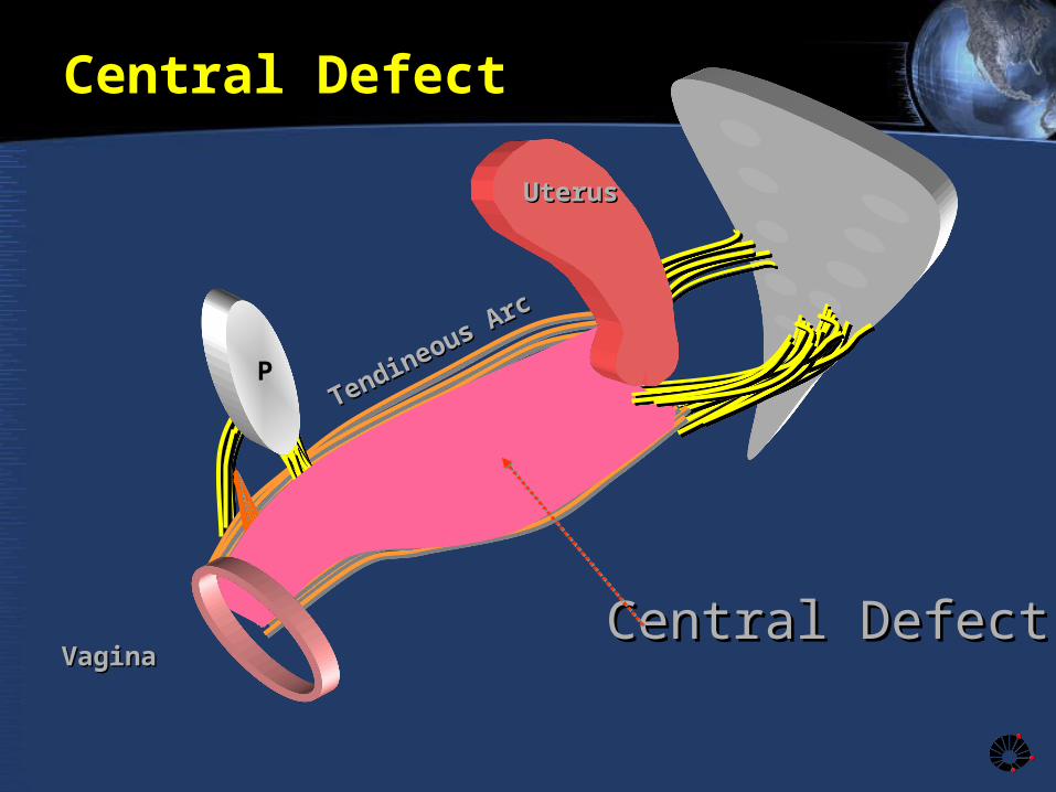
PPTendineous Arc
Tendineous Arc
UterusUterus
VaginaVagina Central DefectCentral Defect
Central Defect

Central Defect
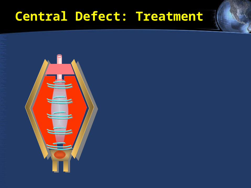
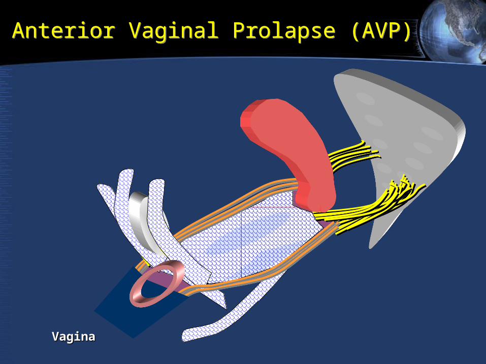
Central Defect: Treatment
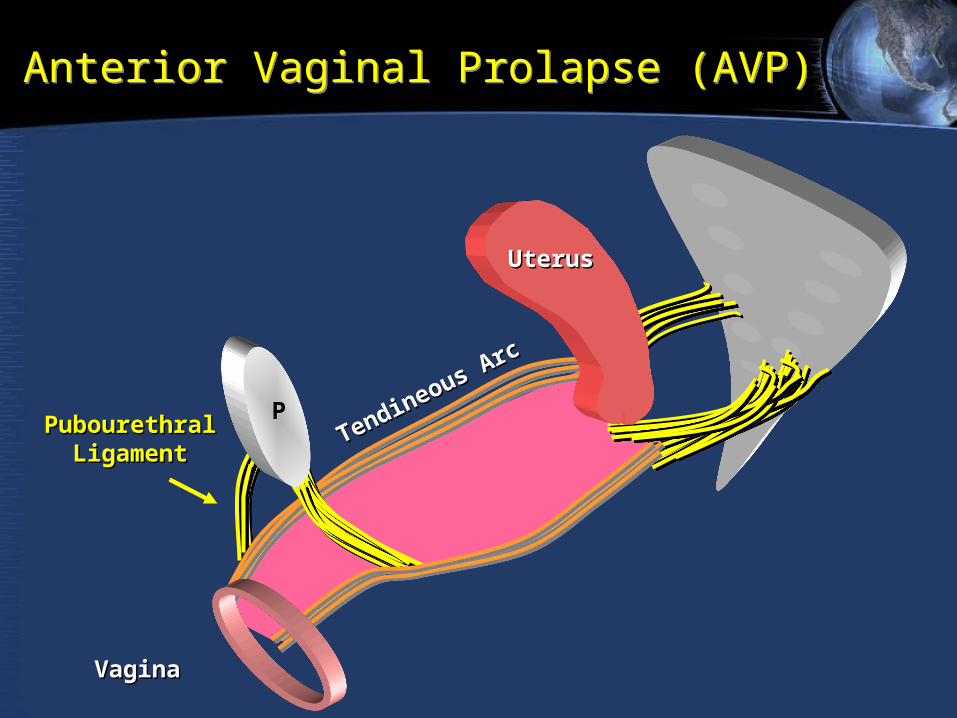
PPTendineous Arc
Tendineous Arc
UterusUterus
VaginaVagina
Anterior Vaginal Prolapse (AVP)Anterior Vaginal Prolapse (AVP)
PubourethralPubourethralLigamentLigament
PP
UterusUterus
VaginaVagina Lateral DefectLateral Defect
Anterior Vaginal Prolapse (AVP)Anterior Vaginal Prolapse (AVP)
Central DefectCentral Defect
Tendineous Arc
Tendineous Arc
PP
UterusUterus
VaginaVagina
Anterior Vaginal Prolapse (AVP)Anterior Vaginal Prolapse (AVP)

Evaluation of pelvic floor reconstructive surgery using tridimentional helical CTEvaluation of pelvic floor reconstructive surgery using tridimentional helical CT
Fig 3. Notice the well supported bladder base and the TOT arms

PPTendineous Arc
Tendineous Arc
RectumRectum
UterusUterus
VaginaVagina
Levator ani
Levator ani
Posterior Posterior DefectDefect
Posterior Vaginal Prolapse (PVP)Posterior Vaginal Prolapse (PVP)

SacrospinalSacrospinalLigamentLigament
PPTendineous Arc
Tendineous Arc
RectumRectum
UterusUterus
VaginaVagina
Levator a
ni
Levator a
ni
Posterior Vaginal Prolapse RepairPosterior Vaginal Prolapse Repair

Evaluation of pelvic floor reconstructive surgery using tridimentional helical CTEvaluation of pelvic floor reconstructive surgery using tridimentional helical CT
Fig 4. Infracoccigeal sacropexy showing indirectly the “neo rectovagianl fascia”
Posterior sling
Anchoring
Tails
BladderBladderRectum
MeshMesh