3% saline versus mannitol in treatment of cerebral edema
TRANSCRIPT
3% SALINE VERSUS MANNITOL IN TREATMENT OF CEREBRAL EDEMA IN
CHILDREN
DISSERTATION SUBMITTED FOR M.D DEGREE (PEDIATRICS)
BRANCH VII
THE TAMILNADU DR. M.G.R. MEDICAL UNIVERSITY
CHENNAI
INSTITUTE OF CHILD HEALTH
AND HOSPITAL FOR CHILDREN
MADRAS MEDICAL COLLEGE CHENNAI
APRIL 2011
Dr.P.JEYACHANDRAN M.D., DCH Chief, Department of Pediatric Intensive Care Institute of Child Health and Hospital for Children Chennai – 600008.
CERTIFICATE
This is to certify that the dissertation titled “3% SALINE
VERSUS MANNITOL IN TREATMENT OF CEREBRAL EDEMA IN
CHILDREN” submitted by Dr.D.KUMARAGURU to the Faculty of
pediatrics, The Tamilnadu Dr. M.G.R. Medical university, Chennai in
partial fulfillment of the requirement for the award of M.D. Degree
(Pediatrics) is a bonafide research work carried out by him under our
direct supervision and guidance, during the academic year 2008-2011
Dr.J.MOHANASUNDARAM, M.D., Ph.D., DNB Dean, Madras Medical College, Chennai - 3.
Dr.P.RAMACHANDRAN, M.D., DNB
Director & Superintendent, Institute of Child Health and
Hospital for Children, Egmore, Chennai - 8.

DECLARATION
I, DR.D.KUMARAGURU, solemnly declare that the dissertation
titled “3% SALINE VERSUS MANNITOL IN TREATMENT OF
CEREBRAL EDEMA IN CHILDREN” has been prepared by me.
This is submitted to The Tamilnadu Dr. M.G.R. Medical
University, Chennai in partial fulfillment of the rules and regulations
for the M.D. Degree Examination in Pediatrics.
Dr.D.KUMARAGURU
Place : Chennai
Date :

SPECIAL ACKNOWLEDGEMENT
My sincere thanks to Prof. Dr.Mohanasundaram M.D., Ph.D,
D.N.B., Dean, Madras Medical College, Chennai for permitting me to
utilize the clinical materials of the hospital
ACKNOWLEDGEMENT
I express my heart full gratitude to Prof.Dr.P.Ramchandran,
Director and Superintendent, Institute of Child health and Hospital for
children, Madras Medical College, Chennai and my unit chief for his
great help from the beginning of the study, his able guidance, inspiration
and encouragement, in conducting my study.
I am very grateful to Prof.Dr.P.Jayachandran, M.D., DCH, Chief,
Pediatric Intensive Care Unit for allowing me and helping me in
conducting my study.
My profound thanks to Dr.S.Thangavelu, M.D., DCH., MRCP.,
Former Reader, Pediatric Intensive Care Unit, Dr.S.Shanthi, M.D.,DCH.,
Dr.V.Poovazhagi, M.D., Dr.Ezhilarasu, M.D, Dr.Sivaraman, MD.,
Asst. Professors , PICU for their meticulous guidance and support
throughout the study
I sincerely thank my unit Assistant professors, Dr.S.Rajendran
M.D.,DCH., Dr.Mekalai Sureshkumar, M.D,DCH, Dr.P.Jeyakumar
M.D for their valuable guidance and support throughout the study.

I am extremely thankful to Dr.Srinivasan, DCH., Registrar, for
his valuable suggestions, invaluable help and guidance in doing this
work.
I am greatly indebted to Dr.K.Nedunchezhian, M.D., DCH, for his
support and guidance in doing this study.
I sincerely thank all the children and their parents who have
submitted themselves for this study and without whom this study would
not have been possible
.

CONTENTS
S.No. Contents Page No.
1. Introduction 1
2. Review of Literature 13
3. Justification of the study 16
4. Aim of the study 18
5. Subjects and Methods 19
6. Observations 25
7. Discussion 42
8. Summary 48
9. Conclusion 49
10. Recommendations 50
11. ANNEXURE 1-PROFORMA
12. ANNEXURE 2 -BIBLIOGRAPHY
13. ANNEXURE 3-ABBREVATIONS

1
INTRODUCTION
Intracranial hypertension is a common feature of many illness
treated in PICU. The early signs and symptoms of intracranial
hypertension tend to be nonspecific. The classic Cushings triad of
bradycardia, hypertension, and apnea occurs late and often not manifest
fully in children.
Most of our understandings and approach to treatment is based
on studies of patients with traumatic brain injuries. Whether those
concepts are directly relevant to the pathophysiologic processes
involved in more global CNS injuries such as hypoxia, infection and
metabolic disorders, remain a matter of debate.
Within constraints of a closed skull, an enlargement of brain
tissue (cerebral edema), an increased volume of CSF, or an increased
volume of blood or the presence of a space-occupying lesion (SOL)
such as a tumour or an abscess will initially reduce the size of the other
compartments and later increase intracranial pressure, once the
compensatory mechanisms fail.

2
Cerebral edema
The brain occupies about 80% of the volume of the skull. Apart
from solid tumours, increase in brain compartment is generally a result
of cerebral edema. Cerebral edema is the most common cause for
intracranial hypertension in children treated in PICU.
Types of cerebral edema
Cerebral edema can be divided into three forms: vasogenic,
hydrostatic, and cytotoxic.
Vasogenic edema occurs in areas of inflamed tissue
characterized by increased capillary permeability, and is most typical
around CNS tumours, abscesses, and infarcts.
Hydrostatic or interstitial edema is a result of elevated CSF
hydrostatic pressures. It occurs primarily in lesions associated with
obstruction to CSF flow, and in a typical periventricular distribution.
Cytotoxic edema is the most common of the three forms seen in
the PICU and is, unfortunately, the least easily treated. It occurs as a
result of direct injury to brain cells, often leading to irreversible cell
swelling and death. This form of cerebral edema is typical of traumatic
brain injuries, infections, hypoxic ischemic injuries and metabolic
disease.

3
Aetiology
Cerebral edema is seen in the following neurological and non-
neurological conditions:
Neurological conditions -
a) Ischemic stroke and intracerebral haemorrhage
b) Brain tumours
c) Meningitis and encephalitis of all etiologies
d) Other brain infections like cysticercosis, tuberculosis and
toxoplasma.
Non-neurological conditions -
a) Diabetic ketoacidosis, lactic acidotic coma
b) Malignant hypertension, hypertensive encephalopathy
c) Fulminant viral hepatitis, hepatic encephalopathy, Reye’s
syndrome
d) Systemic poisoning (carbon monoxide and lead)
e) Hyponatremia, SIADH
4
f) Opioid drug abuse and dependence
g) Bites of certain reptiles and marine animals
h) High altitude cerebral edema
Clinical Features
A high index of suspicion is very important. The features of
cerebral edema add on to and often complicate the clinical features of
the primary underlying condition. Until the ICP reaches a level that
produces local ischemia, cerebral edema per se will not produce clinical
neurological abnormalities.
Signs and symptoms [1]
Early – Poor feeding, vomiting, irritability, lethargy, seizures,
hypertension
Late – GCS < 8, Coma, absent doll’s eye movement, decerebrate
responses, cranial nerve palsies, abnormal respirations, bradycardia,
hypertension, and apnoea

5
Investigations
Urgent imaging is indicated in afebrile coma and in the presence
of focal signs or papilledema, as the diagnosis includes stroke, intra-
cranial bleed, tumour or hydrocephalus. However, any child who does
not have a very obvious metabolic/ toxic cause for the coma generally
requires to be imaged.
CT scan provides an excellent tool for in vivo determination of
abnormalities in brain water content. The areas of edema appear as low
density on unenhanced scan. This is due to the dilution of all the
constituents of the white matter [2]. The anatomical specificity of CT
permits detection of not only the presence but also the type of brain
edema. This is helpful in differentiating nature of underlying lesion eg.
infarction/tumour. However CT scan may be normal initially and it does
not rule out raised ICP.
CT is also an excellent method for following the resolution of
brain edema following therapeutic intervention.
An MRI may be more specific for early changes of herpes
simplex encephalitis (where CT may be normal), posterior fossa and
white matter pathology [1, 3].

6
Invasive ICP monitoring is an important tool to monitor cases
where cerebral edema is present or anticipated. Unfortunately, the direct
measurement of ICP and aggressive measures to counteract high
pressures have not yielded uniformly beneficial results, and after two
decades of popularity - the routine use of ICP monitoring remains
controversial. The problem may be partly a matter of the timing of
monitoring and the proper selection of patients for aggressive treatment
of raised ICP. Whether ICP monitoring adds much to the management
of these patients is still open to question. Clinical signs and imaging data
on shift of brain tissue are probably more useful
Treatment
Treatment of brain edema has not kept up with the advances in
understanding of the mechanism producing the edema.
General measures
When signs of elevated ICP are present certain measures for
management should be initiated.
Position of the patient - Elevation of head end of bed by 15-30 degrees
to promote cerebral venous drainage is advisable and head is kept in
midline to limit neck vein compression [4].

7
Correction of contributory factors - Correction of factors increasing
ICP e.g. hypercarbia, hypoxia, hyperthermia, acidosis, hypotension and
hypervolemia is helpful. Endotracheal intubation and mechanical
ventilation to maintain PaCO2 in low 30s (controversial) [1, 3] may be
helpful in impending herniation.
Hypothermia - Multiple mechanisms for reduced brain temperature-
induced neuroprotection have been identified and include reduced
metabolic rate and energy depletion, decreased excitatory transmitter
release, reduced alterations in ion flux, reduced vascular permeability,
edema and BBB disruption [5].
Fluid restriction - Fluid restriction minimally affects cerebral edema
and, if pursued to excess, may result in episodes of hypotension, which
may decrease cerebral perfusion pressure (mean arterial pressure – ICP)
and is associated with worse neurologic outcome [6]. Fluid restriction is
not warranted as a routine unless in specific situation like SIADH.
Glucose containing solutions should be avoided; euvolemia should be
maintained, 0.9%N or N/2 saline should be used; urinary losses should
be replaced with 0.9%N saline in patients receiving mannitol; eventually
to maintain mean arterial pressure [4].
8
Medical treatment
Osmotherapy
The most rapid and effective means of decreasing tissue water
and brain bulk is osmotherapy [2]. Osmotic therapy is intended to draw
water out of the brain by an osmotic gradient and to decrease blood
viscosity. These changes would decrease ICP and increase cerebral
blood flow (CBF).
Mannitol is the most popular osmotic agent. Mannitol has two
distinct effects. The immediate effect is related to its rheological
properties (decreased blood viscosity) resulting in a transient increased
CBF followed by a more sustained fall in CBF [1]. The delayed osmotic
effects occur after 15-30 minutes and last for 4-6 hours. IV Mannitol is
given in the dosage of 0.25-0.5g/kg repeated 4-6 hourly. Mannitol
should be avoided in case of hypotension, renal failure, serum
osmolarity>320 mOsm/kg.
Hypertonic saline [HTS] is emerging as newer osmotherapy.
Hypertonic saline can possibly affect the volume of the intracranial
structures through various mechanisms. All or several of them are likely
to be interacting to achieve the end result of hypertonic saline therapy:

9
reduction of cerebral edema and elevated ICP. These mechanisms [7]
are summarised below:
• HTS also acts like mannitol by establishing a constant
osmolar gradient in order to draw fluid from the brain
parenchyma but without the risks of dehydration and
tubular damage as in the case of mannitol. Hence it may be
of more benefit in conditions where mannitol should be
avoided.
• Reduced viscosity: Hypertonic saline enhances
intravascular volume and reduces viscosity. The auto
regulatory mechanisms of the brain vasculature have been
shown to respond not only to changes in blood pressure but
also to changes in viscosity. Thus, a decrease in blood
viscosity results in vasoconstriction in order to maintain a
stable cerebral blood flow.
• Increased plasma tonicity: It has been postulated, based on
experimental animal data that increased plasma tonicity,
such as that seen after hypertonic saline administration,
favours more rapid adsorption of CSF.

10
• Increased regional brain perfusion, possibly secondary to
dehydration of cerebral endothelial cells and erythrocytes,
facilitating flow through capillaries.
• Increased cardiac output and mean arterial pressure, with
resultant augmentation of cerebral perfusion pressure, most
likely due to improvement of plasma volume and a positive
inotropic effect.
• Diminished inflammatory response to brain injury, which
has been demonstrated with hypertonic saline
administration.
• Restoration of normal membrane potentials through
normalization of intracellular sodium and chloride
concentrations.
A variety of formulations of hypertonic saline solutions (2, 3, 7.5,
10, and 23%) [8, 9] are used in clinical practice for the treatment of
cerebral edema either as bolus or continuous infusions. The goal in
using hypertonic saline is to increase serum sodium concentration to a
range of 145 to 155 mEq/L (serum osmolality approximately 300–320
mOsm/L) [10]
11
Corticosteroids
Corticosteroids lower intracranial pressure primarily in vasogenic
edema because of their beneficial effect on the blood vessel. They have
been less effective in cytotoxic edema [11].
Other agents
Barbiturates, procaine derivatives, indomethacin, propofol and
THAM (Trihydroxy aminomethane) are some other agents which have
been tried. Barbiturate coma is used in some centres in order to reduce
the cerebral metabolic rate and thus the cerebral blood flow, although
most references pertain to adults.
Surgical treatment
Surgical treatment is occasionally recommended for large
hemispherical infarcts with edema and life threatening brain-shifts.
Temporary venticulostomy or craniotomy may prevent deterioration and
may be lifesaving [12].
Conclusion
Though there has been good progress in understanding of
pathophysiological mechanisms associated with cerebral edema, more

12
effective treatment is required and is still awaited. Certainly, the “ideal”
agent for the treatment of cerebral edema - one that would selectively
mobilize and / or prevent the formation of edema fluid with a rapid
onset and prolonged duration of action, and with minimal side effects,
remains to be discovered.

13
REVIEW OF LITERATURE
I] Yildizdas D, Altunbasak S conducted a retrospective study to
evaluate the efficacy and side effects of hypertonic saline[HTS] and
mannitol use in cerebral edema in the Pediatric Intensive Care Unit
Çukurova University, School of Medicine between June 2002 and May
2004 [13]. Patients treated with mannitol, HTS, or both mannitol and HS
were assigned as Group I, Group II, Group III respectively. Cerebral
edema and increased intracranial pressure were identified based on the
clinical and/or radiological (CT, MR) findings. Clinical findings
included low consciousness level (GCS <8) plus one or more of the
followings: Unequal, dilated or unreactive pupils, loss of brain stem
reflexes (light and oculocephalic), cranial nerve palsies (III, VI) and
Cushing's triad. In Group I, patients were treated with 0.5 g/kg mannitol
for the first two doses and if needed the maintenance doses were 0.25
g/kg/dose. In Group II, Hypertonic saline was given to provide a serum-
Na level of 155-165 mEq/L. Extra boluses were given depending on the
serum Na level. Group III received both mannitol and hypertonic saline.
In Group I – 22, Group II – 25, Group III – 20 were recruited. There was
no statistically significant difference between groups in terms of age (P
= 0.5), gender (P = 0.4), Glasgow coma scale and etiological causes (P =
0.8). Mannitol was given for a total dose of 9.3 ± 5.0 (2-16) doses in

14
Group I, and 6.5 ± 2.8 (2-10) doses in Group III. Hypertonic saline was
infused for 4-25 times in Group II. Although there was no statistically
significant difference in the highest serum Na and osmolarity levels of
the groups, duration of comatose state and mortality rate were
significantly lower in Group II and Group III. Age, gender, cause of
cerebral edema, electrolyte imbalance, hyperglycemia and
hyperventilation had no significant impact on outcome. Limitations of
this study: This was a retrospective study; Outcome variable of “duration
of comatose state” included all patients irrespective of their survival
status. Failure to sensor this time-to-event ‘duration of coma’ variable by
mortality in this study could have been rectified. The p –value 0.003
while comparing the outcome of mortality (proportion) appears to have
error. Using the factual data about mortality and survival given by
authors in this study, the calculated P value is 0.07.

15
II] Piyush Upadhyay and Tripathi conducted a prospective
randomized study to compare the efficacy and side effects of 3% saline
and mannitol in management of raised intracranial pressure in children
at GSVM Medical College, Kanpur [14]. 200 Children aged 2 to 18
months with clinical symptoms and signs of raised intracranial pressure
were recruited. Patients with compromised renal function (increased
serum creatine), hepatic encephalopathy, serum Na + (>150meq/l),
diabetic ketoacidosis, cereberal malaria were excluded. Loading dose
(5ml/kg) was followed by maintenance dose (2ml/kg) of mannitol or 3%
hypertonic saline in corresponding groups for two days was
administered. Comparison of average reduction of mean arterial
pressure (pre and post drug) at definite time (6h) intervals was done to
indirectly assess reduction in intracranial pressure. Decrease in MAP
was found to be highly significant (p<0.001) at 0 hr in males 0, 6 hr in
females and moderately significant at 12, 36 hr in females and
significant (p<0.05) at 6, 24, 42 hr in males in 3% saline group.
Decrease in coma hours was a highly significant finding (p<0.001) in
3% saline group. No difference was noted in osmolality and mortality
between both groups. Limitations of the study: Clinical criteria for
raised intracranial pressure could have been clearly defined; mental
status of the treated children could have been defined and considered for
termination of treatment; side effects encountered during treatment
could have been enumerated.

16
JUSTIFICATION
Cerebral edema is an important and frequent problem in
neurocritically ill patient. This can result from various insults to the
brain. Improving cerebral edema and decreasing ICP have been
associated with improved outcome. However, all current treatment
modalities are far from perfect and are associated with serious adverse
events. Indiscriminate hyperventilation can lead to brain ischemia.
Mannitol can cause intravascular volume depletion, renal insufficiency,
and rebound ICP elevation; barbiturates are associated with
cardiovascular and respiratory depression and prolonged coma; and
cerebrospinal fluid drainage via intraventricular catheter insertion is
highly invasive and may result in intracranial bleeding and infection.
Other treatment modalities have been explored and hypertonic saline
solution appears to be an appealing addition to the current therapeutic
avenues for management of cerebral edema .Mannitol has been used
extensively in treatment of cerebral edema in the past and is the time
tested drug.
In clinical situations there are instances where mannitol is
contraindicated such as when the child is in shock or renal failure.
Mannitol is well known to produce rebound effect, serum electrolyte
disturbances and hypovolemia and this has led to continued search of

17
newer agents. Hence there is a need for the use of alternate drugs like 3%
saline in managing cerebral edema. 3% saline has been regularly used in
patients with intracranial injury, stroke, and DKA and has been found to
be useful. However the role of 3% saline in other causes of cerebral
edema has not been studied in pediatric population. A Retrosepective
study done in Turkey [13] has revealed that 3% saline seems to be more
effective than mannitol in cerebral edema. In our PICU, 3%saline is used
as a treatment for raised ICP since 2004. This study was done to analyze
the effects of hypertonic saline in children with cerebral edema and to
compare this treatment modality with mannitol in terms of efficacy, side
effects and outcome.

18
AIM
To study the effectiveness of 3% saline as an antiedema measure
in children admitted with cerebral edema in the pediatric intensive care
unit of a tertiary care children hospital in a developing country.

19
SUBJECTS AND METHODS
STUDY DESIGN : Randomized Controlled Trial
PLACE OF STUDY : Pediatric Intensive Care Unit
Institute of Child Health and Hospital for
Children, Egmore, Chennai.
PERIOD OF STUDY : November 2008 to October 2010.
STUDY POPULATION :
Children who are suspected to have cereberal edema between 3
months to 12 years.

20
INCLUSION CRITERIA:
Children in the age group of 3 months to 12 years, with cerebral
edema of any etiology who satisfy the criteria of diagnosis, admitted in
the PICU were included.
EXCLUSION CRITERIA:
(i) Children with cerebral edema but presenting with shock, renal
failure or intracranial bleed on admission at PICU were excluded,
as the hospital protocol in these children is to avoid mannitol.
(ii) Child presenting with afebrile status epilepticus, with or without
previous history of seizures, who show clinical recovery in 6 – 8
hours will not be included.
(iii) Children treated prior to PICU admission will be excluded.
(iv) Any of the features in criteria of diagnosis if explainable by other
causes like neuroparalytic disorder (snake bite, GBS, drug
poisoning) will be excluded.

21
CRITERIA OF DIAGNOSIS OF CEREBRAL EDEMA [15]:
Low GCS (<8) with any one of the following
• persistent posturing after correction of shock and hypoxemia
• Unequal, dilated or non-reacting pupil.
• Cranial nerve palsies (3 and 6)
• Bradycardia, Hypertension
• Abnormal respiratory pattern
• Papilledema
• Radiological finding like effacement of the basal cisterns,
thin, slit like or completely obliterated ventricles, obliterated
cortical sulci, shift in midline and temporal lobe or
cerebellar tonsillar herniation.

22
MANEUVER
80 children who satisfied the inclusion criteria were recruited
during the study period. Two groups A and B were chosen. 40 children
were assigned in each group with computer generated random number.
The diagnosis of cerebral edema was made as per diagnostic criteria
mentioned above. The study was approved by institutional ethical
committee. Informed written consent was obtained from parents or
caregivers.
In group A, children were treated with 20% mannitol. In group B,
children were treated with 3% saline. The treatment otherwise were
identical as per the PICU protocol in both groups. 20%mannitol was
given at a dose of 1.5ml/kg IV, over 20 minutes, every 8 hourly [16, 17].
3%saline was given in a dose of 5ml/kg IV, over 20 minutes, every 8
hourly [16, 17]. Serum sodium was targeted to maintain between 145-
155 meq/dl. Treatment for cerebral edema was terminated at a GCS score
> 8 and absence of other signs of cerebral edema as mentioned in
inclusion criteria. Maximum duration of treatment of cerebral edema was
72 hours. Treatment was not continued if child developed complications
necessitating termination of therapy.

23
The outcome was analyzed in terms of survival/death, duration of
coma and complication. Other parameters included were symptoms,
clinical signs, fundus examination, duration of ventilation, CT finding
and lab investigations. Complications recorded were hypernatremia,
coagulopathy, pulmonary edema, subarachnoid hemorrhage, hemolysis,
renal failure. Probable cause of cerebral edema such as CNS infection,
DKA, hepatic encephalopathy, infarct and tumors was arrived using the
clinical criteria and lab investigations. Etiologies were grouped into
infective, metabolic and others. Both group A and B were sub analyzed
in terms of outcome with respect to age and etiological causes.

24
STATISTICAL ANALYSIS
Data were analyzed with SPSS 14.0. Chi square and t test were
used for independent samples; ANOVA test was used for continuous
variables. Brock chart was used where ever necessary.

25
PROFILE OF CEREBRAL EDEMA
Total cases recruited 80 100%
Total cases recruited in Mannitol group
40 50%
Total cases recruited in 3%Saline group
40 50%
Total cases completed the study in Mannitol group
35 43.7%
Total cases completed the study in 3%Saline group
32 40%
Cases not completed the study 13 16.3%
Cases not completed the study include the children who died
before improvement of cerebral edema and within 72 hrs (n-10); and
those who were switched from mannitol to 3% saline due to fluid
refractory shock (n-3) which developed during mannitol therapy.

26
AGE OF PRESENTATION
Mannitol 3%Saline p - value
Mean (months) 59.17 46.47
0.114 SD ± 42.00 ± 36.68
Range 4 - 140 3 - 140
• Age was comparable between both groups.

27
GENDER DISTRIBUTION
Mannitol 3% Saline p - value
Male 22 26
0.49 Female 18 14
Total 40 40
• Gender distribution was comparable in both groups. ( p – value
– 0.49)

28
ETIOLOGICAL DISTRIBUTION
• Predominant cause for cerebral edema in both groups was
infection. 62% in mannitol group and 60% in 3%saline group.
• Metabolic causes include diabetic ketoacidosis, hepatic
encephalopathy and suspected inborn error of metabolism. Other
causes include SOL, infarct etc
Cause Mannitol 3%Saline p - value
Infective 25 24
0.75 Metabolic 12 11
Others 3 5
Total 40 40
29
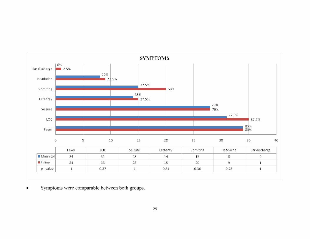
• Symptoms were comparable between both groups.

30
GLASGOW COMA SCALE
• Glasgow coma scale ranged between 3-8 in mannitol group and 4-
8 in 3%saline group. GCS was comparable in both groups.
(p – value – 0.057).
• In intubated children, GCS grimace score were used.
31
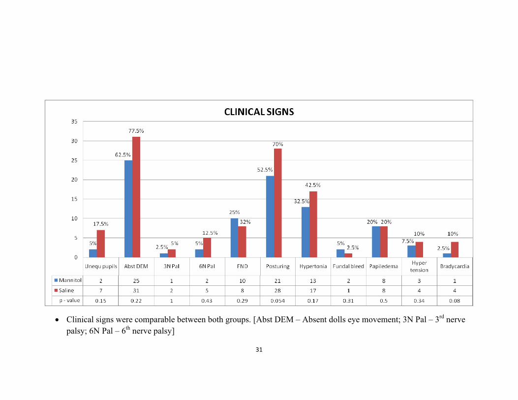
• Clinical signs were comparable between both groups. [Abst DEM – Absent dolls eye movement; 3N Pal – 3rd nerve palsy; 6N Pal – 6th nerve palsy]

32
DURATION OF COMA
No. of cases completed
therapy
Mean (hrs)
Standard deviation
Range (hrs)
Mannitol 35 59.89 29.67 13 - 146
3%Saline 32 78.91 50.84 28 - 260
P – value 0.063
• Duration of coma was compared only for those who completed
the therapy.
• Duration of coma was ranged between 13-146 hrs in mannitol
group and 28-260 hrs in Saline group.
• Even though the duration of coma seems to be more in 3%saline
group, there was no statistical significance.

33
AGE COMPARISION OF DURATION OF COMA
Age
(mon) Group N
Mean
(hrs)
Std.
Deviation p - value
<12
Mannitol 4 67.25 41.70
0.35
3%Saline 7 50.71 15.41
13-60
Mannitol 16 63.12 33.60
0.18
3%Saline 16 84.43 52.66
>60
Mannitol 15 65.06 37.49
0.20
3%Saline 9 91.00 60.97
• Duration of coma in different age groups was not statistically
significant between mannitol and 3%saline.
34
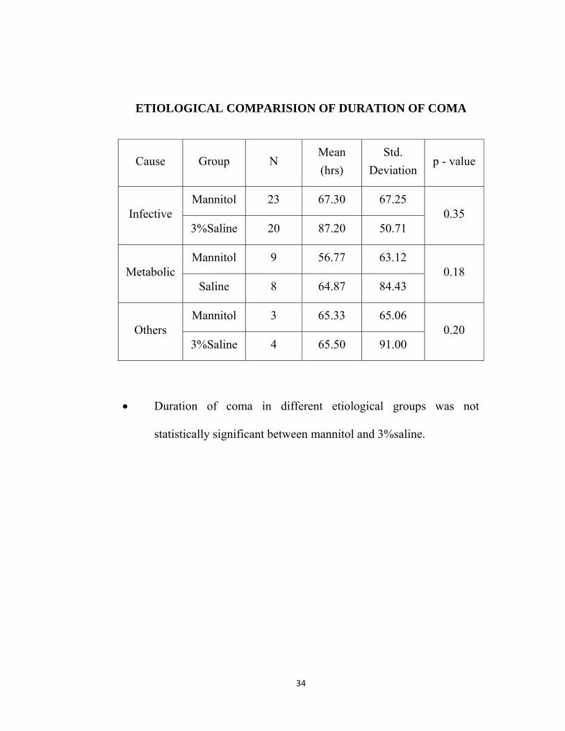
ETIOLOGICAL COMPARISION OF DURATION OF COMA
Cause Group N Mean (hrs)
Std. Deviation
p - value
Infective Mannitol 23 67.30 67.25
0.35 3%Saline 20 87.20 50.71
Metabolic Mannitol 9 56.77 63.12
0.18 Saline 8 64.87 84.43
Others Mannitol 3 65.33 65.06
0.20 3%Saline 4 65.50 91.00
• Duration of coma in different etiological groups was not
statistically significant between mannitol and 3%saline.

35
OVERALL MORTALITY
Outcome Mannitol 3%Saline Total
No % No % No %
Survived
Died
25 62.5% 16 40.0% 41 51.2%
15 37.5% 24 60.0% 39 48.8%
p-value – 0.07
• 62.5% survived in mannitol group. 40% survived in 3%saline
group.
• Mortality between the groups was not statistically significant. (p -
0.07)

36
AGE COMPARISION OF MORTALITY
Age (months) Mannitol 3%saline p - value
≤ 12 4 (10%) 4 (10%) 0.29
13 - 60 7(17.5%) 14 (35%) 0.058
≥ 60 4(10%) 6 (15%) 0.06
• Age wise comparisons of mortality were not statistically
significant between the two groups.

37
ETIOLOGICAL COMPARISION OF MORTALITY
Cause Mannitol 3%saline p -
value Count % Count %
Infective Survived 15 60.0% 10 41.7%
.258 Died 10 40.0% 14 58.3%
Metabolic Survived 8 66.7% 3 27.3%
.100 Died 4 33.3% 8 72.7%
Others Survived 2 66.7% 3 60.0%
1.000 Died 1 33.3% 2 40.0%
• In different etiological groups, mortality was not statistically
significant between the two groups.

38
• Dehydration and shock were significantly higher in mannitol group.
• Hypernatremia was significantly higher in 3%saline group.

39
SERUM SODIUM VALUE
Minimum sodium value Maximum sodium value
• Serum sodium at the time of recruitment into the study was
comparable between the groups: 128 – 135meq/dl in mannitol
group and 129 – 134 meq/dl in 3%saline group.
• Maximum and minimum serum sodium value attained during the
study were significantly higher in 3%saline group (p – 0.00)
• 6 cases in 3%saline group had serum sodium levels > 155meq/dl
during the therapy, hence subsequent doses was skipped until the
serum sodium levels decreased to 155meq/dl.

40
DURATION OF VENTILATION
Total ventilation
(hrs)
Manual
(hrs)
Mechanical
(hrs)
Mannitol
Mean 136.88 11.37 127.90
Range 0 - 648 0 - 103 0 - 648
Std. Dev 155.19 19.72 152.02
3%Saline
Mean 280.45 15.95 264.00
Range 0 – 3336* 0 - 236 0 - 3100
Std. Dev 571.35 37.85 540.38
Total
Mean 208.66 13.66 195.95
Range 0 - 3336* 0 - 236 0 - 3100
Std. Dev 422.21 30.07 400.32
p - value 0.06 0.35 0.06
• Mean ventilation duration was 136 hrs in mannitol group (0-
648hrs) and 280 hrs in saline group (0-3336hrs). Ventilation
duration was not statistically significant between both groups.
∗ In 3%saline group, one child with TBM was ventilated for
3336hrs (4 ½ months) and this child recovered with neurological
sequelae.
41
OTHER OBSERVATIONS
• CT Brain was taken in 80% of mannitol group and in 72.5% of
3%saline group.
• 52.5% of both groups had features of cerebral edema in CT brain.

42
DISCUSSION
The earliest description in the literature of the use of osmotic
agents dates back to 1919 [18]. While studying the transport of salt
solutions into the neuraxis, Weed and McKibben observed that
intravenous administration of a concentrated salt solution resulted in an
inability to withdraw CSF from the lumbar cistern due to a collapse of
the thecal sac. This serendipitous observation was followed by an
elegant set of experiments in an animal model in which they
demonstrated (under direct visualization via a craniotomy) regression of
the brain away from the cranial vault with intravenous infusion of
hypertonic saline solutions and herniation of brain tissue with
administration of hypotonic fluids. This set of observations has formed
the basis for osmotherapy. Concentrated urea was the first agent to be
used clinically as an osmotic agent. [8,9,19]. Its use was short-lived and
is of historic interest only because of several untoward side effects
(nausea, vomiting, diarrhea, and coagulopathy) [19]. The interest in
elevating plasma oncotic pressure as a strategy to ameliorate cerebral
edema with the use of concentrated human plasma proteins, which
appeared briefly in 1940, was short-lived due to several concerns,
including cost, short half-life, cardiopulmonary effects, and allergic
reactions [19]. Glycerol was possibly the second osmotic agent to be

43
used clinically. Mannitol, an alcohol derivative of simple sugar
mannose, was introduced in 1960 and has since remained the major
osmotic agent of choice in clinical practice [8,9,19]. Its long duration of
action (4–6 hours) and relative stability in solution have enhanced its use
over the years. The extra osmotic properties of mannitol have been
studied extensively and may provide additional beneficial effects in
brain injury, including decreases in blood viscosity, resulting in
increases in rCBF and CPP, and a resultant cerebral vasoconstriction
leading to decreased CBV,[20,21] free radical scavenging,[22] and
inhibition of apoptosis [23].
Renewed interest in hypertonic saline solutions reappeared in the
1980s, when they were used in small-volume resuscitation in patients
experiencing hemorrhagic shock. [8,9]. These studies demonstrated that
prehospital restoration of intravascular volume improved morbidity and
mortality rates and physiological parameters (such as systemic blood
pressure, cardiac index, and tissue perfusion) in this subset of patients
[24]. In subsequent studies, cerebral effects of these solutions were
investigated in well-controlled experimental studies in animal models of
acute brain injury. Like mannitol, hypertonic saline also possesses
unique extraosmotic properties, including modulation of CSF production
and resorption and accentuation of tissue oxygen delivery [8,9].

44
The use of hypertonic saline solution in the treatment of cerebral
edema and elevated ICP in the clinical setting is largely based on an
extension of laboratory-based research, a few prospective studies in
humans, and anecdotal case reports. The first report to demonstrate the
efficacy of hypertonic saline in patients with TBI [25] involved two
patients with elevated ICP refractory to mannitol who were treated
successfully with a single intravenous bolus of 30% saline, after which
ICP decreased and systemic perfusion improved. Continuous
intravenous infusion of 2.5 and 5.4% hypertonic saline enhanced CPP
and improved somatosensory evoked potentials after brainstem trauma
[26]. Likewise, in an uncontrolled, nonrandomized study, [27]
reductions in ICP were noted with the use of 7.5% hypertonic saline
treatment following TBI.
Few studies have made direct comparisons between mannitol and
hypertonic saline. In a prospective, randomized comparison of 2.5 ml/kg
of either 20% mannitol (1400 mOsm/kg) or 7.5% hypertonic saline
(2560 mOsm/ kg) in patients undergoing elective supratentorial
procedures,[28] ICP and intraoperative clinical assessment of brain
swelling were similar in both treatment groups.
Most of these study perspectives were from adult population
and only few studies were available in pediatric population. In a double-

45
blind crossover study, in which 3% hypertonic saline for TBI was used
in a pediatric population, ICP was reduced by approximately 5 mmHg
for 2 hours compared with ICP in patients who required equal volumes
of isotonic saline. These available studies have compared population
with traumatic etiology which contribute meagrely to cerebral edema in
pediatric patients. Only two studies done by Yildizdas., et al and Piyush
Upathyay., et al were based on non traumatic etiology in pediatric
populations.
Our study is a prospective analysis done in children between ages
of 3 – 140 months. This was comparable with the study done by
Yildizdas et al., (1-120mon) whereas in study by Upadhyay., et al
included adolescent population. In our study etiological factors included
infective, metabolic, infarct and space occupying lesion. This was
comparable with other two studies.
In our study, male population (55-65%) were predominant similar
to Upadhyay et al. In the study by Yildizdas et al, female population (52-
60%) was predominant. Clinical signs and symptoms in our study were
comparable with other studies.
In our study, Glasgow coma scale in the both groups was
6.77±1.352 (3-8) and 6.04±1.51 (4-8). This was higher compared to
Yildizdas et al. This was probably because in our study, we recruited

46
cases without shock. Cases with low GCS tend to have shock and would
not have been recruited into the study.
The duration of coma in our study was 59.89±29.67 (mannitol) and
78.91±50.84 (3%saline).This was of shorter duration when compared to
study by Yizdizdas et al (123±48.2; 88.6±42.5; 87.5±26.1). In our study,
even though duration was higher in 3%saline group, this did not attain
statistical significance.
In our study, overall mortality, age wise mortality and etiological
mortality comparisons were not statistically significant similar to the
study by Upadhyay et al. whereas study by Yildizdas et al showed
statistically decreased mortality in hypertonic saline group.
In terms of the efficacy and side effect profile of the saline
treatment in cerebral edema, the optimum serum-Na concentration and
osmolarity are still debatable. It was postulated to maintain the serum
sodium between 145 – 155meq/dl (serum osmolality approximately 300-
320 mosm/L). In our study, serum sodium was maintained between 132-
137meq/dl in mannitol and 145-157meq/dl in 3%saline group which was
similar to the study by Upadhyay et al. (123-130; 122-153 meq/dl).
Complications like dehydration, shock were significantly more in
mannitol group and in 3%saline group, hypernatremia was significantly

47
higher. These complications were similar to other studies [13,14]. There
was no significant tendency for hemolysis or haemorrhage observed in
study.
The disadvantage in our study is the fact that ICP monitoring was
not conducted. However clinical efficacy has been compared between
two groups by mortality assessment and duration of coma.

48
SUMMARY
In this prospective randomized control study done at the pediatric
intensive care unit, Institute of child health, Chennai, 40 patients each
were recruited in both mannitol and 3%saline group for treatment of
cerebral edema. The following are the highlights of the study:
a) Age group was between 3 -140 months.
b) Gender distribution showed slight male preponderance.
c) Etiology was similar in both groups; infection being the
predominant underlying cause.
d) GCS was similar in both groups.
e) Clinical signs and symptoms were comparable between both
groups.
f) Duration of coma didn’t reveal statistically significant difference
between the groups.
g) Overall mortality, age wise and etiology wise mortality were
similar between the groups.
h) Shock and dehydration was significantly more in mannitol group
i) Hypernatremia was significantly higher in 3%saline group.
49
CONCLUSION
To conclude, in the treatment of cerebral edema of infectious,
metabolic and non traumatic origin in children, 3%saline is as effective
and safe as mannitol.

50
RECOMMENDATION
3% saline being as effective and safe as mannitol, we need further
research on the following issues related to 3%saline in the treatment of
cerebral edema:
The target serum sodium value.
The duration of treatment of 3%saline.
The preferable mode of administration, either 3%saline
bolus or continuous infusion.

BIBLIOGRAPHY
1) Larsen GY, Vernon DD, Dean JM. Evaluation of the Comatose
Child. In: Rogers MC, ed. Textbook of Pediatric Intensive Care.
3rd edn. Baltimore: Williams and Wilkins; 1996;735-745
2) Pollay M. Blood-Brain Barrier, Cerebral Edema. In: Wilkins RH,
Rengachary SS, editors. Neurosurgery. 2nd ed. New York Mc
Graw Hill Book Co., 1996; 335-44.
3) Kirkham FJ. Non-traumatic coma in children. Arch Dis Child
2001; 85: 303-31
4) Biller J, Bruno A. Acute ischaemic stroke. In: Johnson RT,Griffin
JW, editors. Current Therapy In Neurologic Disease: 5th ed. St
Louis: Mosby, 1997; 191-7.
5) Barone FC, Feuerstein GZ, White RF. Brain cooling during
transient focal ischemia provides complete neuroprotection.
Neurosci Biobehav Rev 1997; 21(1):31-44.
6) 19Zornow MH, Prough DS. Fluid management in patients with
traumatic brain injury. New Horiz 1995; 3:488-98.
7) Alexandros L. Georgiadis and Josè I. Suarez. Hypertonic saline
for cerebral edema. Current Neurology and Neuroscience reports
Volume 3, Number 6, 524-530, DOI: 10.1007/s11910-003-0058-1
8) Bhardwaj A, Ulatowski JA: Cerebral edema: hypertonic saline
solutions. Curr Treat Options Neurol:1999, 1: 179–188

9) Bhardwaj A, Ulatowski JA: Hypertonic saline solutions in brain
injury. Curr Opin Crit Care: 2004; 10:126–131
10) Rogers’ Textbook of pediatric intensive care 4th ed. Pierre
Tissieres, Denis J.Devictor. Chap-88;1541-1543
11) Rosenberg GA. Brain edema and disorders of cerebrospinal fluid
circulation. In: Bradley WG, Daroff RB, Ferichel GM, Marsden
CD, editors. Neurology in clinical practice. Vol 2.3rd ed. Boston:
Butterworth Heinmann 2000; 2:1545-59.
12) Koh MS, Goh KY, Tung MY, Chan C. Is decompressive
craniectomy for acute cerebral infarction of any benefit? Surg
Neurol 2000; 53(3):225-30.
13) Indian Pediatrics. Hypertonic Saline Treatment in Children with
Cerebral Edema. Yildizdas D, Altunbasak S, Celik U; Vol 53
14) Journal of pediatric neuroscience. Piyush Upadhyay,
V.N.Tripathi, R.P.Singh; 2010/Jan-Jun/Vol 5
15) Stack C. Trauma. In: Stack C, Dobbs P eds.Essentials of Pediatric
Intensive Care, 1st edn.London: Greenwich Medical Media
Limited, 2004: 155-161.
16) http://www.ispad.org/FileCenter/10Wolfsdorf_Ped_Diab_2007,8.
22-43.pdf
17) Chapter 4, Evidence-based Paediatric and Adolescent Diabetes,
Eds Allgrove, Swift & Greene, Blackwell Publishing, ISBN 978-
1-4051-5292-1
18) Weed LH, McKibben PS: Experimental alteration of brain bulk.
Am J Physiol:1919; 48:531–555
19) Paczynski RP: Osmotherapy. Basic concepts and controversies.
Crit Care Clin:1997; 13:105–129
20) Rosner MJ, Coley I: Cerebral perfusion pressure: a hemodynamic
mechanism of mannitol and the postmannitol hemogram.
Neurosurgery:1987; 21:147–156
21) Schrot RJ, Muizelaar JP: Mannitol in acute traumatic brain injury.
Lancet:2002; 359:1633–1634
22) Alvarez B, Ferrer-Sueta G, Radi R: Slowing of peroxynitrite
decomposition in the presence of mannitol and ethanol. Free
Radic Biol Med:1998, 24:1331–1337
23) Korenkov AI, Pahnke J, Frei K, Warzok R, Schroeder HW, Frick
R, et al: Treatment with nimodipine or mannitol reduces
programmed cell death and infarct size following focal cerebral
ischemia. Neurosurg Rev:2000; 23:145–150
24) Zornow MH: Hypertonic saline as a safe and efficacious
treatment of intracranial hypertension. J Neurosurg Anesth: 1996;
8:175–177
25) Worthley LI, Cooper DJ, Jones N: Treatment of resistant
intracranial hypertension with hypertonic saline. Report of two
cases. J Neurosurg:1988; 68:478–481

26) Gemma M, Cozzi S, Poccoli C, Magrin S, De Vitis A, Cenzato
M: Hypertonic saline fluid therapy following brain stem trauma. J
Neurosurg Anesthesiol:1996; 8:137–141
27) Weinstabl C, Mayer N, Germann P, Steltzer P, Hammerle AF: Hy
pertonic, hyperoncotic hydroxy ethyl starch decreases in
tracranial pressure following neurotrauma. Anesthesiology: 1992;
75:A201 (Abstract)
28) Gemma M, Cozzi S, Tommasino C, Mungo M, Calvi MR,
Capriani A, et al: 7.5% hypertonic saline versus 20% mannitol
during elective neurosurgical supratentorial procedures. J
Neurosurg Anesth:1997; 9:329–334
ABBREVIATIONS
GCS – Glasgow coma scale
HTS – Hypertonic saline
SPSS – Statistical Package For Social Sciences
DKA – Diabetic Ketoacidosis
CT – Computerised Tomography
THAM – Trihydroxy amminomethane
CBF – Cerebral Blood Flow
ICP – Intracranial pressure
MRI – Magnetic Resonance Imaging
CSF – Cerebrospinal fluid
PICU – Pediatric Intensive Care Unit
TBI – Traumatic Brain Injury
CPP - Cerebral Perfusion Pressure
CBV - Cerebral Blood Volume
DEM - Dolls Eye Movement
SIADH – Syndrome of inappropriate antidiuretic
hormone secretions

BIBLIOGRAPHY
1) Larsen GY, Vernon DD, Dean JM. Evaluation of the Comatose
Child. In: Rogers MC, ed. Textbook of Pediatric Intensive Care.
3rd edn. Baltimore: Williams and Wilkins; 1996;735-745
2) Pollay M. Blood-Brain Barrier, Cerebral Edema. In: Wilkins RH,
Rengachary SS, editors. Neurosurgery. 2nd ed. New York Mc
Graw Hill Book Co., 1996; 335-44.
3) Kirkham FJ. Non-traumatic coma in children. Arch Dis Child
2001; 85: 303-31
4) Biller J, Bruno A. Acute ischaemic stroke. In: Johnson RT,Griffin
JW, editors. Current Therapy In Neurologic Disease: 5th ed. St
Louis: Mosby, 1997; 191-7.
5) Barone FC, Feuerstein GZ, White RF. Brain cooling during
transient focal ischemia provides complete neuroprotection.
Neurosci Biobehav Rev 1997; 21(1):31-44.
6) 19Zornow MH, Prough DS. Fluid management in patients with
traumatic brain injury. New Horiz 1995; 3:488-98.
7) Alexandros L. Georgiadis and Josè I. Suarez. Hypertonic saline
for cerebral edema. Current Neurology and Neuroscience reports
Volume 3, Number 6, 524-530, DOI: 10.1007/s11910-003-0058-1
8) Bhardwaj A, Ulatowski JA: Cerebral edema: hypertonic saline
solutions. Curr Treat Options Neurol:1999, 1: 179–188

9) Bhardwaj A, Ulatowski JA: Hypertonic saline solutions in brain
injury. Curr Opin Crit Care: 2004; 10:126–131
10) Rogers’ Textbook of pediatric intensive care 4th ed. Pierre
Tissieres, Denis J.Devictor. Chap-88;1541-1543
11) Rosenberg GA. Brain edema and disorders of cerebrospinal fluid
circulation. In: Bradley WG, Daroff RB, Ferichel GM, Marsden
CD, editors. Neurology in clinical practice. Vol 2.3rd ed. Boston:
Butterworth Heinmann 2000; 2:1545-59.
12) Koh MS, Goh KY, Tung MY, Chan C. Is decompressive
craniectomy for acute cerebral infarction of any benefit? Surg
Neurol 2000; 53(3):225-30.
13) Indian Pediatrics. Hypertonic Saline Treatment in Children with
Cerebral Edema. Yildizdas D, Altunbasak S, Celik U; Vol 53
14) Journal of pediatric neuroscience. Piyush Upadhyay,
V.N.Tripathi, R.P.Singh; 2010/Jan-Jun/Vol 5
15) Stack C. Trauma. In: Stack C, Dobbs P eds.Essentials of Pediatric
Intensive Care, 1st edn.London: Greenwich Medical Media
Limited, 2004: 155-161.
16) http://www.ispad.org/FileCenter/10Wolfsdorf_Ped_Diab_2007,8.
22-43.pdf
17) Chapter 4, Evidence-based Paediatric and Adolescent Diabetes,
Eds Allgrove, Swift & Greene, Blackwell Publishing, ISBN 978-
1-4051-5292-1

18) Weed LH, McKibben PS: Experimental alteration of brain bulk.
Am J Physiol:1919; 48:531–555
19) Paczynski RP: Osmotherapy. Basic concepts and controversies.
Crit Care Clin:1997; 13:105–129
20) Rosner MJ, Coley I: Cerebral perfusion pressure: a hemodynamic
mechanism of mannitol and the postmannitol hemogram.
Neurosurgery:1987; 21:147–156
21) Schrot RJ, Muizelaar JP: Mannitol in acute traumatic brain injury.
Lancet:2002; 359:1633–1634
22) Alvarez B, Ferrer-Sueta G, Radi R: Slowing of peroxynitrite
decomposition in the presence of mannitol and ethanol. Free
Radic Biol Med:1998, 24:1331–1337
23) Korenkov AI, Pahnke J, Frei K, Warzok R, Schroeder HW, Frick
R, et al: Treatment with nimodipine or mannitol reduces
programmed cell death and infarct size following focal cerebral
ischemia. Neurosurg Rev:2000; 23:145–150
24) Zornow MH: Hypertonic saline as a safe and efficacious
treatment of intracranial hypertension. J Neurosurg Anesth: 1996;
8:175–177
25) Worthley LI, Cooper DJ, Jones N: Treatment of resistant
intracranial hypertension with hypertonic saline. Report of two
cases. J Neurosurg:1988; 68:478–481

26) Gemma M, Cozzi S, Poccoli C, Magrin S, De Vitis A, Cenzato
M: Hypertonic saline fluid therapy following brain stem trauma. J
Neurosurg Anesthesiol:1996; 8:137–141
27) Weinstabl C, Mayer N, Germann P, Steltzer P, Hammerle AF: Hy
pertonic, hyperoncotic hydroxy ethyl starch decreases in
tracranial pressure following neurotrauma. Anesthesiology: 1992;
75:A201 (Abstract)
28) Gemma M, Cozzi S, Tommasino C, Mungo M, Calvi MR,
Capriani A, et al: 7.5% hypertonic saline versus 20% mannitol
during elective neurosurgical supratentorial procedures. J
Neurosurg Anesth:1997; 9:329–334

ABBREVIATIONS
GCS – Glasgow coma scale
HTS – Hypertonic saline
SPSS – Statistical Package For Social Sciences
DKA – Diabetic Ketoacidosis
CT – Computerised Tomography
THAM – Trihydroxy amminomethane
CBF – Cerebral Blood Flow
ICP – Intracranial pressure
MRI – Magnetic Resonance Imaging
CSF – Cerebrospinal fluid
PICU – Pediatric Intensive Care Unit
TBI – Traumatic Brain Injury
CPP - Cerebral Perfusion Pressure
CBV - Cerebral Blood Volume
DEM - Dolls Eye Movement
SIADH – Syndrome of inappropriate antidiuretic
hormone secretions