27 posterior mediastinal lesions
TRANSCRIPT

27 Posterior Mediastinal Lesions

CLINICAL IMAGAGINGAN ATLAS OF DIFFERENTIAL DAIGNOSIS
EISENBERG
DR. Muhammad Bin Zulfiqar PGR-FCPS III SIMS/SHL

• Fig C 27-1 Neurogenic tumor. (A) Frontal and (B) lateral views of the chest demonstrate a large right posterior mediastinal mass.42
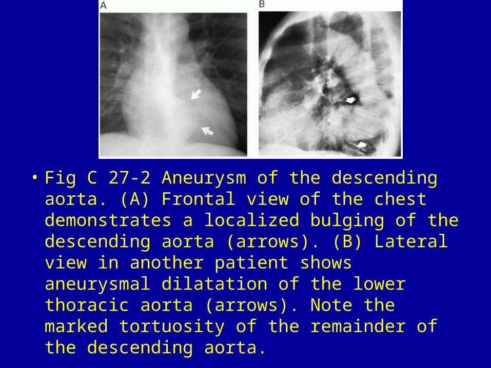
• Fig C 27-2 Aneurysm of the descending aorta. (A) Frontal view of the chest demonstrates a localized bulging of the descending aorta (arrows). (B) Lateral view in another patient shows aneurysmal dilatation of the lower thoracic aorta (arrows). Note the marked tortuosity of the remainder of the descending aorta.

• Fig C 27-3 Hiatal hernia. (A) Frontal and (B) lateral views of the chest demonstrate a huge air-filled hiatal hernia that appears as a posterior mediastinal mass (arrows).

• Fig C 27-4 Megaesophagus. Lateral chest film in a patient with achalasia shows a mixture of fluid and air density in the dilated esophagus (arrows).

• Fig C 27-5 Esophageal varices. (A) Frontal chest radiograph in a patient with severe cirrhosis shows a retrocardiac mass (arrows) that silhouettes the descending aorta and causes abnormal convexity of the azygoesophageal recess. (B) Corresponding MR image reveals extensive paraesophageal vascular channels consistent with varices.19

• Fig C 27-6 Neurenteric cyst. (A) Frontal and (B) lateral views of the chest demonstrate a large, oval, homogeneous mass in the posterior mediastinum. Note the right hydropneumothorax (arrows) with a long air-fluid level that developed as a complication of a diagnostic needle biopsy.

• Fig C 27-7 Tuberculous osteomyelitis of the spine. Large paravertebral abscess produces a fusiform soft-tissue mass about the vertebrae (arrows). There is poorly marginated destruction along with loss of the superior and inferior end plates of the T9 vertebral body.

• Fig C 27-8 Azygos continuation of the inferior vena cava. (A) On the frontal view, there is an irregular paravertebral mass (arrows). (B) Lateral view shows pulmonary vessels in the retrocardiac space but no shadow of the inferior vena cava.


