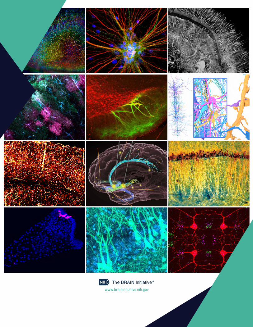
2021 brain initiative calendar
TRANSCRIPT

2021 The BRAIN Initiative®
Show Us Your BRAINs! 12-MONTH CALENDAR
Featuring the top entries from the 2020 BRAIN Initiative Photo & Video Contest
The Brain Research through Advancing Innovative Neurotechnologies® (BRAIN)
Initiative is part of an ambitious, public-private collaborative effort aimed at developing new experimental tools that will
revolutionize our understanding of the brain. www.braininitiative.nih.gov
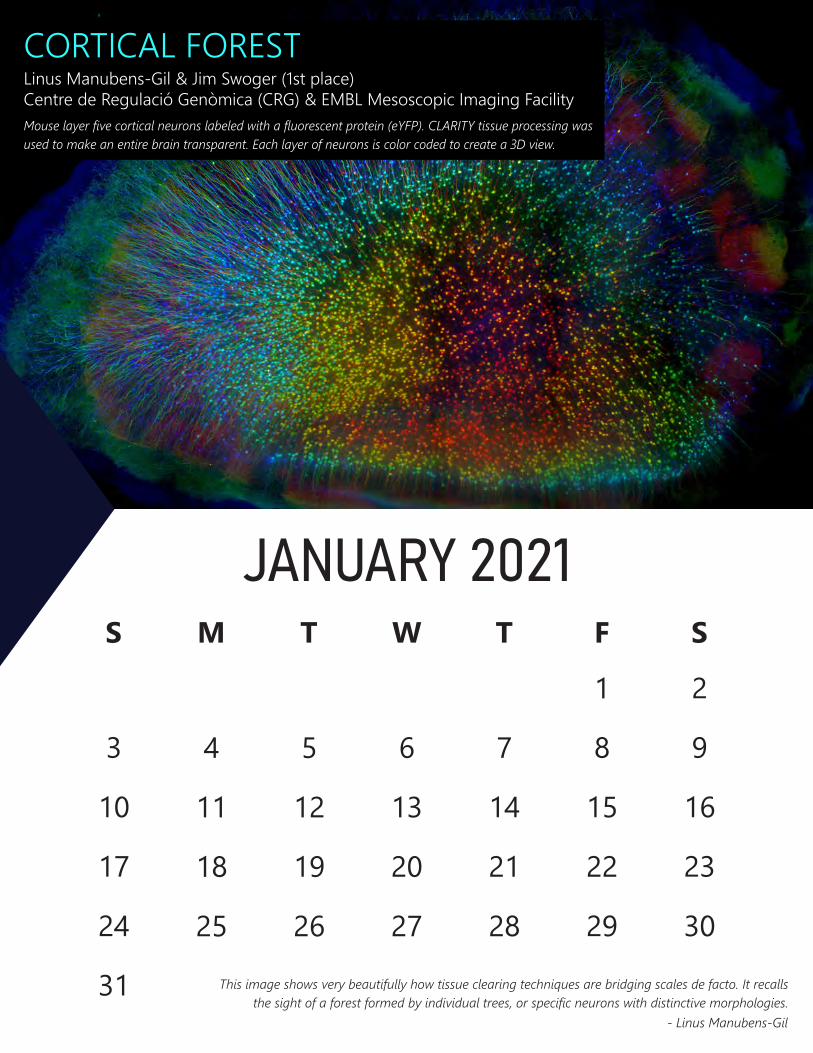
CORTICAL FOREST Linus Manubens-Gil & Jim Swoger (1st place) Centre de Regulació Genòmica (CRG) & EMBL Mesoscopic Imaging Facility Mouse layer five cortical neurons labeled with a fluorescent protein (eYFP). CLARITY tissue processing was used to make an entire brain transparent. Each layer of neurons is color coded to create a 3D view.
S
JANUARY 2021 M T W T F S
1 2
3 4 5 6 7 8 9
10 11 12 13 14 15 16
17 18 19 20 21 22 23
24 25 26 27 28 29 30
31 This image shows very beautifully how tissue clearing techniques are bridging scales de facto. It recalls the sight of a forest formed by individual trees, or specific neurons with distinctive morphologies.
- Linus Manubens-Gil
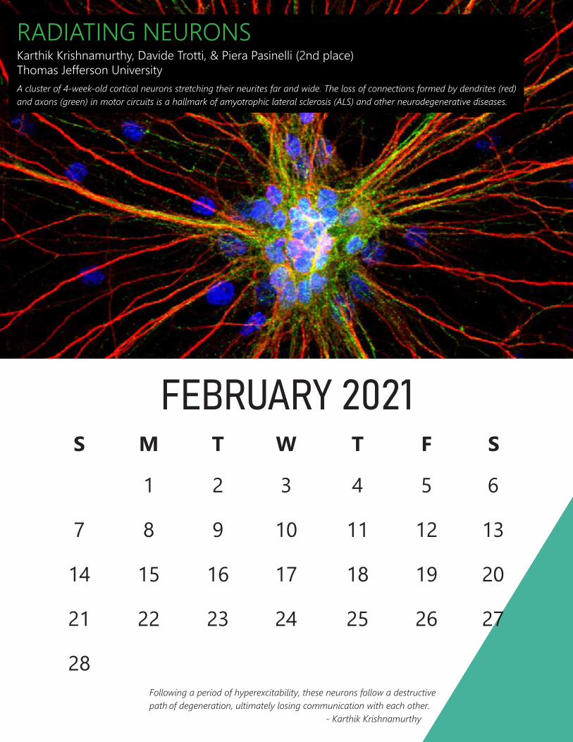
RADIATING NEURONS Karthik Krishnamurthy, Davide Trotti, & Piera Pasinelli (2nd place) Thomas Jefferson University A cluster of 4-week-old cortical neurons stretching their neurites far and wide. The loss of connections formed by dendrites (red) and axons (green) in motor circuits is a hallmark of amyotrophic lateral sclerosis (ALS) and other neurodegenerative diseases.
FEBRUARY 2021 S M T W T F S
1 2 3 4
19
26
20
27
12 13
5 6
7 8 9 10 11
14 15 16 17 18
21 22 23 24 25
28Following a period of hyperexcitability, these neurons follow a destructive path of degeneration, ultimately losing communication with each other.
- Karthik Krishnamurthy
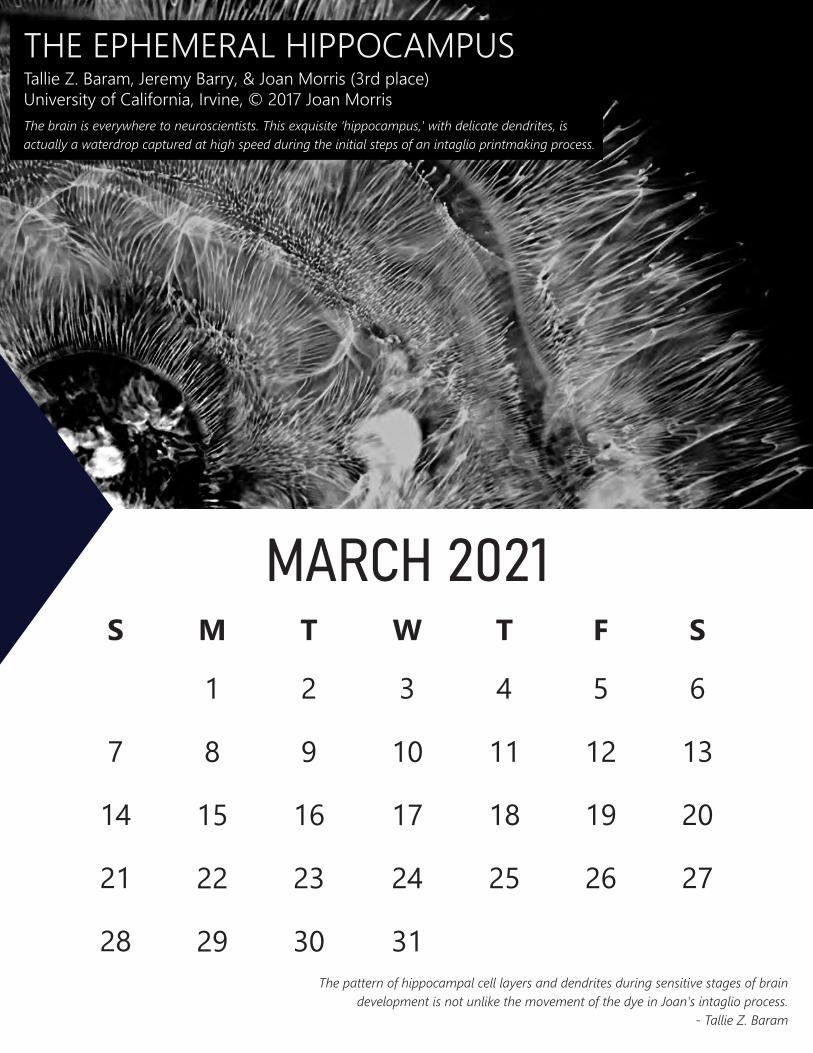
THE EPHEMERAL HIPPOCAMPUS Tallie Z. Baram, Jeremy Barry, & Joan Morris (3rd place) University of California, Irvine, © 2017 Joan Morris The brain is everywhere to neuroscientists. This exquisite ‘hippocampus,' with delicate dendrites, is actually a waterdrop captured at high speed during the initial steps of an intaglio printmaking process.
MARCH 2021 S M T W T F S
1 2 3 4 5 6
7 8 9 10 11 12 13
14 15 16 17 18 19 20
21 22 23 24 25 26 27
28 29 30 31The pattern of hippocampal cell layers and dendrites during sensitive stages of brain
development is not unlike the movement of the dye in Joan's intaglio process. - Tallie Z. Baram
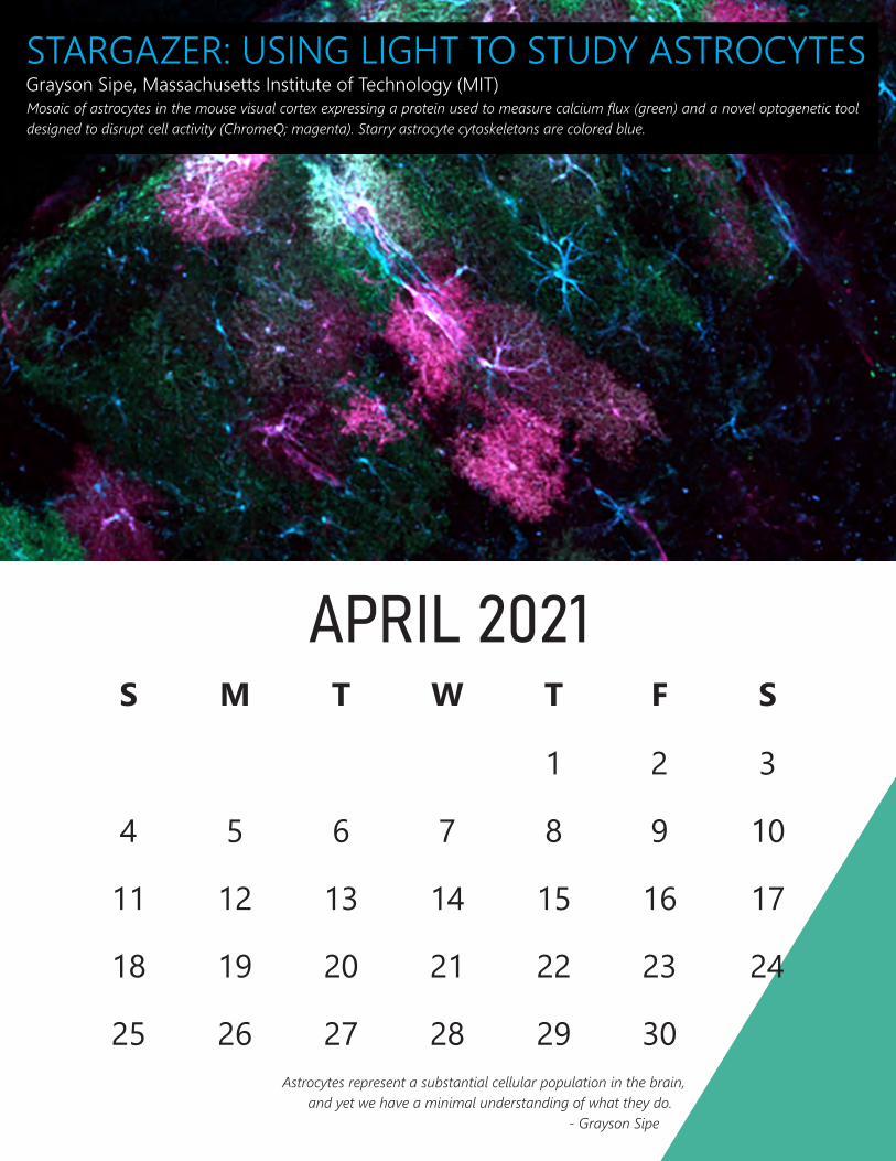
STARGAZER: USING LIGHT TO STUDY ASTROCYTES Grayson Sipe, Massachusetts Institute of Technology (MIT) Mosaic of astrocytes in the mouse visual cortex expressing a protein used to measure calcium flux (green) and a novel optogenetic tool designed to disrupt cell activity (ChromeQ; magenta). Starry astrocyte cytoskeletons are colored blue.
APRIL 2021 S M T W T F S
1 2 3
4 5 6 7 8 9 10
11 12 13 14 15
24
16 17
18 19 20 21 22 23
25 26 27 28 29 30Astrocytes represent a substantial cellular population in the brain,
and yet we have a minimal understanding of what they do. - Grayson Sipe
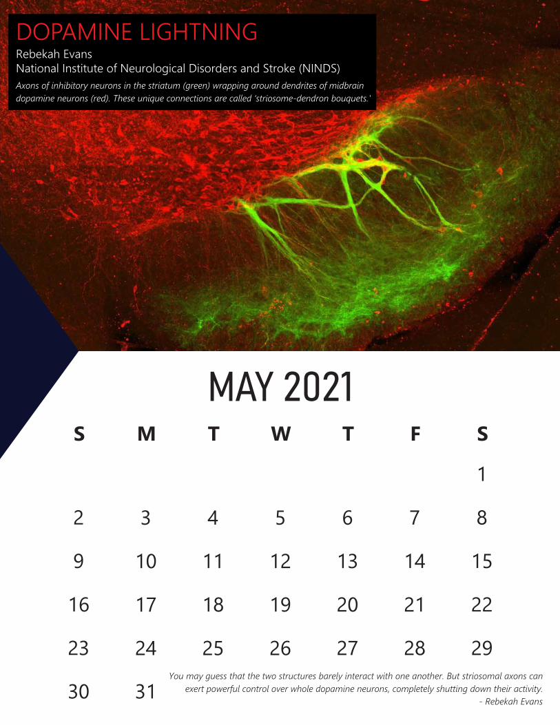
DOPAMINE LIGHTNING Rebekah Evans National Institute of Neurological Disorders and Stroke (NINDS) Axons of inhibitory neurons in the striatum (green) wrapping around dendrites of midbrain dopamine neurons (red). These unique connections are called ‘striosome-dendron bouquets.'
S
MAY 2021 M T W T F S
1
2 3 4 5 6 7 8
9 10 11 12 13 14 15
16 17 18 19 20 21 22
23 24 25 26 27 28 29
30 31You may guess that the two structures barely interact with one another. But striosomal axons can
exert powerful control over whole dopamine neurons, completely shutting down their activity. - Rebekah Evans

COLUMN TO SYNAPSEAmy Sterling, Princeton University 3D reconstruction of a cortical hypercolumn in the mouse brain. This neural structure was recreated by an artificial intelligence pipeline that uses electron microscope images from a cubic mm of mouse visual cortex.
JUNE 2021 S M T W T F S
1 2 3 4 5
6 7 8 9 10 11 12
13 14 15 16 17 18 19
20 21 22 23 24 25 26
27 28 29 30There’s no telling what wonders we will behold in the hidden neuron forest of the brain. - Amy Sterling
7th Annual BRAIN Initiative Investigators Meeting
June 15-17th, 2021

BRAIN IN FLAMES Sripriya Ravindra Kumar, California Institute of Technology (Caltech) An intricate network of blood vessels in the mouse cortex. The newly engineered virus called AAV-PHP.V1 was used to safely encode fiery fluorescent proteins (tdTomato; red) in endothelial cells throughout the brain.
JULY 2021 S M T W T F S
1 2 3
4 5 6 7 8 9 10
11 12 13 14 15 16 17
18 19 20 21 22 23 24
25 26 27 28 29 30 31The brain is a highly vascularized organ and being able to visualize this complex vasculature is very fascinating.
One could only possibly imagine the vital role of vasculature in brain health and disease. - Sripriya (Priya) Kumar

DEEP BRAIN Sameer Sheth, Wayne Goodman, Nader Pouratian, & Kelly Bijanki Baylor College of Medicine & the University of California, Los Angeles Light-based rendering of deep brain stimulation (gray) and stereo electroencephalography (orange) electrodes used to stimulate and record from deep neuronal pathways to study the network basis of depression.
AUGUST 2021 S M T W T F S
1 2 3 4 5 6 7
8 9 10 11 12 13 14
15 16 17 18 19
28
20 21
22 23 24 25 26 27
29 30 31Depression, schizophrenia, and Alzheimer’s disease are network disorders. The better we understand
their brain circuits, the better we’ll be able to treat disorders arising from circuit dysfunction. - Sameer Sheth

FOREST GOLD György Buzsáki, New York University Pyramidal neurons in the CA1 layer of the rat hippocampus. The ‘somatodendritic tree’ of each neuron (gold) was labeled using the Gallyas method, an improved Golgi silver staining technique, and illuminated with dark-field microscopy.
SEPTEMBER 2021 S M T W T F S
1 2 3 4
5 6 7 8 9 10 11
12 13 14 15 16 17 18
19 20 21 22 23 24 25
26 27 28 29 30Dark-field illumination is a long forgotten method of Old World anatomists. The dark field can bring about spectacular
colors, such as gold, and as such, is a great antidote for today's RGB colors typical of multicolor immunostaining. - György Buzsáki
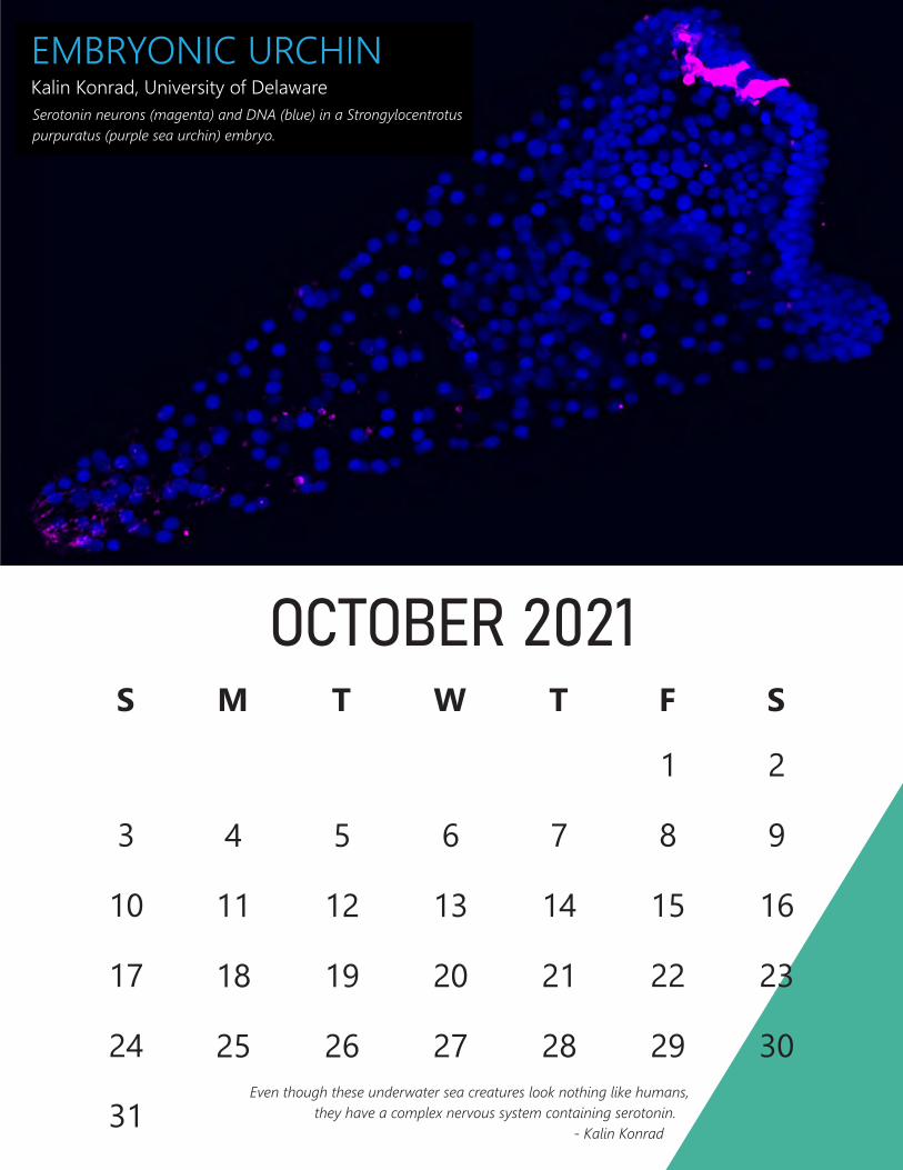
EMBRYONIC URCHIN Kalin Konrad, University of Delaware Serotonin neurons (magenta) and DNA (blue) in a Strongylocentrotus purpuratus (purple sea urchin) embryo.
OCTOBER 2021 S M T W T F S
1 2
3 4 5 6 7 8 9
10 11 12 13 14
23
30
15 16
17 18 19 20 21 22
24 25 26 27 28 29Even though these underwater sea creatures look nothing like humans,
31 they have a complex nervous system containing serotonin. - Kalin Konrad
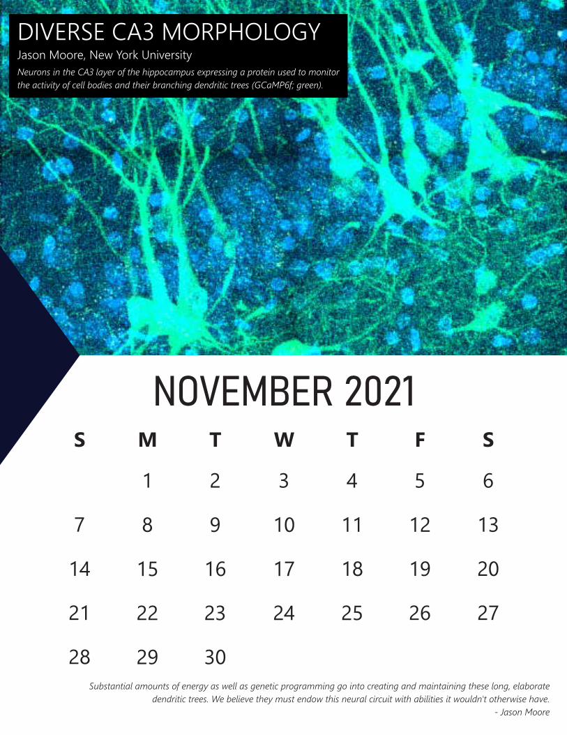
DIVERSE CA3 MORPHOLOGY Jason Moore, New York University Neurons in the CA3 layer of the hippocampus expressing a protein used to monitor the activity of cell bodies and their branching dendritic trees (GCaMP6f; green).
NOVEMBER 2021 S M T W T F S
1 2 3 4 5 6
7 8 9 10 11 12 13
14 15 16 17 18 19 20
21 22 23 24 25 26 27
28 29 30Substantial amounts of energy as well as genetic programming go into creating and maintaining these long, elaborate
dendritic trees. We believe they must endow this neural circuit with abilities it wouldn't otherwise have. - Jason Moore
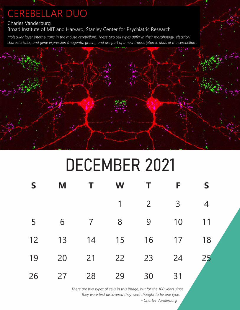
CEREBELLAR DUO Charles Vanderburg Broad Institute of MIT and Harvard, Stanley Center for Psychiatric Research Molecular layer interneurons in the mouse cerebellum. These two cell types differ in their morphology, electrical characteristics, and gene expression (magenta, green), and are part of a new transcriptomic atlas of the cerebellum.
DECEMBER 2021 S M T W T F S
1 2 3 4
5 6 7 8 9 10 11
12 13 14 15 16
25
17 18
19 20 21 22 23 24
26 27 28 29 30 31There are two types of cells in this image, but for the 100 years since
they were first discovered they were thought to be one type. - Charles Vanderburg