2008 3723-3736 3723 the brain, the penis and steroid ... · key words: sex steroids, neurosteroids,...
TRANSCRIPT
Current Pharmaceutical Design, 2008, 14, 3723-3736 3723
1381-6128/08 $55.00+.00 © 2008 Bentham Science Publishers Ltd.
The Brain, the Penis and Steroid Hormones: Clinical Correlates with Endothelial Dysfunction
Abdulmaged M. Traish1,*, Hilal Abu-Zahra
1 and Andre T. Guay
2
1Departments of Biochemistry and Urology, Boston University School of Medicine, Boston, MA, USA and
2Department
of Endocrinology, Center for Sexual Function, Lahey Clinic, Peabody, MA, USA
Abstract: Erectile function is a complex neurovascular process that depends on the health of the central and peripheral
nervous systems and the vasculature. Thus, signaling from the central nervous system (brain) to the peripheral nervous
system (penis) is critical and is modulated by a set of complex interactions that depend on cerebral and vascular circula-
tion. The cerebral and peripheral vasculatures are target tissues for sex steroid hormones. Gonadal, adrenal and neuroster-
oids regulate the function and physiology of the endothelium and modulate vascular and cerebral circulation by genomic
and non-genomic dependent mechanisms. Recent advances in cell and molecular biology have defined a critical role of
endothelium in vascular function. A host of biochemical and clinical markers of endothelium function and dysfunction
have been identified to assess vascular pathology. Emerging evidence suggests that sex steroid hormones play an impor-
tant role in maintaining endothelial health and sex steroid deficiency is associated with endothelial dysfunction, vascular
disease and erectile dysfunction. Such information has important clinical implications in patient management with sex
steroid hormone insufficiency, diabetes, metabolic syndrome, vascular disease and erectile dysfunction. In this review, we
discuss the role of sex steroid hormones in modulation of the biochemical and clinical markers associated with endothelial
dysfunction. Specifically the regulation of endothelial nitric oxide synthase, assymetric dimethylarginine, reactive oxygen
species, endothelin-1, inflammatory cytokines, tumor necrosis factor- , markers of cell adhesion, dysregulation of fibri-
nolytic factors and the inability to regenerate from endothelial progenitor cells concomitant with increased endothelial
apoptosis, increased cellular permeability and increased vascular tone.
Key Words: Sex steroids, neurosteroids, endothelium dysfunction, erectile dysfunction, nitric oxide, nitric oxide synthase, vas-cular tone, vascular disease.
INTRODUCTION
I. Central Nervous System & Erectile Function
Considerable evidence exists on the potential role of neu-rosteroids and neuroactive steroids on sexual function and behavior [1]. In addition, gonadal and adrenal steroids, upon conversion into neuroactive steroids, in the brain, may modulate sexual function and behavior. Furthermore, de novo synthesis of neurosteroids in the central nervous system has been well documented and enzymatic machinery neces-sary for the biosynthetic pathway exists [1]. Sexual desire, arousal, and orgasm are modulated by a complex set of in-teractions between the somatic and autonomic nervous sys-tems, operating at cerebral, spinal, and peripheral levels [2]. Neurosteroids elicit specific responses in select target neu-ronal pathways and modulate sexual function [1]. The exact details of such interactions, however, remain, at best, poorly understood. Neurosteroids and neuroactive steroids as well as peptide hormones modulate neural activities and modify the sexual responses. Further, dopaminergic and serotonergic systems play an important role in various components of the sexual response cycle at the central level. Other neurotrans-mitters including adrenergic, cholinergic, nitergic, gamma-
*Address correspondence to this author at the Department of Biochemistry
& Urology, Boston University School of Medicine, Center for Advanced
Biomedical Research, 700 Albany Street, W607, Boston, MA 02118, USA;
Tel: 617-638-4578; Fax: 617-638-5412; E-mail: [email protected]
aminobutyric acidergic, and neuropeptides also contribute to the sexual response. Temel et al. [3] reviewed data from animal and human studies and proposed that within the cor-tical areas, parts of the frontal lobe (medial and inferior) and cingulate gyrus (anterior) and within the subcortical areas, parts of the amygdala [corticomedial, medial and bed nu-cleus of the stria terminalis (BNST)], thalamus (medial dor-sal, and Cm-parafascicular [Pf] complex), hypothalamus paraventricular nucleus (PVN), medial and lateral, preoptic areas (POAs) and mamillary bodies, nucleus accumbens, fornix and striatum are involved in erection. The authors suggested that brain centers are potent modulators of the spinal centers responsible for generation of penile erection. Salas et al. [4] suggested that the laterodorsal tegmental nu-cleus (LDT) and surrounding region appear to be involved in regulation of penile erection and different anatomical areas in the mesopontine tegmentum may have specific roles in this physiological process. Melis et al. [5] showed that oxy-tocin in the ventral tegmental area (VTA) activates mesolim-bic dopaminergic neurons, which may be involved in the appetitive and rewarding effects of sexual activity. Suzuki et al. [6] investigated the effects of castration and testoster-one (T) replacement on intracavernous pressure (ICP) elic-ited with electrical stimulation of the medial preoptic area (MPOA) and cavernous nerve (CN) in male rats. The authors suggested that T plays an important role not only in the cen-tral nervous system but also in the peripheral neural path-ways for the maintenance and restoration of erectile capacity.
3724 Current Pharmaceutical Design, 2008, Vol. 14, No. 35 Traish et al.
The cerebral vasculature is a target tissue for sex steroid hormones. Estrogens, androgens, and progestins modulate the function and pathophysiology of the cerebral circulation [7]. Estrogens decrease cerebral vascular tone and increase cerebral blood flow by enhancing endothelial nitric oxide synthase (eNOS) expression and activity and facilitating the prostacyclin pathways. Estrogens have important protective effects on cerebral endothelial cells by increasing mitochon-drial efficiency, decreasing free radical production, promot-ing cell survival, and stimulating angiogenesis. Although much has been learned regarding hormonal effects on brain blood vessels, most studies involve young, healthy animals. It is becoming apparent that hormonal effects may be modi-fied by aging or disease states, such as diabetes and athero-sclerosis. Furthermore, the effects of T are complicated be-cause this hormone is also converted to estrogen and DHT, systemically and possibly within the vessels themselves.
Estradiol (E2) regulates Nitric Oxide (NO) synthase (NOS) in the hypothalamus [8] and modulates vascular endothelial growth permeability factor in normal and tumor tissue [9] and glucose transporter-1 expression in blood-brain barrier [10, 11]. E2 has an immediate action on median eminence endothelial cells via non-genomic signaling pathways lead-ing to NO-stimulated GnRH release [12]. Vascular endothe-lial growth factor (VEGF) expression is higher in the neural lobe than in the anterior lobe undetectable in the intermediate lobe and is rapidly up-regulated by E2 in the anterior pitui-tary but remains unchanged in the posterior pituitary [13]. Estrogen receptor alpha (ER ) activation in cerebrovascular tissue resulted in increased eNOS activity and protein levels [14]. Increased NO production by eNOS may contribute to the neuroprotective effects of estrogens. Galea et al. [15] hypothesized that the protective effects of E2 in cerebral ischemia may be attributed to the blockade of leukocyte ad-hesion in cerebral endothelial cells. E2 inhibited the basal and interleukin-1 (IL-1 )-mediated expression of the inter-cellular adhesion molecule type-1 (ICAM1) and NF- acti-vation, in cultured brain endothelial cells. In vivo estrogen treatment leads to a 100% increase in eNOS mRNA copy number and increases eNOS protein levels by 47% in mouse cerebral blood vessels [16]. The authors suggested that es-trogen modulates eNOS at the transcriptional level in blood vessels in vivo. Low E2 results in reduced neuronal nitric oxide synthase (nNOS) and eNOS expression in hippocam-pus and E2 substitution reversed these effects [17] suggesting that E2 increases nNOS and eNOS expression and activity in hippocampus and improves hippocampal function.
II. A Vascular Bed with a Unique Physiological Function:
The Penis
The penis is comprised of two cylindrical chambers, the corpora cavernosa, which comprises the erectile tissue. The tunica albuginea, a thick fibroelastic tissue surrounds the corpora cavernosa. The vascular bed of the erectile tissue encompasses several cellular and non-cellular elements, in-cluding interconnecting sinusoidal spaces, the endothelium lining the lacunar spaces, the trabecular smooth muscle and the fibroelastic connective tissue matrix. The cavernosal ar-teries provide arterial blood flow to the corpora through the resistance helicine arteries and arterioles. Venules located in the subtunical region permit venous blood outflow from the
sinasoids. Corporal smooth muscle relaxation is considered essential for penile erection via increased arterial inflow and restriction of blood out flow. The endothelial cells of the cavernosal arteries, helicine arteries and arterioles as well as the endothelium lining the lacunar spaces play a critical role in regulating the physiological function of the penis. Thus, modulation of endothelium function in the penis by sex ster-oid hormones plays an important physiological role in erec-tile function and dysfunction.
III. Role of Endothelium in Vascular Function
The endothelium is characterized by a dynamic single cell layer, which regulates vascular homeostasis, acts as a semi-permeable layer, and functions as a physical barrier. The endothelium possesses autocrine, paracrine, and endo-crine functions, which play a critical role in regulating vas-cular tone. The endothelium responds to various stimuli such as shear stress by releasing NO [18] and synthesizes and secretes vasoconstrictor molecules such as endothelin-1 (ET-1) and prostaglandin E2 (PGE2). Moreover, the endothelium regulates homeostatic processes including platelet activation, aggregation, inflammation, immune function, vascular per-meability, vascular smooth muscle cell proliferation, and angiogenesis [19]. Endothelial dysfunction is characterized by an imbalance in the expression and activity of the various signaling molecules producing alterations in the biochemical pathways regulating endothelial function therefore resulting in vascular disease such as atherosclerosis and hypertension [20].
IV. Biochemical and Clinical Markers of Endothelial
Dysfunction
Endothelial dysfunction is characterized by significant modifications in the physiological and biochemical parame-ters. These include: vascular stiffness, increased vascular tone, production of inflammatory cytokines, increased per-meability, susceptibility to invasion of immunocytes, a de-crease in endothelial cell growth, and dysregulation of fibri-nolytic factors.
Clinical and biochemical markers of endothelial dysfunc-tion include: a) reduced expression and activity of eNOS, reduced synthesis of NO, and increased production of asymmetric dimethylarginine (ADMA), a competitive, en-dogenous inhibitor of eNOS, b) increased production of re-active oxygen species (ROS) c) increased synthesis and re-lease of the vasoconstrictor peptide ET-1, d) increased pro-duction of inflammatory cytokines such as interluekin-6 (IL-6), C-reactive protein (CRP) and tumor necrosis factor alpha (TNF- ), e) increased expression of markers of cell adhesion such as E-selectin [21], intracellular adhesion molecule (ICAM) [22], and vascular cell adhesion molecule (VCAM), f) dysregulation of fibrinolytic factors such as Von Wille-brand Factor (vWF), tissue plasminogen activator (tPA), and plasminogen activator inhibitor (PAI-1); g) inability to re-generate from endothelial progenitor cells (EPC); h) in-creased endothelial apoptosis; i) increased cellular perme-ability; j) increased vascular tone. In addition to the bio-chemical markers of endothelial dysfunction, diagnostic tools of endothelial dysfunction are characterized by flow mediated dilation (FMD) [23]. This clinical measurement of
Sex Steroids & Endothelial Dysfunction Current Pharmaceutical Design, 2008, Vol. 14, No. 35 3725
endothelial function is strongly linked to coronary endothe-lial dysfunction and predicts cardiovascular events [24-26].
V. Role of Endothelial Dysfunction in Vascular Disease
The mechanisms by which vascular endothelium regu-lates vascular function involve multiple signaling pathways. Expression and activity of eNOS is critical for vascular func-tion and decreased expression or reduced NO synthesis cou-pled with increased scavenging of NO by ROS or increased concentration of the competitive inhibitor, ADMA, contrib-utes to vascular pathology [27]. Reduced NO synthesis and increased ADMA production has been linked to coronary artery disease and erectile dysfunction [28]. Similarly, in-creased production of ET-1 enhances vascular tone and pro-motes loss of vasodilatory properties [29, 30]. Endothelial dysfunction is also characterized by increased expression of markers of cell adhesion, E-selectin [21], soluble intercellu-lar adhesion molecule (sICAM) [22], and VCAM [31]. In-creased serum levels of these factors are indicative of endo-thelial dysfunction. Increased endothelial permeability has also been implicated as a risk factor for cardiovascular dis-ease and this is attributed to invasion of the endothelium by lipoproteins, monocytes, and macrophages. This invasion promotes smooth muscle cell migration and proliferation [32] and facilitates the formation of lesions and atheroscle-rotic plaques [33]. With biochemical insult or injury, the endothelium becomes susceptible to apoptosis and loses its ability to regenerate. In addition, endothelial progenitor cells growth is regulated and may not compensate for the loss of endothelium via apoptosis, thus, exacerbating vascular per-meability. Finally, dysregulation of fibrinolytic factors such as vWF, tPA, and PAI-1 also characterize endothelial dys-function.
V1. Sex Steroid Hormones Regulate Endothelial Func-
tion
Considerable body of evidence exists linking sex steroid hormones deficiency to endothelial dysfunction [34, 35]. Low plasma T level was associated with endothelial dys-function in men independent of other risk factors, suggesting a protective effect of endogenous T on the endothelium [36]. Gonadal hormones affect myogenic tone in male rat cerebral arteries through NOS- and/or endothelium-dependent mech- anisms [37]. Low serum free T, estrone, and free Insulin-like Growth Factor (IGF) were inversely related to intima media thickness (IMT) [38]. Similarly, an inverse relationship ex-ists between T level and thoracic IMT [39]. Examination of the endothelium from castrated rats by transmission electron microscopy demonstrated significant endothelium damage, in which the cell surface appeared crumpled, rough, adhesive and ruptured [40]. This pathology was partially restored by treatment of castrated rats with T or DHT. These observa-tions strongly suggested that low concentrations of T or DHT are associated with ultrastructural damage of the aortic endothelium. Mäkinen et al. [41] have shown that middle-aged men with symptoms of androgen deficiency are at risk of increased carotid IMT and suggested that normal T levels may offer protection against the development of atheroscle-rosis in middle-aged men. Malkin et al. [42, 43] hypothe-sized that the immune-modulating properties of T are impor-tant in inhibiting atheroma formation and progression to
acute coronary syndrome. The authors demonstrated that significant reduction in total cholesterol was recorded with T therapy and demonstrated a shift in the cytokine balance to a state of reduced inflammation. DHEA restored aortic eNOS levels and eNOS activity suggesting that DHEA may have direct genomic and non-genomic effects on the vascular wall [44, 45]. Liu & Dillon [46, 47] demonstrated that physio-logical concentrations of DHEA acutely increase NO release from intact vascular endothelial cells, by a plasma mem-brane-dependent mechanism. This action of DHEA is medi-ated by a steroid-specific, G-protein coupled receptor mech- anism, which activates eNOS in both bovine and human en-dothelial cells. This cellular mechanism may underlie some of the cardiovascular protective effects proposed for DHEA. Parenteral T therapy improves both endothelial-dependent (flow-mediated) and endothelium-independent brachial ar-tery vasodilation in postmenopausal women using long-term estrogen therapy [48]. Suppression of endogenous estrogens with aromatase inhibitors resulted in impairment of FMD without significant changes in lipoproteins, homocysteine or CRP [49], suggesting that endogenous estrogens play a di-rect regulatory role in endothelial function in young healthy men.
VII. Biochemical and Clinical Markers of Endothelial
Dysfunction
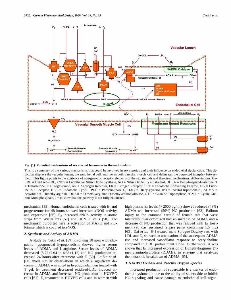
Fig. (1) provides a scheme of the potential mechanisms by which sex steroid hormone modulate endothelial and vas-cular function by multiple, and overlapping, signaling path-ways. These reactions involve genomic and non-genomic mechanisms which stimulate endothelial function to produce endocrine and/or paracrine factors that affect the underlying vascular bed. Sex steroid deficiency contributes to endothe-lial and smooth muscle dysfunction and vascular disease. Fig. (2) illustrates the relationship between endothelial dys-function, biochemical markers and the role of sex steroids on these parameters. In the proceeding sections, we discuss the effects of sex steroid hormones on expression and activities of endothelial biomarkers of function and dysfunction.
1. Endothelial Nitric Oxide Synthase (eNOS) Expression and Activity
Considerable evidence exists suggesting that eNOS is regulated by sex steroid hormones [44, 34, 50]. Marin et al. [51] demonstrated that castration reduced both nNOS and eNOS expression and activity and T treatment restored eNOS in corpora cavernosa. Treatment of bovine aortic en-dothelial cells (BAEC) with DHEA increased expression of eNOS [45, 47, 52] and stimulated an increase in NO secre-tion via PI3 kinase-dependent pathways [53] and in another study via non-genomic pathway with concomitant increase in cyclic guanine monophoshate (cGMP) release from endo-thelial cells [47]. Simoncini et al. [44] showed DHEA treat-ment in human umbilical vein endothelial cells (HUVEC) induced a concentration dependent increase in NO release in cultured medium via activation of eNOS via a non-genomic signaling pathway.
E2 increases eNOS protein expression in rat cerebral mi-crovessels via receptor mediated signal pathways [54] and E2 treatment of BAEC caused eNOS translocation from the in-tracellular membrane to the nucleus via a Ca
2+ dependent
3726 Current Pharmaceutical Design, 2008, Vol. 14, No. 35 Traish et al.
mechanism [55]. Human endothelial cells treated with E2 and progesterone for 48 hours showed increased eNOS activity and expression [56]. E2 increased eNOS activity in aortic strips from Wistar rats [57] and HUVEC cells [58]. The mechanism purported here is activation of MAPK and PI3-Kinase which is coupled to eNOS.
2. Synthesis and Activity of ADMA
A study by Cakir et al. [59] involving 18 men with idio-pathic hypogonadal hypogonadism showed higher serum levels of ADMA and L-arginine. Serum levels of ADMA decreased (5.72±2.64 to 4.36±1.12) and NO production in-creased 24 hours after treatment with T [59]. Leifke et al. [60] made similar observations in which a significant de-crease in ADMA was noted in hypogonadal men treated with T gel. E2 treatment decreased oxidized-LDL induced in-crease in ADMA and increased NO production in HUVEC cells [61]. E2 treatment in HUVEC cells and in women with
high plasma E2 levels (> 2000 pg/ml) showed reduced (48%) ADMA and increased (56%) NO production [62]. Balloon injury to the common carotid of female rats that were bilaterally ovariectomized had an increase of ADMA and a decrease of NO production that was rescued with E2 treat-ment (90 day sustained release pellet containing 1.5 mg) [63]. Dai et al. [64] treated male Sprague-Dawley rats with LDL and E2 showed an attenuation of the subsequent ADMA rise and increased vasodilator response to acetylcholine compared to LDL pretreatment alone. Furthermore, it was shown that E2 increased expression of Dimethylarginine Di-methylaminohydrolase (DDAH), an enzyme that catalyzes the metabolic breakdown of ADMA [65].
3. NADPH Oxidase and Reactive Oxygen Species
Increased production of superoxide is a marker of endo-thelial dysfunction due to the ability of superoxide to inhibit NO signaling and cause damage to endothelial cell organ-
Fig. (1). Potential mechanisms of sex steroid hormones in the endothelium
This is a summary of the various mechanisms that could be involved in sex steroids and their influence on endothelial dysfunction. This de-
piction displays the vascular lumen, the endothelial cell, and the smooth vascular muscle cell and delineates the purported interplay between
them. This figure points to the existence of non-genomic receptor elements of the sex steroids and theorized mechanisms. Abbreviations: Ox-
LDL = Oxidized-LDL, eNOS = Endothelial Nitric Oxide Synthase, NO = Nitric Oxide, E2 = Estradiol, DHEA = Dehydroepiandrosterone, T
= Testosterone, P = Progesterone, AR = Androgen Receptor, ER = Estrogen Receptor, ECE = Endothelin Converting Enzyme, ETA = Endo-
thelin-1 Receptor, ET-1 = Endothelin Type-1, PLC = Phospholipase C, DAG = Diacylglycerol, IP3 = Inositol triphosphate , ADMA =
Assymetrical Dimethylarginine, DDAH = Dimethlyarginine Dimethylaminohydrolase, GTP = Guanine Triphosphate, cGMP = Cyclic Gua-
nine Monophosphate, ? = to show that the pathway is not fully elucidated.
Sex Steroids & Endothelial Dysfunction Current Pharmaceutical Design, 2008, Vol. 14, No. 35 3727
elles. Thus, reduction in superoxide levels represents a pro-tective function. DHEA inhibited macrophage superoxide production as well as neutrophil and granulocyte prolifera-tion [66] and inhibited 12-O-tetradecanoylphorbol-13-acetate (TPA) stimulated superoxide anion (O2-) formation by hu-man neutrophils [67]. Brignardello et al. [68] showed that in ten patients with type-2 diabetes treatment with DHEA re-sulted in decreased ROS. DHEA also decreased TNF- -induced ROS in human endothelial cells [69]. Male Wistar rats undergoing ischemic reperfusion of the kidneys show increased oxidative stress levels, which were reduced upon DHEA treatment [70].
E2 inhibited NADPH oxidase in human monocytic cells and prevents accumulation of ROS [71] by inhibiting precur-sors of the NADPH oxidase reaction whereas the other sex steroids did not have an effect. Also, increased ROS were noted in ovariectomized mice and ROS levels were attenu-ated by E2 treatment [72]. E2 significantly reduced superox-ide induced VSCM proliferation via E2 dependent mecha-nisms in aortic smooth muscle cells of male rats [73]. E2 and P treatment in female porcine coronary arteries decreased
superoxide anion production by 67% while P treatment in-creased superoxide production by 59% respectively [74]. The purported theory for why P increased superoxide may be linked to regulation of NADPH oxidase.
4. ET-1 Expression and Activity
Takahashi et al. [75] demonstrated that endothelin con-verting enzyme and endothelin receptor subtypes A and B were up-regulated in castrated male Sprague-Dawley rats, suggesting an inhibitory effect of T. Kumanov et al. [76] reported that 33 patients with an average age of 21.58±0.69 years with various forms of hypogonadism had significantly higher ET-1 levels than 14 age-matched healthy controls. When these patients were treated with T depot intramuscu-larly over 6 months, ET-1 levels decreased, yet not signifi-cantly. DHEA treatment stimulated increased ET-1 protein expression via a non-genomic MAPK-dependent pathway in BAEC [53]. Peng et al. [77] showed that DHEA reversed ET-1 induced tension in human pulmonary artery ring smooth muscle cells via up-regulation of Kca channel. DHEA also stimulated eNOS production and thus the balance of
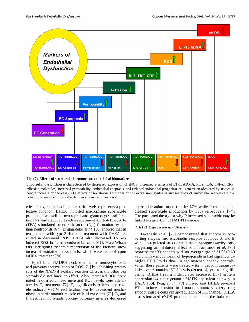
Fig. (2). Effects of sex steroid hormones on endothelial biomarkers
Endothelial dysfunction is characterized by decreased expression of eNOS, increased synthesis of ET-1, ADMA, ROS, IL-6, TNF- , CRP,
adhesion molecules, increased permeability, endothelial apoptosis, and reduced endothelial progenitor cell generation (depicted by arrows to
denote increase or decrease). The effects of sex steroid hormones on the expression, synthesis and secretion of endothelial markers are de-
noted by arrows to indicate the changes (increase or decrease).
3728 Current Pharmaceutical Design, 2008, Vol. 14, No. 35 Traish et al.
DHEA between MAPK and PI 3-kinase signaling pathways may explain DHEA’s conflicting reports discussing benefi-cial cardiovascular effects. Ba et al. [78] showed that E2 at-tenuated ET-1 induced vasoconstriction in male rat aortic vessels following trauma-hemorrhage via an estrogen recep-tor type-beta (ER ) mediated pathway that is independent of endothelium derived eNOS when compared to sham oper-ated controls. E2 administration in male Sprague-Dawley rats with aneurysmal subarachnoid hemorrhage (SAH) showed significantly reduced serum levels of ET-1 as compared to untreated SAH and control groups [79]. As can be seen, E2 treatment consistently reduces ET-1 levels, in vitro and in vivo studies, via an estrogen receptor-dependent mechanism.
5. Vascular Tone
T treatment caused vasodilation in porcine coronary ar-teries [80] and T and DHT caused vasodilation in human umbilical arteries via a non-androgen receptor (AR) pathway [81]. Supraphysiological doses of T relaxed isolated radial arteries via activation of ATP/potassium channels [82] and caused vasodilation in internal mammary arteries [83]. T had a vasodilatory effect on rabbit tracheal smooth muscle medi-ated via eNOS [84]. T caused arterial relaxation via inhibi-tion of Ca
2+ entry into the smooth muscle cells [85, 86]. Fur-
thermore, T has also been able to cause vasodilation in de-nuded vessels [83, 87, 88]. Sader et al. [89] has shown that 23 men with an average age of 32 years old were randomized into three groups receiving either 10 mg of T alone or in combination with 10 or 20 mg of E2 and showed a dose-dependent increase in FMD. Webb et al. [90] showed that T treatment into the left coronary artery caused vasodilation and increased flow. The underlying molecular mechanism for the effect of T has yet to be completely elucidated. Yildiz & Seyrek [91] hypothesized that since denuded vasculature produced the same result the major effect of T is thought to be mediated directly by the vascular smooth muscle. The postulated mechanism suggests that T either activates K
+
channels to increase efflux and/or inhibits Ca2+
channels causing hyperpolarization and subsequent vasodilation. Be-cause these changes occur in seconds to minutes, it is sug-gested that this action is likely to be mediated via the interac-tions with receptors on the membrane (non-genomic effect) rather than interaction with the nucleus (genomic effect). Furthermore, other studies have shown the presence of AR receptors on the membrane of vascular smooth muscle cells [92, 93] suggesting that the proposed mechanism is likely.
DHEA produced vasodilation in human umbilical arteries [81] and in porcine coronary arteries [80]. E2 resulted in arte-rial relaxation via inhibition of Ca
2+ entry into the smooth
muscle cells [85] and inhibited intracellular Ca2+
increase via a non-genomic pathway [94] and E2 produced vasorelaxation in human coronary arteries [95]. The purported mechanism for DHEA, E2 and P are similar to those of T, non-genomic and with wide variation of potential mechanisms most likely due to variations in the vasculature. The major theme is that a Ca
2+ transporter is inhibited, in the case of E2, L-type Ca
2+
channel is inhibited on the membrane of vascular smooth muscle cells [94]. Thus, sex steroid hormones have an im-portant role in regulating vascular tone via endothelium de-pendent and independent pathways.
6. CRP, IL-6 and Inflammatory Markers
In men over the age of 70 years old, total T was inversely correlated with CRP [96]. No correlation was found between total or free T or DHEA and CRP in middle-aged and elderly men [97] and CRP did not change with T or DHT treatment. In 61 eugonadal men (ages 18-35) treated with T-enthanate for 20 weeks no changes in CRP levels were noted [98]. CRP levels also did not change with DHT treatment in an-drogen deficient men over the age of 60 [99]. DHT treatment did not affect IL-6 in human osteoblastic cells [100]. In eugonadal men, exogenous therapy with DHT and T did not increase inflammatory markers [99]. The overall data on CRP is inconclusive. T, DHT, and DHEA either elicits no effect or mitigates release of inflammatory cytokines and thus may have a protective role. Thus far, there is no consen-sus on this issue. In NC/Nga mice, a model for human atopic dermatitis, DHEA treatment prevented an age-induced in-crease in IL-6 production [101]. DHEA and DHEAS inhib-ited IL-6 production in healthy human derived monocytes [102]. Castrated mice receiving DHT treatment showed a decrease in release of pro-inflammatory cytokines interleu-kin IL-1 and IL-6 from splenic and peritoneal macrophages [103].
7. Expression and Function of VCAM, ICA and E-Selectin
T inhibited VCAM-1 mRNA and protein expression in HUVEC most likely via conversion to E2 with endogenous aromatase [104]. Interestingly, T increased TNF- -induced expression of E-selectin and VCAM-1 in HUVEC cells [105]. T treatment of castrated rabbits showed a decrease in sICAM, matrix metalloproteinase (MMP) and reduced plaque atherogenesis and aortic intimal thickness [106]. DHT treatments promoted vascular cell adhesion via up-regulation of VCAM-1, which is thought to occur mainly in the male endothelial cells but not in the female [107]. Incu-bation of HUVEC with DHT for 48 hours caused monocyte adhesion in a dose-dependent manner by increasing expres-sion of VCAM-1. This reaction was blocked by addition of an anti-VCAM-1 antibody [108]. Thus it appears that T ex-hibited an endothelial protective effect whereas DHT had a deleterious effect. Aortic endothelial cells incubated with DHEAS for 48 hours and then treated with TNF- caused up-regulation of ICAM and attenuation of VCAM via inhibi-tion of NF- [109]. DHEA inhibited oxidized LDL induced expression of VCAM/ICAM/PECAM-1 and U937 cells ad-hesion to HUVEC cells [110]. DHEA had no effect on VCAM/ICAM/E-Selectin in HUVEC cells [111]. DHEA inhibited adhesion of HUVEC cells with and without TNF- induction and also inhibited ICAM-1 expression but not E-selectin expression [69]. E2 showed no modulatory effect on ICAM expression in HUVEC [112] but increased TNF- -induced expression of E-selectin and VCAM-1 [105]. E2 reduced VCAM-1 expression via reduction of NF- , activa-tor protein-1 (AP-1), and GATA in human saphenous endo-thelial vein cells [113].
8. Expression and Function of TNF-
T inhibited a myriad of leukocyte cytokine secretions including IL-2, IL-4, IL-10, IFN- , and TNF- on peripheral leukocytes of healthy males [114]. Male rats with low serum T presented with higher incidences of cardiac failure and
Sex Steroids & Endothelial Dysfunction Current Pharmaceutical Design, 2008, Vol. 14, No. 35 3729
also T lowered TNF- mRNA expression [115]. Zhang at-tributed the mechanism to direct action of T on macro-phages. However, a study in macrophages showed that T did not affect TNF- release [116]. Furthermore, T treatment of castrated rabbits showed a decrease in TNF and IL-6 [106]. DHEA and its analogs were shown to inhibit TNF- produc-tion in J774A.1 cells, a murine macrophage cell line [117]. DHEA reduced TNF- and TNF- receptor system [68]. DHEA treatment of RAW 264.7 cells, a murine macrophage culture, significantly reduced TNF- levels [118]. DHEA administration to NMR1 mice following induced sepsis is accompanied by a decrease in TNF- release [119]. Male Wistar rats undergoing ischemic reperfusion of the kidneys showed increased TNF- production that was reduced upon DHEA treatment via improvement of oxidative balance [70]. Lipopolysaccharide (LPS) induced TNF- levels were sig-nificantly decreased with DHEA treatment in CD1 female mice [120]. This result was purported to be obtained by mitigation of endotoxic shock effects of the TNF- pathway.
9. Endothelial Cell Apoptosis
Treatment of human umbilical vein cells (EA.H926) with T reduced Bcl-2 protein expression [121]. DHT had no effect in blocking fluvustatin induced apoptosis of endothelial cell line EA.H926 [122]. Supraphysiological doses of T given to HUVEC cells induced apoptosis [123]. DHEA treatment protects against endothelial cell apoptosis by up-regulating transcription and translation of the anti-apoptotic protein Bcl-2 [124]. HUVEC cells were treated with TNF- and oxidized LDL to induce apoptosis. TNF- induced apopto-sis, which was not altered by E2 treatment after 6 hours, whereas oxidized LDL caused apoptosis at 24 hours and this was attenuated by E2 via increases in the anti-apoptotic pro-teins, Bcl-2 and Bcl-xL [125]. E2 treatment enhanced growth and reduced TNF- induced apoptosis in EC’s [126]. In human endometrial endothelial cells (HEEC), E2 and P in-hibited apoptosis [127]. Intracarotid artery injection of 0.01 mmol/L of hydrogen peroxide into eight-week female old rats caused endothelial cell apoptosis. Treatment with E2 reduced the rate of apoptosis of EC’s by 50%. However, treatment with P did not have an effect [128]. A study with excised resistance arteries from 66 post-menopausal women showed that E2 treatment decreased signs of endothelial cell apoptosis [129]. The majority of findings point to a protec-tive effect of sex steroids by inhibition of apoptotic genes and/or up-regulation of anti-apoptotic genes.
10. Endothelial Progenitor Cell Growth
Treatment of E304 endothelial cells with DHT increased endothelial cells and had a bimodal effect on vascular smooth muscle cells (VSMC’s) in which high doses de-creased proliferation and low doses increased proliferation [130]. T treatment increased endothelial cell proliferation via a MAPK kinase signaling pathway perhaps mediated by a putative G protein-coupled receptor on the plasma membrane [52]. Foresta et al. [131] showed that in 10 men with idio-pathic hypogonadotropic hypogonadism had a lower serum EPC’s compared to normal controls (37.3 and 98.1 cells/ml) and when T was administered the number of EPC’s rose to 170.5 cells/ml in 6 months. The mechanism was attributed to an androgen receptor on CD34-positive cells [132]. DHEAS inhibited endothelial cell growth, whereas none of the other
sex steroids had an effect [133]. DHEA also inhibited human umbilical vein cell proliferation in a dose-dependent manner via up-regulation of p53 and p21 and androgen/estrogen re-ceptor independent mechanisms. E2 increased cell prolifera-tion, where as T inhibited cellular proliferation [134]. DHEA treatment increased EC proliferation independent of andro-gen receptor in BAEC [52]. It appears that DHEA or DHEAS effect on endothelial cell regeneration is pro-atherogenic and is mediated independent of androgen or estrogen pathways. E2 enhances growth and reduces TNF- induced apoptosis in EC’s. The enhanced EC growth may be mediated via telom-erase activity and attenuation of MAPK signaling [126]. Overall, it appears that sex steroids increase cellular regen-eration through a host of mechanisms dependent or inde-pendent of genomic action of sex steroid receptors.
11. Vascular Permeability
Increased endothelial permeability is attributed to in-creased phosphorylation of occludin, the main component of gap junction content. When HUVEC cells were pretreated with E2 and DHT for 24 hours occludin expression was in-creased which decreases permeability via increased MAPK signaling and perhaps via cytochrome C-oxidase modulation and this protects against endothelial dysfunction [135]. DHEAS substantially increased vascular permeability in male ddY mice [136]. This response was blocked by Diphen- hydramine (DPH), a histamine receptor antagonist, implying that DHEAS induces histamine release and this affects per-meability. Interestingly, DHEAS-induced increase in perme-ability was blocked by P. Topical application of Fluasterone (DHEA analog) increased vascular permeability in mouse skin [137]. The proposed mechanism is that DHEA inhibits glucose-6-phosphate dehydrogenase (G6PD), which reduces the supply of NADPH required species, which would subse-quently lower permeability. E2 treatment of HUVEC cells showed a decrease in endothelial cell permeability whereas P reversed the effect of E2 [138]. V-Cadherin, which is known to be associated with vessel permeability [139], was up-regulated by E2 and down-regulated by P. Sex steroids de-crease permeability either with direct influence on occludins, cadherins, tight junctions, and other related compounds as well as indirectly through increased eNOS or reduction in ROS.
12. Expression and Function of PAI-1, vWF factor and tPA
HUVEC cells treated with physiological doses of T de-creased PAI-1 levels [140] and increased the antigen levels of tPA. However, at a larger dose, antigen levels of tPA were decreased. BAEC treated with T showed biphasic modula-tion of PAI-1. At low concentrations PAI-1 was up-regulated and at high concentrations PAI-1 was down-regulated [141]. T treatment with two 2.5 mg patches daily for 12 weeks did not affect tPA and PAI-1 in 46 men (average age 62 years old) with chronic stable angina [142]. A study of 28 hemo-dialysis patients showed that there was no correlation be-tween vWF and T [143]. T treatment in female to male trans-sexuals did not alter tPA or PAI-1 levels. During venous occlusion (VO) tPA increased whereas PAI-1 did not change at baseline and after 4 months of T treatment. Transdermal treatment was not effective compared to oral treatment [144]. In HEEC, E2 and P showed no change in PAI-1 levels [127].
3730 Current Pharmaceutical Design, 2008, Vol. 14, No. 35 Traish et al.
E2 or P treatment of BAEC showed biphasic modulation of PAI-1. At low concentrations PAI-1 was up-regulated and at high concentrations PAI-1 was down-regulated [131]. In male to female transsexuals treated with oral ethinyl E2 and cyproterone acetate (CA), an anti-androgen, reduced tPA and PAI-1 sharply. Serum levels of tPA changed during the (VO) before and 4 months into the ethinyl E2 whereas PAI-1 did not change in either cases. Transdermal treatment was not effective compared to oral treatment [144]. DHEA treatment increased cGMP activity, a marker for NO production, which decreased PAI-1 in 24 healthy elderly men (65 year old)
[52]. DHEA treatment (50 mg 3xper day for 12 days) for 18 men reduced PAI-1 (55.4±3.8 ng/ml to 38.6±3.3 ng/ml) and tPA (from 8.1±1.9 ng/mL to 5.4±1.3 ng/mL) [145]. Serum levels of DHEAS did not change as vWF, PAI-1 and tPA changed [146]. DHEA treatment (150 mg/daily 40 days du-ration) in men with DHEAS levels < 2000 mg/l and verified coronary heart disease (CHD) did not influence PAI-1 and tPA plasma concentrations [147].
VII. Implications of Sex Steroid Hormone Deficiency in
Endothelial Dysfunction
With age, circulating levels of sex steroid hormones de-crease in both men [148, 149] and women [150]. The Rotter-dam study, a population based cohort study, showed low levels of endogenous androgens are associated with in-creased likelihood of atherosclerosis in elderly men [151]. Svartberg demonstrated an inverse relationship between total T levels and carotid IMT [152]. This finding was not inde-pendent of body mass index (BMI). Accumulating evidence has shown an association of low T with cardiovascular mor- tality, morbidity in men of varying age, and cardiovascular risk factors [36, 39, 41, 153, 154]. Men have a higher rate of cardiovascular diseases (CVD) than females. The likely cul-prits appear to be T, DHT, DHEA and their metabolites. Ca-paldo et al. [155] showed that men with sex steroid defi-ciency had a greater IMT thickness. According to Akishita et al. [36], low plasma T levels were associated with endothe-lial dysfunction independent of other factors. Low plasma T has also been associated with cardiovascular risk in healthy men [156]. Akishita et al. [157] also found that DHEAS lev-els correlate with FMD analysis and it was irrespective of other confounding factors in women.
A poignant view on the effects of androgen deficiency on vascular function can be seen with the adverse effects of androgen deprivation therapy (ADT) in prostate cancer pa-tients. ADT, whether via orchiectomy or use of GnRH ago-nists or antagonists results in low circulating T/DHT with concomitant changes in body composition, insulin resistance and vascular disease [158]. There is a decrease in lean mus-cle mass and an increase in fat mass. A long-term study (1-8 months) comparing men undergoing ADT to eugonadal men found an increase in fat mass compared to controls of eugo-nadal men [159]. ADT has been implicated in inducing metabolic syndrome [160]. Overall, ADT in men with pros-tate cancer has been shown to increase risk of cardiovascular events [161]. Keating et al. [162] reported that men undergo-ing ADT had 25% increase in risk of coronary artery disease compared to non-ADT. In a large study consisting of 23,000 men undergoing ADT for at least 12 months showed an in-crease of cardiovascular morbidity by 20% compared to non-
ADT men after controlling for confounding factors [163]. A recent report found that men receiving ADT were approxi-mately 2.6 times at greater risk of cardiovascular mortality than non-ADT controls after adjusting for confounding fac-tors [164]. Interestingly a new study by D’amico et al. [165] have suggested that elderly men with T-1 to T-2 localized prostate cancer should not be given primary ADT due to reduced overall survival in these patients. Montalcini et al. [166] showed that post-menopausal women in the lowest T tertile had the least FMD which implies that not only does estrogen deficiency play a role in cardiovascular disease, but T deficiency as well.
VIII. Endothelium Dysfunction Contributes to Erectile
Dysfunction
Erectile dysfunction (ED) and atherosclerosis share simi-lar risk factors [167]. It has been hypothesized that ED may be an early warning marker for cardiovascular disease [168, 169] Gazzaruso et al. [170] has shown a higher incidence of ED among men with diabetes and overt and silent cardiovas-cular artery disease (CAD). In patients with CAD, the preva-lence of ED was 8 times more likely [171]. Montorsi et al. [172] showed that in men with CAD, the incidence of ED was approximately 49%. It was also shown that there was a correlation between ED and cardiovascular morbidity in 132 men [173]. Nurkalem et al. [174] showed reduced coronary blood flow in ED patients. In a study comprising 9,000 men, ED was found to independently predict cardiovascular dis-ease at a rate similar to smokers, patients with familial car-diovascular disease or hypercholesteremia [169]. It is known that ED and CAD both arise from the underlying endothelial dysfunction [175, 176]. An important cause of both ED and endothelial dysfunction arises out of a decreased production of NO or down-regulation of eNOS, NO is vital in vasore-laxation as well as modulating smooth muscle cells and in-hibiting cellular adhesion [177].
DISCUSSION AND CONCLUSIONS
Erectile dysfunction is a neurovascular process that re-quires healthy central and peripheral nervous systems and the peripheral vascular beds. Erectile dysfunction has re-ceived great attention over the past decades and this is attrib-uted to the advances in research made in vascular biology of the erectile tissue. Further, new information is emerging sug-gesting that the central nervous system play a critical role in the regulation of the mechanism involved in sexual function and behavior via neurosteroids.
The endothelium plays a critical role in the physiological function of all vascular beds, maintaining vascular homeo-stasis thus preventing initiation or progression of vascular disease. Any insult or injury to the endothelium may produce pathological states and dysfunction. Synthesis and release of vasodilators from the endothelium such as NO, and EDHF are integral to maintenance of physiological function. Endo-thelial damage due to various insults contribute to vascular disease and erectile dysfunction.
Considerable body of literature is available indicating that steroid hormones modulate endothelial function in all vascular beds including the brain and the penis and their de-ficiency promote endothelial dysfunction. Androgens and
Sex Steroids & Endothelial Dysfunction Current Pharmaceutical Design, 2008, Vol. 14, No. 35 3731
estrogens produce specific and marked biological effects on endothelial function as demonstrated by the changes in the endothelial markers of function and dysfunction. Low T and DHT are associated with ultrastructural damage of the aortic endothelium. Also, endothelial dysfunction in men is associ-ated with low plasma testosterone level independent of other risk factors, suggesting a protective effect of testosterone on the endothelium. Furthermore, free testosterone level is in-versely correlated with VCAM-1 concentration and IMT, which are indicators of endothelial function. Several studies have also corroborated that DHEA also improved endothelial function in vascular beds. These observations point to the clinical relevance of sex steroid in vascular health and to treating patients with hormonal deficiencies with appropriate physiological hormone levels formulations. Better under-standing of the role of sex steroid hormones in regulating endothelial function is critical to translation of the basic re-search into treatment of patients with metabolic syndrome, vascular disease and erectile dysfunction.
ACKNOWLEDGEMENTS
This work was supported by the Departments of Bio-chemistry and Urology, Boston University School of Medi-cine and Department of Endocrinology, Center for Sexual Function, Lahey Clinic, Peabody, MA.
ABBREVIATIONS
ADMA = Assymetric Dimethylarginine
ADT = Androgen Deprivation Therapy
AP-1 = Activator Protein-1
AR = Androgen Receptor
ATP = Adenosine Triphosphate
BAEC = Bovine Aortic Endothelial Cells
Bcl-2 = Anti-Apoptotic Protein
Bcl-xL = Anti-Apoptotic Protein
BMI = Body Mass Index
BNST = Bed Nucleus of the Stria Terminalis
CAD = Cardiovascular Artery Disease
cGMP = Cyclic Guanine Monophosphate
CHD = Coronary Heart Disease
CN = Cavernous Nerve
CRP = C-Reactive Protein
CVD = Cardiovascular Disease
DDAH = Dimethlyarginine Dimethylaminohydrolase
DHEA = Dihydroepiandrosterone
DHT = Dihydrotestosterone
DPH = Diphenhydramine
E2 = Estradiol or 17 -estradiol
EA.H926 = Human Umbilical Vein Cells
EC = Endothelial Cells
ED = Erectile Dysfunction
eNOS = Endothelial Nitric Oxide Synthase
EPC = Endothelial Progenitor Cells
ER = Estrogen Receptor Alpha
ER = Estrogen Receptor Beta
ET-1 = Endothelin-1
FMD = Flow Mediated Dilation
G6PD = Glucose-6-phosphate-dehydrogenase
GATA = GATA Transcription Factor
GnRH = Gonadotropin Releasing Hormone
HEEC = Human Endometrial Endothelial Cells
HUVEC = Human Umbilical Vascular Endothelial Cells
ICAM1 = Intercellular Adhesion Molecular Type 1
ICP = Intracavernous Pressure
INF- = Interferon-
IGF = Insulin-like Growth Factor
IL-1 = Interleukin-1 Beta
IL-2 = Interleukin-2
IL-4 = Interleukin-4
IL-6 = Interleukin-6
IL-10 = Interleukin-10
IMT = Intima Media Thickness
J774A.1 = Murine Macrophage cell line
LDL = Low Density Lipoprotein
LDT = Laterodorsal Tegmental Nucleus
LPS = Lipopolysaccharide
MAPK = Mitogen-Activated Protein (MAP) Kinases
MMP = Matrix Metalloproteinase
MPOA = Medial Preoptic Area
NADPH = Nicotinamide Adenine Dinucleotide Phosphate
NC/Nga = A model animal for human atopic dermatitis
nNOS = Neuronal Nitric Oxide Synthase
NO = Nitric Oxide
O2- = Superoxide anion
P = Progesterone
P21 = Cyclin-Dependent Kinase Inhibitor 1A
P53 = Tumor Protein 53
PAI-1 = Plasminogen Activator Inhibitor type 1
PECAM-1 = Platelet Endothelial Cell Adhesion Molecule
PGE2 = Prostaglandin E2
PI-3 Kinase = Phosphate Inisitol-3-Kinase
POA = Preoptic Area
3732 Current Pharmaceutical Design, 2008, Vol. 14, No. 35 Traish et al.
PVN = Paraventricular Nucleus
RAW 264.7 = A murine Macrophage Cell Line
ROS = Reactive Oxygen Species
SAH = Aneurysmal Subarachnoid Hemorrhage
T = Testosterone
TNF- = Tumor Necrosis Factor-alpha
tPA = Tissue Plasminogen Activator
TPA = Tetradecanoylphorbol-13-acetate
VCAM = Vascular Cell Adhesion Molecule
VEGF = Vascular Endothelial growth factor
VO = Venous Occlusion
VSMC = Vascular Smooth Cell Muscle
vWF = Von Willebrand Factor
REFERENCES
[1] King SR. Emerging roles for neurosteroids in sexual behavior and function. J. Andrology 2008; 29: 524-533.
[2] Rowland DL. Neurobiology of sexual response in men and women. CNS Spectr 2006; 11(8 Suppl 9): 6-12.
[3] Temel Y, Hafizi S, Tan S, Visser-Vandewalle V. Role of the brain in the control of erection. Asian J Androl 2006; 8: 259-64.
[4] Salas JC, Iwasaki H, Jodo E, Schmidt MH, Kawauchi A, Miki T, et al. Penile erection and micturition events triggered by electrical
stimulation of the mesopontine tegmental area. Am J Physiol Regul Integr Comp Physiol 2008; 294: R102-11.
[5] Melis MR, Melis T, Cocco C, Succu S, Sanna F, Pillolla G, et al. Oxytocin injected into the ventral tegmental area induces penile
erection and increases extracellular dopamine in the nucleus ac-cumbens and paraventricular nucleus of the hypothalamus of male
rats. Eur J Neurosci 2007; 26: 1026-35. [6] Suzuki N, Sato Y, Hisasue S, Kato R, Suzuki K, Tsukamoto T.
Effect of testosterone on intracavernous pressure elicited with elec-trical stimulation of the medial preoptic area and cavernous nerve
in male rats J Androl. 2007; 28: 218-22. [7] Krause DN, Duckles SP, Pelligrino DA. Influence of sex steroid
hormones on cerebrovascular function. J Appl Physiol 2006; 101: 1252-61.
[8] Ceccatelli S, Grandison L, Scott RE, Pfaff DW, Kow LM. Estradiol regulation of nitric oxide synthase mRNAs in rat hypothalamus.
Neuroendocrinology 1996; 64: 357-63. [9] Nakamura J, Savinov A, Lu Q, Brodie A. Estrogen regulates vascu-
lar endothelial growth/permeability factor expression in 7,12-dimethylbenz(a)anthracene-induced rat mammary tumors. Endo-
crinology 1996; 137: 5589-96. [10] Shi J, Simpkins JW. 17 beta-Estradiol modulation of glucose trans-
porter 1 expression in blood-brain barrier. Am J Physiol 1997; 272(6 Pt 1): E1016-22.
[11] Shi J, Zhang YQ, Simpkins JW. Effects of 17beta-estradiol on glucose transporter 1 expression and endothelial cell survival fol-
lowing focal ischemia in the rats. Exp Brain Res 1997; 117: 200-6. [12] Prevot V, Croix D, Rialas CM, Poulain P, Fricchione GL, Stefano
GB, et al. Estradiol coupling to endothelial nitric oxide stimulates gonadotropin-releasing hormone release from rat median eminence
via a membrane receptor. Endocrinology 1999; 140: 652-9. [13] Ochoa AL, Mitchner NA, Paynter CD, Morris RE, Ben-Jonathan
N. Vascular endothelial growth factor in the rat pituitary: differen-tial distribution and regulation by estrogen. J Endocrinol 2000; 165:
483-92. [14] McNeill AM, Zhang C, Stanczyk FZ, Duckles SP, Krause DN.
Estrogen increases endothelial nitric oxide synthase via estrogen receptors in rat cerebral blood vessels: effect preserved after con-
current treatment with medroxyprogesterone acetate or progester-one. Stroke 2002; 33: 1685-91.
[15] Galea E, Santizo R, Feinstein DL, Adamsom P, Greenwood J, Koenig HM, et al. Estrogen inhibits NF kappa B-dependent in-
flammation in brain endothelium without interfering with I kappa B
degradation. Neuroreport 2002; 13: 1469-72. [16] Stirone C, Chu Y, Sunday L, Duckles SP, Krause DN. 17 Beta-
estradiol increases endothelial nitric oxide synthase mRNA copy number in cerebral blood vessels: quantification by real-time po-
lymerase chain reaction. Eur J Pharmacol 2003; 478: 35-8. [17] Grohé C, Kann S, Fink L, Djoufack PC, Paehr M, van Eickels M,
et al. 17 Beta-estradiol regulates nNOS and eNOS activity in the hippocampus. Neuroreport 2004; 15: 89-93.
[18] Kadi A, de Isla N, Lacolley P, Stoltz JF, Menu P. Potential relation between cytoskeleton reorganization and e-NOS activity in sheared
endothelial cells (Effect of rate and time of exposure). Clin Hemor-heol Microcirc 2007; 37: 131-40.
[19] Herrmann J, Lerman A. The endothelium: dysfunction and beyond, J Nucl Cardiol 2001; 8: 197-206.
[20] Chapman MJ, Sposito AC. Hypertension and dyslipidaemia in obesity and insulin resistance: pathophysiology, impact on athero-
sclerotic disease and pharmacotherapy. Pharmacol Ther 2008; 117: 354-73.
[21] Blankenberg S, Rupprecht HJ, Bickel C, Peetz D, Hafner G, Tiret L, et al. Circulating cell adhesion molecules and death in patients
with coronary artery disease. Circulation 2001; 104: 1336-42. [22] Witte DR, Broekmans WM, Kardinaal AF, Klopping-Ketelaars IA,
van Poppel G, Bots ML. Soluble intercellular adhesion molecule 1 and flow-mediated dilatation are related to the estimated risk of
coronary heart disease independently from each other. Atheroscle-rosis 2003; 170: 147-53.
[23] Corretti MC, Anderson TJ, Benjamin EJ, Celermajer D, Charbon-neau F, Creager MA. Guidelines for the ultrasound assessment of
endothelial-dependent flow-mediated vasodilation of the brachial artery: a report of the International Brachial Artery Reactivity Task
Force. J Am Coll Cardiol 2002; 39: 257-65. [24] Anderson TJ, Uehata A, Gerhard MD, Meredith IT, Knab S, Dela-
grange D. Close relation of endothelial function in the human coro-nary and peripheral circulations. J Am Coll Cardiol 1995; 26:
1235-41. [25] Wu WC, Sharma SC, Choudhary G, Coulter L, Coccio E, Eaton
CB. Flow-mediated vasodilation predicts the presence and extent of coronary artery disease assessed by stress thallium imaging. J Nucl
Cardiol 2005; 12: 538-44. [26] Yavuz BB, Yavuz B, Sener DD, Cankurtaran M, Halil M, Ulger Z,
et al. Advanced age is associated with endothelial dysfunction in healthy elderly subject. Gerontology 2008; 54: 153-6.
[27] Palmer RM, Ferrige AG, Moncada S. Nitric oxide release accounts for the biological activity of endothelium-derived relaxing factor.
Nature 1987; 327: 524-6. [28] Elesber AA, Solomon H, Lennon RJ, Mathew V, Prasad A, Pumper
G, et al. Coronary endothelial dysfunction is associated with erec-tile dysfunction and elevated asymmetric dimethylarginine in pa-
tients with early atherosclerosis. Eur Heart J 2006; 27: 824-31. [29] Yanagisawa M, Kurihara H, Kimura S, Goto K, Masaki T. A novel
peptide vasoconstrictor, endothelin, is produced by vascular endo-thelium and modulates smooth muscle Ca2+ channels. J Hypertens
Suppl 1988; 6: S188-91. [30] Kedzierski RM, Yanagisawa M. Endothelin system: the double-
edged sword in health and disease. Ann Rev Pharmacol Toxicol 2001; 41: 851-76.
[31] Silvestro A, Brevetti G, Schiano V, Scopacasa F, Chiariello M. Adhesion molecules and cardiovascular risk in peripheral arterial
disease. Soluble vascular cell adhesion molecule-1 improves risk stratification. Thromb Haemost 2005; 93: 559-63.
[32] Celermajer D.S. Endothelial dysfunction: does it matter? Is it reversible? J Am Coll Cardiol 1997; 30: 325-33.
[33] Ross R. Atherosclerosis is an inflammatory disease, Am. Heart J 1999; 138: S419-20.
[34] Miller VM, Mulvagh SL. Sex steroids and endothelial function: translating basic science to clinical practice. Trends Pharmacol Sci
2007; 28: 263-70. [35] Sader MA, Griffiths KA, Skilton MR, Wishart SM, Handelsman
DJ, Celermajer DS. Physiological testosterone replacement and ar-terial endothelial function in men. Clin Endocrinol (Oxf) 2003; 59:
62-7. [36] Akishita M, Hashimoto M, Ohike Y, Ogawa S, Iljima K, Eto M,
et al. Low testosterone level is an independent determinant of en-dothelial dysfunction in men. Hypertens Res 2007; 30: 1029-34.

Sex Steroids & Endothelial Dysfunction Current Pharmaceutical Design, 2008, Vol. 14, No. 35 3733
[37] Geary GG, Krause DN, Duckles SP. Gonadal hormones affect
diameter of male rat cerebral arteries through endothelium-dependent mechanisms. Am J Physiol Heart Circ Physiol 2000;
279: H610-8. [38] Van den Beld AW, Bots ML, Janssen JA, Pols HA, Lamberts SW,
Grobbee DE. Endogenous hormones and carotid atherosclerosis in elderly men. Am J Epidemiol 2003; 157: 25-31.
[39] Demirbag R, Yilmaz R, Ulucay A, Unlu D. The inverse relation-ship between thoracic aortic intima media thickness and testoster-
one level. Endocr Res 2005; 31: 335-44. [40] Lu YL, Zhu H, Wu H, Wang XF, Pang YP, Wang NJ, et al.
Changes in aortic endothelium ultrastructure in male rats following castration, replacement with testosterone and administration of 5-
alpha-reductase inhibitor. Asian J Androl 2007; 9: 843-7. [41] Mäkinen J, Järvisalo MJ, Pöllänen P, Perheentupa A, Irjala K,
Koskenvuo M, et al. Increased carotid atherosclerosis in andro-pausal middle-aged men. J Am Coll Cardiol 2005; 45: 1603-8.
[42] Malkin CJ, Pugh PJ, Jones RD, Jones TH, Channer KS. Testoster-one as a protective factor against atherosclerosis-immunomodula-
tion and influence upon plaque development and stability. J Endo-crinol 2003; 178: 373-80. Review.
[43] Malkin CJ, Pugh PJ, Jones RD, Kapoor D, Channer KS, Jones TH. The effect of testosterone replacement on endogenous inflamma-
tory cytokines and lipid profiles in hypogonadal men. J Clin Endo-crinol Metab 2004; 89: 3313-8.
[44] Simoncini T, Mannella P, Fornari L, Varone G, Caruso A, Genazzani AR. Dehydroepiandrosterone modulates endothelial ni-
tric oxide synthesis via direct genomic and nongenomic mecha-nisms. Endocrinology 2003; 144: 3449-55.
[45] Simoncini T, Genazzani AR. Dehydroepiandrosterone, the endo-thelium, and cardiovascular protection. Endocrinology 2007; 148:
3065-7. [46] Liu D, Dillon JS. Dehydroepiandrosterone activates endothelial cell
nitric-oxide synthase by a specific plasma membrane receptor cou-pled to Galpha(i2,3). J Biol Chem 2002; 277: 21379-88.
[47] Liu D, Dillon JS. Dehydroepiandrosterone stimulates nitric oxide release in vascular endothelial cells: evidence for a cell surface
receptor. Steroids 2004; 69: 279-89. [48] Worboys S, Kotsopoulos D, Teede H, McGrath B, Davis SR. Evi-
dence that parenteral testosterone therapy may improve endothe-lium-dependent and -independent vasodilation in postmenopausal
women already receiving estrogen. J Clin Endocrinol Metab 2001; 86: 158-61.
[49] Lew R, Komesaroff P, Williams M, Dawood T, Sudhir K. Endoge-nous estrogens influence endothelial function in young men. Circ
Res 2003; 93: 1127-33. [50] Rosenfeld CR, Chen C, Roy T, Liu X. Estrogen selectively up-
regulates eNOS and nNOS in reproductive arteries by transcrip-tional mechanisms. J Soc Gynecol Investig 2003; 10: 205-15.
[51] Marin R, Escrig A, Abreu P, Mas M. Androgen-dependent nitric oxide release in rat penis correlates with levels of constitutive nitric
oxide synthase isoenzymes. Biol Reprod 1999; 61: 1012-6. [52] Williams MR, Dawood T, Ling S, Dai A, Lew R, Myles K, et al.
Dehydroepiandrosterone increases endothelial cell proliferation in vitro and improves endothelial function in vivo by mechanisms in-
dependent of androgen and estrogen receptors. J Clin Endocrinol Metab 2004; 89: 4708-15.
[53] Formoso G, Chen H, Kim JA, Montagnani M, Consoli A, Quon MJ. Dehydroepiandrosterone mimics acute actions of insulin to
stimulate production of both nitric oxide and endothelin 1 via dis-tinct phosphatidylinositol 3-kinase- and mitogen-activated protein
kinase-dependent pathways in vascular endothelium. Mol Endocri-nol 2006; 20: 1153-63.
[54] McNeill AM, Kim N, Duckles SP, Krause DN, Kontos HA. Chronic estrogen treatment increases levels of endothelial nitric ox-
ide synthase protein in rat cerebral microvessels. Stroke 1999; 30: 2186-90.
[55] Goetz RM, Thatte HS, Prabhakar P, Cho MR, Michel T, Golan DE. Estradiol induces the calcium-dependent translocation of endothe-
lial nitric oxide synthase. Proc Natl Acad Sci USA 1999; 96: 2788-93.
[56] Simoncini T, Caruso A, Garibaldi S, Fu XD, Giretti MS, Baldacci C, et al. Activation of nitric oxide synthesis in human endothelial
cells using nomegestrol acetate. Obstet Gynecol 2006; 108: 969-78.
[57] Selles J, Polini N, Alvarez C, Massheimer V. Progesterone and 17
beta-estradiol acutely stimulate nitric oxide synthase activity in rat aorta and inhibit platelet aggregation. Life Sci 2001; 69: 815-27.
[58] Simoncini T, Fu XD, Caruso A, Garibaldi S, Baldacci C, Giretti MS, et al. Drospirenone increases endothelial nitric oxide synthesis
via a combined action on progesterone and mineralocorticoid re-ceptors. Hum Reprod 2007; 22: 2325-34.
[59] Cakir E, Ozcan O, Yaman H, Akgul EO, Bilgi C, Erbil MK, et al. Elevated plasma concentration of asymmetric dimethylarginine that
is reduced by single dose testosterone administration in idiopathic hypogonadotropic hypogonadism patients. J Clin Endocrinol Metab
2005; 90: 1651-4. [60] Leifke E, Kinzel M, Tsikas D, Gooren L, Frölich JC, Brabant G.
Effects of normalization of plasma testosterone levels in hypogo-nadal men on plasma levels and urinary excretion of asymmetric
dimethylarginine (ADMA). Horm Metab Res 2008; 40: 56-9. [61] Monsalve E, Oviedo PJ, García-Pérez MA, Tarín JJ, Cano A,
Hermenegildo C. Estradiol counteracts oxidized LDL-induced asymmetric dimethylarginine production by cultured human endo-
thelial cells. Cardiovasc Res 2007; 73: 66-72. [62] Cevik D, Unay O, Durmusoglu F, Yurdun T, Bilsel AS. Plasma
markers of NO synthase activity in women after ovarian hyper-stimulation: influence of estradiol on ADMA. Vasc Med 2006; 11:
7-12. [63] Ishibahshi T, Obayashi S, Sakamoto S, Aso T, Ishizaka M, Azuma
H. Estrogen replacement effectively improves the accelerated inti-mal hyperplasia following balloon injury of carotid artery in the
ovariectomized rats. J Cardiovasc Pharmacol 2006; 47: 37-45. [64] Dai Z, Zhu HQ, Jiang DJ, Jiang JL, Deng HW, Li YJ. 17beta-
estradiol preserves endothelial function by reduction of the en-dogenous nitric oxide synthase inhibitor level. Int J Cardiol 2004;
96: 223-7. [65] Holden DP, Cartwright JE, Nussey SS, Whitley GS. Estrogen
stimulates dimethylarginine dimethylaminohydrolase activity and the metabolism of asymmetric dimethylarginine. Circulation 2003;
108: 1575-80. [66] Perner A, Nielsen SE, Rask-Madsen J. High glucose impairs
superoxide production from isolated blood neutrophils. Intensive Care Med 2003; 29: 642-5.
[67] Whitcomb JM, Schwartz AG. Dehydroepiandrosterone and 16 alpha-Br-epiandrosterone inhibit 12-O-tetradecanoylphorbol-13-
acetate stimulation of superoxide radical production by human po-lymorphonuclear leukocytes. Kidney Int 2003; 64: 836-43.
[68] Brignardello E, Runzo C, Aragno M, Catalano MG, Cassader M, Perin PC, et al. Dehydroepiandrosterone administration counteracts
oxidative imbalance and advanced glycation end product formation in type 2 diabetic patients. Diabetes Care 2007; 30: 2922-7.
[69] Gutiérrez G, Mendoza C, Zapata E, Montiel A, Reyes E, Montaño LF, et al. Dehydroepiandrosterone inhibits the TNF-alpha-induced
inflammatory response in human umbilical vein endothelial cells. Atherosclerosis 2007; 190: 90-9.
[70] Aragno M, Cutrin JC, Mastrocola R, Perrelli MG, Restivo F, Poli G, et al. Oxidative stress and kidney dysfunction due to ische-
mia/reperfusion in rat: attenuation by dehydroepiandrosterone. Kidney Int 2003; 64: 836-43.
[71] Sumi D, Hayashi T, Matsui-Hirai H, Jacobs AT, Ignarro LJ, Iguchi A. 17beta-estradiol inhibits NADPH oxidase activity through the
regulation of p47phox mRNA and protein expression in THP-1 cells. Biochim Biophys Acta 2003; 1640: 113-8.
[72] Dantas AP, Tostes RC, Fortes ZB, Costa SG, Nigro D, Carvalho MH. In vivo evidence for antioxidant potential of estrogen in
microvessels of female spontaneously hypertensive rats. Hypertension 2002; 39: 405-11.
[73] Cathapermal S, Lavigne MC, Leong-Son M, Alibadi T, Ramwell PW. Eoisomer-specific inhibition of superoxide anion-induced rat
aortic smooth-muscle cell proliferation by 17beta-estradiol is estro-gen receptor dependent. J Cardiovasc Pharmacol 1998; 31: 499-
505. [74] Cox MW, Fu W, Chai H, Paladugu R, Lin PH, Lumsden AB, et al.
Effects of progesterone and estrogen on endothelial dysfunction in porcine coronary arteries. J Surg Res 2005; 124: 104-11.
[75] Takahashi W, Yono M, Wada Y, Ikeda K, Weiss RM, Latifpour J. Regulatory effect of castration on endothelins, their receptors and
endothelin-converting enzyme in rat seminal vesicle. BJU Int 2003; 92: 803-9.
3734 Current Pharmaceutical Design, 2008, Vol. 14, No. 35 Traish et al.
[76] Kumanov P, Tomova A, Kirilov G. Testosterone replacement ther-
apy in male hypogonadism is not associated with increase of endo-thelin-1 levels. Int J Androl 2007; 30: 41-7.
[77] Peng W, Michael JR, Hoidal JR, Karwande SV, Farrukh IS. ET-1 modulates KCa-channel activity and arterial tension in normoxic
and hypoxic human pulmonary vasculature. Am J Physiol 1998; 275: L729-39.
[78] Ba ZF, Lu A, Shimizu T, Szalay L, Schwacha MG, Rue LW 3rd, et al. 17beta-Estradiol modulates vasoconstriction induced by en-
dothelin-1 following trauma-hemorrhage. Am J Physiol Heart Circ Physiol 2007; 292: H245-50.
[79] Lin CL, Dumont AS, Wu SC, Wang CJ, Howng SL, Huang YF, et al. 17beta-estradiol inhibits endothelin-1 production and attenu-
ates cerebral vasospasm after experimental subarachnoid hemor-rhage. Exp Biol Med (Maywood) 2006; 231: 1054-7.
[80] Hutchison SJ, Browne AE, Ko E, Chou TM, Zellner C, Komesaroff PA, et al. Dehydroepiandrosterone sulfate induces acute vasodila-
tion of porcine coronary arteries in vitro and in vivo. J Cardiovasc Pharmacol 2005; 46: 325-32.
[81] Perusquía M, Navarrete E, González L, Villalón CM. The modula-tory role of androgens and progestins in the induction of vasore-
laxation in human umbilical artery. Life Sci 2007; 81: 993-1002. [82] Seyrek M, Yildiz O, Ulusoy HB, Yildirim V. Testosterone relaxes
isolated human radial artery by potassium channel opening action. J Pharmacol Sci. 2007; 103: 309-16.
[83] Yildiz O, Seyrek M, Gul H, Un I, Yildirim V, Ozal E, et al. Testos-terone relaxes human internal mammary artery in vitro. J Cardio-
vasc Pharmacol 2005; 45: 580-5. [84] Kouloumenta V, Hatziefthimiou A, Paraskeva E, Gourgoulianis K,
Molyvdas PA. Non-genomic effect of testosterone on airway smooth muscle. Br J Pharmacol 2006; 149: 1083-91.
[85] Crews JK, Khalil RA. Gender-specific inhibition of Ca2+ entry mechanisms of arterial vasoconstriction by sex hormones. Clin Exp
Pharmacol Physiol 1999; 26: 707-15. [86] Giannattasio C, Failla M, Grappiolo A, Stella ML, Del Bo A,
Colombo M, et al. Fluctuations of radial artery distensibility throughout the menstrual cycle. Arterioscler Thromb Vasc Biol
1999; 19: 1925-9. [87] Murphy JG, Khalil RA. Decreased [Ca(2+)](i) during inhibition of
coronary smooth muscle contraction by 17beta-estradiol, proges-terone, and testosterone. J Pharmacol Exp Ther 1999; 291: 44-52.
[88] Yue P, Chatterjee K, Beale C, Poole-Wilson PA, Collins P. Testos-terone relaxes rabbit coronary arteries and aorta. Circulation 1995;
91: 1154-60. [89] Sader MA, McCredie RJ, Griffiths KA, Wishart SM, Handelsman
DJ, Celermajer DS. Oestradiol improves arterial endothelial func-tion in healthy men receiving testosterone. Clin Endocrinol (Oxf)
2001; 54: 175-81. [90] Webb CM, Adamson DL, de Zeigler D, Collins P. Effect of acute
testosterone on myocardial ischemia in men with coronary artery disease. Am J Cardiol 1999; 83: 437-9.
[91] Yildiz O, Seyrek M. Vasodilating mechanisms of testosterone. Exp Clin Endocrinol Diabetes 2007; 115: 1-6. Review.
[92] Fujimoto R, Morimoto I, Morita E, Sugimoto H, Ito Y, Eto S. Androgen receptors, 5 alpha-reductase activity and androgen- de-
pendent proliferation of vascular smooth muscle cells. J Steroid Biochem Mol Biol 1994; 50: 169-74.
[93] Benten WP , Lieberherr M, Stamm O, Wrehlke C, Guo Z, Wunder-lich F. Testosterone signaling through internalizable surface recep-
tors in androgen receptor-free macrophages. Mol Biol Cell 1999; 10: 3113-23.
[94] Castillo C, Ceballos G, Rodríguez D, Villanueva C, Medina R, López J, et al. Effects of estradiol on phenylephrine contractility
associated with intracellular calcium release in rat aorta. Am J Physiol Cell Physiol 2006; 291: C1388-94.
[95] Mugge, A, Riedel, M, Barton, M, Kuhn M, Lichtlen P.R., Endothe-lium independent relaxation of human coronary arteries by 17 -
oestradiol in vitro. Cardiovasc Res 1993; 27: 1939-42. [96] Tang YJ, Lee WJ, Chen YT, Liu PH, Lee MC, Sheu WH. Serum
testosterone level and related metabolic factors in men over 70 years old. J Endocrinol Invest 2007; 30: 451-8.
[97] Nakhai Pour HR, Grobbee DE, Muller M, van der Schouw YT. Association of endogenous sex hormone with C-reactive protein
levels in middle-aged and elderly men. Clin Endocrinol (Oxf) 2007; 66: 394-8.
[98] Singh AB, Hsia S, Alaupovic P, Sinha-Hikim I, Woodhouse L,
Buchanan TA, et al. The effects of varying doses of testosterone on insulin sensitivity, plasma lipids, apolipoproteins and C-reactive
protein in healthy young men. J Clin Endocrinol Metab 2002; 87: 136-43.
[99] Ng MK, Liu PY, Williams AJ, Nakhla S, Ly LP, Handelsman DJ, et al. Prospective study of effect of androgens on serum inflamma-
tory markers in men. Arterioscler Thromb Vasc Biol 2002; 22: 1136-41.
[100] Hierl T, Börcsök I, Sommer U, Ziegler R, Kasperk C. Regulation of interleukin-6 expression in human osteoblastic cells in vitro. Exp
Clin Endocrinol Diabetes 1998; 106: 324-33. [101] Sudo N, Yu XN, Kubo C. Dehydroepiandrosterone attenuates the
spontaneous elevation of serum IgE level in NC/Nga mice. Immunol Lett 2001; 79: 177-9.
[102] Straub RH, Konecna L, Hrach S, Rothe G, Kreutz M, Schölmerich J, et al. Serum dehydroepiandrosterone (DHEA) and DHEA sulfate
are negatively correlated with serum interleukin-6 (IL-6), and DHEA inhibits IL-6 secretion from mononuclear cells in man in vi-
tro: possible link between endocrinosenescence and immunosenes-cence. J Clin Endocrinol Metab 1998; 83: 2012-7.
[103] Angele MK, Knöferl MW, Schwacha MG, Ayala A, Cioffi WG, Bland KI, et al. Sex steroids regulate pro- and anti-inflammatory
cytokine release by macrophages after trauma-hemorrhage. Am J Physiol 1999; 277: C35-42.
[104] Mukherjee TK, Dinh H, Chaudhuri G, Nathan L. Testosterone attenuates expression of vascular cell adhesion molecule-1 by con-
version to estradiol by aromatase in endothelial cells: implications in atherosclerosis. Proc Natl Acad Sci USA 2002; 99: 4055-60.
[105] Zhang X, Wang L, Dou Y, Zhao J, Jiang T, Qiao Z, et al. Testos-terone and estradiol modulate TNF-alpha-induced expression of
adhesion molecules in endothelial cells. Methods Find Exp Clin Pharmacol 2002; 24: 125-30.
[106] Li S, Li X, Li Y. Regulation of atherosclerotic plaque growth and stability by testosterone and its receptor via influence of inflamma-
tory reaction. Vascul Pharmacol 2008; 49: 14-8. [107] Death AK, McGrath KC, Sader MA, Nakhla S, Jessup W,
Handelsman DJ, et al. Dihydrotestosterone promotes vascular cell adhesion molecule-1 expression in male human endothelial cells
via a nuclear factor-kappaB-dependent pathway. Endocrinology 2004; 145: 1889-97.
[108] McCrohon JA, Jessup W, Handelsman DJ, Celermajer DS. Andro-gen exposure increases human monocyte adhesion to vascular en-
dothelium and endothelial cell expression of vascular cell adhesion molecule-1. Circulation 1999; 99: 2317-22.
[109] Altman R, Motton DD, Kota RS, Rutledge JC. Inhibition of vascu-lar inflammation by dehydroepiandrosterone sulfate in human aor-
tic endothelial cells: roles of PPARalpha and NF-kappaB. Vascul Pharmacol 2008; 48: 76-84.
[110] López-Marure R, Huesca-Gómez C, Ibarra-Sánchez Mde J, Zentella A, Pérez-Méndez O. Dehydroepiandrosterone delays LDL
oxidation in vitro and attenuates several oxLDL-induced inflamma-tory responses in endothelial cells. Inflamm Allergy Drug Targets
2007; 6: 174-82. [111] Ng MK, Nakhla S, Baoutina A, Jessup W, Handelsman DJ,
Celermajer DS. Dehydroepiandrosterone, an adrenal androgen, in-creases human foam cell formation: a potentially pro-atherogenic
effect. J Am Coll Cardiol 2003; 42: 1967-74. [112] Chen W, Lee JY, Hsieh WC. Effects of dexamethasone and sex
hormones on cytokine-induced cellular adhesion molecule expres-sion in human endothelial cells. Eur J Dermatol 2002; 12: 445-8.
[113] Simoncini T, Maffei S, Basta G, Barsacchi G, Genazzani AR, Liao JK, et al. Estrogens and glucocorticoids inhibit endothelial vascular
cell adhesion molecule-1 expression by different transcriptional mechanisms. Circ Res 2000; 87: 19-25.
[114] Janele D, Lang T, Capellino S, Cutolo M, Da Silva JA, Straub RH. Effects of testosterone, 17beta-estradiol, and downstream estrogens
on cytokine secretion from human leukocytes in the presence and absence of cortisol. Ann N Y Acad Sci 2006; 1069: 168-82.
[115] Zhang YZ, Xing XW, He B, Wang LX. Effects of testosterone on cytokines and left ventricular remodeling following heart failure.
Cell Physiol Biochem 2007; 20: 847-52. [116] Chao TC, Van Alten PJ, Greager JA, Walter RJ. Steroid sex hor-
mones regulate the release of tumor necrosis factor by macro-phages. Cell Immunol 1995; 160: 43-9.
Sex Steroids & Endothelial Dysfunction Current Pharmaceutical Design, 2008, Vol. 14, No. 35 3735
[117] Ramírez JA, Bruttomesso AC, Michelini FM, Acebedo SL, Alché
LE, Galagovsky LR. Syntheses of immunomodulating androstanes and stigmastanes: comparison of their TNF-alpha inhibitory activ-
ity. Bioorg Med Chem 2007; 15: 7538-44. [118] Padgett DA, Loria RM. Endocrine regulation of murine macro-
phage function: effects of dehydroepiandrosterone, androstenediol, and androstenetriol. J Neuroimmunol 1998; 84: 61-8.
[119] Oberbeck R, Dahlweid M, Koch R, van Griensven M, Emmendör-fer A, Tscherne H, et al. Dehydroepiandrosterone decreases mortal-
ity rate and improves cellular immune function during polymicro-bial sepsis. Crit Care Med 2001; 29: 380-4.
[120] Danenberg HD, Alpert G, Lustig S, Ben-Nathan D. Dehydroepian-drosterone protects mice from endotoxin toxicity and reduces tu-
mor necrosis factor production. Antimicrob Agents Chemother 1992; 36: 2275-9.
[121] Ling S, Dai A, Williams MR, Myles K, Dilley RJ, Komesaroff PA, et al. Testosterone (T) enhances apoptosis-related damage in hu-
man vascular endothelial cells. Endocrinology 2002; 143: 1119-25. [122] Newton CJ, Xie YX, Burgoyne CH, Adams I, Atkin SL, Abidia A,
et al. Fluvastatin induces apoptosis of vascular endothelial cells: blockade by glucocorticoids. Cardiovasc Surg 2003; 11: 52-60.
[123] D'Ascenzo S, Millimaggi D, Di Massimo C, Saccani-Jotti G, Botrè F, Carta G, et al. Detrimental effects of anabolic steroids on human
endothelial cells. Toxicol Lett 2007; 169: 129-36. [124] Liu D, Si H, Reynolds KA, Zhen W, Jia Z, Dillon JS. Dehydroepi-
androsterone protects vascular endothelial cells against apoptosis through a Galphai protein-dependent activation of phosphatidyli-
nositol 3-kinase/Akt and regulation of antiapoptotic Bcl-2 expres-sion. Endocrinology 2007; 148: 3068-76.
[125] Florian M, Magder S. Estrogen decreases TNF-alpha and oxidized LDL induced apoptosis in endothelial cells. Steroids 2008; 73: 47-
58. [126] Ling S, Zhou L, Li H, Dai A, Liu JP, Komesaroff PA, et al. Effects
of 17beta-estradiol on growth and apoptosis in human vascular en-dothelial cells: influence of mechanical strain and tumor necrosis
factor-alpha. Steroids 2006; 71: 799-808. [127] Sha GH, Lin SQ. A potential mechanism of breakthrough bleeding
associated with progestin: involvement in alteration of endometrial endothelial cells. Chin Med Sci J 2008; 23: 32-7.
[128] Sudoh N, Toba K, Akishita M, Ako J, Hashimoto M, Iijima K, et al. Estrogen prevents oxidative stress-induced endothelial cell
apoptosis in rats. Estrogen prevents oxidative stress-induced endo-thelial cell apoptosis in rats. Circulation 2001; 103: 724-9.
[129] Kublickiene K, Fu XD, Svedas E, Landgren BM, Genazzani AR, Simoncini T. Effects in postmenopausal women of estradiol and
medroxyprogesterone alone and combined on resistance artery function and endothelial morphology and movement. J Clin Endo-
crinol Metab 2008; 93: 1874-83. [130] Somjen D, Kohen F, Jaffe A, Amir-Zaltsman Y, Knoll E, Stern N.
Effects of gonadal steroids and their antagonists on DNA synthesis in human vascular cells. Hypertension 1998; 32: 39-45.
[131] Foresta C, Caretta N, Lana A, De Toni L, Biagioli A, Ferlin A, et al. Reduced number of circulating endothelial progenitor cells in
hypogonadal men. J Clin Endocrinol Metabolism 2006; 91: 4599-602.
[132] Mantalaris A, Panoskaltsis N, Sakai Y, Bourne P, Chang C, Mess-ing EM, et al. Localization of androgen receptor expression in hu-
man bone marrow. J Pathol 2001; 193: 361-6. [133] Hinson JP, Khan M. Dehydroepiandrosterone sulphate (DHEAS)
inhibits growth of human vascular endothelial cells. Endocr Res 2004; 30: 667-71.
[134] Zapata E, Ventura JL, De la Cruz K, Rodriguez E, Damián P, Massó F, et al. Dehydroepiandrosterone inhibits the proliferation of
human umbilical vein endothelial cells by enhancing the expression of p53 and p21, restricting the phosphorylation of retinoblastoma
protein, and is androgen- and estrogen-receptor independent. FEBS J 2005; 272: 1343-53.
[135] Sumanasekera WK, Zhao L, Ivanova M, Morgan DD, Noisin EL, Keynton RS, et al. Effect of estradiol and dihydrotestosterone on
hypergravity-induced MAPK signaling and occludin expression in human umbilical vein endothelial cells. Cell Tissue Res 2006; 324:
243-53. [136] Uchida H, Mizuno K, Yoshida A, Ueda H. Neurosteroid-induced
hyperalgesia through a histamine release is inhibited by progester-one and p,p'-DDE, an endocrine disrupting chemical. Neurochem
Int 2003; 42: 401-7.
[137] Schwartz AG, Pashko LL. Suppression of 12-O-tetradecanoyl-
phorbol-13-acetate-induced epidermal hyperplasia and inflamma-tion by the dehydroepiandrosterone analog 16alpha-fluoro-5-
androsten-17-one and its reversal by NADPH liposomes. Cancer Lett 200; 168: 7-14.
[138] Fujimoto J, Sakaguchi H, Hirose R, Tamaya T. Sex steroidal regu- lation of vessel permeability associated with vessel endothelial cad-
herin (V-cadherin). J Steroid Biochem Mol Biol 1998; 67: 25-32. [139] Lampugnani MG, Resnati M, Raiteri M, Pigott R, Pisacane A,
Houen G, et al. A novel endothelilal-specific membrane protein is a marker of cell cell contacts. J Cell Biol 1992; 118: 703-14.
[140] Jin H, Lin J, Fu L, Mei YF, Peng G, Tan X, et al. Physiological testosterone stimulates tissue plasminogen activator and tissue fac-
tor pathway inhibitor and inhibits plasminogen activator inhibitor type 1 release in endothelial cells. Biochem Cell Biol 2007; 85:
246-51. [141] Sobel MI, Winkel CA, Macy LB, Liao P, Bjornsson TD. The regu-
lation of plasminogen activators and plasminogen activator inhibi-tor type 1 in endothelial cells by sex hormones. Am J Obstet Gyne-
col 1995; 173: 801-8. [142] Smith AM, English KM, Malkin CJ, Jones RD, Jones TH, Channer
KS. Testosterone does not adversely affect fibrinogen or tissue plasminogen activator (tPA) and plasminogen activator inhibitor-1
(PAI-1) levels in 46 men with chronic stable angina. Eur J Endo-crinol 2005; 152: 285-91.
[143] Baumann G, Reza P, Chatterton R, Green D, Krumlovsky F. Plasma estrogens, androgens, and von Willebrand factor in men on
chronic hemodialysis. Int J Artif Organs 1988; 11: 449-53. [144] Van Kesteren PJ, Kooistra T, Lansink M, van Kamp GJ,
Asscheman H, Gooren LJ, et al. The effects of sex steroids on plasma levels of marker proteins of endothelial cell functioning.
Thromb Haemost 1998; 79: 1029-33. [145] Beer NA, Jakubowicz DJ, Matt DW, Beer RM, Nestler JE. Dehy-
droepiandrosterone reduces plasma plasminogen activator inhibitor type 1 and tissue plasminogen activator antigen in men. Am J Med
Sci 1996; 311: 205-10. [146] Thögersen AM, Jansson JH, Boman K, Nilsson TK, Weinehall L,
Huhtasaari F, et al. High plasminogen activator inhibitor and tissue plasminogen activator levels in plasma precede a first acute myo-
cardial infarction in both men and women: evidence for the fibri-nolytic system as an independent primary risk factor. Circulation
1998; 98: 2241-7. [147] Rabijewski M, Zgliczy ski W. Dehydroepiandrosterone therapy in
men with angiographically verified coronary heart disease: the ef-fects on plasminogen activator inhibitor-1 (PAI-1), tissue plasmi-
nogen activator (tPA) and fibrinogen plasma concentrations. Endokrynol Pol 2007; 58: 213-9.
[148] Gray A, Berlin JA, McKinlay JB, Longcope C. An examination of research design effects on the association of testosterone and male
aging: results of a meta-analysis. J Clin Epidemiol 1991; 44: 671-84.
[149] Ferrini RL, Barrett-Connor E. Sex hormones and age: a cross-sectional study of testosterone and estradiol and their bioavailable
fractions in community-dwelling men. Am J Epidemiol 1998; 147: 750-4.
[150] Davis SR, Burger HG. Clinical review 82: androgens and the post-menopausal woman. J Clin Endocrinol Metab 1996; 81: 2759-63.
[151] Hak AE, Witteman JC, de Jong FH, Geerlings MI, Hofman A, Pols HA. Low levels of endogenous androgens increase the risk of athe-
rosclerosis in elderly men: the Rotterdam study. J Clin Endocrinol Metab 2002; 87: 3632-9.
[152] Svartberg J, von Mühlen D, Mathiesen E, Joakimsen O, Bønaa KH, Stensland-Bugge E. Low testosterone levels are associated with ca-
rotid atherosclerosis in men. J Intern Med 2006; 259: 576-82. [153] Yang YM, Xu ZR, Wu LJ, Li Z, Gu HF, Zhao XH. Atherosclerosis
and androgen levels in elderly males. Zhejiang Da Xue Xue Bao Yi Xue Ban 2005; 34: 547-50, 556.
[154] Laughlin GA, Barrett-Connor E, Bergstrom J. Low serum testos-terone and mortality in older men. J Clin Endocrinol Metab 2008;
93: 68-75. [155] Capaldo B, Patti L, Oliviero U, Longobardi S, Pardo F, Vitale F,
et al. Increased arterial intima-media thickness in childhood-onset growth hormone deficiency. J Clin Endocrinol Metab 1997; 82:
1378-81. [156] Simon D, Charles MA, Nahoul K, Orssaud G, Kremski J, Hully V,
et al. Association between plasma total testosterone and cardiovas-
3736 Current Pharmaceutical Design, 2008, Vol. 14, No. 35 Traish et al.
cular risk factors in healthy adult men: the Telecom Study. J Clin
Endocrinol Metab 1997; 82: 682-5. [157] Akishita M, Hashimoto M, Ohike Y, Ogawa S, Iijima K, Eto M,
et al. Association of plasma dehydroepiandrosterone-sulfate levels with endothelial function in postmenopausal women with coronary
risk factors. Hypertens Res 2008; 31: 69-74. [158] Chodak GW, Keane T, Klotz L. Critical evaluation of hormonal
therapy for carcinoma of the prostate. Urology 2002; 60: 201-8. [159] Basaria S, Lieb J 2nd, Tang A, DeWeese T, Carducci M, Eisen-
berger M, et al. Long-term effects of androgen deprivation therapy in prostate cancer patients. Clin Endocrinol 2002; 56: 779-86.
[160] Shahani S, Braga-Basaria M, Basaria Shehzad. Androgen Depriva-tion therapy in prostate cancer & metabolic risk for atherosclerosis.
J Clin Endocrin Metab 2008; 93: 2042-9. [161] D'Amico AV, Denham JW, Crook J, Chen MH, Goldhaber SZ,
Lamb DS, et al. Influence of androgen suppression therapy for prostate cancer on the frequency and timing of fatal myocardial in-
farctions. J Clin Oncol 2007; 25: 2420-5. [162] Keating NL, O’Malley AJ, Smith MR. Diabetes and cardiovascular
disease during androgen deprivation therapy for prostate cancer. J Clin Oncol 2006; 24: 4448-56.
[163] Saigal CS, Gore JL, Krupski TL, Hanley J, Schonlau M, Litwin MS. Androgen deprivation therapy increases cardiovascular mor-
bidity in men with prostate cancer. Cancer 2007; 110: 1493-500. [164] Tsai HK, D'Amico AV, Sadetsky N, Chen MH, Carroll PR. Andro-
gen deprivation therapy for localized prostate cancer and the risk of cardiovascular mortality. J Natl Cancer Inst 2007; 99: 1516-24.
[165] D'Amico AV, Chen MH, Renshaw AA, Loffredo M, Kantoff PW. Androgen suppression and radiation vs radiation alone for prostate
cancer: a randomized trial. JAMA 2008; 299: 289-95. [166] Montalcini T, Gorgone G, Gazzaruso C, Sesti G, Perticone F, Pujia
A. Endogenous testosterone and endothelial function in postmeno-pausal women. Coron Artery Dis 2007; 18: 9-13.
[167] Montorsi P, Ravagnani PM, Galli S, Rotatori F, Briganti A, Salonia A, et al. Common grounds for erectile dysfunction and coronary ar-
tery disease. Curr Opin Urol 2004; 14: 361-5.
[168] Billups KL. Erectile dysfunction as a marker for vascular disease.
Curr Urol Rep 2005; 6: 439-44. [169] Thompson IM, Tangen CM, Goodman PJ, Probstfield JL,
Moinpour CM, Coltman CA. Erectile dysfunction and subsequent cardiovascular disease. JAMA 2005; 294: 2996-3002.
[170] Gazzaruso C, Giordanetti S, De Amici E, Bertone G, Falcone C, Geroldi D, et al. Relationship between erectile dysfunction and si-
lent myocardial ischemia in apparently uncomplicated type 2 dia-betic patients. Circulation 2004; 110: 22-6.
[171] Gazzaruso C, Pujia A, Solerte SB, Amici ED, Emanuele E, Falcone C, et al. Erectile dysfunction and angiographic extent of coronary
artery disease in type II diabetic patients. Int J Impot Res 2006; 18: 311-5.
[172] Montorsi F, Briganti A, Salonia A, Rigatti P, Margonato A, Macchi A, et al. Erectile dysfunction prevalence, time of onset and associa-
tion with risk factors in 300 consecutive patients with acute chest pain and angiographically documented coronary artery disease. Eur
Urol 2003; 44: 360-4. [173] Solomon H, Man JW, Wierzbicki AS, Jackson G. Relation of erec-
tile dysfunction to angiographic coronary artery disease. Am J Car-diol 2003; 91: 230-1.
[174] Nurkalem Z, Kaya C, Orhan AL, Zencirci E, Alper AT, Kucuk E, et al. Erectile dysfunction and coronary slow flow: distinct presen-
tations of endothelial disease. Int J Impot Res 2007; 19: 610-4. [175] Guay AT. ED2: erectile dysfunction endothelial dysfunction. En-
docrinol Metab Clin North Am 2007; 36: 453-63. [176] Billups KL, Bank AJ, Padma-Nathan H, Katz S, Williams R. Erec-
tile dysfunction is a marker for cardiovascular disease: results of the Minority Health Institute expert advisory panel. J Sex Med
2005; 2: 40-50. [177] Ross R. Atherosclerosis—an inflammatory disease. N Engl J Med
1999; 340: 115-26.