2-introduction and basic structural organization of the
TRANSCRIPT
Introduction and Basic Structural Organization of the
Nervous System

Lecture Objectives
• Describe the organization of the NS.• Overview of the main parts of the CNS.• Identify the main parts of the brain in CT scan and MRI.• Describe the surface anatomy of the brain.• Briefly describe the brain ventricles and meninges.• Explain the concept of nuclei, fasciculi, lemnisci, tracts, laminae, white and gray matter inputs (afferent) and outputs (efferent).

INTRODUCTION
• The nervous system, along with the endocrine system, helps to keep controlled conditions within limits that maintain health and helps to maintain homeostasis.
• The nervous system is responsible for all our behaviors, memories, and movements.
• The branch of medical science that deals with the normal functioning and disorders of the nervous system is called neurology.
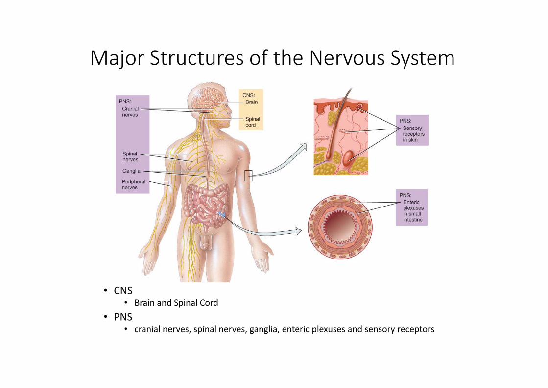
Major Structures of the Nervous System
• CNS• Brain and Spinal Cord
• PNS• cranial nerves, spinal nerves, ganglia, enteric plexuses and sensory receptors
Nervous System Divisions
• Central nervous system (CNS) • consists of the brain and spinal cord
• Peripheral nervous system (PNS)• consists of cranial and spinal nerves that contain both sensory and motor fibers
• connects CNS to muscles, glands & all sensory receptors
Functions of the Nervous Systems
• The sensory function of the nervous system is to sense changes in the internal and external environment through sensory receptors. • Sensory (afferent) neurons serve this function.
• The integrative function is to analyze the sensory information, store some aspects, and make decisions regarding appropriate behaviors. • Association or interneurons serve this function.
• The motor function is to respond to stimuli by initiating action. • Motor(efferent) neurons serve this function.

Subdivisions of the PNS
• Somatic (voluntary) nervous system (SNS)• neurons from cutaneous and special sensory receptors to the CNS
• motor neurons to skeletal muscle tissue
• Autonomic (involuntary) nervous systems• sensory neurons from visceral organs to CNS• motor neurons to smooth & cardiac muscle and glands
• sympathetic division (speeds up heart rate)• parasympathetic division (slow down heart rate)


Gray and White Matter
• White matter = myelinated processes (white in color)• Gray matter = nerve cell bodies, dendrites, axon terminals, bundles of unmyelinated axons and neuroglia (gray color)• In the spinal cord = gray matter forms an H‐shaped inner core surrounded by white matter
• In the brain = a thin outer shell of gray matter covers the surface & is found in clusters called nuclei inside the CNS

The Spinal Cord & Spinal Nerves
• Together with brain forms the CNS• Functions
• spinal cord reflexes• integration (summation of inhibitory and excitatory) nerve impulses
• highway for upward and downward travel of sensory and motor information

Spinal Cord Protection
By the vertebral column, meninges, cerebrospinal fluid, and vertebral ligaments.

Structures Covering the Spinal Cord
• Bones (Vertebrae)Epidural space filled with fat• Meninges oDura mater
• dense irregular CT tubeSubdural space filled with interstitial fluid
oArachnoid = spider web of collagen fibersSubarachnoid space = CSFoPia mater
• thin layer covers BV• denticulate ligs hold in place

Gray Matter of the Spinal Cord
• Gray matter is shaped like the letter H or a butterfly• contains neuron cell bodies, unmyelinated axons & dendrites• paired dorsal and ventral gray horns • lateral horns only present in thoracic spinal cord• gray commissure crosses the midline
• Central canal continuous with 4th ventricle of brain

White Matter of the Spinal Cord
• White matter covers gray matter• Anterior median fissure deeper than Posterior median sulcus• Anterior, Lateral and Posterior White Columns contain axons that form ascending & descending tracts

Spinal Nerves
• 31 Pairs of spinal nerves• Named & numbered by the cord level of their origin• 8 pairs of cervical nerves• 12 pairs of thoracic nerves • 5 pairs of lumbar nerves• 5 pairs of sacral nerves • 1 pair of coccygeal nerves
• Mixed sensory & motor nerves

The Brain and Cranial Nerves

Principal Parts of the Brain
• Cerebrum• Cerebral hemispheres
• Corpus callosum
• Diencephalon• thalamus, hypothalamus, & epithalamus
• Cerebellum• Brainstem
• medulla, pons & midbrain

Principal Parts of the Brain

Brain CT scan

Brain MRI
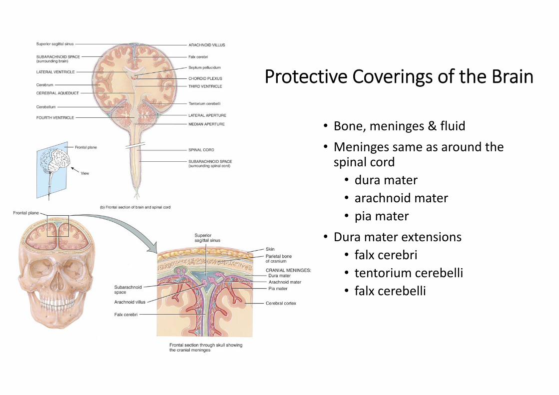
Protective Coverings of the Brain
• Bone, meninges & fluid• Meninges same as around the spinal cord• dura mater• arachnoid mater• pia mater
• Dura mater extensions• falx cerebri• tentorium cerebelli• falx cerebelli

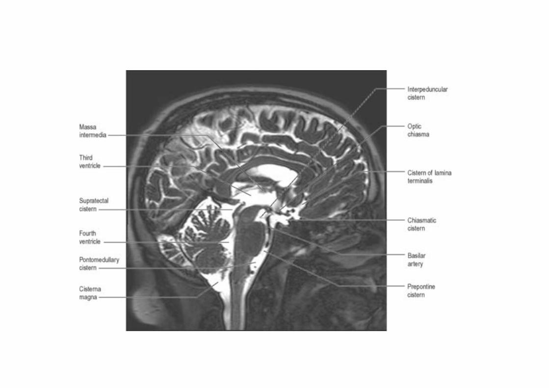
Ventricles
• 2 lateral ventricles, one within each cerebral hemisphere• 3rd ventricle• fourth ventricle

Ventricles
• Choroid plexus = capillaries covered by ependymal cells• Production CSF

Cranial Nerves• The cranial nerves are part of the peripheral nervous system
• There are 12 pairs of Cranial nerves • All cranial nerves travel through foramina of the skull
• 10 pairs originate from the brain stem (III‐XII)
• The cranial nerves are designated by:• Roman numerals which indicate the order in which the nerves arise from the brain from anterior to posterior
• Names which indicate the distribution or function

Cranial Nerves• Two cranial nerves (I and II) contain only sensory fibers and are therefore called sensory nerves
• The other cranial nerves contain both sensory and motor fibers and are therefore called mixed nerves• Some of the mixed nerves are primarily motor in function (but contain proprioceptive fibers).
• The cell bodies of sensory fibers are located in ganglia outside the brain.
• The cell bodies of motor fibers are located in nuclei within the brain• Some cranial nerves include both somatic motor and parasympathetic fibers of the autonomic nervous system.

Cranial Nerves: Types of Fibers

Cranial Nerves
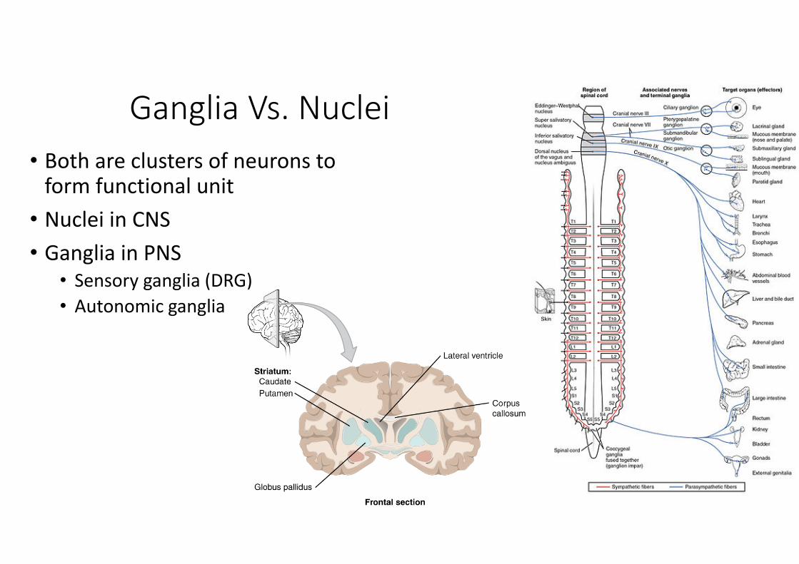
Ganglia Vs. Nuclei• Both are clusters of neurons to form functional unit
• Nuclei in CNS• Ganglia in PNS
• Sensory ganglia (DRG)• Autonomic ganglia

Fasciculi, Lemnisci, & Tracts
• All are bundles of nerve fibers in the CNS
• Lemniscus is a bundle of nerve fibers of second order sensory neurons• Medial & lateral lemnisci
• Tracts or fasciculi• Gracile & cuneate fasciculi• Ascending and descending tracts

Afferent Vs. Efferent
• PNS• Afferent – Sensory fibers• Efferent – Motor fibers
• SC• Afferent – Ascending tracts• Efferent – Descending tracts
• Brain parts• Afferent – Inputs • Efferent – Outputs

Laminae• Thin plates or layers of the gray matter of CNS• Laminae of cerebral cortex• Rexed laminae of spinal cord