2 barrett’s esophagus and esophageal neoplasms
TRANSCRIPT

21
BARRETT’S ESOPHAGUS AND ESOPHAGEAL NEOPLASMS
2
Many biopsies from the esophagus are taken to address Barrett’s esophagus or other colum-nar epithelium found in the esophagus. The risk factors for Barrett’s esophagus are chronic gastroesophageal re!ux (more than 5 years), age over 50 years, male gender, smoking, central obesity and Caucasian race. Alcohol use is not a signi"cant factor and some data suggest a pro-tective effect for wine drinking (1). Bottoms up!
Many of the issues surround the source of the biopsy (proximal stomach versus tubular esophagus). There are many articles concern-ing special stains to address these issues but a good hematoxylin and eosin (H&E) stain is all
you need once you recognize the "ndings (2). Knowing the endoscopic appearance is import-ant but recording precisely which epithelial types are encountered is the main issue. There are a number of types of mucosa that can be encountered: 1) oxyntic mucosa ("g. 2-1); 2) cardiac mucosa ("g. 2-2); 3) pancreatic het-erotopia/metaplasia ("g. 2-3); 4) multilayered epithelium ("gs. 2-4, 2-5) (3); 5) squamous ep-ithelium; and 6) esophageal ducts (see chapter 1, "g. 1-3).
Finding either multilayered epithelium or ducts on a biopsy con"rms that a sample is from the esophagus rather than the stomach.
Figure 2-1
OXYNTIC MUCOSAThis is gastric type mucosa of the type found in the gastric
body and fundus. The gastric pits are convoluted tubules that connect to the surface such that a two dimensional section shows crowded glands lined by parietal cells (the pink ones in the center of the "eld). The deep portion of the mucosa contains bluish chief cells. The surface foveolar cells each have an apical mucin cap.
Figure 2-2
GASTRIC CARDIAC MUCOSAThere are no parietal cells and there is lamina propria
chronic inflammation, a common finding in patients who undergo biopsies after presenting re!ux symptoms. Since re!ux results in cycles of damage and repair, there is disorganized smooth muscle in the lamina propria between cardiac type glands, which produce mucin. The surface has foveolar cells just as the oxyntic mucosa does. Some samples show a combination (“cardio-oxyntic mucosa”).
Survival Guide to Gastrointestinal Mucosal Biopsies
22
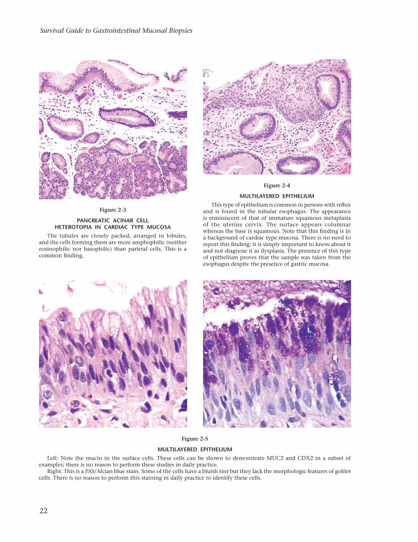
Figure 2-3
PANCREATIC ACINAR CELL HETEROTOPIA IN CARDIAC TYPE MUCOSA
The tubules are closely packed, arranged in lobules, and the cells forming them are more amphophilic (neither eosinophilic nor basophilic) than parietal cells. This is a common "nding.
Figure 2-4
MULTILAYERED EPITHELIUMThis type of epithelium is common in persons with re!ux
and is found in the tubular esophagus. The appearance is reminiscent of that of immature squamous metaplasia of the uterine cervix. The surface appears columnar whereas the base is squamous. Note that this "nding is in a background of cardiac type mucosa. There is no need to report this "nding; it is simply important to know about it and not diagnose it as dysplasia. The presence of this type of epithelium proves that the sample was taken from the esophagus despite the presence of gastric mucosa.
Figure 2-5
MULTILAYERED EPITHELIUMLeft: Note the mucin in the surface cells. These cells can be shown to demonstrate MUC2 and CDX2 in a subset of
examples; there is no reason to perform these studies in daily practice.Right: This is a PAS/Alcian blue stain. Some of the cells have a bluish tint but they lack the morphologic features of goblet
cells. There is no reason to perform this staining in daily practice to identify these cells.