barrett’s esophagus and associated adenocarcinoma
TRANSCRIPT

Department of Surgery, Cardiothoracic Division,Division of General Thoracic and Esophageal SurgeryHelsinki University Central Hospital, Helsinki, Finland
BARRETT’S ESOPHAGUS AND ASSOCIATED ADENOCARCINOMA
- Studies on pathogenesis, clinical staging, and cost-utility of cancer treatment
Jari Räsänen
ACADEMIC DISSERTATION
To be presented, with the permission of the Faculty of Medicine,
University of Helsinki, for public examination in the Lecture Hall of the
Surgical Unit in Meilahti Hospital, Haartmaninkatu 4, Helsinki,
on October 11th, 2007, at 12 noon.
HELSINKI 2007

Supervised by:
Professor h.c. Jarmo A. Salo MD, PhDUniversity of Helsinki
Reviewed by:
Docent Pekka Nuutinen, MD, PhDUniversity of Kuopio
Docent Markku Voutilainen, MD, PhDUniversity of Helsinki
Opponent:
Professor Heikki Joensuu MD, PhDUniversity of Helsinki
ISBN 978-952-92-2640-5 (paperback)ISBN 978-952-10-4183-9 (PDF)
Helsinki 2007Yliopistopaino

3
TABLE OF CONTENTS
LIST OF ORIGINAL PUBLICATIONS ........................................................................ 5
ABBREVIATIONS .......................................................................................................... 6
ABSTRACT ..................................................................................................................... 8
INTRODUCTION ......................................................................................................... 10
REVIEW OF THE LITERATURE ................................................................................. 12
1. Barrett’s esophagus .................................................................................................... 121.1. Defi nition .......................................................................................................... 121.2. Pathogenesis ..................................................................................................... 121.3. Diagnosis ........................................................................................................... 161.4. Epidemiology .................................................................................................... 171.5. Treatment of gastroesophageal refl ux disease on Barrett’s esophagus patients 181.6. Dysplasia in Barrett’s esophagus ...................................................................... 18
2. Adenocarcinoma associated with Barrett’s esophagus ............................................ 212.1. Epidemiology .................................................................................................... 212.2. Classifi cation of esophageal and gastroesophageal junction
adenocarcinomas .............................................................................................. 212.3. Diagnosis ........................................................................................................... 232.4. Treatment of esophageal and gastroesophageal junctional adenocarcinomas 262.5. Oxidative stress in development of Barrett’s esophagus and
adenocarcinoma ............................................................................................... 302.6. Cost-effectiveness, cost-utility, and cost-benefi t of treatment of
esophageal and esophagogastric junction carcinomas ................................... 31
AIMS OF THE STUDY .................................................................................................. 34
PATIENTS AND METHODS ........................................................................................ 35
1. Patients ...................................................................................................................... 352. Methods ..................................................................................................................... 35
2.1. Tissue sample collection .................................................................................. 352.2. Analysis of superoxide dismutase (SOD) and myeloperoxidase (MP)
activities and glutathione content .................................................................... 362.3. Analysis of 8-hydroxydeoxyguanosine (8-OHdG) .......................................... 362.4. Positron emission tomography (PET) imaging ............................................... 372.5. Health-related quality of life (HRQoL) ........................................................... 372.6. Cost-utility ........................................................................................................ 382.7. Statistical methods ............................................................................................ 38
4
RESULTS ........................................................................................................................ 39
1. Antirefl ux surgery and oxidative stress in the distal and proximal esophagus ...... 392. Expression of 8-hydroxydeoxyguanosine in esophageal tissues and tumors ......... 423. Impact of positron emission tomography on clinical staging and prognostication
of adenocarcinoma of the esophagus and the esophagogastric junction .............. 434. Cost-utility of treatment of carcinoma of the esophagus or esophagogastric
junction ...................................................................................................................... 47
DISCUSSION ................................................................................................................. 50
1. Oxidative stress and antirefl ux surgery .................................................................... 502. Oxidative DNA damage and pathogenesis of esophageal adenocarcinoma .......... 513. Imaging and optimal treatment and prognosis ...................................................... 524. Cost-utility of treatment of carcinoma of the esophagus or esophagogastric
junction ...................................................................................................................... 53
CONCLUSIONS ............................................................................................................ 54
YHTEENVETO (FINNISH SUMMARY) ..................................................................... 55
ACKNOWLEDGMENTS .............................................................................................. 57
REFERENCES ................................................................................................................ 58
ORIGINAL PUBLICATIONS ........................................................................................ 79
5
LIST OF ORIGINAL PUBLICATIONS
This thesis is based on the following original publications, which are referred to in the text by their Roman numerals.
I Rantanen TK, Räsänen JV, Sihvo EIT, Ahotupa MO, Färkkilä MA, Salo JA. The impact of antirefl ux surgery on oxidative stress of esophageal mucosa caused by gastroesophageal refl ux disease: 4-yr follow-up study. Am J Gastroenterol 2006;101:222-8.
II Räsänen JV, Sihvo EIT, Rantanen TK, Ahotupa MO, Färkkilä MA, Harjula A, Salo JA. Gastroesophageal refl ux patients’ defective anti-oxidative capacity in the proximal esophageal mMucosa before antirefl ux surgery and also after 4-year follow-up (in press, Ann Med).
III Räsänen JV, Sihvo EIT, Ahotupa MO, Färkkilä MA, Salo JA. The expression of 8-hydroxydeoxyguanosine in oesophageal tissues and tumours. Eur J Surg Oncol (2007; Apr 27; [Epub ahead of print]. Doi:10.1016/j.ejso.2007.03.003.(in press)
IV Räsänen JV, Sihvo EIT, Knuuti MJ, Minn HR, Luostarinen ME, Laippala P, Viljanen T, Salo JA. Prospective analysis of accuracy of positron emission tomography, computed tomography, and endoscopic ultrasonography in staging of adenocarcinoma of the esophagus and the esophagogastric junction. Ann Surg Oncol 2003;10:954-60.
V Sihvo EIT, Räsänen JV, Knuuti MJ, Minn HR, Luostarinen ME, Viljanen T, Färkkila MA, Salo JA. Adenocarcinoma of the esophagus and the esophagogastric junction: positron emission tomography improves staging and prediction of survival in distant but not in locoregional disease. J Gastrointest Surg 2004;8:988-96.
VI Räsänen JV, Räsänen PR, Sintonen H, Aronen P, Roine RP, Sihvo EIT, Salo JA. Cost-utility of treatment of carcinoma of oesophagus or oesophagogastric junction – a prospective decision tree analysis (Submitted).
These publications are reprinted with permission of the copyright holders.(In addition, some unpublished material is presented.)

6
ABBREVIATIONS
AB-PAS Alcian blue-periodic acid-SchiffAMACR A-methylacyl-CoAracemaseA:T transition Adenine:thymine transitionBE Barrett´s esophagusCBA Cost-benefi t analysisCC Cytosine-cytosineCEA Cost-effectiveness analysisCLE Columnar-lined esophagusCM Cardia type of mucosa CpG Cytosine and guanine are connected by a phosphodiester bondCRT Chemoradiation therapyCT Computed tomography CUA Cost-utility analysisEAC Esophageal adenocarcinomaERD Erosive refl ux diseaseEUS Endoscopic ultrasound FDG 18F-fl uorodeoxy-D-glucoseFDG-PET 18F-fl uorodeoxy-D-glucose positron emission tomographyFNA Fine-needle aspirationG1 Grade 1G3 Grade 2G3 Grade 3 G:C transition Guanine:cytosine transitionGEJ Gastroesophageal junctionGER Gastroesophageal refl uxGERD Gastroesophageal refl ux disease GSH Glutathione content HPLC High-performance liquid chromatographyHRQoL Health related quality of life ICUR Incremental cost-effectiveness ratioLES Lower esophageal sphincterMBq MegabecquerelMPA Myeloperoxidase activityMRI Magnetic resonance imagingMUC-2 Mucin 2NERD Nonerosive refl ux diseaseOR Odds ratioPET Positron emission tomographyPPI Proton pump inhibitorQALY Quality-adjusted life-year

7
ROS Reactive oxygen speciesR 0 resection Complete surgical resectionRR Risk ratioSOD Superoxide dismutaseTT Thymine-thymineZ line Squamocolumnar junction8-OHdG 8-hydroxydeoxyguanosine

8
ABSTRACT
Esophageal and gastroesophageal junction adenocarcinoma (GEJ) is a rapidly increasing deadly disease with a pathophysiology that is connected to oxidative stress. Exact pretreatment clinical staging is essential for optimal care of this lethal malignancy. Because of the rising incidence of the disease and the expense of new treatments, the cost-effectiveness of treatment is increasingly important. The aim of this study was to assess the protective effect of antirefl ux surgery against oxidative stress of esophageal mucosa, the role of oxidative DNA-damage (8-hydroxydeoxyguanosine) in the pathogenesis of Barrett’s esophagus and adenocarcinoma of the distal esophagus and GEJ, and the accuracy and a prognostic value of positron emission tomography (PET) in preoperative staging of adenocarcinoma of the esophagus and GEJ. In addition, we estimated the cost-utility of the present treatment schema for adenocarcinoma of the esophagus and GEJ.
We measured oxidative metabolism in the distal and proximal esophagus by myeloperoxidase activity (MPA), glutathione content (GSH), and superoxide dismutase (SOD) in 20 patients operated on with Nissen fundoplication and in 9 controls during a 4-year follow-up. Further, we assessed the oxidative damage of DNA by 8-hydroxydeoxyguanosine (8-OHdG) in esophageal samples of 51 subjects (13 Barrett’s metaplasia, 6 Barrett’s esophagus with high-grade dysplasia, 18 adenocarcinoma of the distal esophagus/GEJ, and 14 normal controls). We estimated the accuracy (42 patients) and preoperative prognostic value (55 patients) of PET compared with computed tomography (CT) and endoscopic ultrasound (EUS) in patients with adenocarcinoma of the esophagus or the GEJ. Finally, we clarifi ed the specialty-related costs and the utility of either radical (30 patients) or palliative (23 patients) treatment of esophageal/GEJ carcinoma by the 15D health-related quality-of-life (HRQoL) questionnaire and survival rate. The cost-utility of radical treatment of esophageal/GEJ carcinoma was investigated using a decision tree analysis model comparing radical, palliative, and hypothetical new treatment.
Despite successful antirefl ux surgery, oxidative stress (measured by MPA) in the distal esophagus remained higher in patients than in controls at 6 months and 4 years postoperatively. Antioxidative capacity of distal esophageal mucosa measured by GSH levels was lower than control levels both pre- and postoperatively. In spite of decreased oxidative stress (MPA) in the proximal esophagus, GERD patients had defi cient antioxidative capacity before and after fundoplication compared with controls, refl ected as decreased GSH and SOD levels both preoperatively and 4 years postoperatively. Oxidative stress-related DNA damage (as 8-OHdG) in the distal esophagus was markedly increased in Barrett’s epithelium and in high-grade dysplasia as well as in adenocarcinoma of the esophagus/GEJ compared with controls. Barrett’s patients had similar 8-OHdG levels in their distal and proximal esophageal samples.
9
PET was no better in detection locoregional lymph node metastasis than CT or EUS, but PET was more sensitive than CT in identifying distant organ metastases. Unfortunately, some false-negative diagnoses of stage IV disease (distant metastases) were made even with PET. Positive PET for distant metastasis predicted well the poor survival of these patients. Despite increasing costs, taking into consideration the improved survival and quality of life of treated patients, the incremental cost-utility ratio of the radical surgery strategy compared with non-surgical options was favorable during the 2-year follow-up period.
Elevated oxidative stress (MPA) and decreased antioxidant defense (GSH) after antirefl ux surgery in the distal esophagus indicate that antirefl ux surgery is not a perfect solution for oxidative stress of the esophageal mucosa. Elevated oxidative stress in turn may partly explain why adenocarcinoma of the distal esophagus is found even after successful fundoplication. In GERD patients, the proximal esophageal mucosal anti-oxidative defense seems to be defective before and even years after successful antirefl ux surgery. In addition, antirefl ux surgery apparently does not change the level of oxidative stress in the proximal esophagus, suggesting that defective mucosal anti-oxidative capacity plays an important role in development of oxidative damage to the esophageal mucosa in GERD. In the malignant transformation of Barrett’s esophagus, an important component appears to be oxidative stress. DNA damage may to be mediated by 8-OHdG, and the entire esophagus of Barrett’s patients suffers from increased oxidative stress. PET is a useful tool in the staging and prognostication of adenocarcinoma of the esophagus/GEJ, detecting organ metastases better than CT, although its accuracy in staging of paratumoral and distant lymph nodes is limited. Radical surgery for esophageal/GEJ carcinoma provides the greatest benefi t in terms of survival, and its cost-utility appers to be the best currently available treatments. While waiting for new, more effective treatments, radical surgery serves as the gold standard for all other treatments.

10
INTRODUCTION
Once a very uncommon tumor, adenocarcinoma of the esophagus is currently a cancer with a rapidly rising incidence in several Western countries, including Finland (Powell et al. 1990, Armstrong and Borman 1996, Devesa et al. 1998, Botterweck et al. 2000, Sihvo et al. 2000, Bollschweiler et al. 2001, van Blankenstein et al. 2005, Voutilainen and Juhola 2005). It has replaced squamous cell carcinoma as the most common esophageal malignancy in the many industrialized countries (Pohl et al. 2005). The highest reported incidence is 7/100,000 in the United Kingdom, and the average incidence in the United States is 2.5/100 000, although in some regions the incidence in white men is as high as 5.3/100 000 (Bollschweiler et al. 2001). In Finland, the incidence rose from 0.28 to 0.77/100 000 in males during 1976-95 (Sihvo et al. 2000). Similar to esophageal adenocarcinoma, the incidence of adenocarcinoma of the gastroesophageal junction (GEJ) has increased signifi cantly since the mid-1970s (Blot et al. 1991), although the average rate after the late 1980s in the United States (El-Serag et al. 2002) as well as in Finland has stabilized (Sihvo et al. 2000, Finnish Cancer Registry 2005). The overall 5-year survival can be as high as 40-50% in selected patients in the best surgical series (Hagen 2001, Sihvo 2004). Unfortunately, up to 60% of patients end up receiving palliative treatment because of disseminated disease or comorbidities and have a median survival of only 3-4 months (Sihvo 2004).
The specifi c etiological factor behind the dramatic increase in the prevalence of esophageal and GEJ adenocarcinomas is unknown, but an undeniable risk factor for adenocarcinoma of the distal esophagus is gastroesophageal refl ux disease (GERD) and its consequence, Barrett’s esophagus (Chow et al. 1998, Lagergren et al. 1999). A major risk for cancer among those who have GERD is Barrett’s esophagus (Solaymani-Dodaran et al. 2004). Patients with Barrett’s esophagus have a 30- to 400-fold increased risk for developing adenocarcinoma (Spechler and Goyal 1986, Drewitz et al. 1997). The annual risk for adenocarcinoma in Barrett’s patients is 0.5% (Shaheen et al. 2000). While the increasing length of Barrett’s esophagus may slightly increase the cancer risk, the progression of Barrett’s metaplasia to dysplasia is the major risk (DeMeester and DeMeester 2000, Rudolph et al. 2000, Conio et al. 2001). Other risk factors that may contribute to the rising incidence of esophageal adenocarcinoma are cigarette smoking, low intakes of fruit, vegetables, and cereal fi bers, and obesity (Pera 2005).
The risk factors for GEJ adenocarcinoma are controversial. The risk has been suggested to invariably be related to Helicobacter pylori infection (Goldblum 2002, Goldblum et al. 2002,), but strong evidence indicates that the risk is at least partly connected to GERD (Bowrey et al. 1999, Couvelard et al. 2001, DeMeester et al. 2002, Balaji et al. 2003).
The development and maintenance of metaplastic epithelium (Barrett’s esophagus) are associated with infi ltration of infl ammatory cells (Weston et al. 1997, Goldblum et al. 1998, Harrison et al. 2000). This infi ltration of infl ammatory cells in turn causes the increased production of reactive oxygen species (ROS), which can lead to oxidative damage to proteins, cell membranes, or most importantly to DNA, producing pro-mutagenic lesions like 8-hydroxydeoxyguanosine (8-OHDG) (Kasai et al. 1997, Olliver et

11
al. 2003, Boonstra and Post 2004). Oxidative damage seems to be strongly connected to the malignant transformation of Barrett’s esophagus (Olyaee et al. 1995, Oh et al. 2001, Sihvo et al. 2002).
The prevention of infl ammation and its sequelae is the ultimate goal of treatment of GERD and Barrett’s esophagus. Antirefl ux surgery is very effective in diminishing symptoms and macroscopic esophagitis (Desai et al. 2003) and may even induce regression of Barrett’s esophagus (Gurski et al. 2002). It may reduce oxidative stress in the distal esophagus in the short term (Wetscher et al. 1995), but the long-term effect of fundoplication on oxidative stress is unknown. In addition, the preventive effect on adenocarcinoma of the esophagus has been refuted by several studies (McDonald et al. 1996, Spechler et al. 2001, Corey et al. 2002). The role of antirefl ux surgery is still unclear in the prevention of oxidative stress in the esophagus. Proton pump inhibitor (PPI) medication has been suggested to slow the progression of Barrett’s esophagus to dysplasia and cancer, but not prevent the process entirely (Ouatu-Lascar et al. 1999, El-Serag et al. 2004).
New, stage-dependent treatment protocols require the most complete and accurate staging possible. For most patients with advanced disease present at diagnosis, the key factor for successful treatment of adenocarcinomas near the esophagogastric junction is exact pretreatment staging. Unfortunately, the classic combination of endoscopic ultrasound (EUS) and thoracic and abdominal computed tomography (CT) scanning can detect primary tumors with an accuracy of no more than 66-79%, lymph node involvement of 72-82 %, and systemic disease 64% compared with histopathologic results obtained from surgery (Salminen et al. 1999, Flamen et al. 2000a, Heidemann et al. 2000). Positron emission tomography (PET) is a new staging method based on the accumulation of a fl uorinated glucose analog (18F-fl uorodeoxy- D-glucose; FDG) in malignant cells (Pauwels et al. 1998), which be observed by a positron camera. PET thus provides the opportunity to detect altered tissue metabolism in malignant tumors. PET in combination with CT, provides improved diagnostic accuracy for solid-organ systemic metastases (Flamen et al. 2000a, Lerut et al. 2000, Lowe et al. 2005). EUS with fi ne-needle aspiration of regional nodes is still investigational (Vazquez-Sequeiros et al. 2003), are thoracoscopy and laparoscopy, which can cause tumor seeding of a port site (Freeman et al. 2001, Suntharalingam et al. 2001).
Functional well-being of patients has become increasingly important when assessing the treatment results of esophageal carcinoma (Blazeby et al. 2001). Treatment is costly because of the many different investigations and treatment modalities needed (Soni et al. 2001). Despite this, cost and cost-utility analyses have been performed to date by very few authors in the fi eld of esophageal cancer surgery (Hulscher et al. 2002, Sihvo et al. 2002).

12
REVIEW OF THE LITERATURE
1. Barrett’s esophagus
1.1. Defi nition
The current defi nition of Barrett’s esophagus includes both endoscopically evident displacement of the squamocolumnar junction proximal to the GEJ and the typical histological fi nding of normal stratifi ed squamous epithelium lining the esophagus being replaced by metaplastic columnar epithelium-containing goblet cells. This epithelium is called specialized intestinal metaplasia (Trier 1970, Sharma et al. 2004). The rationale for nomenclature is that dysplasia and adenocarcinoma seem to occur only in intestinal mucosa (Lee et al. 1985, Hamilton et al. 1987, Hameeteman et al. 1989, Haggitt 1994).
1.2. Pathogenesis
The relationship between gastroesophageal refl ux (GER) and the development of Barrett’s esophagus is nowadays generally accepted (Hayward 1961, Bremner et al. 1970, Conio et al. 2002). The precise mechanism leading to metaplastic changes in the distal esophagus is, however, unclear. Animal studies have clarifi ed that the columnar epithelium is acquired when the squamous epithelium is injured; during repair the squamous epithelium undergoes columnar metaplasia (Bremner et al. 1970). Although damage to squamous mucosa is a necessary factor for the development of columnar metaplasia, a chronically abnormal esophageal environment during the period of mucosal repair is essential as well (Bremner et al. 1970, Wong and Finckh 1971, Dresner et al. 2003). Gillen et al. (1998) have shown that columnar regrowth does not have to ascend from the gastric cardia, as previously thought, and Li et al. (1994) suggested that the depth of damage caused by GER determines whether regenerating epithelium is columnar or squamous. The acquisition of Barrett’s mucosa after the onset of GER following esophagogastrostomy, Heller myotomy, and esophagojejunostomy provides further evidence for a relationship between GER and Barrett’s esophagus (Naef et al. 1975, Hamilton and Yardley 1977, Meyer et al. 1979, Kortan et al. 1981, Westhoff et al. 2004).
Lower esophageal sphincter (LES) hypotension
A defective barrier mechanism between the stomach and the esophagus enables the noxious gastroduodenal juice to enter the esophageal mucosa, and this together with failing esophageal defenses leads to gastroesophageal refl ux disease (GERD). The failure of the barrier can be transient or permanent. Several authors (Dodds et al. 1982, Dent et al. 1988, Mittal and McCallum 1988, Mittal et al. 1995) have shown that transient lower esophageal sphincter (LES) relaxation is the single most common mechanism in GERD,

13
accounting for 65% of refl ux episodes in refl ux esophagitis patients. The reason for refl ux patients having more frequent transient LES relaxation than controls is unknown. In 1983, Iascone et al. found patients with Barrett’s esophagus to have worse acid refl ux and weaker LES than patients with uncomplicated esophagitis or asymptomatic controls. They suggested that Barrett’s esophagus develops as a result of long-standing refl ux esophagitis.
Attwood et al. (1989) reported that 90% of patients with a columnar-lined esophagus (CLE) had a mechanically defective LES, and 93% had increased esophageal exposure to gastric juice on esophageal pH monitoring. Stein et al. (1990) noted that for GER the critical LES pressure is less than 6 mm Hg, overall LES length is less than 2 cm, and abdominal LES length is less than 1 cm. It has also been demonstrated by multichannel ambulatory 24-h pH measurement that as the rate of recorded acid exposure values increase from the proximal to distal esophagus, the length of Barrett’s esophagus increases signifi cantly (Tharalson et al. 2002).
Hiatal hernias are common among patients with Barrett’s esophagus and are larger than among controls or GERD patients with or without esophagitis (Cameron 1999). They impair esophageal clearance, cause permanent lowering in LES pressure, and reduce the protective effect of the crural diaphragm, thereby predisposing the esophageal mucosa to an increased effect of refl uxate (Mittal et al. 1987, Sloan and Kahrilas 1991, Sloan et al. 1992, Mittal and Balaban 1997).
Motility disturbances in esophagus and stomach
Delayed esophageal clearance in the distal esophagus is common among patients with Barrett’s esophagus and thus refl ued material may have a prolonged contact with the esophageal mucosa. Kahrilas et al. (1986) showed positive correlation exists between the grade of peristaltic dysfunction in the distal esophagus and the grade of esophagitis. It is unclear whether this is a primary defect or secondary to acid-induced injury (Eastwood et al. 1975, Eckardt 1988, Howard et al. 1994). Fass et al. (2001) has demostrated a defi nite positive correlation between the length of Barrett’s esophagus and the duration of esophageal acid exposure. Recent Swedish epidemiological studies confi rm the correlation between esophageal adenocarcinoma and the duration, frequency, and severity of GER symptoms (Lagergren et al. 1999, Ye et al. 2001).
The role of gastric emptying seems to be more unclear. While McCallum et al. (1981) found that up to 57% of patients with GERD have impaired gastric emptying, more recent studies have demonstrated no difference in the rate of gastric emptying between GERD patients with or without esophageal mucosal injury and asymptomatic controls (Shay et al. 1987, Keshavarzian et al. 1991). Furthermore, patients with Barrett’s esophagus seem to have normal gastric emptying for both solids and liquid bolus (Kogan et al. 1985, Johnson et al. 1986). More recently, a multivariate analysis of pathophysiological factors in refl ux esophagitis has shown that impaired esophageal acid clearance and hypotonic lower esophageal sphincter are the two main independent pathophysiological factors of esophagitis, and gastric emptying seems to have no signifi cant role in the development of esophagitis (Cadiot et al. 1997).

14
Acid, pepcin, and bile
Compared with patients with erosive and nonerosive GERD, patients with Barrett’s esophagus typically have greater esophageal acid exposure based on 24-h pH monitoring (Iascone et al. 1983, Champion et al. 1994, Coenraad et al. 1998, Neumann et al. 1994, Singh et al. 1994, Salminen et al. 1999), but do not have increased basal acid output or 24-h or daytime patterns of gastric pH compared with healthy controls (Hirschowitz 1996, Savarino et al. 1996). Evidence favoring the important role of acid in development of Barrett’s esophagus is that after mucosal ablation of Barrett’s esophagus the substitutive mucosa is squamous epithelium with acid suppression, and recurrent Barrett’s epitehlium without it (Brandt and Kauvar 1992, Salo et al. 1998, Haag et al. 1999). The role of pepcin seems to be less signifi cant in the development of mucosal injury (Hirschowitz 1996).
Bile refl ux into the esophagus has been suggested by many investigators to be an important causative factor in acquisition of Barrett’s esophagus (Halvorsen et al. 1975, Hamilton and Yardley 1977). Experimental studies have shown that bile salts and duodenal contents can cause esophageal mucosal damage (Kivilaakso et al. 1980, Salo and Kivilaakso 1983, Martinez de Haro et al. 2001). Duodenogastroesophageal refl ux is also increased in Barrett’s esophagus patients, especially in patients with concomitant ulcers, strictures, or dysplasia (Attwood et al. 1989). In addition, these same patients seem to have simultaneous acid refl ux. Obviously, therefore, both gastric and duodenal contents have a role in the pathogenesis of Barrett’s esophagus. The combination of acid and bile acid caused the worst mucosal damage according to a recent survey (Oh et al. 2006).
Cell of origin
Epithelial metaplasia in the distal esophagus is a poorly understood process. There are several theories regarding the origin of metaplastic cells. One is that Barrett’s esophagus is a result of the upward migration of gastric epithelium after denudation of esophageal squamous epithelium (Bremner et al. 1970). This theory is opposed by Gillen et al. (1988) who stated that the cell of origin of Barrett’s epithelium is located in esophageal gland ducts and is likely to be a multipotent stem cell. The presence of squamous epithelium-related cytoceratin 13 in Barrett’s metaplasia supports the origin being the native epithelium (Salo et al. 1996). In recent studies, a unique surface cell at the squamocolumnar junction was found that has features of both glandular epithelium and squamous epithelium (Shields et al. 1993). Furthermore, Boch et al. (1997) reported a new multilayered epithelium within Barrett’s epithelium that has histologic characteristics of both squamous and columnar epithelia. A study of cytokeratin expression in this multilayered epithelium detected columnar and squamous cell markers, indicating an intermediate nature of this epithelium (Glickman et al. 2001). This cell type could be the missing link in the metaplastic process. Cameron and Arora (2002) suggest that Barrett’s esophagus may develop after loss of a long segment of squamous epithelium, with columnar replacement in the presence of continuing acid refl ux rather than directly from areas of esophagitis. The defi nitive answer of why this stem cell differentiates into columnar cells eludes us.

15
Mucosal resistance and the role of saliva
An important supplemental defense against acid in the distal esophagus is the neutralizing effect of the saliva (Helm et al. 1984). Saliva also contains several growth factors, including epidermal growth factor and transforming growth factor, which have a role in the healing of esophageal mucosa (Kongara and Soffer 1999) Saliva may also participate in carcinogenesis near the esophagogastric junction (McColl 2005). The precise role of saliva in esophageal defense remains unclear and requires further investigation.
Healthy esophageal mucosa has pre-epithelial, epithelial, and postepithelial defenses. Pre-epithelia defenses include the mucus and the unstirred water layer along with surface bicarbonate ions. Epithelial defenses consist of the apical cell membrane, junction barriers, intracellular and extracellular buffers, and pH regulatory processes. Postepithelial defenses involve blood fl ow and the tissue acid-base balance. Patients who are predisposed to GER–related injury may have suboptimal mucosal defense (Sarosiek and McCallum 1995, Kongara and Soffer 1999).
Helicobacter pylori infection
The simultaneous disappearance of H. pylori infection and an increase in refl ux-related GEJ adenocarcinoma has raised the question of a possible link between these two events. Most studies have found no causal relationship between H. pylori infection and Barrett’s esophagus, but some evidence suggests a protective role of H. pylori infection (Werdmuller and Loffeld 1997, Varanasi et al. 1998, Vicari et al. 1998).
H. pylori infection has been proposed to protect against the development of Barrett’s esophagus because corpus-predominant gastritis is associated with decreased acid secretion (El-Serag et al. 1999). This protective effect is especially linked to cagA-positive H. pylori strains (Vicari et al. 1998).
Diet, smoking, and alcohol
Low intake or defective absorption of antioxidants such as vitamin C may play a role in the development of Barrett’s esophagus (Fountoulakis et al. 2004). Dietary nitrates can be converted to nitrosating species by bacteria, saliva, and acidic gastric juice (Mirvish 1995), which in turn may deplete antioxidant species, producing oxidative stress. This can cause damage to DNA and lead to mutagenesis. The role of alcohol and tobacco smoking in the development of Barrett’s esophagus is uncertain, although a few reports connect both of these to refl ux disease (Kadakia et al. 1995, Hirota et al. 1999, Pehl et al. 2006).
In conclusion, the pathogenesis of refl ux esophagitis and Barrett’s esophagus depends on defects in either the esophageal antirefl ux barrier or the luminal clearance mechanisms, which expose the epithelium gastroduodenal refl uxate for prolonged periods. Ingested products that directly impair the epithelium’s intrinsic defenses add to the damage, compounding vulnerability to injury from refl ux and potentially leading to development of Barrett’s esophagus. Genetic defects in mucosal defense may also play a role in this process.

16
1.3. Diagnosis
The endoscopic recognition of Barrett’s esophagus may be diffi cult. Normally, the squamous cell junction (Z line) is at the same level as the GEJ, and this can be identifi ed as the proximal limit of the linear gastric mucosal folds. When the Z line has transferred upwards, the length of Barrett’s esophagus is the distance between the Z line and the proximal limit of the linear gastric mucosal folds (Armstrong 2004). The current way of describing the extent of Barrett’s esophagus is the Prague C & M criteria, which includes assessment of the circumferential (C) and maximum (M) extent of the endoscopically visualized BE segment (Sharma et al. 2006a). The columnar epithelium in the esophagus has a characteristic red color and a velvet-like texture that contrasts sharply with the pale, glossy appearance of adjacent squamous epithelia. A large hiatal hernia may complicate identifi cation of the length of Barrett’s esophagus. Although endoscopic examination can usually distinguish columnar epithelium from squamous epithelium in the esophagus, the three different subtypes of columnar epithelium lining the esophagus: fundic-type, cardiac-type, and specialized columnar epithelium (Paull et al. 1976), cannot be differentiated based on endoscopic appearance alone. The classical distribution of Barrett’s esophagus has been based on the length of Barrett’s metaplasia. Barrett’s exceeding 3 cm has been thought to carry a higher risk for malignant transformation than shorter measures, but recent studies have shown that the length of Barrett’s esophagus is not signifi cantly related to the risk for adenocarcinoma (Rudolph et al. 2000, Weston et al. 2000). The clinical relevance of the length of Barrett’s esophagus is thus disputable. The histological diagnosis of Barrett’s esophagus often requires both hematoxylin and eosin staining (Weinstein et al. 1996) and Alcian blue staining, which is specifi c for goblet cells and helps to discriminate between different types of intestinal metaplasias (Lee 1984).
Another diffi culty in diagnosis is the patchy nature of intestinal metaplasia. A linear relationship has been demontrated between the prevalence of intestinal metaplasia and the length of columnar-lined esophagus. All patients a columnar-lined esophagus exceeding 5 cm have intestinal metaplasia, in contrast to only 15% of those whitcolumnar segment of less than 1 cm (Chandrasoma et al. 2003). Generally, the number of biopsies taken in short-segment Barrett’s esophagus remains so low that the probability of fi nding intestinal metaplasia at initial endoscopy is only 35-45 % (Kim et al. 1994). With increasing length, the accuracy improves (Weinstein and Ippoliti 1996). The accuracy of detection of intestinal metaplasia also depends on how experienced the endoscopist is (Padda and Ramirez 2001), and repeated endoscopies improve the accuracy signifi cantly (Oberg et al. 2001). Correct diagnosis of intestinal metaplasia involves a combination of endoscopy and histology, and often repeated endoscopies are required to rule out intestinal metaplasia if suspected at initial endoscopy.
There are a few molecular markers that can help in the diagnosis of Barrett’s esophagus. Cdx2 protein is a transcription factor for which expression in normal tissues is restricted to intestinal-type epithelium. Its sensitivity seems to be high for Barrett’s esophagus (Groisman et al. 2004). The biochemical marker of cellular differentiation mucin 2 (MUC-2) found in immunohistochemistry can detect intestinal metaplasia as reliably as

17
the Alcian blue-periodic acid-Schiff (AB-PAS) stain detects goblet cells (Lopes et al. 2004). The pattern of cytokeratins 7/20 immunoreactivity may help to distinguish cardia-type intestinal metaplasia from Barrett’s esophagus, although the results are controversial (El-Zimaity and Graham 2001).
1.4. Epidemiology
In 1987, Winters et al. showed that previously undetected Barrett’s esophagus was common in people with heartburn. By using the classic 3-cm rule Barrett’s esophagus appears in up to 0.5-2% of the general population submitted to esophagogastroduodenoscopy, and up to 10% of patients with chronic symptoms of GER (Phillips and Wong 1991).
Based on an autopsy series, Cameron et al. (1990) estimated the prevalence of traditional Barrett’s esophagus in the general population to be 376/100 000. Dulai et al. (2002) suggested that for every known patient with Barrett’s, 20 or more unrecognized cases may exist in the general population. If all patients with a biopsy showing intestinal metaplasia, regardless of length, were included in the defi nition, then the incidence increases from 9% to 32% of unselected patients undergoing upper endoscopy (Cameron et al. 1997). In a recent survey, Ronkainen et al. (2005) found the prevalence of Barrett’s esophagus of the general Swedish population to be 1.6%.
According to an epidemiologic survey, the median age for developing Barrett’s esophagus is 40 years, although the mean age at diagnosis is 63 years (Cameron and Lomboy 1992). Men are overrepresented among Barrett’s esophagus patients, the radio being 2-2.5/1 (Gruppo Operativo per lo Studio delle Precancerosi dell’Esofago (GOSPE) 1991, Cameron and Lomboy 1992, Cook et al. 2005). The length but not the severity of symptoms of GERD may predict the development of Barrett’s esophagus (Lieberman et al. 1997). The prevalence of Barrett’s esophagus, in a large multicenter study, was reported to be 25 times more common among those with refl ux symptoms than among those without (GOSPE 1991). On the other hand, up to 40% of Barrett’s esophagus patients with simultaneous adenocarcinoma may have no refl ux symptoms (Williamson et al. 1991).
The prevalence of Barrett’s esophagus is signifi cantly lower among Asians and blacks than among Caucasians living in Western countries, suggesting a role for genetic factors in this phenomenon (Hirota et al. 1999, Ford et al. 2005). The role of a genetic predisposition in GERD and further in Barrett’s esophagus is supported by the fi nding that fi rst-degree relatives of patients with Barrett’s esophagus have up to 4.8-fold more weekly heartburn symptoms than matched controls (Trudgill et al. 1999), and long-segment Barrett’s esophagus is 2 times more common in relatives of patients with Barrett’s esophagus who have refl ux symptoms than in controls with the same symptoms (Romero et al. 2002). In an epidemiologic study, familial Barrett’s esophagus could be confi rmed in 7.3% of persons with Barrett’s esophagus (Chak et al. 2006).
Although adenocarcinoma of the distal esophagus is found with careful pathological investigation to be connected to Barrett’s esophagus in 60-95% of cases (Hamilton et al. 1988, Cameron et al. 1995, Ruol et al. 2000, Theisen et al. 2002), and the incidence of this adenocarcinoma has exploded during the last two decades (Powell et al. 1990, Armstrong

18
et al. 1996, Devesa et al. 1998, Sihvo et al. 2000, Bollschweiler et al. 2001), there is no conclusive evidence that the prevalence of Barrett’s esophagus has increased markedly compared with the number of gastroscopies performed (Caygill et al. 1999, Conio et al. 2001, Todd et al. 2002).
1.5. Treatment of gastroesophageal refl ux disease on Barrett’s esophagus patients
The treatment of GERD in Barrett’s esophagus patients follows the same guidelines as for other refl ux patients. The aim of antirefl ux therapy is to remove the symptoms and signs of GERD and to prevent its complications. Usually this approach involves suppressing the secretion of gastric acid through the administration of H2-receptor antagonists, or more importantly proton-pump inhibitors (DeVault and Castell 1999). Antirefl ux surgery creates a barrier to GER through fundoplication (Hinder et al. 1999). These two therapies are highly effective in improving or eliminating the symptoms and signs of GERD, but no antirefl ux therapy has yet proven to decrease the risk for esophageal adenocarcinoma.
1.6. Dysplasia in Barrett’s esophagus
Abundant evidence suggests that esophageal/GEJ adenocarcinoma develops through a dysplasia-carcinoma sequence. Mapping studies have shown dysplasia in mucosa adjacent to esophageal/GEJ adenocarcinoma in resected specimens (Spechler and Goyal 1986). Follow-up studies have revealed a progression from dysplasia to adenocarcinoma in repeated endoscopies with biopsy (Hameeteman et al. 1989, Reid et al. 1992, Sharma et al. 2006). Therefore, dysplasia in Barrett’s epithelium, and specifi cally high-grade dysplasia, is today considered the major risk factor for esophageal/GEJ adenocarcinoma (Reid et al. 1988, Sharma et al. 2006).
Definition of dysplasia
Dysplasia is defi ned as neoplastic changes in the epithelium that are confi ned to the basement membrane of the gland from which they arise (Riddell et al. 1983). Dysplastic mucosal changes can be visible or indistinguishable in standard endoscopy. Histologically, the typical appearance of dysplasia is hyperchromatic (darker) because dysplastic cells have less cytoplasmic mucin and are therefore more basophilic than normal cells. Furthermore, nuclear enlargement and crowding that extend a beyond the crypts onto the mucosal surface are typical of dysplastic epithelium. The classifi cation of dysplasia of Barrett’s esophagus is based on the observation of infl ammatory bowel disease, where the presence of dysplasia is defi ned as negative, indefi nite, or positive (Riddell et al. 1983).
19
Diagnosis
With a higher grade of dysplasia, cytologic atypia and architectural distortion become more apparent. When this distortion is severe, intramucosal adenocarcinoma is certain. In practice, it may be impossible for a pathologist to distinguish between high-grade dysplasia and intramucosal adenocarcinoma, especially from endoscopic biopsies (Ormsby et al. 2002). Inter-observer variation is very common in diagnoses of low-grade dysplasia (Reid et al. 1988, Montgomery et al. 2001). The diffi culty in achieving the correct diagnosis does not only apply to the pathologist. Because dysplasia may be present anywhere along a Barrett’s esophageal segment and foci can be small or invisible, there is a risk for sampling error (Falk et al. 1999). Immunohistochemistry may help to distinguish between dysplastic and nondysplastic Barrett’s esophagus by using a-methylacyl-CoAracemase (AMACR), an antibody often utilized in the assessment of diagnostically diffi cult atypical and potentially neoplastic lesions of the prostate (Dorer et al. 2006).
To improve the reliability of endoscopy fi nding dysplastic foci in Barrett’s esophagus, tissue staining methods have been developed in conjunction with magnifying and high-resolution endoscopes, but their clinical usefulness remains controversial (Connor and Sharma 2004).
Treatment of dysplasia
Dysplasia in Barrett’s epithelium is a concern for both the patient and the treating clinician. The probability of progression low-grade dysplasia to cancer is rather low (Sharma et al. 2006), although it seems to be higher than in patients with nondysplastic Barrett’s esophagus (1/78 versus 1/278 patient-years of follow-up) (Dulai et al. 2005). In long-term surveillance studies (up to 10 years), the frequency of adenocarcinoma in patients with high-grade dysplasia was reported to range from 16% to 27% (Weston et al. 2000, Schnell et al. 2001). On the other hand, the risk for cancer is as high as 60% among those with high-grade dysplasia at initial endoscopy who have a visible lesion in the esophagus (Tharavej et al. 2006). The extent of dysplasia appears to have a role in the risk of cancer (Buttar et al. 2001). In addition, pathological examinations after resection have shown unrecognized cancers in 38-73% of all patients undergoing surgery for high-grade dysplasia (Peters et al. 1994, Falk et al. 1999, Collard et al. 2002). However, no detectable cancers were found within one year of intensive searching, following the diagnosis of high-grade dysplasia by Schnell et al.(2001). Endoscopic treatments have been suggested to be intermediate options between follow-up and surgery (Sharma et al. 1999, Ell et al. 2000, Morris et al. 2001, Overholt et al. 2003).
Because of the diffi culty for pathologists to distinguish adenocarcinoma from high-grade dysplasia in endoscopic biopsies, new jumbo biopsies have been advocated to improve the accuracy of diagnosis (Ormsby et al. 2002). Despite these new biopsies, up to 33% of patients undergoing esophagectomy were found to have invasive adenocarcinoma (Falk et al. 1999). Therefore, esophagectomy continues to be the gold standard in the management of high-grade dysplasia since it removes all Barrett’s esophageal cells, thereby decreasing the risk of metacromous and syncronous cancers (Pera et al. 1992, Rice et al.

20
1993, 1998, Stein et al. 2005). Unfortunately, in-hospital mortality can soar as high as 14% (mean 2.7%) and morbidity to 28% (Altorki et al. 1991, Pera et al. 1992, Rice et al. 1993, Peters et al. 1994, Heitmiller et al. 1996, Stein et al. 1996, Falk et al. 1999). In addition, some patients are poor candidates for major surgery because of their age and comorbidity. Endoscopic surveillance strategies are favored because of the low progression rate of dysplasia and effective follow-up programs that can detect the development of cancer in time (Schnell et al. 2001). The weakness of this strategies is that cancer and dysplasia can be multifocal and scattered in patches, and thus can be missed even with numerous random biopsies (Cameron et al. 1997).
Endoscopic treatments are attractive alternatives because of the resultant expected low mortality and morbidity. Photodynamic therapy is a nonthermal chemical method involving the activation of a photosensitier given to the patient in advance. The photosensitizer is activated by a laser light that causes the production of oxygen molecules cytotoxic to the mucosa, leading to necrosis. The eradication of high-grade dysplasia can be reached in 88% of patients by using a porfi mer or haematoporphyrin derivative and neodymium: yttrium-aluminium-garnet laser therapy (Overholt et al. 1999, Wang 2000). It is uncertain, however, whether this treatment reduces the incidence of carcinoma, and severe strictures can complicate the treatment in up to 60% of cases (Overholt et al. 1999). A few studies, involving a small number of patients, have analyzed the effectiveness of laser therapy or argon plasma coagulation therapy in the treatment of high-grade dysplasia (Sharma et al. 1999, Morris et al. 2001, Van Laethem et al. 2001, Weston et al. 2002). Although reported results have been fairly good, with the exception of a couple of major complications, these treatments have not become popular.
The mainstream in endoscopic treatments of high-grade dysplasia is endoscopic mucosal resection techniques. Several resection techniques have been introduced: with or without suction, with or without submucosal injection, cap-assisted, using a variceal-band ligator, in a single piece (en bloc) or in several fragments (piecemeal) (Ell et al. 2000, Nijhawan and Wang 2000, May et al. 2002, May et al. 2003, Seewald et al. 2003, Giovannini et al. 2004, Rajan et al. 2004, Vieth et al. 2004). The strength of mucosal resection is that removal of full-thickness mucosa enables histological assessment of the lesion. It leads to reclassifi cation of the pathological stage in up to 75% of patients, possibly owing to biopsy sampling error and inconsistent observer interpretation (Nijhawan and Wang 2000, Seewald et al. 2003). Endoscopic mucosal resection seems to be a promising tool in the treatment of high-grade dysplasia, although long-term follow-up results are needed.

21
2. Adenocarcinoma associated with Barrett’s esophagus
2.1. Epidemiology
The incidence of adenocarcinoma of the esophagus is rapidly rising in most Western countries (Armstrong and Borman 1996, Devesa et al. 1998, Sihvo et al. 2000, Bollschweiler et al. 2001, El-Serag et al. 2002, Powell et al. 2002, Voutilainen and Juhola 2005). The highest estimated incidence rates have been recorded in white males. Between 1992 and 1996 in the United States, Caucasians were affected fi ve times more than Blacks, and men eight times more than women (El-Serag et al. 2002). In 2000, the rates in Great Britain was 5.0– 8.7/100 000 and in Australia 4.8/100 000 followed by the Netherlands 4.4 cases/100 000, the United States (3.7/100 000, and Denmark 2.8/100 000. Low rates 1.0/100 000 were found in Eastern Europe (Bollschweiler et al. 2001). In Finland, the rate in white males had risen from 0.2/100 000 in 1960, to 0.9/100 000 in 1995 (Sihvo et al. 2000). Further, the incidence of esophageal adenocarcinoma in men rose tenfold from the 1970s, being 1.10/100 000/year in 1998-2002. In women, a 4.5-fold increase was observed (0.11/100 000/year). In 1998-2002, the mean annual number of new esophageal adenocarcinoma cases was 57.4 (79.8% men) (Voutilainen and Juhola 2005). Overall, the 5-year survival has remained dismal, less than 10%, because the majority of patients present with advanced disease at diagnosis, and less than 50% undergo curative treatment (Sihvo et al. 2004).
Risk and preventive factors
The few known risk factors for adenocarcinoma of the esophagus and cardia, besides GERD (Lagergren et al. 1999) and its consequence Barrett’s esophagus (Solaymani-Dodaran et al. 2004), are obesity (Lagergren et al. 1999, Samanic et al. 2004, Samanic et al. 2006) and male gender (Hansson et al. 1993, Botterweck et al. 2000), although the exact mechanisms by which they increase the risk remain obscure.
Some factors have been suggested to protect against adenocarcinoma of the distal esophagus and cardia. Helicobacter pylori infection may protect against esophageal adenocarcinoma (Ye et al. 2004). The role of anti-infl ammatory drugs is controversial (Corley et al. 2003, Gonzalez-Perez et al. 2003, Jankowski and Anderson 2004, Lindblad et al. 2005, Lagergren 2006). It is generally accepted that high intake of fruit and vegetables is inversely associated with the risk of esophageal adenocarcinoma (Terry et al. 2000, Chen et al. 2002, Wong and Fitzgerald 2005). Neither antirefl ux medication (Chow et al. 1995, Farrow al. 2000) nor antirefl ux surgery (Ye et al. 2001) signifi cantly protects against adenocarcinoma associated with Barrett’s esophagus. Identifi ed risk factors to date are insuffi cient to devise a truly effective prevention program (Lagergren 2006).
2.2. Classifi cation of esophageal and gastroesophageal junctional adenocarcinomas
There is general consensus that adenocarcinomas located clearly in the distal esophagus and associated with Barrett’s esophagus are of esophageal origin. When an adenocarcinoma

22
crosses the GEJ, it is very diffi cult to conclude whether it is of esophageal or gastric origin. The glandular elements found in tumors are common in the normal proximal stomach, the normal distal esophagus, the gastric-type columnar epithelium, and Barrett’s esophagus (Spechler 1999). It is therefore impossible to indisputably prove the origin of adenocarcinomas at the GEJ. The mucosal line (Z line) between squamous and columnar epithelium does not always coincide with the level at which the gastric mucosal folds (GEJ) starts. Cardiac mucosa lining of the distal esophagus may be present (Hayward 1961, Paull et al. 1976). A recent study has shown that the cardiac epithelium may exist already in childhood (Kilgore et al. 2000), and another study revealed that CM develops during pregnancy and is present at birth as a normal structure (De Hertogh et al. 2003) Paull et al. (1976) postulated that the cardiac epithelium can extend in some cases to several centimeters above the GEJ . However a more recent study showed that the cardiac epithelium does not normally extend more than 2-4 mm below the Z line (Kilgore et al. 2000). Opinions against the inborn nature of cardiac epithelium have also been presented (Chandrasoma et al. 2000a, 2000b).
Adenocarcinoma in the distal esophagus arises from Barrett’s esophagus in the vast majority if not in all cases (Hamilton et al. 1988, Clark et al. 1994, Haggitt 1994, Cameron et al. 1995, Theisen et al. 2002). The same causality has been suggested for cardiac cancers (Clark et al. 1994, Ruol et al. 2000). Many similarities exist concerning GERD and H. pylori between carcinomas in the distal esophagus and cardia (MacDonald and MacDonald 1987, Parsonnet et al. 1991, Zhang et al. 1996). According to several authors, H. pylori is not risk factor for GEJ adenocarcinoma, unlike for gastric cancer (Abbas et al. 1995, Ricaurte et al. 1996, Asaka et al. 1997). The relationship between adenocarcinoma of the esophagus and the GEJ remains controversial. The often asked clinical question has the distal esophageal tumor grown downward into the cardia or has the GEJ tumor extended proximally into the distal esophagus has no defi nite answer. For practical use, the Siewert classifi cation according to the tumor’s location relative to the GEJ provides a common language for clinicians. In type I cases, the tumor’s epicenter is located at least 1 cm (but no more than 5 cm) above the GEJ, in type II the epicenter is from 1 cm above to 2 cm below the GEJ and in type III the epicenter is between 2 and 5 cm below the GEJ (Siewert and Stein 1998). Other investigators have given similar classifi cations based on the location of the epicenter of the tumor (Kalish et al. 1984, Mori et al. 1987, Husemann 1989, Misumi et al. 1989, Heidl et al. 1993, Clark et al. 1994). The problem with this classifi cation is that it presumes that the growth of the tumor is symmetric, which is not necessarily the case. Uneven growth may explain the diverse features of cardiac cancer which include features of both gastric and esophageal cancers (Clark et al. 1994). Furthermore, the pathophysiology does not make a clear differentiation between adenocarcinoma of the esophagus and the GEJ (Dolan et al. 1999, Cameron et al. 2002). The most important factor uniting adenocarcinomas of the distal esophagus and the GEJ is the similar distribution of lymph nodes (Dolan et al. 1999, Wijnhoven et al.1999); the optimal surgical treatment for both seems to be the same (Nigro et al.1999, Barbour et al. 2007), although Siewert et al. (2000) has advocated gastrectomy and more intensive lymphadenectomy intra-abdominally for Type II tumors.

23
2.3. Diagnosis
Clinical characteristics and diagnosis
Predominant symptoms before diagnosis are dysphagia, weight loss, and abdominal pain. No symptoms are usually present when the tumor is found at an early stage. Weight loss of more than 10% predicts a worse outcome and earlier recurrence after treatment (Mal et al. 2005). The diagnosis is typically achieved by endoscopy with biopsy. Barium swallow may help to identify the length of stenosis when endoscopy is not feasible because the stricture is too tight to pass (Levine et al. 1997).
Imaging
T stage
Pretreatment staging of esophageal/GEJ adenocarcinoma comprises assessment of thedepth of tumor invasion (T stage), nodal evaluation (N stage), and distant stage evaluation(M stage) (Sobin and Wittekind 1997). Endoscopic ultrasound (EUS) is the most useful tool in T stage assessment, offering an accuracy of 75-95% of that of histopathology in recent studies (Kienle et al. 2002, Luketich et al. 2000). The inability to transverse tight malignant strictures, which may occur in up to 45% of patients, decreases the overall accuracy of staging (Kelly et al. 2001). EUS plays a very important role in identifying patients with advanced locoregional disease (T3, T4, or N1 stage) who may benefi t from neoadjuvant therapy. The relatively low spatial and contrast resolution of computed tomography (CT) makes it unreliable for assessment of a tumor’s local spreading, except in cases where a tracheo-esophageal fi stula or tumor extension into the lumen of the airway is present (Hansen et al. 2000, Kienle et al. 2002). Magnetic resonance imaging (MRI) is effective, but has not demonstrated any added value over CT and EUS in staging of esophageal/GEJ tumors, and is costly compared with EUS (Dave et al. 2004). Although functional imaging using PET (positron emission tomography) has been shown to be very sensitive 82-100% in detecting primary tumors, its role in locoregional staging is still controversial (Block et al. 1997, Flanagan et al. 1997, Luketich et al. 1997, Kole et al. 1998, Yeung et al. 1999, Flamen et al. 2000a, Lowe VJ et al. 2005).
N stage
The detection of correct N stage is very important in the clinical practice. The increasing use of neo-adjuvant chemotherapy and new treatment modalities, such as endoscopic mucosal resections, requires an exact knowledge of N stage. CT’s accuracy ranges from 45% to 88% in the staging of mediastinal N disease compared with histopathology (Lerut et al. 2000, Nakamura et al. 2002, Weaver et al. 2004). A limitation of CT is that lymph nodes may be categorized as suspected malignancies merely because of their size. Lymph nodes larger than 10 mm in short-axis diameter on the axial plane are considered suggestive of malignancy (Levine et al. 1997). Unfortunately, there may be microscopic tumors in normal-sized nodes and an absence of tumors in enlarged, reactive infl ammatory nodes.
24
The accuracy of EUS in N staging has been reported to be between 72% and 77% (Salminen et al. 1999, Lowe AS et al. 2005), and in combination with helical CT up to 90% (Lerut et al. 2000, Kienle et al. 2002). EUS-guided fi ne-needle aspiration (FNA) biopsy from lymph nodes seems to further enhance the accuracy (Eloubeidi et al. 2001, Romagnuolo et al. 2002). The recent development of lymph node-specifi c contrast agents may improve the usefulness of MRI in the staging of medistinal lymph nodes (Imano et al. 2004).
PET’s spatial resolution is 6 mm, which makes it diffi cult for it to discriminate small (< 1 cm) lymph nodes near the primary tumor with intense 18-fl uorodeoxyglucose (FDG) uptake. The FDG uptake is proportional to the utilization of glucose in the tumor; well-differentiated carcinomas (G1) are therefore harder to distinguish poorly differentiated carcinomas (G3) (Kato et al. 2005, Miyazaki et al. 2005). Heterogeneous FDG uptake in the primary tumor and infl ammatory changes may cause false-positive results. A number of reports suggest that PET’s avarage sensitivity and specifi city are 51% and 84%, respectively (Block et al. 1997, Flanagan et al. 1997, Luketich 1997, Kole et al. 1998, Yeung et al. 1999, Flamen et al. 2000a, Lerut et al. 2000, van Westreenen et al. 2004).
All of the previously presented results highlight the shortcomings of the existing imaging modalities in identifying locoregional lymph node metastases (N1) in esophageal/GEJ adenocarcinoma. Under and overstaging is very common, which limits the accurate selection of patients for appropriate therapy. However, keeping in mind the strengths and limitations of each diagnostic and staging modality, a rational management strategy can be developed for individual patients with esophageal/GEJ adenocarcinoma.
M-Stage
Patients with distant metastases (stage IV disease) the diagnosis do not benefi t from surgical treatment, and it is therefore important to identify these patients. Metastases most commonly occur in distant lymph nodes (celiac, cervical, supraclavicular), solid organs (liver, lung, adrenals), and bone (Quint et al. 1985). The diagnosis of cervical and supraclavicular node metastases can be made with a high accuracy (88-89%) using ultrasound (van Overhagen et al. 1993, Natsugoe et al. 1999). The recognition of pathological celiac nodes is challenging. The accuracy of CT with advances in helical technology is around 80% (sensitivity 50%, specifi city up to 90%) (van Overhagen et al. 1993, Reed et al. 1999, Romagnuolo et al. 2002).
For diagnosis of distant nodal metastases, FDG-PET alone may be superior to combined use of CT and EUS (although accuracy 62% vs. 86%, respectively) due to its higher sensitivity and specifi city (Lerut et al. 2000). The best available method for the diagnosis of celiac lymph node metastases to date is EUS-guided FNA biopsy applied by an expert (sensitivity 98%, specifi city 100%, accuracy 98%) (Eloubeidi et al. 2001).
Several studies have shown that FDG-PET (mean sensitivity 67%, specifi city 97%, accuracy 82-94%) is superior to both CT and CT combined with EUS in the identifi cation of stage IV disease (Block et al. 1997, Flanagan et al. 1997, Luketich et al. 1997, Kole et al. 1998, Flamen et al. 2000a, Lerut et al. 2000, van Westreenen et al. 2004, Lowe VJ et al. 2005). PET seems to have a signifi cant role in choosing the correct management strategy

25
in 3-20% patients (Block et al. 1997, Luketich et al. 1997, Kole et al. 1998, Yeung et al. 1999, Flamen et al. 2000a, Lowe VJ et al. 2005). The shortcoming of PET here again is its lack of sensitivity in fi nding distant metastatic sites of less than 1 cm (i.e. liver, pancreas, peritoneum, micrometastastic deposits in lymph nodes) leading to false-negatives fi ndings (Flanagan et al. 1997, Luketich et al. 1999, Flamen et al. 2000a, Kinkel et al. 2002, Lowe VJ et al. 2005). Another limitation is that false-positive results in cervical lymph nodes and liver are typically due to infl ammatory or infectious processes (Lerut et al. 2000). A false-positive FDG-PET fi nding could inaccurately exclude patients from curative surgery, and therefore, potential metastases need to be confi rmed by histology or cytology. Despite its limitations, FDG-PET is currently the most sensitive noninvasive imaging modality for the evaluation of non-nodal metastatic disease, although the size of metastasis matters in detection of hepatic metastases (Kinkel et al. 2002). Whole-body MRI in a single session with the latest generation of multichannel scanners may compete with PET in the detection of hepatic and osseous metastases (Lauenstein et al. 2004). On the other hand, the new hybrid PET/CT may improve the usefulness of FDG-PET (Larson et al. 2004). Although PET seems to be effective in certain cases, ranked according to cost-effi cacy is CT, followed by EUS with FNA (Harewood et al. 2002, Wallace et al. 2002, Kneist et al. 2003). Invasive staging methods, like thoracoscopy and laparoscopy, have been shown to be effective, but are probably too expensive and laborious for general use (Krasna et al. 2001).
prognostic value of preoperative staging
Despite its limitations, EUS in T and N stagingt is currently the most precise method for predicting complete surgical (R0) resection, and thus, the outcome of surgically treated esophageal cancer patients (Mariette et al. 2003). CT, by contrast, has limited value in predicting the completeness of surgical resection, with an accuracy reaching only 65% (Kole et al. 1998). The value of FDG-PET is uncertain. It has been advocated to quite accurately predict prognosis based on the intensity of FDG uptake in the primary tumor (Fukunaga et al. 1998, Blackstock et al. 2006), but it fails to discriminate between mucosal and submucosal tumors on the basis of the intensity of FDG uptake (Little et al. 2007).
Restaging after neoadjuvant therapy
Multimodality treatments that include surgery and chemotherapy with or without radiation therapy are used increasingly to treat of esophageal/GEJ adenocarcinoma. Choosing the best treatment for each patient is crucial. The response to chemotherapy and/or radiotherapy has been estimated by using CT and PET. CT seems to be able to identify patients with a large amount of residual disease after chemoradiation therapy (CRT) (Swisher et al. 2004). However, its ability to recognize the tumor response after induction CRT is limited (Jones et al. 1999). The functional characters of FDG–PET provide an opportunity to measure tumor activity and response in adenocarcinoma patients before, shortly after beginning (2 weeks), and at completion of neoadjuvant therapy (Weber et al. 2001, Arslan et al. 2002, Flamen et al. 2002, Downey et al. 2003, Wieder et al. 2005). These authors confi rm that a PET scan is much more sensitive than a CT scan in assessing tumor

26
response after chemotherapy as early as 2 weeks after initiation of chemotherapy and at all time-points. FDG uptake seems to decrease signifi cantly after successful chemotherapy or CRT which has a signifi cant impact on disease-free survival (Downey et al. 2003). PET may enable responders to be reliably distinguished from non-responders already after 2 weeks of treatment (Weber et al. 2001). This has a signifi cant clinical and economic impact on the treatment. These fi ndings have been confi rmed in other studies (Kroep et al. 2003, Swisher et al. 2004, , Levine et al. 2006, Ott et al. 2006). The shortcomings of PET after chemo and/or radiation therapy are that it cannot exclude the presence of residual microscopic disease, and therefore, it also cannot exclude the need for esophageal resection after defi nitive CRT in eligible patients (Swisher et al. 2004). Another limitation is the high rate of false-positive fi ndings, probably because of therapy-induced esophagitis (Arslan et al. 2002, Swisher et al. 2004).
Imaging of tumor recurrence
A whole-body CT is the most common method used in follow-up after defi nitive therapy for esophageal/GEJ adenocarcinoma (Carlisle et al. 1993, Kantarci et al. 2004). In a preliminary report the sensitivities of FDG-PET for the diagnosis of a perianastomotic recurrence, diagnosis of regional and distant recurrences were stated to be 100% and 94%, respectively (Flamen et al. 2000b). Unfortunately, FDG-PET has not been shown to provide any survival advantage following earlier treatment of recurrent disease (Flamen et al. 2000b).
2.4. Treatment of esophageal and gastroesophageal junctional adenocarcinomas
Adenocarcinomas are almost without exception located in the distal esophagus and at the GEJ (Devesa et al. 1998, Botterweck et al. 2000, Siewert et al. 2000, Bollschweiler et al. 2001, Siewert et al. 2001, Pohl and Welch 2005, van Blankenstein et al. 2005). Patterns of spread of esophageal/GEJ adenocarcinoma have been well characterized. The adenocarcinoma invades beyond the esophageal wall (T3-T4) to enter the mediastinum (trachea, pericardium, and aorta). Spread to cervical, thoracic, and especially upper abdominal lymph nodes is common and can skip contiguous stations (Hosch et al. 2001, Mariette et al. 2003). Spread can also occur hematogenously, particularly to the liver (Quint et al. 1995).
With surgery an overall 5-year survival of up to 40% can be achieved in eligible patients (Hulscher et al. 2001, Johansson et al. 2004), but surgery is accompanied by morbidity as high as 60% and in-hospital mortality up to 5% (Hulscher et al. 2002, Johansson et al. 2004). Advances in surgical techniques together with improvements in perioperative care have reduced in-hospital mortality to under 10% in high-volume expert centers (Dimick et al. 2005).
The overall prognosis, even for surgically treated patients, is poor because very often of diagnosis the tumor has already passed through the wall of the esophagus/cardia, and spread of the disease to lymph nodes and/or distant organs has occurred. This poor

27
prognosis is mainly due to patients remaining asymptomatic until dysphagia develops from obstruction of the esophageal lumen, a frequent symptom in patients with advanced disease. Despite thorough preoperative staging to choose patients for potentially curative surgery, many patients experience recurrences within 2 years of esophagectomy (Hulscher et al. 2000, de Manzoni et al. 2003, Mariette et al. 2003), and 5-year survival rates rarely exceed 25% (Orringer et al. 1999, Hulscher et al. 2001, Hulscher et al. 2002, van Sandick et al. 2002). Even with early stage tumors (e.g. submucosal T1b), as many as 30-40% of patients will have lymph node metastasis. In T3 tumors, lymph node involvement is reported in up to 80% of cases. In addition, the esophageal wall has an extensive submucosal lymphatic plexus, which facilitates early dissemination and gives rise to skip metastases (Clark et al. 1994).
Recently, 5-year survival rates in excess of 40% after esophagectomy have been presented by specialized centers (Ellis et al. 1997, Hulscher et al. 2002, Sihvo et al. 2004). There are also reports which showing favorable trends in postoperative mortality and long-term survival of large, unselected patient populations who underwent esophagectomy for esophageal cancer (Ellis et al. 1997, Hofstetter et al. 2002). Large hospital volume, early detection, improved patient selection based on novel staging modalities, and increased use of preoperative neoadjuvant therapy are potential explanations for this (Walsh et al. 1996, Ellis et al. 1997, Stein et al. 2001, Urschel et al. 2003).
Type of operation
It is consistently accepted that patients with adenocarcinoma in the distal esophagus (Siewert type I tumors) should undergo esophagectomy. However, the extent of surgical resection necessary is disputed (Hulscher et al. 2001). Some authors have suggested that better survival will result from aggressive surgery with extended two- or three-fi eld en bloc resection (Lerut et al. 1992), while others argue that similar survival with less morbidity can be achieved with limited resection (Gockel et al. 2005). The superiority of an extended lymphadenectomy is obvious in staging (Lerut et al. 1992, 1999, Hulscher et al. 2001). The survival advantage of transthoracic resection seems to be clearer for Siewert type I tumors than for type II tumors (Lerut et al. 1999,2004, Hulscher et al. 2001, Altorki et al. 2002, Hulscher et al. 2002, D’Journo et al. 2005). Extended total gastrectomy (with lower morbidity rates than transhiatal resection) has also been suggested adequate in patients with type II tumors (Lerut et al. 1992, 1999, Hulscher et al. 2001). Further randomized studies are, however, needed on this issue (Siewert et al. 2005). Irrespective of the chosen esophagectomy method, the completeness of surgical resection (R 0 resection) is a uniform determinant of long-term survival after potentially curative resection (Hölscher et al. 1995, Nigro et al. 1999). Siewert et al. (2000) found on 1 002 consecutive patients with resected adenocarcinoma of the esophagogastric junction the 5-year survival of resected patients in R0 (both macroscopically and microscopically tumor-free resection marginals) vs. R1 (only macroscopically tumor-free resection marginals) to be 40% and 10 %, respectively.

28
Multimodality therapy
The relative 5-year survival rate for patients with a diagnosis of esophageal or GEJ adenocarcinoma in the US from 1995 to 2000 for all stages was 14.3%. The respective rates for local, regional, and distant disease at diagnosis were 29.3%, 13.6%, and 3.1% (Jemal et al. 2004). Even for those with potentially surgically curative disease, 5-year survival rates are only 40% at best in unselected series (Kelsen 2001, Brenner et al. 2004). Interest in multimodality treatments, including surgery and chemotherapy with or without radiation therapy, has therefore increased.
Preoperative Radiotherapy versus surgery alone
Several studies comparing neoadjuvant radiation plus surgery with surgical resection alone have been performed on esophageal squamous cell carcinoma patients. A meta-analysis of all available trials concluded that neoadjuvant radiotherapy did not improve survival and was not recommended (Arnott et al. 1998).
Preoperative chemotherapy versus surgery alone
The idea behind providing the chemotherapy before surgery is to obtain downstaging of the tumor, thus that increasing the proportion of possible R 0 resections. Given beforehand, chemotherapy is also belived to be better tolerated with the tumor reacting to therapy more effectively because tumor tissue oxygenation is better, and when the therapy is given at an earlier time-point in treatment it prevents further systemic spread (Burak et al. 2003, Lordick el al. 2004). Two large phase 3 trials have reported contradictory results concerning pre-operative chemotherapy in esophageal cancer. The US Intergroup trial found no signifi cant advantage for neoadjuvant chemotherapy, whereas a positive effect was observed in the United Kingdom in the Medical Research Council’s (MRC) study with improved survival at 2 years (43% vs. 34%) (Kelsen et al. 1998, Oesophageal Medical Research Council Oesophageal Cancer Working Party 2002). Preliminary results from the United Kingdom MRC trial including potentially resectable adenocarcinoma of the stomach, GEJ, and lower esophagus also indicate that preoperative chemotherapy is benefi cial, with 5-year survival rates of 36% for the preoperative chemotherapy group and 23% for the surgery group (Mooney et al. 2005).
Meta-analyses have also produced discrepant fi ndings on this issue. Malthaner et al. (2003) reported a survival advantage for preoperative chemotherapy in the 5-year risk ratio. In another meta-analysis, statistically signifi cant differences were noted in 1, 2, and 3-year survival rates (Urschel et al. 2002).
In conclusion, the role of neoadjuvant therapy in the treatment of the esophageal and GEJ adenocarcinoma is unclear and warrants further investigations before any conclusive recommendations can be made.

29
Preoperative chemoradiation and surgery versus surgery alone
Two randomized trials have evaluated preoperative chemoradiation and surgery compared with surgery alone in patients with both squamous cell carcinoma and adenocarcinoma (Urba et al. 2001, Burmeister et al. 2005), and a third trial limited enrollment to patients with adenocarcinoma only (Walsh et al. 1996). Of these three trials, only one found an improvement in survival associated with preoperative chemoradiation (Walsh et al. 1996), but its results have been criticized because of methodological defi ciencies. An Australasian Clinical Trials Group reported their results with 256 patients (mixed cell type) who had received preoperative chemoradiation. No signifi cant differences were noted in overall or disease-free survival (Burmeister et al. 2005). A meta-analysis of nine randomized clinical trials (with mixed cell type) was performed on this issue, revieling a statistically signifi cant advantage in both 3-year survival rate (OR 0.66, 95%CI 0.47 - 0.92), and loco-regional recurrence (OR 0.38, 95% CI 0.23 - 0.63) (Urschel et al. 2003).
These mixed results, as with preoperative chemotherapy, allow no defi nitive recommendations to be made regarding preoperative chemoradiation.
Postoperative chemoradiation therapy and surgery versus surgery alone
No randomized clinical trial exists that compares postoperative chemoradiation and surgery with surgery alone for patients with adenocarcinoma of the esophagus. A US Intergroup trial, INT-0116, assessed this combined treatment alternative in a postoperative setting in patients with adenocarcinoma of the stomach and GEJ. Approximately 20% of the of 552 patients had tumors located in the cardia or GEJ areas.. Signifi cantly better median survival was found in the combined therapy arm (27 months vs. 36 months, hazard ratio for death 1.35 (95%CI 1.09 -1.66; P=0.005) (Macdonald et al. 2001). Whether it is possible to generalize the results of this trial, which include adenocarcinoma of the GEJ, to adenocarcinoma of the thoracic esophagus is unclear.
Postoperative chemotherapy and surgery versus surgery alone
There is no randomized clinical trial study comparing post-operative chemotherapy and surgery with surgery alone for patients with adenocarcinoma of the esophagus. Three randomized clinical trials compared these modalities in patients with squamous cell carcinoma of the esophagus, however, and found no improvement in survival with postoperative chemotherapy (Malthaner et al. 2004).
Definitive chemoradiation
Defi nitive chemoradiation therapy refers to chemoradiation therapy given with a curative intent without any surgery involved. Currently, defi nitive chemoradiation is used for nonsurgical patients if (1.) comorbidity excludes surgery, (2.) the tumor is located in the cervical esophagus, and (3.) the disease is too extensive for surgery. In such patients, a 2-year survival rate of 38% can be achieved in series including mostly squamous cell

30
carcinomas with chemoradiation alone (al-Sarraf et al. 1997, Minsky et al. 2002). Recently, in locally advanced operable esophageal cancers responding to chemoradiation, defi nitive chemoradiation has been described as an alternative to surgery because overall survival is equal and early mortality including length of hospital care is better with chemo-radiation alone (Stahl et al. 2005, Michel et al. 2006). However, these studies have been carried out almost solely with squamous carcinoma patients. Further studies are needed to conclude whether a patient population exists that will not benefi t more from a three modality (surgery included) treatment instead of two-modality treatment (chemoradiation) and whether these results are also applicable toadenocarcinoma patients.
In conclusion, the choice of treatment for the esophageal/GEJ adenocarcinoma is not straightforward. The recent trend is to fi nd a tailor-made treatment solution for each patient depending on the stage of the disease and existing comorbidities. Localized esophageal carcinomas can be removed by using an open transthoracic approach (combination of laparatomy and right tharcotomy), an open transhiatal approach, the combined use of laparoscopy and thoracoscopy, or endoscopic mucosal resections in intramucosal tumors. Reconstructive methods include stomach tube, colon interponate, and jejunum interponate. An upper anastomosis can be placed intrathoracically or in the neck. Multimodality treatments (surgery with chemotherapy with or without radiation therapy) may improve overall survival in eligible patients. Choosing the optimal treatment for each patient demands precise pretreatment staging. These kinds of highly sophisticated treatment plans can be executed only in specialized centers.
2.5. Oxidative stress in development of Barrett’s esophagus and adenocarcinoma
Oxidative damage has been suggested as a likely mechanism for human GERD and possibly also for Barrett’s esophagus (Olyaee et al. 1995, Dvorak et al. 2007). Oxidative damage is a result of an imbalance between oxidative stress and antioxidative defense. Oxidative stress on the esophageal mucosa is caused by reactive oxygen species (ROS), which are chemicals including several oxygen metabolites that form by a one- or two-electron reduction of oxygen, such as superoxide anion or hydrogen peroxide. Other endogenous ROS, the like hydroxyl radicals, can be generated in the presence of such transition metal ions as Fe2+. The major source of ROS seems to be infl ammatory cells (Naya et al. 1997, Yamaguchi et al. 2005), although a role of esophageal epithelial cells in the production of ROS has been proposed (Olyaee et al. 1995).
Wetscher et al. (1997) found that oxidative stress increased with the grade of esophagitis and was highest in Barrett’s esophagus. Antioxidant capacity and superoxide dismutase (SOD) activity, decreased as the grade of esophagitis increased, being lowest in Barrett’s esophagus with severe esophagitis. Antirefl ux surgery prevented oxidative damage in the esophagus. The authors believed that GERD was mediated by oxidative damage, and Barrett’s metaplasia was the result of severe oxidative damage. Decreased manganese superoxide dismutase (MnSOD) enzyme expression and activity led to esophagitis in an animal model (Li et al. 2007). Jimenez et al. (2005) found SOD mucosal
31
activity signifi cantly decreased in patients with esophagitis and Barrett’s esophagus. They concluded that a decrease in SOD antioxidant activity, leading to increased mucosal levels of superoxide anion and peroxynitrite radicals, may contribute to the development of esophageal damage and Barrett’s esophagus in patients with GER (Jimenez et al. 2005). In addition, Inayama et al. (2007) showed that besides decreased SOD activity, the reduced glutathione content was lowered in rats in experimentally induced refl ux esophagitis and esophageal cancer. Furthermore, Hermann et al. (2005) showed that SOD can protect against carcinogenesis in Barrett’s esophagus.
ROS can cause formation of oxidative base adducts such as 8-hydroxydeoxyguanosine (8-OHdG), which in turn can lead to DNA mutations, such as cytosine-cytosine (CC) -> thymine-thymine (TT) (Reid et al. 1993,Walch et al. 2000). In addition, guanine:cytosine (G:C) to adenine:thymine (A:T) transitions at cytosine and guanine phosphodiester bond (CpG) sites of the the p53 gene occur frequently in human esophageal adenocarcinoma. In fact, C to T transition is the most commonly seen mutation that is relatively specifi c to oxidative damage (Reid et al. 1992, Loeb 1996). A combination of gastric and bile acids has been postulated to cause mutations in mitochondrial DNA as a result of oxidative stress (Cocco et al. 1999, Miyazono et al. 2002,). Dvorak et al. (2007) have shown that bile acids and low pH induce oxidative DNA damage (8-OHdG) and mitochondrial oxidative stress in esophageal cells. In another study, Dvorak et al. (2006) had found Barrett’s esophagus patients to suffer from increased esophageal acid exposure, leading to increased oxidative stress.
Other studies have demonstrated that oxidative damage plays an important role in the development of esophageal adenocarcinoma (Wetscher et al. 1995, Cheng and Yang 2001, Sihvo et al. 2003), and that a higher intake of antioxidants, such as vitamin C, beta-carotene, and alpha-tocopherol, is linked with a decreased risk for esophageal adenocarcinoma (Li and Mobarhan 2000, Terry et al. 2000, Fountoulakis et al. 2004). Recent animal studies using a surgical model of Barrett’s esophagus and esophageal adenocarcinoma to induce duodenogastroesophageal refl ux also confi rm the importance of oxidative stress in the pathogenesis of Barrett’s esophagus (Chen et al. 2000, Piazuelo et al. 2005, Bondeet et al. 2007).
2.6. Cost-effectiveness, cost-utility, and cost-benefi t of treatment of esophageal and esophagogastric junction carcinomas
Because of limited resources, health care policy-makers need to take into account the economic impact of their decisions. Thus, there is a growing demand for economic evaluations of health technologies. The aim of economic evaluations is to aid the process of decision-making by establishing the trade-offs for selecting one treatment over another. Only a few studies of cancer treatments have investigated the economic impact of treatments in the fi eld of esophageal cancer.
There are three main types of economic evaluation. In a cost-effectiveness analysis (CEA), effects are determined by a single clinical outcome, such as survival (in terms of years or months). Survival differences established in CEA studies of esophageal cancer are often small (a matter of months) (Xinopoulos et al. 2004), and it is therefore important

32
to take into account the patients’ quality of life before and after treatments (Homs et al. 2004).
A cost-utility analysis (CUA) may therefore be the more suitable method of evaluation. CUA is a special form of cost-effectiveness analysis that evaluates incremental costs and impacts of an intervention by assessing health effects using quality-adjusted life-years (QALYs) (Drummond et al. 1997). QALYs incorporate both length of life and quality of life into a single metric and are calculated by summing the time periods an individual spends in different health states, weighted by the qualities of the health states (Gold et al. 1996). Because new therapies are typically more expensive than standard therapies, CUA has gained prominence as a method to inform decision-makers who seek to compare the trade-off in incremental costs and gains in health conferred by new treatment choices within and across disease states.
CUA requires that health outcomes are translated into utilities (Drummond et al. 1997). Utilities can be elicited directly from patients or clinicians or the general public by survey techniques known as “time trade-off” and “standard gamble”. This approach was used by McNamee et al. (2004) with esophageal cancer outcomes. Another approach is to use a validated instrument, such as the 15D (Sintonen 2001), to record patients’ own judgments of their health experience by mailing them specially designed questionnaires at intervals throughout the study. The reported estimations (health states) are then assigned utility values that have previously been measured in surveys of the general public (Sintonen 2001). Questionaire 15D is very well suited for calculation of utilities. The uncertainties around the utility values should, however, be subjected to sensitivity analyses to explore their impact on the direction and magnitude of results.
Cost-benefi t analysis (CBA) by defi nition measures consequences in monetary terms, thus enabling a direct comparison between the costs and consequences of a health program. This type of analysis directly answers the question of whether a program is worthwhile by assessing its net benefi t. CBA studies in the health care literature are rare because of diffi culties in assigning a monetary value to health.
Measuring costs
The assessment of costs includes identifi cation of signifi cant items to be costed, measurement of the quantities of resources used, and valuation; i.e. assigning costs or prices to resources. Essential to the cost analysis is the perspective of the study. The study can be carried out from the viewpoint of service providers, when only direct costs of the treatment of patients including hospital costs or primary care costs are considered or from a wider perspective, which includes the views of the patients, their families, or carers, costs of traveling to and from the hospital, lost earnings during the time taken off work, and other related costs. Other perspectives include those of the government or individual ministries, employers, or health insurers. The broadest perspective of all is societal. Economic evaluations do not, and cannot, measure all possible costs. For example, diagnostic or treatment costs considered to be the same in both arms of a trial will have no impact on the cost differences and should be excluded from the analysis. Other costs may be disqualifi ed on the grounds that they will not affect preferences.

33
When valuing the resources, it is possible to use either true costs or prices as charged by hospitals, which will include a profi t margin, although the latter may introduce biases to the study because they also refl ect local demand for services. A common approach to deal with this problem is to defl ate prices by a cost-to-charge ratio (Gold et al. 1996).
Incremental cost-utility ratio (icur) analysis
A strategy that is more effective is also often more costly, thus, the question arises: “How much extra do we have to pay for the extra benefi t?” This is addressed by the incremental CUA, which identifi es the cost of an extra unit of benefi t in the form of an incremental cost-utility ratio (ICUR).
When the measure of benefi t is in QALYs, as in a CUA, the ratio will be measured in cost per QALY gained. In the illustrative CUA example by Martin et al. (2003), the costs in euros and the health benefi ts were measured in terms of QALYs gained. The ICUR for epoetin-alfa treatment against cancer treatment-induced anemia was £8 851 per QALY with a 99% probability of a positive net benefi t in QALYs and a 94% probability of being acceptable using a threshold ICUR of £30 000/QALY.
Sensitivity analysis
Sensitivity analysis investigates in economic evaluations the relative impact of key variables and assumptions on the fi ndings. In a one-way sensitivity analysis, the infl uence of a particular variable is studied by varying its value across a credible range, while all other variables are held at their baseline values. Similarly, a two-way or a multiway sensitivity analysis can be executed. Another method is scenario analysis, in which variables are simultaneously set to either the most optimistic or the most pessimistic values, thus creating “best-case” or “worst- case” scenarios. The most recent technique is probabilistic sensitivity analysis. It is based on a large number of simulations and examines the effect on the results by varying basic variables simultaneously in compliance with predefi ned parameter estimate distributions. The use of sensitivity analysis can increase the generalizability of the study and its value to decision-makers.
Role of modeling in an economic evaluation
Modeling is a technique of merging effectiveness and cost data from different sources.There are several reasons why modeling in the form of decision trees or state transition models (Markov models) is useful in economic evaluation (Buxton et al. 1997), including 1) to extrapolate outcomes beyond the trial timeframe (e.g. survival), 2) to transform intermediate results into fi nal outcomes (e.g. to relate diagnosis to survival), 3) to assemble together data from various sources to explore hypothetical options or options which, for ethical reasons, cannot be studied in a trial (e.g. a “no treatment” group), and 4) to investigate how results of a trial may vary from setting to setting or in a different population. Modeling can be used as a supplement, but it is not a substitute for clinical trials. Models are based on existing data and are only as good as the data and the underlying hypothesis.

34
AIMS OF THE STUDY
Aims of this study were to assess:
1) the impact of antirefl ux surgery on oxidative stress of esophageal mucosa caused by gastroesophageal refl ux disease.
2) the anti-oxidative capacity and oxidative stress of proximal squamous esophageal mucosa before and after antirefl ux surgery.
3) the role of oxidative DNA damage (8-hydroxydeoxyguanosine) in the pathogenesis of Barrett’s esophagus and adenocarcinoma of the distal esophagus and esophagogastric junction.
4) the accuracy of preoperative staging of the adenocarcinoma of esophagus and esophagogastric junction by positron emission tomography (PET).
5) PET’s role in prognostication and treatment allocation.
6) the cost-utility of present treatment schema of esophageal and esophagogastric junction carcinoma.
35
PATIENTS AND METHODS
1. Patients
Studies I and II, 20 gastroesophageal refl ux disease (GERD) patients with typical symptoms scheduled for Nissen fundoplication at Kanta-Häme Central Hospital, Hämeenlinna, Finland underwent a normal clinical work-up with manometry to exclude disturbances in esophageal motility, 24-hour pH measurement, and endoscopy. The grade of esophagitis was recorded according to Savary-Miller’s grading system (Savary and Miller 1978). Controls were 9 subjects who underwent gastroscopy because of diverse dyspepsia.
In Study III, subjects consisted of 51 patients treated at Helsinki University Hospital: 13 had Barrett’s metaplasia (at least 3 cm), 6 had Barrett’s esophagus with high-grade dysplasia, 18 had adenocarcinoma of the distal esophagus/esophagogastric junction (10 in histologically proven Barrett’s esophagus, and 14 were normal controls. Controls were patients with neither symptoms nor endoscopic evidence of esophageal pathology.
In Study IV, 42 consecutive operable patients and in Study V, 55 operable patients with histologically proven adenocarcinoma of the esophagus or the esophagogastric junction treated at the Division of General Thoracic and Esophageal Surgery in the Department of Cardiothoracic Surgery of Helsinki University Central Hospital were examined prior to radical esophagectomy and two-fi eld lymphadenectomy by PET in addition to normal clinical staging. The sensitivity, specifi city, and accuracy of this new investigation were compared with those of CT and EUS using histopathology as a gold standard. In addition, in Study V, the long-term survival of patients was evaluated against the results of clinical staging.
In Study VI, 53 patients entering Helsinki University Central Hospital for treatment of esophageal or esophagogastric junction carcinoma between May 2002 and October 2003 were invited to participate and to fi ll in the 15D HRQoL (health-related quality-of-life) questionnaire. Of these, 30 were treated by radical surgery and 23 by palliative means, mostly by self–expandable metallic stents. Approximately 3, 12, and 24 months after treatment, a follow-up questionnaire was mailed to all patients who had returned the baseline questionnaire.
2. Methods
2.1. Tissue sample collection
Six biopsy samples (four for analysis of oxidative metabolism, two for histopathologic examination) for analysis of oxidative metabolism from the distal esophagus and the proximal esophagus were taken at 5 cm and 20 cm above the esophagogastric junction at gastroscopy before surgery, as well as at 6 months and 4 years after surgery for Studies I and II. For analysis of oxidative metabolism, specimens were snap-frozen and stored at
36
-70oC. Histopathological specimens were stained with hematoxylin and eosin by standard techniques. Results were confi rmed in questionable cases with Alcian blue stainingpH 2.5.
For Study III, all samples were taken either at endoscopy (controls, Barrett’s metaplasia, or high-grade dysplasia) with Olympus FB-53U-1 forceps (made specifi cally for esophageal biopsies) from the distal (5 cm above the esophagogastric junction) and proximal esophagus (20 cm above it), or during surgery from the resected specimen (cancer patients). From the same esophageal area (the same square centimeter), biopsies for measurement of 8-OHdG, and conventional histology were taken. For high-performance liquid chromatography (HPLC)-based 8-OHdG analysis, specimens were snap-frozen and stored at -70oC. In cases of high-grade dysplasia, the diagnosis was confi rmed by two independent pathologists.
2.2. Analysis of superoxide dismutase (SOD) and myeloperoxidase (MP) activities and glutathione content
Myeloperoxidase activity (MPA) was determined by modifi cation of the method of Suzuki et al. (1983), in which the enzyme catalyzes the oxidation of 3, 3’, 5, 5’,-tetramethylbenzidine by H2O2 to yield a blue chromogen with a maximum wavelength of 655 nm. MP activity is expressed as units/milligram protein (U/mg protein). SOD activity, as U/mg protein, was determined by the method of Laihia et al. (1983), in which xanthine/xanthine oxidase-dependent chemiluminescence was enhanced by both lucinogenin and linoleate. Glutathione (GSH) content, expressed as nmol/mg protein, was estimated by Saville’s method (Saville 1958).
2.3. Analysis of 8-hydroxydeoxyguanosine (8-OHdG)
DNA from tissue samples was isolated and purifi ed by a DNA purifi cation kit (NucleoSpin Tissue, Machereye Nagel and Düren, Germany). Pure DNA (25-70 mg) was solubilized in 200 ml of HPLC-grade bottled water with 0.1 mM of DFAM (deferoxamine mesylate) added to protect the DNA from artifi cial oxidation. These DNA samples were stored at -70˚ C until hydrolyzed and analysed.
The pH of DNA samples was adjusted to 5 with 20 ml of 20 mM sodium acetate buffer (pH 5.0). DNA was hydrolyzed to nucleotides on incubation with 5.7 U of nuclease P1 at 65˚C for 10 minutes. Thereafter, the pH was adjusted to 8 with 20 ml of 1 M Tris-HCl buffer (pH 8.5), and samples were further hydrolyzed to nucleosides with alkaline phosphatase at 37˚ C for 1 hour. Proteins were removed from the sample with centrifugal Micropure-EZ fi lters (Millipore, Bedford, MA). Digested DNA samples were stored at 4o
C before HPLC analysis, which took place within 24 hours. The amount of 8-OHdG was determined with HPLC equipped with an electrochemical detector, and deoxyguanosine with a UV-detector. The HPLC confi guration was as follows: system controller SCL-10Avp, solvent delivery module LC-10ADvp, degasser PGU-14A and Uv-VIS-detector SPD-10Avp from Shimadzu, Kyoto, Japan, and electrochemical detector Intro from ANTEC Leyden, the Netherlands.

37
Data from both detectors were acquired by Shimadzu CLASS-VP software. Injection volume was 200 ml, and samples were diluted with HPLC-grade water if necessary. The nucleosides were separated by a C18 reverse-phase column (Phenomenex Luna C18, 3 mm, 4.6 × 150 mm). The elution solution was 50 mM citric acid sodium citrate buffer, pH 3.75, with 10% methanol (HPLC-grade) and 2 mM NaCl; fl ow rate was 0.8 ml/min, and the column was maintained at 30˚ C in a column oven. The cell potential of the electrochemical detector was 700 mV and its range 0.2 nA/V. The absorbance of the UV-detector was290 nm, which increased the amount of deoxyguanosine that could be quantifi ed.
Retention time for 8-OHdG was 9.5 min and for dG 7.2 min. After 12 min of elution at a fl ow rate of 0.8 ml/min, the column was washed with a higher fl ow rate of 1.7 ml/min for 40 min. After washing, the system was allowed to re-equilibrate for 8 min at a running fl ow rate; 8-OHdG and dG standards were injected before and after the samples. The8-OHdG concentration was expressed as the ratio of 8-OHdG per 105 dG.
2.4. Positron emission tomography (PET) imaging
The radiochemical synthesis of FDG was a modifi cation of the method reported by Hamacher et al. (1986). All PET studies were performed after a minimum fast of 6 h. A median dose of 370 MBq of FDG was injected into the vein of the forearm, and after a 50-min uptake period, patients were positioned supine on a scanner couch. PET acquisition commenced with a GE Advance scanner (GE Medical Systems, Milwaukee, WI), which has an axial fi eld of view of 15 cm and a spatial resolution of 6 mm. The emission scan was obtained in 4-5 bed positions (5 min per position), starting from the level of the maxilla and moving down to the mid-abdomen. The fi rst 19 patients were imaged without transmission correction for photon attenuation. At the beginning of November 2000, the imaging protocol was fulfi lled, and post-emission transmission scans after 3 min in the same bed positions were acquired. All images were corrected for decay, dead time, and photon attenuation and reconstructed in a 128 × 128 matrix, with an ordered subsets expected maximum likelihood reconstruction algorithm and four iterations. For patients without transmission- corrected scans, standard Hanning-fi ltered back-projection with a 0.3 cutoff level was applied for image reconstruction. Transaxial, coronal, and sagittal views were visually evaluated on a high-resolution display monitor (SUN workstation; Sun Microsystems, Inc., Mountain View, CA). Corresponding diagnostic CT scans of the chest and abdomen, as well as radiology reports, were always available, but no direct co-registration of PET and CT images was performed. All focally increased FDG uptake not associated with a known physiological accumulation of tracer was scored on a three-grade scale as defi nitively positive, potentially positive, or unlikely positive for cancer. After co-reading of CT and/or transmission scans, the anatomical localization of the focus was included in the evaluation.
2.5. Health-related quality of life (HRQoL)
HRQoL was measured by the 15D, a generic, 15-dimensional, standardized, self-administered HRQoL instrument that can serve as both a profi le and a single index score

38
measure. The 15D questionnaire consists of the following 15 dimensions: Moving, seeing, hearing, breathing, sleeping, eating, speech, eliminating, usual activities, mental function, discomfort and symptoms, depression, distress, vitality, and sexual activity. A set of utility or preference weights, elicited from the general public through a three-stage valuation procedure, is used in an additive aggregation formula to generate the utility score, i.e., the 15D score (single index number) over all dimensions. The maximum score is 1 (no problems on any dimension), and minimum score 0 (being dead).
2.6. Cost-utility
The utility of treatment was estimated by survival and HRQoL counting quality-adjusted life-years (QALYs). The costs used for analysis covered all relevant specialty-related costs from the Ecomed® clinical patient administration system (Datawell Ltd., Finland) including pre- and postoperative outpatient visits to Helsinki University Central Hospital because of esophageal cancer. The cost-utility of radical treatment of esophageal carcinoma was investigated using a decision tree analysis model comparing radical and palliative treatment on one hand and current practice and hypothetical treatment on the other. Incremental cost-utility ratio (ICUR) analysis and sensitivity analyses were performed.
2.7. Statistical methods
Values are expressed as median and range unless otherwise stated. All reported p-values are based on two-tailed tests without adjustment for multiple comparisons. Statistical calculations were carried out with SPSS software, version 11.0 for Windows™ (SPSS, Inc., Chicago, IL).
Studies I and II: Differences at different time-points between controls and patients were compared using Mann-Whitney U-test while changes between time-points were analyzed using Friedman or Wilcoxon test. Spearman rank correlations were used to detect associations between changes in pH measurements and MPA, GSH, and SOD. Nonparametric methods were applied because of the asymmetrical shape of the distributions of variables.
Study III: Differences between controls and patients were compared by Mann-Whitney U-test. Nonparametric methods were applied because of the shape of the distributions of variables. Values are expressed as median and range unless otherwise stated.
Studies IV and V: Sensitivity, specifi city, and accuracy were also calculated by standard defi nitions and compared by use of the McNemar test. Median survival was calculated according to the Kaplan-Meier method, and comparisons of survival times between groups were made by the log-rank test.
Study VI: Costs were compared between groups by the Mann-Whitney U-test. Disease-free survival and overall survival were estimated according to the Kaplan-Meier method. For measures of overall survival, the comparisons were performed with the log-rank test.

39
RESULTS
1. Antireflux surgery and oxidative stress in the distal and proximal esophagus
Pathological 24-hour pH measurements were detectable in every patient preoperatively in Studies I and II. Manometry showed no severe disturbances in esophageal motility. Preoperatively, all patients had typical symptoms. Endoscopy revealed by Savary-Miller’s grading system (Savary and Miller 1978) nonerosive refl ux disease in 8 and erosive refl ux disease in 12 patients with either Barrett’s esophagus changes or granulation tissue due to infl ammation: grade I in 1, grade II in 4, and grade IV esophagitis in 7 (6 Barrett’s esophagus, 1 esophageal ulcer) patients.
After 6 months, no patient had refl ux symptoms. Esophageal acid exposure was normalized in all patients after fundoplication. At endoscopy, erosive esophagitis had healed in all cases. After 4 years, 19 patients were re-interviewed, 16 of whom underwent endoscopy. Of these 16 patients, 15 were asymptomatic. None of the 16 patients showed erosive esophagitis. The three interviewed patients who refused to undergo endoscopy were asymptomatic.
Though MPA in the distal esophagus decreased signifi cantly (p<0.05) 4 years after successful antirefl ux surgery, it remained higher than that of controls at both 6 months and 4 years after surgery (p<0.05) (Figure 1). GSH levels also decreased signifi cantly at both 6 months and 4 years (p<0.05) after surgery compared with baseline. At all time-points, GSH levels in the distal esophagus in patients were signifi cantly lower than in controls (p<0.01) (Figure 2). In the distal esophagus, SOD values neither changed nor differed signifi cantly from those of controls.
Figure 1. Myeloperoxidase activity (MPA) at the distal esophagus in controls and in patients before treatment (preop) and after 6 months (6 mo postop) and 4 years (4 yrs postop) of follow-up. NERD = nonerosive refl ux disease. Controls vs. patients p<0.05 (all time-points). Patients preop vs. 4 yrs postop: p<0.05.
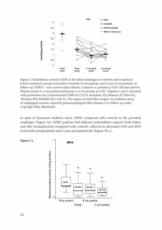
40
Figure 3 a
Figure 2. Glutathione content (GSH) at the distal esophagus in controls and in patients before treatment (preop) and aafter 6 months (6 mo postop) and 4 years (4 yrs postop) of follow-up. NERD = non-erosive refl ux disease. Controls vs. patients p<0.01 (all time-points). Patients preop vs. 6 mo postop and preop vs. 4 yrs postop: p<0.05. (Figures 1 and 2 reprinted with permission Am J Gastroenterol 2006;101:222-8. Rantanen TK, Räsänen JV, Sihvo EI, Ahotupa MO, Färkkilä MA, Salo JA. The impact of antirefl ux surgery on oxidative stress of esophageal mucosa caused by gastroesophageal refl ux disease: 4-yr follow-up study. . Copyrigh Wiley-Blackwell)
In spite of decreased oxidative stress (MPA) compared with controls in the proximal esophagus (Figure 3a), GERD patients had defi cient antioxidative capacity both before and after fundoplication compared with controls, refl ected as decreased GSH and SOD levels both preoperatively and 4 years postoperatively (Figure 3b, c).

41
Figure 3, a, b, c.Myeloperoxidase activity (MPA), superoxide dismutase (SOD) and glutahione content (GSH) in the proximal esophagus of controls (Prox control) and patients preoperatively (Preop), and after 6 months (6 mo postop) and 4 years (4 yrs postop) of follow-up. * p<.05, ** p<.01 prox control vs patients preop, 6 mo postop and 4 yrs postop, Median and inter-quartile in box, 10th and 90th percentile shown by horizontal bars.
Figure 3 b
Figure 3 c

42
At all time-points, MPA of the distal esophagus was also signifi cantly higher than that of the proximal esophagus in patients but not in controls (Figure 4) (p < 0.01 and p = NS, respectively).
Figure 4.
Figure 4. Myeloperoxidase activity (MPA) in the proximal and distal esophagus of controls (Prox control and Dist control) and patients preoperatively (Prox preop and Dist preop), and after 6 months (Prox 6 mo and Dist 6 mo) and 4 years (Prox 4 yrs and Dist 4 yrs) of follow-up.Plots as in Fig 3. Prox control vs Dist control, p = NS, Prox preop vs Dist preop, Prox 6 mo vs Dist 6 mo, and Prox 4 yrs vs Dist 4 yrs, ** p < .01 for all
2. Expression of 8-hydroxydeoxyguanosine in esophageal tissues and tumors
The amount of oxidative stress-related DNA damage (as 8-OHdG) was signifi cantly increased in the distal esophagus both in Barrett’s epithelium 1.26 (0.08-29.47) and in high-grade dysplasia 1.35 (1.04-1.65), as well as in adenocarcinoma of the esophagus/esophagogastric junction 1.08 (0.59-1.94) compared with controls 0.06 (0-4.08) (p=0.002, p=0.012, p=0.001, respectively) (Figure 5). Barrett’s patients had similar 8-OHdG levels in their distal and proximal esophageal samples.

43
Figure 5. Oxidative DNA damage (8-OHdG/105dG) levels in the distal esophagus of controls (Controls) and patients with adenocarcinoma of the distal esophagus/esophagogastric junction (Carcinoma), Barrett’s esophagus with high-grade dysplasia (Dysplasia), or Barrett’s metaplasia (Barrett). *p<0.05 and **p<0.01, comparison between controls and patients. No signifi cant differences were found between samples from Barrett’s epithelium patients with or without high-grade dysplasia and adenocarcinoma. Plots display median (horizontal bars), 25th and 75th percentiles (lower and upper limits of boxes) and lowest and highest values excluding outliers (error bars).
3. Impact of positron emission tomography on clinical staging and prognostication of adenocarcinoma of the esophagus and esophagogastric junction
Diagnostic sensitivity for the primary tumor was 83% for PET and 67% for CT; for local peritumoral lymph node metastasis, it was 37% for PET and 89% for EUS; and for distant metastasis, it was 47% for PET and 33% for CT compared with histopathology. Diagnostic specifi city for local lymph node metastasis was 100% with PET and 54% with EUS, and for distant metastasis, it was 89% with PET and 96% with CT. Accuracy for locoregional lymph node metastasis was 63% for PET, 66% for CT, and 75% for EUS, and for distant metastasis, it was 74% for PET and 74% for CT (Table 1). Of the 10 patients who were considered inoperable during surgery, PET identifi ed 7 and CT 4 (Table 2). The false-negative diagnoses of stage IV disease in PET were peritoneal carcinomatosis in two patients, abdominal para-aortic cancer growth in one, metastatic lymph nodes by the celiac artery in four, and metastases in the pancreas in one. PET showed false-positive lymph nodes at the jugulum in three patients.

44
Table 1. Detection of distant metastases by positron emission tomography (PET) and computed tomography (CT) in 42 patients with adenocarcinoma of the esophagus or esophagogastric junction compared with histopathology.
Sensitivity Specifi city Accuracy
No./Total no. % No./Total no. % No/Total no. %
PET 7/15 47 24/27 89 31/42 74
CT 5/15 33 26/27 96 31/42 74
Table 2. Findings of computed tomography (CT), positron emission tomography (PET), and endoscopic ultrasonography (EUS) for patients with inoperable disease.
Sites of disease demonstrated by:
PatientNo.
Site of metastatic disease which rendered tumor unresectable
Biopsyproven CT PET EUS
13 Liver Yes No distant metastasis Liver No distant metastasis
16 Left adrenal gland Yes Left adrenal gland Left adrenal gland No passage
20 Carcinomatosis Yes Celiac lymph node No distant metastasis No distant
metastasis
26 Carcinomatosis Yes No distant metastasis Rib No passage
31 Carcinomatosis Yes No distant metastasis No distant metastasis No passage
39 Left supraclavicular
lymph nodes and
wide retroperitoneal
lymphatic growth
Yes Retroperitoneal
growth
Left supraclavicular
lymph nodes and
retroperitoneal growth
No distant
metastasis
42 Liver Yes No distant metastasis Liver No passage
43 Pancreas Yes No distant metastasis No distant metastasis No passage
44 Liver Yes No distant metastasis Liver No passage
49 Liver Yes Liver Liver No passage
(Table1 and 2 reprinted with permission from Ann Surg Oncol 2003;10:954-60. Räsänen JV, Sihvo EI, Knuuti MJ, Minn HR, Luostarinen ME, Laippala P, Viljanen T, Salo JA. Prospective analysis of accuracy of positron emission tomography, computed tomography, and endoscopic ultrasonography in staging of adenocarcinoma of the esophagus and the esophagogastric junction. Copyrigh Springer Science and Business Media).

45
Positron emission tomography (pet) and survival
Median survival was not reached in histopathologically confi rmed non-distant metastases (pM-negative) disease (Figure 6a). In the clinical stage without distant metastases, median survival was 24 months when disease was detected by any of the imaging techniques alone or in combination: CT (95% CI 13-36 months), PET (14-35 months), EUS or CT (13-36 months), EUS, CT, or PET (14-35 months), and EUS or CT and PET (14-35 months). Median survival in clinical or histopathological stage IV disease is shown in Figure 6b. Positive PET for distant metastasis together either EUS or CT accurately predicted the poor survival of these patients.
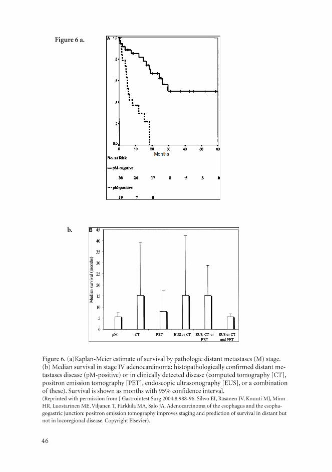
46
Figure 6 a.
Figure 6 Figure 6 b.
Figure 6. (a)Kaplan-Meier estimate of survival by pathologic distant metastases (M) stage. (b) Median survival in stage IV adenocarcinoma: histopathologically confi rmed distant me-tastases disease (pM-positive) or in clinically detected disease (computed tomography [CT], positron emission tomography [PET], endoscopic ultrasonography [EUS], or a combination of these). Survival is shown as months with 95% confi dence interval.(Reprinted with permission from J Gastrointest Surg 2004;8:988-96. Sihvo EI, Räsänen JV, Knuuti MJ, Minn HR, Luostarinen ME, Viljanen T, Färkkila MA, Salo JA. Adenocarcinoma of the esophagus and the esopha-gogastric junction: positron emission tomography improves staging and prediction of survival in distant but not in locoregional disease. Copyright Elsevier).

47
4. Cost-utility of treatment of carcinoma of the esophagus or esophagogastric junction
Of the 30 radically treated patients, 27, 23, and 16 were alive at 3, 12, and 24 months, respectively (median survival 24 months). The corresponding mean 15D scores of survivors in the radically treated group at each point of time were 0.82, 0.87, and 0.86. The median cost of radical treatment was 32 271 € over a two-year study period. Of the 23 palliatively treated patients 17, 4, and 2 were alive at 3, 12, and 24 months (median survival 6 months) (p<0.001 compared with the radical group) (Figure 7). The corresponding mean 15D scores of survivors in the palliative group at each point of time were 0.80, 0.72 and 0.75, respectively. The median cost of palliative treatment was 18 565 € (p<0.01 compared with the radical group) over the two year follow-up or until death. The current practice model suggested that, on average, radically treated patients would experience 1.198 quality-adjusted life-years (QALYs) at a cost of 43 000 € over the entire 24-month time-horizon of the model and palliatively treated patients 0.418 QALYs at a cost of 25 000 €. The incremental cost-utility ratio (ICUR) of the radical treatment was 22 893 € /QALY gained compared with palliative treatment.
Figure 7. Survival of patients with carcinoma of the esophagus or esophagogastric junction treated either with radical surgery or palliatively within current practice.
Sensitivity analyses
Sensitivity analyses showed that in 46% of the simulated cases radical surgery seems to be cost-saving, while in 53% of the cases was more costly and more effective than palliative treatment for all patients (Figure 8). If willingness to pay for a QALY is 30 000 €, the probability of the current treatment is being acceptable is 68% (Figure 9).

48
Figure 8. Scatter plot of estimated joint density of incremental costs and incremental effects of current practice versus hypothetical treatment by bootstrap resampling (base case). Probabilistic sensitivity analysis: a scatter plot that illustrates the uncertainty in the expected incremental costs and QALYs gained for current practice versus hypothetical treatment for the base case. Practically all bootstrap replicates lie above zero on the QALYs gained axis, indicating a high degree of certainty that current practice where eligible patients are treated by radical surgery is more effective than hypothetical treatment where all patients are pro-vided only palliative treatment. Data points from bootstrapping that lie in the “northeast” quadrant of the cost-effectiveness plane represent QALYs gain from hypothetical treatment at an additional cost. Points that lie in the “southeast” quadrant represent lower costs from hypothetical treatment. The base case result is shown by a box.
-100 000
-50 000
0
50 000
100 000
150 000
-0,20 -0,10 0,00 0,10 0,20 0,30 0,40 0,50 0,60 0,70 0,80
Incremental QALYs gained
Quadrant I Quadrant II
Quadrant III Quadrant IV
Incremental cost (€)
49
Figure 9. Probabilistic sensitivity analysis: cost-effectiveness acceptability curve representing the probability that current practice by radical surgery for all eligible patients vs. hypotheti-cal treatment (where all patients provided only palliative treatment) for different values of willingness-to-pay for a quality-adjusted life-year (QALY) saved.
0,00
0,10
0,20
0,30
0,40
0,50
0,60
0,70
0,80
0,90
1,00
0 10 000 20 000 30 000 40 000 50 000 60 000 70 000 Willingness to pay for a QALY (€)
Prob
abili
ty o
f cur
rent
pra
ctic
e be
ing
acce
ptab
le

50
DISCUSSION
The adenocarcinoma of the esophagus and esophagogastric junction in Western men has received increasing attention because of its rapidly incidence and deadly nature (Powell et al. 1990, Armstrong and Borman 1996, Devesa et al. 1998, Botterweck et al. 2000, Sihvo et al. 2000, van Bollschweiler et al. 2001, Blankenstein et al., Voutilainen and Juhola 2005). Unfortunately, despite new treatment forms, in the advanced stages of this disease, survival after diagnosis remains dismal (Orringer et al. 1999, Hulscher et al. 2001, 2002, van Sandick et al. 2002). Only the disease stages limited to the wall of the esophagus or cardia and the surrounding lymph tissue warrant radical surgical treatment (Clark et al. 1994); exact preoperative clinical staging is therefore essential for optimal treatment of this lethal malignancy. The precise mechanism behind the development of this malignancy is unknown, although gastroesophageal refl ux disease (GERD) and Barrett’s esophagus have been recognized as risk factors for years (Lagergren et al. 1999). Because of the rising incidence and expense of new treatments, the cost-effectiveness of treatment is becoming increasingly important (Xinopoulos et al. 2004).
1. Oxidative stress and antireflux surgery
This study shows that GER patients suffer from increased oxidative stress and decreased anti-oxidative defense in their distal esophagus before antirefl ux surgery. In addition, their anti-oxidative defense throughout the esophagus seems to be defective. It was surprising to fi nd that although macroscopic esophagitis after antirefl ux surgery was already healed at 6 months, reduction in MPA and healing at the cellular level apparently take a much longer period and do not reach control levels even at 4 years. We can therefore assume that even successful fundoplication cannot fully reverse the oxidative stress in the esophagus of patients with GERD during a 4-year follow-up. This is contrary to the conclusion of Wetscher et al. (1995) who found in their short-term follow-up study that anti-refl ux surgery can prevent oxidative stress in esophageal mucosa. However, MPA in the distal esophagus decreased signifi cantly after successful antirefl ux surgery.
Our patients had signifi cantly lower GSH values both before and after surgery in the distal esophagus than controls, in agreement with van Lieshout et al. (1999), who reported signifi cantly lower glutathione content in Barrett’s epithelium than in normal esophageal mucosa. Sido et al. (1998) have shown that patients with infl ammatory bowel disease have an intestinal GSH defi ciency in the infl amed and noninfl amed ileum because of impairment of GSH synthesis. Our results suggest a similar phenomenon in the distal esophagus of patients with GERD. Imbalance between anti-oxidative defense and increased oxidative stress can lead to increased reactive oxygen species (ROS) (Farhadi et al. 2002, Jimenez et al. 2005, Li et al. 2007). In addition to the lack of recovery from oxidative stress of the esophageal mucosa during short- to mid-term follow-up, the increasing failure rates of fundoplication with recurrent GERD during long-term follow-up (Luostarinen 1993) indicate that antirefl ux surgery is not a perfect solution for oxidative stress of the
51
esophageal mucosa. Further studies are needed to compare the effect of medical treatment versus antirefl ux surgery on oxidative stress. Keeping in mind that oxidative stress is closely related to DNA damage, our results may also partially explain why adenocarcinoma of the distal esophagus is found even after successful fundoplication (Ye et al. 2001, Tran et al. 2005).
Based on our study in GERD patients, anti-oxidative capacity (GSH, SOD) of the proximal esophagus was diminished before and even 4 years after successful antirefl ux surgery. Oxidative stress (MPA) in the proximal esophagus was preoperatively lower in controls and remained so both 6 months and 4 years after fundoplication. The proximal esophageal mucosa in patients with GERD therefore appears to be characterized by a low level of oxidative stress and a diminished anti-oxidative defense mechanism. Several explanations are possible for the continued difference in levels of oxidative stress between the distal and proximal esophagus even years after successful antirefl ux surgery. First, higher MPA in the distal esophagus may be an irreversible change after a long period of GERD. Second, the protective effects of saliva in the upper and lower esophagus may differ. Third, this difference may reveal uneven myeloperoxidase activity of neutrophils in these parts of the esophagus in response to chemotactic stimuli, as previously shown in gingival tissue (Kowolik and Grant 1983). The reason for the lower MPA in the proximal esophagus than in controls, even before surgery, remains unclear. The volume, neutralizing capacity, or components of saliva may explain the difference between GERD patients and controls (Kongara and Soffer 1999). Most importantly, our fi ndings suggest that the impaired anti-oxidative capacity of the proximal esophagus may be caused not by increased oxidative stress, but rather by changes in the anti-oxidative defense system itself. The cause of this phenomenon may be genetic. The defective antioxidative defense in the proximal esophagus of GERD patients may have an important role in the development of complicated GERD.
2. Oxidative DNA damage and pathogenesis of esophageal adenocarcinoma
ROS can cause formation of oxidative base adducts, such as 8-OHdG which in turn can lead to DNA mutations. Dvorak et al. (2006) have shown that bile acids compined with low pH induce oxidative DNA damage (8-OHdG) and mitochondrial oxidative stress in esophageal cells.
We observed signs of oxidative DNA damage in Barrett’s metaplasia, in high-grade dysplasia, and in adenocarcinoma of the esophagus/esophagogastric junction. The leading hypothesis regarding carcinogenesis in adenocarcinoma of the esophagus/esophagogastric junction is a metaplasia-dysplasia-carcinoma sequence of malignant transformation (Jankowski et al. 2000), in which the molecular mechanisms remain in many respects unclear. Tissue damage in esophagitis and in Barrett’s metaplasia has been associated with oxidative stress (Olyaee et al. 1995, Oh et al. 2001), and an inverse epidemiological link exists between risk for esophageal adenocarcinoma and anti-oxidants (Terry et al. 2000). Furthermore, in an animal model of refl ux-related esophageal damage these anti-oxidants

52
have been lower (Oh et al. 2001). Strong evidence thus suggests that oxidative stress may cause changes in DNA bases, leading to mutations and further to the development of adenocarcinoma.
3. Imaging and optimal treatment and prognosis
Different options, from mucosal resections (Ell et al. 2000, Nijhawan and Wang 2000, May et al. 2002, May et al. 2003, Seewald et al. 2003, Giovannini et al. 2004, Rajan et al. 2004, Vieth et al. 2004) to multimodal therapy (Burak et al. 2003, Lordick el al. 2004), are currently available treat esophageal/GEJ adenocarcinoma. Accurate pretreatment staging is crucial to enable optimal choice of treatment. Positron emission tomography (PET) is based on the accumulation of a fl uorinated glucose analog (18F-fl uorodeoxy-D-glucose; FDG) in malignant cells (Pauwels et al. 1998). This can be observed by a positron camera. Thus, PET provides the opportunity to detect altered tissue metabolism in malignant tumors. This has been thought to detect tumors and metastases more precisely than with conventional staging methods, such as computed tomography (CT) and endoscopic ultrasonography (EUS). According to our results, PET failed to identify very small primary tumors or to detect small-sized metastatic lesions (spatial resolution 6 mm), such as intra-abdominal carcinomatosis. Moreover its sensitivity for detecting lymph node metastases was inferior to that of EUS. Our fi ndings show that the overall accuracy of PET to detect stage IV disease is no better than that of CT. This is mostly because of a lack of sensitivity in fi nding distant lymph node metastases and false-positive judgments of cervical lymph nodes.
In conclusion, all of the staging methods used in this study have shortcomings. EUS has problems with accuracy in detecting the T and N stages. The development of new high-frequency probes and EUS with fi ne-needle biopsy may solve these problems in the future. CT is insuffi ciently sensitive in detecting distant metastatic lesions and locoregional nodal metastasis. PET can identify organ metastases fairly well, but its lack of overall accuracy in predicting distant metastatic (M1) disease and its inability to identify locoregional metastases limit its reliability. PET seems, however, to detect organ metastases better than CT.
In prognostication and treatment allocation for adenocarcinoma of the esophagus/GEJ, one prerequisite is accurate pretreatment staging. This staging is hoped to be improved by the use of PET. We found that adding PET to standard staging does improve detection of stage IV disease and its associated poor survival. In sum in locally advanced tumors, PET is recommended to exclude patients from unnecessary surgery. Moreover adding PET to standard staging does improve detection of stage IV disease and its associated poor survival.

53
4. Cost-utility of treatment of carcinoma of the esophagus or esophagogastric junction
Esophageal carcinoma is one of the deadliest cancer forms known. Its incidence is increasing also in Finland (Sihvo et al. 2000, Voutilainen and Juhola 2005). Increased resources are needed for its treatment. Our cost-utility analysis shows the incremental cost-utility ratio (ICUR) of the radical surgical treatment was 22 893 € /QALY gained compared with palliative treatment according to current practices, which is well below the often used threshold of £30 000/QALY (Martin et al. 2003). Our hypothetical model where all patients were treated by palliative means was not competitive with current practice. Sensitivity analyses showed that in current practice where all eligible patients are treated by radical surgery seems to be cost-saving in 46% of the simulated cases, while in 53% of the cases more costly and effective compared with palliative treatment for all patients. If willingness to pay for a QALY is 30 000 €, the probability of current treatment being acceptable is 68%; using a threshold value of 50 000 €, it would be almost 80%.
Radical treatment produces better survival and HRQoL than palliative treatment, and consequently, better effi ciency, since the ICUR of radical treatment compared with palliative treatment remain reasonably low despite the cost of treatment in the radical group being signifi cantly higher. The sensitivity analysis demonstrates that cost-effectiveness of treatment of esophageal carcinoma cannot be improved by choosing a palliative treatment option for all patients over the current of treatment strategy.

54
CONCLUSIONS
Elevated oxidative stress (MPA) and decreased antioxidant defense (GSH) after antirefl ux surgery in the distal esophagus indicate that antirefl ux surgery is not a perfect solution for oxidative stress of the esophageal mucosa, although it is effective against symptoms and macroscopic esophagitis. Elevated oxidative stress may in part explain why adenocarcinoma of the distal esophagus is found even after successful fundoplication.
In GERD patients, proximal esophageal mucosal anti-oxidative defense seems to be defective before and even years after successful antirefl ux surgery. In addition, antirefl ux surgery seems not to change the level of oxidative stress in the proximal esophagus, suggesting that defective mucosal anti-oxidative capacity plays an important role in the development of oxidative damage to the esophageal mucosa in GERD.
In the malignant transformation of Barrett’s esophagus, an important component is oxidative stress. DNA damage seems to be mediated via 8-OHdG and the entire esophagus of Barrett’s patients suffers from increased oxidative stress.
PET is a useful tool in the staging of adenocarcinoma of the esophagus and esophagogastric junction because it seems to detects organ metastases better than CT, although its accuracy in staging of paratumoral and distant lymph nodes is limited.
Despite its limited accuracy, positive PET for distant metastasis with either positive EUS or CT accurately predicts the poor survival of these patients. Adding PET to standard staging therefore improves detection of stage IV disease and its associated poor survival.
The current practice in the treatment of esophageal carcinoma where eligible patients are treated by radical surgery seems to offer the greatest benefi t in terms of survival and cost-utility. Multimodality treatments may further improve effectiveness, but at increased cost.

55
YHTEENVETO (FINNISH SUMMARY)
Ruokatorven ja mahansuun rauhassyöpä on nopeasti lisääntyvä huonoennusteinen sairaus, jonka syntymekanismi on liitetty oksidatiiviseen stressiin. Taudin tarkka kliininen levinneisyysluokitus on äärimmäisen tärkeää hoidon onnistumiseksi. Koska tauti on niin huonoennusteinen luonteeltaan, ja sen yleisyys lisääntyy nopeasti, on sen hoidon paras mahdollinen kustannustehokkuus entistä tärkeämpää. Tämän tutkimuksen tavoitteena oli selvittää 1) onko ruokatorven refl uksia estävällä fundoplikaatioleikkauksella ruokatorven limakalvon oksidatiivista stressiä vähentävä vaikutus, 2) mikä on oksidatiivisen DNA vaurion osuus Barrettin ruokatorven ja ruokatorven rauhassyövän synnyssä, 3) mikä on positroniemissiotomografi an (PET) arvo ruokatorven rauhassyövän ennen hoidon aloittamista tehtävässä kliinisessä luokituksessa, ja auttaako se taudin ennusteen laatimisessa sekä 4) mikä on nykyisen ruokatorven rauhassyövän hoitokäytännön kustannustehokkuus.
Ensimmäisessä ja toisessa osatyössä määritimme ruokatorven oksidatiivista metaboliaa ruokatorven ylä- ja alapäästä otetuista näytteistä. Oksidatiivisen metabolian merkkiaineina toimivat kudoksen myeloperoksidaasi-entsyymin ja superoksidi-entsyymin aktiivisuus sekä glutathioni-pitoisuus. Ensimmäiset näytteet otettiin ennen fundoplikaatioleikkausta ja potilaita seurattiin aina 4 vuoteen asti. Kolmannessa osatyössä analysoimme 51 potilaan ruokatorven kudosnäytteistä oksidatiivisen stressin aiheuttamaa DNA muutosta, 8-hydroksideoksiguanosiinia (8-OHdG). Neljännessä ja viidennessä osatyössä tarkastelimme ensin 42 potilaan ennen leikkaushoitoa tehtyä kliinistä levinneisyysluokitusta verrattuna leikkauksen jälkeen tiedossa olevaan patologiseen luokitukseen. Jatkoimme edelleen potilaskeräystä aina 55 potilaaseen saakka ja tälle joukolle teimme analyysin positroniemissiotomografi an hyödyllisyydestä ennusteen laatimiseen. Kuudennessa osatyössä selvitimme ruokatorven syövän kustannuksia ja sen hoitoon liittyvää elämänlaatua hoidon eri vaiheissa 53:lla potilaalla. Laadimme kustannustehokkuusanalyysin näistä tiedoista.
Tutkimuksemme löydökset olivat seuraavat: 1) onnistunutkaan refl uksinestokirurgia ei kykene täysin häivyttämään oksidatiivista stressiä ruokatorven limakalvolta 2) koko ruokatorven limakalvon antioksidatiivinen puolustusmekanismi vaikuttaa olevan puutteellinen, 3) oksidatiiviseen stressiin liittyvä DNA muutos (8-OHdG) näyttää olevan mukana ruokatorven rauhassyövän kehittymisessä, 4) PET parantaa muihin elimiin levinneen ruokatorven rauhassyövän toteamista ja näin ollen helpottaa huonon ennusteen potilaiden tunnistamista 5) kirurginen, parantamiseen tähtäävä hoito on kustannustehokkain vaihtoehto oikein valituille ruokatorven rauhassyöpäpotilaille.
Johtopäätöksinä löydöksistämme on tehtävissä: 1) refl uksinestokirurgia ei pysty normalisoimaan oksidatiivisen stressin vaurioittamaa limakalvoa ja näin ollen syövän kehittyminen on edelleen mahdollista vialliselle limakalvolle huolimatta toimivasta fundoplikaatiosta 2) uusia tutkimuksia tarvitaan - voisiko ruokatorven antioksidatiivista puolustusta auttaa refl uksitaudista kärsivillä esim. uusilla lääkeaineilla? 3) todetut oksidatiiviset DNA muutokset ruokatorven rauhaskarsinooman synnyssä kannustavat

56
jatkamaan uusissa tutkimuksissa syövän syntymekanismin selvittämistä tältä suunnalta 4) PET puoltaa paikkaansa ruokatorven rauhassyövän kliinisessä luokittelussa ja ennusteen tunnistamisessa erityisesti huonon ennusteen potilailla. Tämä auttaa resurssien järkevässä suuntaamisessa. 5) ruokatorven rauhasyövän parantamiseen tähtäävä kirurginen hoito on kannattavaa sekä elämänlaatua parantavana että myös kustannustehokkaana hoitomuotona oikein valituille potilaille.
57
ACKNOWLEDGMENTS
This study was carried out in the Division of General Thoracic and Esophageal Surgery of the Deparment of Cardiothoracic Surgery of Helsinki University Central Hospital. I wish to express my deepest gratutude to a number of people who made this work possible:
To Professor Ari Harjula for providing me the opportunity to perform this work.
To my supervisor, Professor Jarmo Salo, for excellent guidance and encouraging attitude both in scientifi c and clinical work in the fi eld of thoracic and esophageal surgery.
Docent Pekka Nuutinen and Docent Markku Voutilainen for constructive criticism and the enhancement of this thesis.
Eero Sihvo and Tuomo Rantanen for their valuable cooperation.
Docent Markku Ahotupa for biochemical analysis.
Professor Juhani Knuuti, Docent Heikki Minn, , MSc Tapio Viljanen for their analysis of positron emission tomography and valuable advices.
Docent Markku Luostarinen for an excellent study idea.
Late docent Pekka Laippala and M.Sc Anna Koivisto for statistical assistance.
Professor Martti Färkkilä for providing important control material.
Professor Harri Sintonen, MSocSc Pasi Aronen and docent Risto Roine for familiarization with the secrets of health economics.
Yvonne Sundström for skillful and invaluable secretarial assistance.Merjä Räsänen for helping to collect material for studies.Carol Pelli for author-editing of the language.My cousins in the U.S.A for their exhortation.My mother and my sisters and brothers for their love and support.
Most of all, my dear wife, Pirjo, my lovely daughter, Lotta, and my brisk son, Lauri, without you the whole work would have been worthless.
This study was supported by grants from The Research Foundation (EVO) of Helsinki University Central Hospital, the Ida Montin Foundation, and Finska Läkaresällskapet.

58
REFERENCES
Abbas Z, Hussainy AS, Ibrahim F, Jafri SM, Shaikh H, Khan AH. Barrett’s oesophagus and Helicobacter pylori. J Gastroenterol Hepatol 1995;10:331-3.
al-Sarraf M, Martz K, Herskovic A, Leichman L, Brindle JS, Vaitkevicius VK, Cooper J, Byhart R, Davis L, Emami B. Progress report of combined chemoradiotherapy versus radiotherapy alone in patients with esophageal cancer: an intergroup study. J Clin Oncol 1997;15:277-84.
Altorki NK, Sanagawa M, Little AG, Skinner DB. High-grade dysplasia in the columnar lined esophagus. Am J Surg 1991;161:97-9.
Armstrong D. Review article: towards consistency in the endoscopic diagnosis of Barrett‘s oesophagus and columnar metaplasia. Aliment Pharmacol Ther 2004;20(Supp 5):40-7; discussion 61-2.
Armstrong RW, Borman B. Trends in incidence rates of adenocarcinoma of the oesophagus and gastric cardia in New Zealand, 1978-1992. Int J Epidemiol 1996;25:941-7.
Arnott SJ, Duncan W, Gignoux M, Girling DJ, Hansen HS, Launois B, Nygaard K, Parmar MK, Roussel A, Spiliopoulos G, Stewart LA, Tierney JF, Mei W, Rugang Z. Preoperative radiotherapy in esophageal carcinoma: a meta-analysis using individual patient data (Oesophageal Cancer Collaborative Group). Int J Radiat Oncol Biol Phys 1998; 41:579-83.
Arslan N, Miller TR, Dehdashti F, Battafarano RJ, Siegel BA. Evaluation of response to neoad-juvant therapy by quantitative 2-deoxy-2-[18F]fl uoro-D-glucose with positron emission tomography in patients with esophageal cancer. Mol Imaging Biol 2002;4:301-10.
Asaka M, Takeda H, Sugiyama T, Kato M. What role does Helicobacter pylori play in gastric cancer? Gastroenterology 1997;113(Suppl 6):S56-60.
Attwood SE, DeMeester TR, Bremner CG, Barlow AP, Hinder RA. Alkaline gastroesophageal refl ux: implications in the development of complications in Barrett’s columnar-lined lower esophagus. Surgery 1989;106:764-70.
Balaji NS, DeMeester SR, Wickramasinghe KS, Hagen JA, Peters JH, DeMeester TR. Etiology of intestinal metaplasia at the gastroesophageal junction. Surg Endosc 2003;17:43-8.
Barbour AP, Rizk NP, Gonen M, Tang L, Bains MS, Rusch VW, Coit DG, Brennan MF. Adeno-carcinoma of the gastroesophageal junction: infl uence of esophageal resection margin and operative approach on outcome. Annals of Surgery 2007 246:1-8.
Blackstock AW, Farmer MR, Lovato J, Mishra G, Melin SA, Oaks T, Aklilu M, Clark PB, Levine EA. A prospective evaluation of the impact of 18-F-fl uoro-deoxy-D-glucose positron emission tomography staging on survival for patients with locally advanced esophageal cancer. International Journal of Radiation Oncology, Biology, Physics 2006;64:455-60.
Block MI, Patterson GA, Sundaresan RS, Bailey MS, Flanagan FL, Dehdashti F, Siegel BA, Cooper JD. Improvement in staging of esophageal cancer with the addition of positron emission tomography. Ann Thorac Surg 1997;64:770-7.
Blot WJ, Devesa SS, Kneller RW, Fraumeni JF, Jr. Rising incidence of adenocarcinoma of the esophagus and gastric cardia. JAMA 1991;265:1287-9.
Boch JA, Shields HM, Antonioli DA, Zwas F, Sawhney RA, Trier JS. Distribution of cytokeratin markers in Barrett’s specialized columnar epithelium. Gastroenterology 1997;112:760-5.
Bollschweiler E, Wolfgarten E, Gutschow C, Holscher AH. Demographic variations in the rising incidence of esophageal adenocarcinoma in white males. Cancer 2001;92:549-55.
Bonde P, Gao D, Chen L, Miyashita T, Montgomery E, Harmon JW, Wei C. Duodenal refl ux leads to down regulation of DNA mismatch repair pathway in an animal model of esophageal cancer. Annals of Thoracic Surgery 2007;83:433-40.
59
Boonstra J, Post JA. Molecular events associated with reactive oxygen species and cell cycle progression in mammalian cells. Gene 2004;337:1-13.
Botterweck AA, Schouten LJ, Volovics A, Dorant E, van Den Brandt PA. Trends in incidence of adenocarcinoma of the oesophagus and gastric cardia in ten European countries. Int J Epidemiol 2000;29:645-54.
Bowrey DJ, Clark GW, Williams GT. Patterns of gastritis in patients with gastro-oesophageal refl ux disease. Gut 1999;45:798-803.
Brandt LJ, Kauvar DR. Laser-induced transient regression of Barrett’s epithelium. Gastrointest Endosc 1992;38:619-22.
Bremner CG, Lynch VP, Ellis FH, Jr. Barrett’s esophagus: congenital or acquired? An experimen-tal study of esophageal mucosal regeneration in the dog. Surgery 1970;68:209-16.
Brenner B, Ilson D, Minsky BD. Treatment of localized esophageal cancer. Semin Oncol 2004;31:554-65.
Briggs AH, O’Brien BJ. The death of cost-minimisation analysis? Health Econ Lett 2000;4:3-10.Burak WE. Is neoadjuvant therapy the answer to adenocarcinoma of the esophagus? Am J Surg
2003;186:296-300.Burmeister BH, Smithers BM, Gebski V, Fitzgerald L, Simes RJ, Devitt P, Ackland S, Gotley DC,
Joseph D, Millar J, North J, Walpole ET, Denham JW. Trans-Tasman Radiation Oncology Group. Australasian Gastro-Intestinal Trials Group. Surgery alone versus chemora-diotherapy followed by surgery for resectable cancer of the oesophagus: a randomised controlled phase III trial. Lancet Oncology 2005;6:659-68.
Buttar NS, Wang KK, Sebo TJ, Riehle DM, Krishnadath KK, Lutzke LS, Anderson MA, Petterson TM, Burgart LJ. Extent of high-grade dysplasia in Barrett‘s esophagus correlates with risk of adenocarcinoma. Gastroenterology 2001;120:1630-9.
Buxton MJ, Drummond MF, Van Hout BA, Prince RL, Sheldon TA, Szucs T, Vray M. Modelling in economic evaluation: An unavoidable fact of life. Health Econ 1997;6:217-27.
Cadiot G, Bruhat A, Rigaud D, et al. Multivariate analysis of pathophysiological factors in refl ux oesophagitis. Gut. 1997;40:167-174.
Cameron AJ, Souto EO, Smyrk TC. Small adenocarcinomas of the esophagogastric junction: association with intestinal metaplasia and dysplasia. Am J Gastroenterol 2002;97:1375-80.
Cameron AJ. Arora AS. Barrett’s esophagus and refl ux esophagitis: is there a missing link? American Journal of Gastroenterology 2002;97:273-8.
Cameron AJ. Barrett’s esophagus: prevalence and size of hiatal hernia. Am J Gastroenterol 1999;94:2054-9.
Cameron AJ. Epidemiology of columnar-lined esophagus and adenocarcinoma. Gastroenterol Clin North Am 1997;26:487–94.
Cameron AJ, Carpenter HA. Barrett’s esophagus, high-grade dysplasia, and early adenocarci-noma: a pathological study. Am J Gastroenterol 1997;92:586-91.
Cameron AJ, Lomboy CT, Pera M, Carpenter HA. Adenocarcinoma of the esophagogastric junction and Barrett’s esophagus. Gastroenterology 1995;109:1541-6.
Cameron AJ, Lomboy CT. Barrett’s esophagus: age, prevalence, and extent of columnar epithe-lium. Gastroenterology 1992;103:1241-5.
Cameron AJ, Zinsmeister AR, Ballard DJ, Carney JA. Prevalence of columnar-lined (Barrett’s) esophagus. Comparison of population-based clinical and autopsy fi ndings. Gastroenterol-ogy 1990;99:918–22.
Carlisle JG, Quint LE, Francis IR, Orringer MB, Smick JF, Gross BH. Recurrent esophageal carcinoma: CT evaluation after esophagectomy. Radiology 1993;189:271-5.
Caygill CP, Reed PI, Johnston BJ, Hill MJ, Ali MH, Levi S. A single centre’s 20 years’ experi-ence of columnar-lined (Barrett’s) oesophagus diagnosis. Eur J Gastroenterol Hepatol 1999;11:1355-8.

60
Chak A, Ochs-Balcom H, Falk G, Grady WM, Kinnard M, Willis JE, Elston R, Eng C. Familiality in Barrett´s esophagus, adenocarcinoma of the esophagus, and adenocarcinoma of the gastroesophageal junction. Cancer Epidemiol Biomarkers Prev 2006;15:1668-73.
Champion G, Richter JE, Vaezi MF, Singh S, Alexander R. Duodenogastroesophageal refl ux: re-lationship to pH and importance in Barrett’s esophagus. Gastroenterology 1994;107:747-54.
Chandrasoma PT, Der R, Ma Y. Peters J, DeMeester T. Histologic classifi cation of patients based on mapping biopsies of the gastroesophageal junction. Am J Surg Pathol 2003;27:929-36.
Chandrasoma PT, Der R, Ma Y, Dalton P, Taira M. Histology of the gastroesophageal junction: an autopsy study. Am J Surg Pathol 2000a;24:402-9.
Chandrasoma PT, Lokuhetty DM, DeMeester TR, Bremmer CG, Peters JH, Oberg S, Groshen S. Defi nition of histopathologic changes in gastroesophageal refl ux disease. Am J Surg Pathol 2000b;24:344-51.
Chen H, Tucker KL, Graubard BI, Heineman EF, Markin RS, Potischman NA, Russell RM, Weisenburger DD, Ward MH. Nutrient intakes and adenocarcinoma of the esophagus and distal stomach. Nutr Cancer 2002;42:33-40.
Chen X, Ding YW, Yang G, Bondoc F, Lee MJ, Yang CS. Oxidative damage in an esophageal adenocarcinoma model with rats. Carcinogenesis 2000;21:257-63.
Cheng X, Yang CY. Esophageal adenocarcinoma: a review and perspectives on the mechanism of carcinogenesis and chemoprevention. Carcinogenesis 2001;22:1119-29.
Chow WH, Blot WJ, Vaughan TL, Risch HA, Gammon MD, Stanford JL, Dubrow R, Schoenberg JB, Mayne ST, Farrow DC, Ahsan H, West AB, Rotterdam H, Niwa S, Fraumeni JF, Jr. Body mass index and risk of adenocarcinomas of the esophagus and gastric cardia. J Natl Cancer Inst 1998;90:150-5, 1998.
Chow WH, Finkle WD, McLaughlin JK, Franki H, Ziel HK, Fraumeni JF, Jr. The relation of gastroesophageal refl ux disease and its treatment to adenocarcinomas of the esophagus and gastric cardia. JAMA 1995;274:474-7.
Clark GW, Smyrk TC, Burdiles P, Hoeft SF, Peters JH, Kiyabu M, Hinder RA, Bremner CG, DeMeester TR. Is Barrett‘s metaplasia the source of adenocarcinomas of the cardia? Arch Surg 1994;129:609-14.
Cocco T, Di Paola M, Papa S, Lorusso M. Arachidonic acid interaction with the mitochondrial electron transport chain promotes reactive oxygen species generation. Free Radic Biol Med 1999;27:51-9.
Coenraad M, Masclee AA, Straathof JW, Ganesh S, Griffi oen G, Lamers CB. Is Barrett’s esophagus characterized by more pronounced acid refl ux than severe esophagitis? Am J Gastroenterol 1998;93:1068-72.
Collard JM. High-grade dysplasia in Barrett’s esophagus: the case for esophagectomy. Chest Surg Clin N Am 2002;12:7-2.
Conio M, Filiberti R, Blanchi S, Ferraris R, Marchi S, Ravelli P, Lapertosa G, Iaquinto G, Sablich R, Gusmaroli R, Aste H, Giacosa A. Gruppo Operativo per lo Studio delle Precancerosi Esofagee (GOSPE). Risk factors for Barrett’s esophagus: a case-control study. International Journal of Cancer 2002;97:225-9.
Conio M, Cameron AJ, Romero Y, Branch CD, Schleck CD, Burgart LJ, Zinsmeister AR, Melton LJ, 3rd., Locke GR, 3rd. Secular trends in the epidemiology and outcome of Barrett’s oesophagus in Olmsted County, Minnesota. Gut 2001;48:304-9.
Connor MJ, Sharma P. Chromoendoscopy and magnifi cation endoscopy for diagnosing esophageal cancer and dysplasia. Thorac Surg Clin 2004;14:87-94.
Cook MB. Wild CP, Forman D. A systematic review and meta-analysis of the sex ratio for Barrett’s esophagus, erosive refl ux disease, and nonerosive refl ux disease. Am J Epidemiol 2005;162:1050-61.

61
Corey KE, Schmitz SM, Shaheen NJ. Does a surgical antirefl ux procedure decrease the incidence of esophageal adenocarcinoma in Barrett’s esophagus? A meta-analysis. Am J Gastroen-terol 2002;98:2390-4.
Corley DA, Kerlikowske K, Verma R, Buffl er P. Protective association of aspirin/NSAIDs and esophageal cancer: a systematic review and meta-analysis. Gastroenterology 2003;124:47-56.
Couvelard A, Cauvin JM, Goldfain D, Rotenberg A, Robaszkiewicz M, Flejou JF, Groupe d’Etude l’Oesophage de Barrett. Cytokeratin immunoreactivity of intestinal metaplasia at normal oesophagogastric junction indicates its etiology. Gut 2001;49:761-6.
Dave UR, Williams AD, Wilson JA, Amin Z, Gilderdale DJ, Larkman DJ, Thursz MR, Taylor-Robinson SD, deSouza NM. Esophageal cancer staging with endoscopic MR imaging: pilot study. Radiology 2004;230:281-6.
De Hertogh G, Van Eyken P, Ectors N, Tack J, Geboes K. On the existence and location of cardiac mucosa: an autopsy study in embryos, fetuses, and infants. Gut 2003;52:791-6.
DeMeester SR, Wickramasinghe KS, Lord RV, Friedman A, Balaji NS, Chandrasoma PT, Hagen JA, Peters JH, DeMeester TR. Cytokeratin and DAS-1 immunostaining reveal similarities among cardiac mucosa, CIM, and Barrett’s esophagus. Am J Gastroenterol 2002;97:2514-23.
DeMeester SR, DeMeester TR. Columnar mucosa and intestinal metaplasia of the esophagus: fi fty years of controversy. Ann Surg 2000;231:303-21.
Dent J, Holloway RH, Toouli J, Dodds WJ. Mechanisms of lower oesophageal sphincter incom-petence in patients with symptomatic gastrooesophageal refl ux. Gut 1988;29:1020-8.
Desai KM, Frisella MM, Soper NJ. Clinical outcomes after laparoscopic antirefl ux surgery in patients with and without preoperative endoscopic esophagitis. J Gastrointest Surg 2003;7:44-51.
DeVault KR, Castell DO. Practice Parameters Committee of the American College of Gastroen-terology. Updated guidelines for the diagnosis and treatment of gastroesophageal refl ux disease. Am J Gastroenterol 1999;94:1434-42.
Devesa SS, Blot WJ, Fraumeni JF, Jr. Changing patterns in the incidence of esophageal and gastric carcinoma in the United States. Cancer 1998;83:2049-53.
Dimick JB, Wainess RM. Upchurch GR Jr. Iannettoni MD. Orringer MB. National trends in outcomes for esophageal resection. Annals of Thoracic Surgery 2005;79:212-6
D’Journo XB, Doddoli C, Miechelet P, Loundou A, Trousse D, Giudicelli R, Funetes PA, Thomas PA. Transthoracic esophacectomy for adenocarcinoma of the esophagus: standard versus extended two-fi eld mediastinal lymphadenectomy? Eur J Cardiothorac Surg 2005;27:698-704.
Dodds WJ, Dent J, Hogan WJ, Helm JF, Hauser R, Patel GK, Egine MS. Mechanisms of gastro-esophageal refl ux in patients with refl ux esophagitis. N Engl J Med 1982;307:1547-52.
Dolan K, Sutton R, Walker SJ, Morris AI, Campbell F, Williams EM. New classifi cation of oesophageal and gastric carcinomas derived from changing patterns in epidemiology. Brit J Cancer 1999;80:834-42.
Downey RJ, Akhurst T, Ilson D, Ginsberg R, Bains MS, Gonen M, Koonq H, Gollub M, Minsky BD, Zakowski M, Turnbull A, Larson SM, Rusch V. Whole body 18FDG-PET and the response of esophageal cancer to induction therapy: Results of a prospective trial. J Clin Oncol 2003;21:428-32.
Dresner SM, Griffi n SM, Wayman J, Bennett MK, Hayes N, Raimes SA. Human model of duodenogastro-oesophageal refl ux in the development of Barrett’s metaplasia. British Journal of Surgery 2003;90:1120-8
Drewitz DJ, Sampliner RE, Garewal HS. The incidence of adenocarcinoma in Barrett‘s esophagus: a prospective study of 170 patients followed 4.8 years. Am J Gastroenterol 1997;92:212-5.
62
Drummond MF, O’Brien BJ, Stoddart GL, Torrance GW. Methods for the economic evaluation of health care programmes. 2nd edition. Oxford, NY, Oxford University Press; 1997.
Dulai GS, Guha S, Kahn KL, Gornbein J, Weinstein WM. Preoperative prevalence of Barrett’s esophagus in esophageal adenocarcinoma: a systematic review. Gastroenterology 2002;122:26-33.
Dulai GS, Shekelle PG, Jensen DM, Spiegel BM, Chen J, Oh D, Kahn KL. Dysplasia and risk of further neoplastic progression in a regional Veterans Administration Barrett’s Cohort. Am J Gastroenterol 2005;100:775-83.
Dvorak K, Payne CM, Chavarria M, Ramsey L, Dvorakova B, Bernstein H, Holubec H, Sam-pliner RE, Guy N, Condon A, Bernstein C, Green SB, Prasad A, Garewal HS. Bile acids in combination with low pH induce oxidative stress and oxidative DNA damage: relevance to the pathogenesis of Barrett’s oesophagus. Gut 2007;56:763-71.
Dvorak K, Fass R, Dekel R, Payne CM, Chavarria M, Dvorakova B, Bernstein H, Bernstein C, Garewal H. Esophageal acid exposure at pH < or = 2 is more common in Barrett’s esophagus patients and is associated with oxidative stress. Diseases of the Esophagus 2006;19:366-72.
Eastwood GL, Castell DO, Higgs RH. Experimental esophagitis in cats impairs lower esophageal sphincter pressure. Gastroenterology 1975;69:146-53.
Eckardt VF. Does healing of esophagitis improve esophageal motor function? Dig Dis Sci 1988;33:161-5.
Ell C, May A, Gossner L, Pech O, Gunter E, Mayer G, Heinrich R, Vieth M, Muller H, Seitz G, Stolte M. Endoscopic mucosal resection of early cancer and high-grade dysplasia in Barrett’s esophagus. Gastroenterology 2000;118:670-7.
Ellis FH, Heatley GJ, Krasna MJ, Williamson WA, Balogh K. Esophagogastrectomy for carci-noma of the esophagus and cardia: a comparision of fi ndings and results after standard resection in three consecutive eight-year intervals with improved staging criteria. J Thorac Cardiovasc Surg 1997;113:836-48.
Eloubeidi MA, Wallace MB, Reed CE, Hadzijahic N, Lewin DN, Van Velse A, Leveen MB, Etemad B, Matsuda K, Patel RS, Hawes RH, Hoffman BJ. The utility of EUS and EUS-guided fi ne needle aspiration in detecting celiac lymph node metastasis in patients with esophageal cancer: A single center experience. Gastrointest Endosc 2001;54:714-9.
El-Serag HB, Mason AC, Petersen N, Key CR. Epidemiological differences between adenocar-cinoma of the oesophagus and adenocarcinoma of the gastric cardia in the USA. Gut 2002;50:368-72.
El-Serag HB, Sonnenberg A, Jamal MM, Inadomi JM, Crooks LA, Feddersen RM. Corpus gastritis is protective against refl ux oesophagitis. Gut 1999;45:181-5.
El-Zimaity HM, Graham DY. Cytokeratin subsets for distinguishing Barrett‘s esophagus from intestinal metaplasia in the cardia using endoscopic biopsy specimens. Am J Gastroenterol 2001;96:1378-82.
Falk GW, Rice TW, Goldblum JR, Richter JE. Jumbo biopsy forceps protocol still misses unsuspected cancer in Barrett‘s esophagus with high-grade dysplasia. Gastrointest Endosc 1999;49:170-6.
Falk GW, Chittajallu R, Goldblum JR, Biscotti CV, Geisinger KR, Petras RE, Birgisson S, Rice TW, Richter JE. Surveillance of patients with Barrett‘s esophagus for dysplasia and cancer with balloon cytology. Gastroenterology 1997;112:1787-97.
Farhadi A, Fields J, Banan A, Keshavarzian A. Reactive oxygen species: are they involved in the pathogenesis of GERD, Barrett‘s esophagus, and the latter‘s progression toward esophageal cancer? Am J Gastroenterol 2002;97:22-6.
63
Farrow DC, Vaughan TL, Sweeney C, Gammon MD, Chow WH, Risch HA, Stanford JL, Hansten PD, Mayne ST, Schoenberg JB, Rotterdam H, Ahsan H, West AB, Dubrow R, Fraumeni JF, Jr., Blot WJ. Gastroesophageal refl ux disease, use of H2 receptor antagonists, and risk of esophageal and gastric cancer. Cancer Causes Control 2000;11:231-8.
Finnish Cancer Registry, Cancer Statistics at www.cancerregistry.fi Flamen P, Van Cutsem E, Lerut A, Cambier JP, Haustermans K, Bormans G, De Leyn P, Van
Raemdonck D, De Wever W, Ectors N, Maes A, Mortelmans L. Positron emission tomog-raphy for assessment of the response to induction radiochemotherapy in locally advanced oesophageal cancer. Ann Oncol 2002;13:361-8.
Flamen P, Lerut A, Van Cutsem E, De Wever W, Peeters M, Stroobants S, Dupont P, Bormans G, Hiele M, De Leyn P, Van Raemdonck D, Coosemans W, Ectors N, Haustermans K, Mortelmans L. Utility of positron emission tomography for the staging of patients with potentially operable esophageal carcinoma. J Clin Oncol 2000a;18:3202-10.
Flamen P, Lerut A, Van Cutsem E, Cambier JP, Maes A, De Wever W, Peeters M, De Leyn P, Van Raemdonck D. The utility of positron emission tomography for the diagnosis and staging of recurrent esophageal cancer. J Thorac Cardiovasc Surg 2000b;120:1085-92.
Flanagan FL, Dehdashti F, Siegel BA, Trask DD, Sundaresan SR, Patterson GA, Cooper JD. Staging of esophageal cancer with 18F-fl uorodeoxyglucose positron emission tomography. Am J Roentgenol 1997;168:417-24.
Ford AC, Forman D, Reynolds PD, Cooper BT, Moayyedi P. Ethnicity, gender, and socioeco-nomic status as risk factors for esophagitis and Barrett‘s esophagus. Am J Epidemiol 2005;162:454-60.
Fountoulakis A, Martin IG, White KL, Dixon MF, Cade JE, Sue-Ling HM, Wild CP. Plasma and esophageal mucosal levels of vitamin C: role in the pathogenesis and neoplastic progres-sion of Barrett‘s esophagus. Dig Dis Sci 2004;49:914-9.
Freeman RK, Wait MA. Port site metastasis after laparoscopic staging of esophageal carcinoma. Ann Thorac Surg 2001;71:1032-4.
Fukunaga T, Okazumi S, Koide Y, Isono K, Imazeki K. Evaluation of esophageal cancers using fl uorine-18-fl uorodeoxyglucose PET. J Nucl Med 1998;39:1002-7.
Gillen P, Keeling P, Byrne PJ, West AB, Hennessy TP. Experimental columnar metaplasia in the canine oesophagus. Br J Surg 1988;75:113-5.
Giovannini M, Bories E, Pesenti C, Moutardier V, Monges G, Danisi C, Lelong B, Delpero JR. Circumferential endoscopic mucosal resection in Barrett’s esophagus with high-grade intraepithelial neoplasia or mucosal cancer: preliminary results in 21 patients. Endoscopy 2004;36:782-7.
Glickman JN, Chen YY, Wang HH, Antonioli DA, Odze RD. Phenotypic characteristics of a distinctive multilayered epithelium suggests that it is a precursor in the development of Barrett’s esophagus. American Journal of Surgical Pathology 2001;25:569-78.
Gold MR, Siegel JE, Russell LB, Weinstein MC. Cost-effectiveness in health and medicine. New York, NY, Oxford University Press; 1996.
Goldblum JR, Richter JE, Vaezi M, Falk GW, Rice TW, Peek RM. Helicobacter pylori infection, not gastroesophageal refl ux, is the major cause of infl ammation and intestinal metaplasia of gastric cardiac mucosa. Am J Gastroenterol 2002a;97:302-11.
Goldblum JR. The signifi cance and etiology of intestinal metaplasia of the esophagogastric junction. Ann Diagn Path 2002b;6:67-73.
Goldblum JR, Vicari JJ, Falk GW, Rice TW, Peek RM, Easley K, Richter JE. Infl ammation and intestinal metaplasia of the gastric cardia: the role of gastroesophageal refl ux and H. pylori infection. Gastroenterol 1998;114:633-9.
Gollub MJ, Lefkowitz R, Moskowitz CS, Ilson D, Kelsen D, Felderman H. Pelvic CT in patients with esophageal cancer. Am J Roentgenol 2005;184:487-90.

64
Gonzalez-Perez A, Garcia Rodriguez LA, Lopez-Ridaura R. Effects of non-steroidal anti-infl am-matory drugs on cancer sites other than the colon and rectum: a meta-analysis. BMC Cancer 2003;3:28.
Groisman GM, Amar M, Meir A. Expression of the intestinal marker Cdx2 in the colum-nar-lined esophagus with and without intestinal (Barrett‘s) metaplasia. Mod Pathol 2004;17:1282-8.
Gruppo Operativo per lo Studio delle Precancerosi dell‘Esofago (GOSPE). Barrett‘s esophagus: epidemiological and clinical results of a multicentric survey. Int J Cancer 1991;48:364-8.
Gurski RR, Peters JH, Hagen JA, DeMeester SR, Bremner CG, Chandrasoma PT, DeMeester TR. Barrett’s esophagus can and does regress after antirefl ux surgery: a study of prevalence and predictive features. J Am Coll Surg 2002;196:706-12.
Haag S, Nandurkar S, Talley NJ. Regression of Barrett’s esophagus: the role of acid suppression, surgery, and ablative methods. Gastrointest Endosc 1999;50:229-40.
Hagen, J A, DeMeester S R, Peters J H, Chandrasoma P, DeMeester T R. Curative resection for esophageal adenocarcinoma: analysis of 100 en bloc esophagectomies. Ann Surg 2001; 234:520-31.
Haggitt RC. Barrett’s esophagus, dysplasia, and adenocarcinoma. Hum Pathol 1994;25:982-93.Halvorsen JF, Semb BK. The “Barrett syndrome” (the columnar-lined lower oesophagus): an
acquired condition secondary to refl ux oesophagitis. A case report with discussion of pathogenesis. Acta Chir Scand 1975;141:683-7.
Hameeteman W, Tytgat GN, Houthoff HJ, van den Tweel JG. Barrett’s esophagus: development of dysplasia and adenocarcinoma. Gastroenterology 1989;96(5 Pt 1):1249-56.
Hamilton SR, Smith RR, Cameron JL. Prevalence and characteristics of Barrett esophagus in patients with adenocarcinoma of the esophagus or esophagogastric junction. Hum Pathol 1988;19:942-8.
Hamilton SR, Smith RR. The relationship between columnar epithelial dysplasia and invasive adenocarcinoma arising in Barrett’s esophagus. Am J Clin Pathol 1987;87:301-12.
Hamilton SR, Yardley JH. Regnerative of cardiac type mucosa and acquisition of Barrett mucosa after esophagogastrostomy. Gastroenterology 1977;72:669-75.
Hansen CP, Oskarsson K, Mortensen D. Computed tomography for staging of oesophageal cancer. Ann Chir Gynaecol 2000;89:14-18.
Hansson LE, Sparen P, Nyren O. Increasing incidence of both major histological types of esophageal carcinomas among men in Sweden. Int J Cancer 1993;54:402-7.
Harewood GC, Wiersema MJ. A cost analysis of endoscopic ultrasound in the evaluation of esophageal cancer. Am J Gastroenterol 2002;97:452-8.
Harrison RF, Perry I, Jankowski JA. Barrett’s mucosa: remodelling by the microenvironment. J Pathol 2000;192:1-3.
Hayward J. The lower end of the esophagus. Thorax 1961;16:36-41.Heidemann J, Schilling MK, Schmassmann A, Maurer CA, Buchler MW. Accuracy of endoscopic
ultrasonography in preoperative staging of esophageal carcinoma. Dig Surg 2000;17:219-24.
Heidl G, Langhans P, Krieg V, Mellin W, Schilke R, Bunte H. Comparative studies of cardia carcinoma and infracardial gastric carcinoma. J Cancer Res Clin Oncol 1993;120:91-4.
Heitmiller RF, Redmond M, Hamilton SR. Barrett’s esophagus with high-grade dysplasia. An indication for prophylactic esophagectomy. Ann Surg 1996;224:66-71.
Helm JF, Dodds WJ, Pelc LR, Palmer DW, Hogan WJ, Teeter BC. Effect of esophageal emptying and saliva on clearance of acid from the esophagus. N Engl J Med 1984;310:284-8.
Hermann B, Li Y, Ray MB, Wo JM, Martin RC, 2nd. Association of manganese superoxide dismutase expression with progression of carcinogenesis in Barrett esophagus. Arch Surg 2005;140:1204-9; discussion 1209.
65
Hinder RA, Libbey JS, Gorecki P, Bammer T. Antirefl ux surgery: indications, preoperative evalu-ation, and outcome. Gastroenterol Clin North Am 1999;28:987-1005.
Hirota WK, Loughney TM, Lazas DJ, Maydonovitch CL, Rholl V, Wong RK. Specialized intestinal metaplasia, dysplasia, and cancer of the esophagus and esophagogastric junction: prevalence and clinical data. Gastroenterology 1999;116:277-85.
Hirschowitz BI. Gastric acid and pepsin secretion in patients with Barrett’s esophagus and appropriate controls. Dig Dis Sci 1996;41:1384-91.
Hofstetter W, Swischer SG, Correa AM, Hess K, Putnam JR, Jr., Ajani JA, Dolormente M, Franscisco R, Komaki R, Komaki RR, Lara A, Martin F, Rice DC, Sarabia AJ, Smythe WR, Vaporciyan AA, Walsch GL, Roth JA. Treatment outcomes of resected esophageal cancer. Ann Surg 2002; 236:-84; discussion 384-5.
Homs MY, Steyerberg EW, Eijkenboom WM, Tilanus HW, Stalpers LJ, Bartelsman JF, van Lanschot JJ, Wijrdeman HK, Mulder CJ, Reinders JG, Boot H, Aleman BM, Kuipers EJ, Siersema PD. Single-dose brachytherapy versus metal stent placement for the palliation of dysphagia from oesophageal cancer: multicentre randomised trial. Lancet 2004;364:1497-504.
Hosch SB, Stoecklein NH, Pichlmeier U, Rehders A, Scheunemann P, Niendorf A, Knoefel WT, Izbicki JR. Esophageal cancer: the mode of lymphatic tumor cell spread and its prognostic signifi cance. J Clin Oncol 2001;19:1970-5.
Howard JM, Reynolds RP, Frei JV, Flowers MA, McDonald TJ, Tilbe K, Bondy DC. Macroscopic healing of esophagitis does not improve esophageal motility. Dig Dis Sci 1994;39:648-54.
Hulscher JB, Tijssen JG, Obertop H, van Lanschot JJ. Transthoracic versus transhiatal resection for carcinoma of the esophagus: A meta-analysis. Ann Thorac Surg 2001;72:306-13.
Hulscher JB, van Sandick JW, de Boer AG, Wijnhoven BP, Tijssen JG, Fockens P, Stalmeier PF, ten Kate FJ, van Dekken H, Obertop H, Tilanus HW, van Lanschot JJ. Extended trans-thoracic resection compared with limited transhiatal resection for adenocarcinoma of the esophagus. N Engl J Med 2002;347:1662-9.
Husemann B. Cardia carcinoma considered as a distinct clinical entity. Br J Surg 1989;76:136-9.Hölscher AH, Bollschweiler E, Bumm R, Bartels H, Höfl er H, Siewert JR. Prognostic factors of
resected adeonocarcinoma of the esophagus. Surgery 1995;118:845-55.Iascone C, DeMeester TR, Little AG, Skinner DB. Barrett’s esophagus: functional assessment,
proposed pathogenesis, and surgical therapy. Arch Surg 1983;118:543-9.Imano H, Motoyama S, Saito R, Minamiya Y, Katoyose Y, Okuyama M, Nakamura M, Ishiyama
K, Sashi R, Ogawa J. Superior mediastinal and neck lymphatic mapping in mid- and lower-thoracic esophageal cancer as defi ned by ferumoxides-enhanced magnetic reso-nance imaging. Jpn J Thorac Cardiovasc Surg 2004;52:445-50.
Inayama M, Hashimoto N, Tokoro T, Shiozaki H. Involvement of oxidative stress in experi-mentally induced refl ux esophagitis and esophageal cancer. Hepatogastroenterology. 2007;54:761-5
Jankowski JA. Anderson M. Review article: management of oesophageal adenocarcinoma -- control of acid, bile and infl ammation in intervention strategies for Barrett’s oesophagus. Alimentary Pharmacology & Therapeutics 2004;20 Suppl 5:71-80
Jankowski JA, Harrison RF, Perry I, Balkwill F, Tselepis C. Barrett’s metaplasia. Lancet 2000;356:2079–85.
Jemal A, Clegg LX, Ward E, Ries LA, Wu X, Jamison PM, Wingo PA, Howe HL, Anderson RN, Edwards BK. Annual report to the nation on the status of cancer, 1975-2001, with a special feature regarding survival. Cancer 2004;1:101:3-27.
Jimenez P. Piazuelo E. Sanchez MT. Ortego J. Soteras F. Lanas A. Free radicals and antioxidant systems in refl ux esophagitis and Barrett’s esophagus. World Journal of Gastroenterology 2005;11:2697- 703.

66
Johansson J, DeMeester TR, Hagen JA, DeMeester SR, Peters JH, Oberg S, Bremner CG. En bloc vs transhiatal esophagectomy for stage T3 N1 adenocarcinoma of the distal esophagus. Archives of Surgery 2004;139:627-31
Johnson DA, Winters C, Drane WE, Cattau EL, Jr., Karvelis KC, Silverman ED, Spurling TJ, Chobanian SJ, Dubois A. Solid-phase gastric emptying in patients with Barrett’s esopha-gus. Dig Dis Sci 1986;31:1217-20.
Jones DR, Parker LA, Jr., Detterbeck FC, Egan TM. Inadequacy of computed tomography in assessing patients with esophageal carcinoma after induction chemoradiotherapy. Cancer 1999;85:1026-32.
Kadakia SC, Kikendall JW, Maydonovitch C, Johnson LF. Effect of cigarette smoking on gastroesophageal refl ux measured by 24-h ambulatory esophageal pH monitoring. Am J Gastroenterol 1995;90:1785-90.
Kahrilas PJ, Dodds WJ, Hogan WJ, Kern M, Arndorfer RC, Reece A. Esophageal peristaltic dysfunction in peptic esophagitis. Gastroenterology 1986;91:897-904.
Kalish RJ, Clancy PE, Orringer MB, Appelman HD. Clinical, epidemiologic, and morphologic comparison between adenocarcinomas arising in Barrett’s esophageal mucosa and in the gastric cardia. Gastroenterology 1984;86:461-7.
Kantarci M, Polat P, Alper F, Eroglu A, Eren S, Okur A, Onbas O. Comparison of CT and MRI for the diagnosis recurrent esophageal carcinoma after operation. Dis Esophagus 2004;17:32-7.
Kasai H. Analysis of a form of oxidative DNA damage, 8-hydroxy-2’-deoxyguanosine, as a marker of cellular oxidative stress during carcinogenesis. Mutat Res 1997;387:147-63.
Kato H, Miyazaki T, Nakajima M, Takita J, Kimura H, Faried A, Sohda M, Fukai Y, Masuda N, Fukuchi M, Manda R, Ojima H, Tsukada K, Kuwano H, Oriuchi N, Endo K. The incremental effect of positron emission tomography on diagnostic accuracy in the initial staging of esophageal carcinoma. Cancer 2005;103:148-56.
Kelly S, Harris KM, Berry E, Hutton J, Roderick P, Cullingworth J, Gathercole L, Smith MA. A systematic review of the staging performance of endoscopic ultrasound in gastro-oesopha-geal carcinoma. Gut 2001;49:534-9.
Kelsen D. Preoperative chemoradiotherapy for esophageal cancer. J Clin Oncol 2001;19:283-5.Keshavarzian A, Bushnell DL, Sontag S, Yegelwel EJ, Smid K. Gastric emptying in patients with
severe refl ux esophagitis. Am J Gastroenterol 1991;86:738-42.Kienle P, Buhl K, Kuntz C, Dux A, Hartmann C, Axel B, Herfarth C, Lehnert T. Prospective
comparison of endoscopy, endosonography and computed tomography for staging of tumours of the oesophagus and gastric cardia. Digestion 2002;66:230-6.
Kilgore SP, Ormsby AH, Gramlich TL, Rice TW, Richter JE, Falk GW, Goldblum JR. The gastric cardia: fact or fi ction? Am J Gastroenterol 2000;95:921-4.
Kim SL, Waring JP, Spechler SJ, Sampliner RE, Doos WG, Krol WF, Williford WO. Diagnostic inconsistencies in Barrett’s esophagus. Department of Veterans Affairs Gastroesophageal Refl ux Study Group. Gastroenterology 1994;107:945-9.
Kinkel K, Lu Y, Both M, Warren RS, Thoeni RF. Detection of hepatic metastases from cancers of the gastrointestinal tract by using noninvasive imaging methods (US, CT, MR imaging, PET): A meta-analysis. Radiology 2002;224:748-56.
Kivilaakso E, Fromm D, Silen W. Effect of bile salts and related compounds on isolated esopha-geal mucosa. Surgery 1980;87:280-5.
Kneist W, Schreckenberger M, Bartenstein P, Grunwald F, Oberholzer K, Junginger T. Positron emission tomography for staging esophageal cancer: Does it lead to a different therapeutic approach? World J Surg 2003;27:1105-12.
Kogan FJ, Kotler J, Sampliner R. Normal gastric emptying in Barrett’s esophagus. Gastroenterol-ogy 1985;88:A1451.

67
Kongara KR, Soffer EE. Saliva and esophageal protection. Am J Gastroenterol 1999;94:1446-52. Kortan P, Warren RE, Gardner J, Ginsberg RJ, Diamant NE. Barrett‘s esophagus in a patient
with surgically tested achalasia. J Clin Gastroenterol 1981;3:357-60. Kowolik MJ. Grant M. Myeloperoxidase activity in human gingival crevicular neutrophils.
Archives of Oral Biology 1983;28:293-5.Krasna MJ, Reed CE, Nedzwiecki D, Hollis DR, Luketich JD, DeCamp MM, Mayer RJ, Sugar-
baker DJ, CALGB Thoracic Surgeons. CALGB 9280: A prospective trial of the feasibility of thoracoscopy/laparoscopy in staging esophageal cancer. Ann Thorac Surg 2001;71:1073-9.
Kroep JR, Van Groeningen CJ, Cuesta MA, Craanen ME, Hoekstra OS, Comans EF, Bloema E, Hoekstra CJ, Golding RP, Twist JW, Peters GJ, Pinedo HM, Lammertsma AA. Positron emission tomography using 2-deoxy-2-[18F]-fl uoro-D-glucose for response monitoring in locally advanced gastroesophageal cancer: A comparison of different analytical meth-ods. Mol Imaging Biol 2003;5:337-46.
Lagergren J, Bergström R, Lindgren A, Nyren O. Symptomatic gastroesophageal refl ux as a risk factor for esophageal adenocarcinoma. N Engl J Med 1999;340:825-831.
Lagergren J, Bergström R, Nyren O. Association between body mass and adenocarcinoma of the esophagus and gastric cardia. Ann Intern Med 1991;130:883-90.
Lagergren J. Oesophageal adenocarcinoma: possibilities for chemoprophylaxis? Best Pract Res Clin Gastroenterol 2006;20,803-12.
Larson SM, Schoder H, Yeung H. Positron emission tomography/computerized tomography functional imaging of esophageal and colorectal cancer. Cancer J 2004;10:243-50.
Lauenstein TC, Goehde SC, Herborn CU, Goyen M, Oberhoff C, Debatin JF, Ruehm SG, Barkhausen J. Whole-body MR imaging: Evaluation of patients for metastases. Radiology 2004; 233:139-48.
Lee RG. Dysplasia in Barrett’s esophagus. A clinicopathologic study of six patients. Am J Surg Pathol 1985;9:845-52.
Lee RG. Mucins in Barrett’s esophagus: a histochemical study. Am J Clin Pathol 1984;81:500-3.Lerut T, Coosemans W, De Leyn P, Deneffe G, Topal B, Van de Ven C, Van Raemdonck D.
Refl ections on three fi eld lymphadenectomy in carcinoma of the esophagus and gastro-esophageal junction. Hepatogastroenterology 1999;46:717-25.
Lerut T, Flamen P, Ectors N, Van Cutsem E, Peeters M, Hiele M, De Wever W, Coosemans W, Decker G, De Leyn P, Deneffe G, Van Raemdonck D, Mortelmans L. Histopathologic validation of lymph node staging with FDG-PET scan in cancer of the esophagus and gastroesophageal junction: A prospective study based on primary surgery with extensive lymphadenectomy. Ann Surg 2000;232:743-52.
Levine EA, Farmer MR, Clark P, Mishra G, Ho C, Geisinger KR, Melin SA, Lovato J, Oaks T, Blackstock AW. Predictive value of 18-fl uoro-deoxy-glucose-positron emission tomogra-phy (18F-FDG-PET). Annals of Surgery 2006;243:472-8.
Levine MS, Chu P, Furth EE, Rubesin SE, Laufer I, Herlinger H. Carcinoma of the esophagus and esophagogastric junction: sensitivity of radiographic diagnosis. Am J Roentgenol 1997;168:1423-6.
Li H, Walsh TN, O’Dowd G, Gillen P, Byrne PJ, Hennessy TP. Mechanisms of columnar metaplasia and squamous regeneration in experimental Barrett’s esophagus. Surgery 1994;115:176-81.
Li S, Mobarhan S. Association between body mass index and adenocarcinoma of the esophagus and gastric cardia. Nutr Rev 2000;58:54-9.
Li Y. Wo JM. Su RR. Ray MB. Martin RC. Loss of manganese superoxide dismutase expression and activity in rat esophagus with external esophageal perfusion. Surgery 2007;141:359-67.
Lieberman DA, Oehlke M, Helfand M. Risk factors for Barrett‘s esophagus in community-based practice. GORGE consortium. Gastroenterology Outcomes Research Group in Endoscopy. Am J Gastroenterol 1997;92:1293-7.

68
Lindblad M, Lagergren J, Garcia Rodriguez LA. Nonsteroidal anti-infl ammatory drugs and risk of esophageal and gastric cancer. Cancer Epidemiol Biomarkers Prev 2005;14:444-50.
Little SG, Rice TW, Bybel B, Mason DP, Murthy SC, Falk GW, Rybicki LA, Blackstone EH. Is FDG-PET indicated for superfi cial esophageal cancer? European Journal of Cardio-Tho-racic Surgery. 2007;31:791-6.
Loeb LA. Many mutations in cancer. Cancer Surv 1996;28:329-42.Lopes CV, Pereira-Lima JC, Hartmann AA. Correlation between Alcian blue-periodic acid-Schiff
stain and immunohistochemical expression of mucin 2 in Barrett’s oesophagus. Histopa-thology 2004;45:198-9.
Lordick F, Stein HJ, Peschel C, Siewert JR. Neoadjuvant therapy for oesophagogastric cancer. Br J Surg 2004;91:540-51.
Lowe AS, Kay CL. Noninvasive competition for endoscopic ultrasound. Gastrointest Endosc Clin N Am 2005;15:209-24.
Lowe VJ, Booya F, Fletcher JG, Nathan M, Jensen E, Mullan B, Rohren E, Wiersema MJ, Vazquez-Sequeiros E, Murray JA, Allen MS, Levy MJ, Clain JE. Comparison of positron emission tomography, computed tomography, and endoscopic ultrasound in the initial staging of patients with esophageal cancer. Mol Imaging Biol 2005;7:422-30.
Luketich JD, Friedman DM, Weigel TL, Meehan MA, Keenan RJ, Townsend DW, Meltzer CC. Evaluation of distant metastases in esophageal cancer: 100 consecutive positron emission tomography scans. Ann Thorac Surg 1999;68:1133-7.
Luketich JD, Meehan M, Nguyen NT, Christie N, Weigel T, Yousem S, Keenan RJ, Schauer PR. Minimally invasive surgical staging for esophageal cancer. Surg Endosc 2000;14:700-2.
Luketich JD, Schauer PR, Meltzer CC, Landreneau RJ, Urso GK, Townsend DW, Ferson PF, Keenan RJ, Belani CP. Role of positron emission tomography in staging esophageal cancer. Ann Thorac Surg 1997;64:765–9.
Luostarinen M. Nissen fundoplication for refl ux esophagitis. Long-term clinical and endoscopic results in 109 of 127 consecutive patients. Ann Surg 1993;217:329-37.
Macdonald JS, Smalley SR, Benedetti J, Hundahl SA, Estes NC, Stemmermann GN, Haller DG, Ajani JA, Gunderson LL, Jessup JM, Martenson JA. Chemoradiotherapy after surgery compared with surgery alone for adenocarcinoma of the stomach or gastroesophageal junction. N Engl J Med 2001;345:725-30.
MacDonald WC, MacDonald JB. Adenocarcinoma of the esophagus and/or gastric cardia. Cancer 1987;60:1094-8.
Mal F, Perniceni T, Levard H, Denet C, Validire P, Gayet B. Pre-operative predictive factors of early recurrence after resection of adenocarcinoma of the esophagus and cardia. Gastroen-terol Clin Biol 2005;29:1275-8.
Malthaner R, Fenlon D. Preoperative chemotherapy for respectable thoracic esophageal cancer. Cochrane Database Syst Rev 2003, Issue:4: Art. No:CD001556.
Malthaner RA, Wong RKS, Rumble RB, Zuraw L. Members of the Gastrointestinal Cancer Disease Site Group of Cancer Care Ontario´s Program in Evidence-Care Neoadjuvant or adjuvant therapy for resectable esophageal cancer: A systematic review and meta-analysis. BMC Med 2004;2:35.
Mariette C, Balon J, Piessen G, Fabre S, Van Seuningen I, Triboulet J. Pattern of recurrence following complete resection of esophageal carcinoma and factors predictive of recurrent disease. Cancer 2003;97:1616-23.
Martin SC, Gagnon DD, Zhang L, Bokemeyer C, van Marwijk Kooy M, van Hout B. Cost-utility analysis of survival with epoetin-alfa versus placebo in stage IV breast cancer. Multicenter Study. Pharmacoeconomics 2003;21:1153-69.
Martinez de Haro L, Ortiz A, Parilla P, Munitiz V, Molina J, Bermejo J, Rios A. Intestinal metaplasia in patients with columnar lined esophagus is associated with high level of duodenogastroesophageal refl ux. Ann Surg 2001;233:34-8.
69
May A, Gossner L, Behrens A, Kohnen R, Vieth M, Stolte M, Ell C. A prospective randomized trial of two different endoscopic resection techniques for early stage cancer of the esopha-gus. Gastrointest Endosc 2003;58:167-75.
May A, Gossner L, Pech O, Fritz A, Gunter E, Mayer G, Muller H, Seitz G, Vieth M, Stolte M, Ell C. Local endoscopic therapy for intraepithelial high-grade neoplasia and early adenocar-cinoma in Barrett’s oesophagus: acute-phase and intermediate results of a new treatment approach. Eur J Gastroenterol Hepatol 2002;14:1085-91.
McCallum RW, Berkowitz DM, Lerner E. Gastric emptying in patients with gastroesophageal refl ux. Gastroenterology 1981;80:285-91.
McColl KE. When saliva meets acid: chemical warfare at the oesophagogastric junction. Gut 2005;54:1-3.
McDonald ML, Trastek VF, Allen MS, Deschamps C, Pairolero PC, Pairolero PC. Barrett’s esophagus: does an antirefl ux procedure reduce the need for endoscopic surveillance?. J Thorac Cardiovasc Surg 1996;111:1135-8; discussion 1139-40.
McNamee P, Glendinning S, Shenfi ne J, Steen N, Griffi n SM, Bond J. Chained time trade-off and standard gamble methods. Applications in oesophageal cancer. Eur J Health Econ 2004;5:81-6.
Meyer W, Vollmar F, Bar W. Barrett-esophagus following total gastrectomy. A contribution to its pathogenesis. Endoscopy 1979;11:121-6.
Michel P, Adenis A, Di Fiore F, Boucher E, Galais MP, Dahan L, Mirabel X, Hamidou H, Raoul JL, Jacob JH, Hellot MF, Prod’Homme S, Paillot B. Induction cisplatin–irinotecan followed by concurrent cisplatin–irinotecan and radiotherapy without surgery in oesopha-geal cancer: multicenter phase II FFCD trial. Br J Cancer 2006;95:705-9.
Minsky BD, Pajak TF, Ginsberg RJ, Pisansky TM, Martensen J, Komaki R, Okawara G, Rosenthal SA, Kelsen DP. INT 0123 (Radiation Therapy Oncology Group 94-05) phase III trial of combined-modality therapy for esophageal cancer: high-dose versus standard-dose radia-tion therapy. J Clin Oncol 2002;20:1167-74.
Mirvish SS. Role of N-nitroso compounds (NOC) and N-nitrosation in etiology of gastric, esophageal, nasopharyngeal and bladder cancer and contribution to cancer of known exposures to NOC. Cancer Lett 1995;93:17-48.
A, Harada K, Baba K, Akagi M. Defi nition of carcinoma of the gastric cardia. Langenbecks Arch Chir 1989;374:221-6.
Mittal RK, Balaban DH. The esophagogastric junction. N Engl J Med 1997;336:924-32.Mittal RK, Holloway RH, Penagini R, Blackshaw LA, Dent J. Transient lower esophageal
sphincter relaxation. Gastroenterology 1995;109:601-10.Mittal RK, Lange RC, McCallum RW. Identifi cation and mechanism of delayed esophageal acid
clearance in subjects with hiatus hernia. Gastroenterology 1987;92:130-5.Mittal RK, McCallum RW. Characteristics and frequency of transient relaxations of the lower
esophageal sphincter in patients with refl ux esophagitis. Gastroenterology 1988;95:593-9.Miyazono F, Schneider PM, Metzger R, Warnecke-Eberz U, Baldus SE, Dienes HP, Aikou T,
Hoelscher AH. Mutations in the mitochondrial DNA D-Loop region occur frequently in adenocarcinoma in Barrett’s esophagus. Oncogene 2002;21:3780-3.
Montgomery E, Bronner MP, Goldblum JR, Greenson JK, Haber MM, Hart J, Lamps LW, Lauwers GY, Lazenby AJ, Lewin DN, Robert ME, Toledano AY, Shyr Y, Washington K. Reproducibility of the diagnosis of dysplasia in Barrett esophagus: a reaffi rmation. Hum Pathol 2001;32:368-78.
Mooney MM. Neoadjuvant and adjuvant chemotherapy for esophageal adenocarcinoma. J Surg Oncol 2005;92:230-8.
Mori M, Kitagawa S, Iida M, Sakurai T, Enjoji M, Sugimachi K, Ooiwa T. Early carcinoma of the gastric cardia. A clinicopathologic study of 21 cases. Cancer 1987;59:1758-66.

70
Morris CD, Byrne JP, Armstrong GR, Attwood SE. Prevention of the neoplastic progression of Barrett’s oesophagus by endoscopic argon beam plasma ablation. Br J Surg 2001;88:1357-62.
Naef AP, Savary M, Ozzello L. Columnar-lined lower esophagus: an acquired lesion with malig-nant predisposition. Report on 140 cases of Barrett’s esophagus with 12 adenocarcinomas. J Thorac Cardiovasc Surg 1975;70:826-35.
Natsugoe S, Yoshinaka H, Shimada M, Shirao K, Nakano S, Kusano C, Baba M, Fukumoto T, Takao S, Aikou T. Assessment of cervical lymph node metastasis in esophageal carcinoma using ultrasonography. Ann Surg 1999;229:62-6.
Naya MJ, Pereboom D, Ortego J, Alda JO, Lanas A. Superoxide anions produced by infl amma-tory cells play an important part in the pathogenesis of acid and pepsin induced oesopha-gitis in rabbits. Gut 1997;40:175-81.
Neumann CS, Cooper BT. 24 hour ambulatory oesophageal pH monitoring in uncomplicated Barrett’s oesophagus. Gut 1994;35:1352-5.
Nigro JJ, DeMeester SR, Hagen JA, DeMeester TR, Peters JH, Kiyabu M, Campos GM, Oberg S, Gastal O, Crookes PF, Bremner CG. Node status in transmural esophageal adenocarcino-ma and outcome after en bloc esophagectomy. J Thorac Cardiovasc Surg 1999;117:960-8.
Nijhawan PK, Wang KK. Endoscopic mucosal resection for lesions with endoscopic features sug-gestive of malignancy and high-grade dysplasia within Barrett’s esophagus. Gastrointest Endosc 2000;52:328-32.
O’Connor JB, Falk GW, Richter JE. The incidence of adenocarcinoma and dysplasia in Barrett’s esophagus. Am J Gastroenterol 1999;94;2037–2042.
Oberg S, Johansson J, Wenner J, Johnsson F, Zilling T, von Holstein CS, Nilsson J, Walther B. Endoscopic surveillance of columnar-lined esophagus: frequency of intestinal metaplasia detection and impact of antirefl ux surgery. Ann Surg 2001;234:619-26.
Oesophageal Medical Research Council Oesophageal Cancer Working Party. Surgical resec-tion with or without preoperative chemotherapy in oesophageal cancer: A randomized controlled trial. Lancet 2002:359:1727-33.
Oh DS, Hagen JA, Fein M, Bremner CG, Dunst CM, DeMeester SR, Lipham J, DeMeester TR. The impact of refl ux composition on mucosal injury and esophageal function. J Gastroin-testinal Surg 2006;10:787-96; discussion 796-7.
Oh TY, Lee JS, Ahn BO, Cho H, Kim WB, Kim YB, Surh YJ, Cho SW, Hahm KB. Oxidative damages are critical in pathogenesis of refl ux esophagitis:implication of antioxidants in its treatment. Free Radic Biol Med 2001;30:905-15.
Olliver JR, Hardie LJ. Dexter S. Chalmers D. Wild CP. DNA damage levels are raised in Barrett‘s oesophageal mucosa relative to the squamous epithelium of the oesophagus. Biomarkers 2003;8:509-21.
Olyaee M, Sontag S, Salman W, Schnell T, Mobarhan S, Eiznhamer D, Keshavarzian A. Mucosal reactive oxygen species production in oesophagitis and Barrett‘s oesophagus. Gut. 1995;37:168-73.
Ormsby AH, Petras RE, Henricks WH, Rice TW, Rybicki LA, Richter JE, Goldblum JR. Observer variation in the diagnosis of superfi cial oesophageal adenocarcinoma. Gut 2002;51:671-6.
Ott K, Weber WA, Lordick F, Becker K, Busch R, Herrmann K, Wieder H, Fink U, Schwaiger M, Siewert JR. Metabolic imaging predicts response, survival, and recurrence in adenocarci-nomas of the esophagogastric junction. J Clin Oncol 2006;24:4692-8.
Overholt BF, Panjehpour M, Halberg DL. Photodynamic therapy for Barrett’s esophagus with dysplasia and/or early stage carcinoma: long-term results. Gastrointest Endosc 2003;58:183-8.
Overholt BF, Panjehpour M, Haydek JM. Photodynamic therapy for Barrett’s esophagus: follow-up in 100 patients. Gastrointest Endosc 1999;49:1-7.
71
Ouatu-Lascar R, Fitzgerald RC, Triadafi lopoulus G. Differentiation and proliferation in Barrett’s esophagus and the effects of acid suppression. Gastroenterology 1999;117:1509-10.
Padda S, Ramirez FC. Accuracy in the diagnosis of short-segment Barrett‘s esophagus: the role of endoscopic experience. Gastrointest Endosc 2001;54:605-8.
Parsonnet J, Friedman GD, Vandersteen DP, Chang Y, Vogelman JH, Orentreich N, Sibley RK. Helicobacter pylori infection and the risk of gastric carcinoma. N Engl J Med 1991;325:1127-31.
Paull A, Trier JS, Dalton MD, Camp RC, Loeb P, Goyal RK. The histologic spectrum of Barrett’s esophagus. N Engl J Med 1976;26;295:476-80.
Pauwels EK, McCready VR, Stoot JH, van Deurzen DF. The mechanism of accumulation of tumour-localising radiopharmaceuticals. Eur J Nucl Med 1998;25:277–305.
Pehl C, Wendl B, Pfeiffer A. White wine and beer induce gastro-oesophageal refl ux in patients with refl ux disease. Aliment Pharmacol Ther 2006;23:1581-6.
Pera M. Manterola C. Vidal O. Grande L. Epidemiology of esophageal adenocarcinoma. Journal of Surgical Oncology 2005;92:151-9.
Pera M, Trastek VF, Carpenter HA, Allen MS, Deschamps C, Pairolero PC. Is Barrett’s esophagus with high-grade dysplasia an indication for esophagectomy? Ann Thorac Surg 1992;54:199-204.
Peters JH, Clark GWB, Ireland AP, Chandrasoma P, Smyrk TC, DeMeester TR. Outcome of adenocarcinoma arising in Barrett’s esophagus in endoscopically surveyed and nonsur-veyed patients. J Thorac Cardiovasc Surg 1994;108:813-22.
Phillips RW, Wong RK. Barrett’s esophagus. Natural history, incidence, etiology, and complica-tions. Gastroenterol Clin North Am 1991;20:791–816.
Piazuelo E, Cebrian C, Escartin A, Jimenez P, Soteras F, Ortego J, Lanas A. Superoxide dismutase prevents development of adenocarcinoma in a rat model of Barrett’sesophagus. World J Gastroenterol 2005;11:7436-43.
Pohl H, Welch HG. The role of overdiagnosis and reclassifi cation in the marked increase of esophageal adenocarcinoma incidence. J Natl Cancer Inst 2005;97:142-6.
Powell J, McConkey CC, Gillison EW, Spychal RT. Continuing rising trend in oesophageal adenocarcinoma. International Journal of Cancer 2002;102:422-7.
Quint LE, Glazer GM, Orringer MB, Gross BH. Esophageal carcinoma: CT fi ndings. Radiology 1985;155:171-5.
Quint LE, Hepburn LM, Francis IR, Whyte RI, Orringer MB. Incidence and distribution of distant metastases from newly diagnosed esophageal carcinoma. Cancer 1995;76:1120-5.
Rajan E, Gostout CJ, Feitoza AB, Leontovich ON, Herman LJ, Burgart LJ, Chung S, Cotton PB, Hawes RH, Kalloo AN, Kantsevoy SV, Pasrich PJ. Widespread EMR: a new technique for removal of large areas of mucosal. Gastrointest Endosc 2004;60:623-7.
Reed CE, Mishra G, Sahai AV, Hoffman BJ, Hawes RH. Esophageal cancer staging: Improved ac-curacy by endoscopic ultrasound of celiac lymph nodes. Ann Thorac Surg 1999;67:319-22.
Reid BJ, Blount PL, Rubin CE, Levine DS, Haggitt RC, Rabinovitch PS. Flow-cytometric and histological progression to malignancy in Barrett‘s esophagus: prospective endoscopic surveillance of a cohort. Gastroenterology 1992;102(4 Pt 1):1212-9.
Reid BJ, Haggitt RC, Rubin CE, Roth G, Surawicz CM, Van Belle G, Lewin K, Weinstein WM, Antonioli DA, Goldman H, MacDonald W, Owen D. Observer variation in the diagnosis of dysplasia in Barrett’s esophagus. Human Pathology 1988;19:166-78.
Reid BJ, Weinstein WM, Lewin KJ, Haggitt RC, VanDeventer G, DenBesten L, Rubin CE. Endoscopic biopsy can detect high-grade dysplasia or early adenocarcinoma in Barrett’s esophagus without grossly recognizable neoplastic lesions. Gastroenterology 1988;94:81-90.
Reid TM, Loeb LA. Mutagenic specifi city of oxygen radicals produced by human leukemia cells. Cancer Res 1992;53, 1082-6.

72
Reid TM, Loeb LA. Tandem double CC-->TT mutations are produced by reactive oxygen species. Proc Natl Acad Sci U S A 1993;90:3904-7.
Ricaurte O, Flejou JF, Vissuzaine C, Goldfain D, Rotenberg A, Cadiot G, Potet F. Helicobacter pylori infection in patients with Barrett’s oesophagus: a prospective immunohistochemical study. J Clin Pathol 1996;49:176-7.
Rice TW, Falk GW, Achkar E, Petras RE. Surgical management of high-grade dysplasia in Barrett’s esophagus. Am J Gastroenterol 1993;88:1832-6.
Rice TW, Zuccaro G, Jr., Adelstein DJ, Rybicki LA, Blackstone EH, Goldblum JR. Esophageal carcinoma: depth of tumor invasion is predictive of regional lymph node status. Ann Thorac Surg 1998;65:787-92.
Riddell RH, Goldman H, Ransohoff DF, Appelman HD, Fenoglio CM, Haggitt RC, Ahren C, Correa P, Hamilton SR, Morson BC, Sommers SC, Yardley JH. Dysplasia in infl ammatory bowel disease: standardized classifi cation with provisional clinical applications. Hum Pathol 1983;14:931-68.
Romagnuolo J, Scott J, Hawes RH, Hoffman BJ, Reed CE, Aithal GP, Breslin NP, Chen RY, Gumustop B, Hennessey W, Van Velse A, Wallace MB. Helical CT versus EUS with fi ne needle aspiration for celiac nodal assessment in patients with esophageal cancer. Gastroin-test Endosc 2002;55:648-54.
Romero Y, Cameron AJ, Schaid DJ, McDonnell SK, Burgart LJ, Hardtke CL, Murray JA, Locke GR, 3rd. Barrett’s esophagus: prevalence in symptomatic relatives. Am J Gastroenterol 2002;97:1127-32.
Ronkainen J, Aro P, Storskrubb T, Johansson SE, Lind T, Bolling-Sternevald E, Vieth M, Stolte M, Talley NJ, Agreus L. Prevalence of Barrett’s esophagus in the general population: an endoscopic study. Gastroenterology 2005;129:1825-31
Rudolph RE, Vaughan TL, Storer BE, Haggitt RC, Rabinovitch PS, Levine DS, Reid BJ. Effect of segment length on risk for neoplastic progression in patients with Barrett esophagus. Ann Int Med 2000;132:612-20.
Ruol A, Parenti A, Zaninotto G, Merigliano S, Costantini M, Cagol M, Alfi eri R, Bonavina L, Peracchia A, Ancona E. Intestinal metaplasia is the probable common precursor of adenocarcinoma in Barrett esophagus and adenocarcinoma of the gastric cardia. Cancer 2000;88:2520-8.
Salminen JT, Färkkila MA, Rämö OJ, Toikkanen V, Simpanen J, Nuutinen H, Salo JA. En-doscopic ultrasonography in the preoperative staging of adenocarcinoma of the distal oesophagus and oesophagogastric junction. Scand J Gastroenterol 1999;34:1178-82.
Salminen JT, Tuominen JA, Rämö OJ, Färkkila MA, Salo JA. Oesophageal acid exposure: higher in Barrett’s oesophagus than in refl ux oesophagitis. Ann Med 1999;31:46-50.
Salo JA, Kivilaakso E. Role of bile salts and trypsin in the pathogenesis of experimental alkaline esophagitis. Surgery 1983;93:525-32.
Salo JA, Kivilaakso EO, Kiviluoto TA, Virtanen IO. Cytokeratin profi le suggests metaplastic epithelial transformation in Barrett’s oesophagus. Ann Med 1996;28:305-9.
JT, Kiviluoto TA, Nemlander AT, Rämö OJ, Färkkila MA, Kivilaakso EO, Mattila SP. Treatment of Barrett’s esophagus by endoscopic laser ablation and antirefl ux surgery. Ann Surg 1998;227:40-4.
Samanic C, Chow WH, Gridley G, Jarvholm B, Fraumeni JF, Jr. Relation of body mass index to cancer risk in 362,552 Swedish men. Cancer Causes Control 2006;17:901-9.
Samanic C, Gridley G, Chow WH, Lubin J, Hoover RN, Fraumeni JF, Jr. Obesity and cancer risk among white and black United States veterans. Cancer Causes Control 2004;15:35-43.
Sarosiek J, McCallum RW. What role do salivary inorganic components play in health and disease of the esophageal mucosa? Digestion 1995;56(Suppl 1):24-31.

73
Savarino V, Mela GS, Zentilin P, Mele MR, Mansi C, Remagnino AC, Vigneri S, Malesci A, Belcchi M, Lapertosa G, Celle G. Time pattern of gastric acidity in Barrett’s esophagus. Dig Dis Sci 1996;41:1379-83.
Savary M, Miller G, The Esophagus. Handbook and Atlas of Endoscopy. Solothurn, Switzerland: Gassman AG, 1978:135-9.
Saville B. A scheme for the colorimetric determination of microgram amounts of thiols. Analyst1958;83:670-2.
Fass R, Hell RW, Garewal HS, Martinez P, Pulliam G, Wendel C, Sampliner RE. Correlation of oesophageal acid exposure with Barrett‘s oesophagus length.Gut 2001;48:310-3.
Schnell TG, Sontag SJ, Chejfec G, Aranha G, Metz A, O´Connell S, Sedel UJ, Sonnenberg A. Long-term nonsurgical management of Barrett’s esophagus with high-grade dysplasia. Gastroenterology 2001;120:1607-19.
Seewald S, Akaraviputh T, Seitz U, Brand B, Groth S, Mendoza G, He X, Thonke F, Stole M, Schroeder S, Soehendra N. Circumferential EMR and complete removal of Barrett’s epithelium: a new approach to management of Barrett’s esophagus containing high-grade intraepithelial neoplasia and intramucosal carcinoma. Gastrointest Endosc 2003;57:854-9.
Shaheen NJ. Crosby MA. Bozymski EM. Sandler RS. Is there publication bias in the reporting of cancer risk in Barrett’s esophagus?Gastroenterology 2000;119:333-8.
Sharma P, Dent J, Armstrong D, Bergman JJ, Gossner L, Hoshihara Y, Jankowski JA, Junghard O, Lundell L, Tytgat GN, Vieth M. The development and validation of an endoscopic grading system for Barrett’s esophagus: the Prague C & M criteria. Gastroenterology 2006;131:1392-9
Sharma P, Falk GW, Weston AP, Reker D, Johnston M, Sampliner RE. Dysplasia and cancer in a large multicenter cohort of patients with Barrett’s esophagus. Clin Gastroenterol Hepatol 2006;4:566-72.
Sharma P, Jafe PE, Bhattacharyya A, Sampliner RE. Laser and multipolar electrocoagulation ablation of early Barrett’s adenocarcinoma: long-term follow-up. Gastrointest Endosc 1999;49:442-6.
Sharma P, McQuaid K, Dent J, Fennerty MB, Sampliner R, Spechler S, Cameron A, Corley D, Falk G, Goldblum J, Hunter J, Jankowski J, Lundell L, Reid B, Shaheen NJ, Sonnenberg A, Wang K, Weinstein W, AGA Chicago Workshop. A critical review of the diagnosis and management of Barrett’s esophagus: the AGA Chicago Workshop. Gastroenterology 2004;127:310-30.
Shay SS, Eggli D, McDonald C, Johnson LF. Gastric emptying of solid food in patients with gastroesophageal refl ux. Gastroenterology 1987;92:459-65.
Shields HM, Zwas F, Antonioli DA, Doos WG, Kim S, Spechler SJ. Detection by scanning electron microscopy of a distinctive esophageal surface cell at the junction of squamous and Barrett’s epithelium. Dig Dis Sci 1993;38:97-108.
Sido B, Hack V, Hochlehnert A, Lipps H, Herfarth C, Droge W. Impairment of intestinal glutathione synthesis in patients with infl ammatory bowel disease. Gut 1998;42:485–9.
Siewert JR, Feith M, Stein HJ. Biologic and clinical variations of adenocarcinoma at the esophago-gastric junction: relevance of a topographic-anatomic subclassifi cation. J Surg Oncol 2005;90:139-46; discussion 146.
Siewert JR, Feith M, Werner M, Stein HJ. Adenocarcinoma of the esophagogastric junction: Results of surgical therapy based on anatomical/topographic classifi cation in 1,002 consecutive patients. Ann Surg 2000;232:353-61.
Siewert JR, Stein HJ, Feith M, Bruecher BL, Bartels H, Fink U. Histologic tumor type is an independent prognostic parameter in esophageal cancer: Lessons from more than 1,000 consecutive resections at a single center in the Western world. Ann Surg 2001;234:360-7.
Siewert JR, Stein HJ. Classifi cation of adenocarcinoma of the oesophagogastric junction. British Journal of Surgery 1998; 85:1457-9.
74
Sihvo EI. Luostarinen ME. Salo JA. Fate of patients with adenocarcinoma of the esophagus and the esophagogastric junction: a population-based analysis. American Journal of Gastroen-terology 2004;99:419-24.
Sihvo EI, Ruohtula T, Auvinen MI, Koivistoinen A, Harjula AL, Salo JA. Simultaneous progres-sion of oxidative stress and angiogenesis in malignant transformation of Barrett esopha-gus. J Thorac Cardiovasc Surg 2003;126:1952-7.
Sihvo EI, Salminen JT, Rämö OJ, Salo JA. The epidemiology of oesophageal adenocarcinoma: has the cancer of gastric cardia an infl uence on the rising incidence of oesophageal adenocarcinoma? Scand J Gastroenterol 2000;35:1082-6.
Sihvo EI, Salminen JT, Rantanen TK, Rämö OJ, Ahotupa M, Färkkilä M, Auvinen MI, Salo JA. Oxidative stress has a role in malignant transformation in Barrett’s oesophagus. Int J Cancer 2002;102:551-5.
Singh P, Taylor RH, Colin-Jones DG. Esophageal motor dysfunction and acid exposure in refl ux esophagitis are more severe if Barrett’s metaplasia is present. Am J Gastroenterol 1994;89:349-56.
Sintonen H. The 15D instrument of health-related quality of life: properties and applications. Ann Med 2001;33:328-36.
Sloan S, Kahrilas PJ. Impairment of esophageal emptying with hiatal hernia. Gastroenterology 1991;100:596-605.
Sloan S, Rademaker AW, Kahrilas PJ. Determinants of gastroesophageal junction incompetence: hiatal hernia, lower esophageal sphincter, or both? Ann Int Med 1992;117:977-82.
Sobin LH, Wittekind C, eds. TNM classifi cation of malignant tumors. 5th ed. New York: J. Wiley & Sons, 1997;54-8.
Solaymani-Dodaran M, Logan RF, West J, Card T, Coupland C. Risk of oesophageal cancer in Barrett’s oesophagus and gastro-oesophageal refl ux. Gut 2004;53:1070-4.
Spechler SJ, Goyal RK. Barrett’s esophagus. N Engl J Med 1986;315:362-71.Spechler SJ, Lee E, Ahnen D, Goyal RK, Hirano I, Ramirez F, Raufman JP, Sampliner R, Schnell
T, Sontag S, Vlahcevic ZR, Young R, Williford W. Long-term outcome of medical and surgical therapies for gastroesophageal refl ux disease: follow-up of a randomized control-led trial. JAMA 2001;285:2331-8.
Spechler SJ. The role of gastric carditis in metaplasia and neoplasia at the gastroesophageal junction. Gastroenterology 1999;117:218-28.
Stahl M, Stuschke M, Lehmann N, Meyer HJ, Walz MK, Seeber S, Klump B, Budach W, Teich-mann R, Schmitt M, Schmitt G, Franke C, Wilke H. Chemoradiation with and without surgery in patients with locally advanced squamous cell carcinoma of the esophagus. J Clin Oncol 2005;23:2310-7.
Stein HJ, Eypasch EP, DeMeester TR, Smyrk TC, Attwood SE. Circadian esophageal motor func-tion in patients with gastroesophageal refl ux disease. Surgery 1990;108:769-77; discussion 777-8.
Stein HJ, Feith M, Bruecher BL, Naehrig J, Sarbia M, Siewert JR. Early esophageal cancer: pattern of lymphatic spread and prognostic factors for long-term survival after surgical resection. Ann Surg 2005;242:566-73; discussion 573-5.
Stein HJ. Esophageal cancer: screening and surveillance. Results of a consensus conference held at the VIth World Congress of the International Society for Diseases of the Esophagus. Dis Esophagus 1996;9:S3–S19.
Suntharalingam M, Haas ML, Sonett JR, Doyle LA, Hausner PF, Schuetz J, Greenwald B, Krasna MJ. Accurate lymph node assessment prior to trimodality therapy for esophageal carci-noma. Cancer J 2001;7:509-15.
Suzuki K, Ota H, Sasagawa S, SakataniT, Fujikura T. Assay method for myeloperoxidase in human polymorphonuclear leukocytes. Ann Biochem 1983;132:345-52.

75
Swisher SG, Erasmus J, Maish M, Correa AM, Macapinlac H, Ajani JA, Cox JD, Komaki RR, Hong D, Lee HK, Putman JB, Jr., Rice DC, Smythe WR, Thai L, Vaporciyan AA, Walsch GL, Wu TT, Roth JA. 2-Fluoro-2-deoxy-D-glucose positron emission tomography imag-ing is predictive of pathologic response and survival after preoperative chemoradiation in patients with esophageal carcinoma. Cancer 2004;101:1776-85.
Terry P, Lagergren J, Ye W, Nyren O, Wolk A. Antioxidants and cancers of the esophagus and gastric cardia. Int J Cancer 2000;87:750-4.
Tharalson EF, Martinez SD, Garewal HS, Sampliner RE, Cui H. Pulliam G, Fass R. Relation-ship between rate of change in acid exposure along the esophagus and length of Barrett‘s epithelium. American Journal of Gastroenterology 2002;97:851-6
Tharavej C, Hagen JA, Peters JH, Portale G, Lipham J, DeMeester SR, Bremner CG, DeMeester TR. Predictive factors of coexisting cancer in Barrett‘s high-grade dysplasia. Surg Endosc 2006;20:439-43.
Theisen J, Stein HJ, Dittler HJ, Feith M, Moebius C, Kauer WK, Werner M, Siewert JR. Preop-erative chemotherapy unmasks underlying Barrett‘s mucosa in patients with adenocarci-noma of the distal esophagus. Surg Endosc 2002;16:671-3.
Todd JA, Johnston DA, Dillon JF. The changing spectrum of gastroesophageal refl ux disease. Eur J Cancer Prev 2002;11:215-9.
Tran T, Spechler SJ, Richardson P, El-Seraq HB. Fundoplication and the risk of esophageal cancer in gastroesophageal refl ux disease: A Veterans Affairs cohort study. Am J Gastroen-terol 2005;100:1002-8.
Trier JS. Morphology of the epithelium of the distal esophagus in patients with midesophageal peptic strictures. Gastroenterology1970;58:444-61.
Trudgill NJ, Kapur KC, Riley SA. Familial clustering of refl ux symptoms. Am J Gastroenterol 1999;94:1172-8.
Urba SG, Orringer MB, Turrisi A, Tannettoni A, Forastiere A, Strawderman M. Randomized trial of preoperative chemoradiation versus surgery alone in patients with locoregional esophageal carcinoma. J Clin Oncol 2001;19:305-13.
Urschel JD, Vasan H, Blewett CJ. A meta-analysis of randomized controlled trials that compared neoadjuvant chemotherapy and surgery to surgery alone for resectable esophageal cancer. Am J Surg 2002;183:274-9.
Urschel JD, Vasan H. A meta-analysis of randomized controlled trials that compared neoadju-vant chemoradiation and surgery to surgery alone for resectable esophageal cancer. Am J Surg 2003;185:538-43.
van Blankenstein M, Looman CW, Hop WC, Bytzer P. The incidence of adenocarcinoma and squamous cell carcinoma of the esophagus: Barrett’s esophagus makes a difference. Am J Gastroenterol 2005;100:766-74.
Van Laethem JL, Jagodzinski R, Peny MO, Cremer M, Deviere J. Argon plasma coagulation in the treatment of Barrett’s high grade dysplasia and in situ adenocarcinoma. Endoscopy 2001;33:257-61.
van Lieshout EM, Tiemessen DM, Witteman BJ, Jansen JB, Peters WH. Low glutathione and glutathione S-transferase levels in Barrett’s esophagus as compared to normal esophageal epithelium. Jpn J Cancer Res 1999;90:81-5.
van Overhagen H, Berger MY, Meijers H, Tilanus HW, Kok TC, Stijnen T, Lameris JS. Infl uence of radiologically and cytologically assessed distant metastases on the survival of patients with esophageal and gastroesophageal junction carcinoma. The Rotterdam Esophageal Tumor Study Group. Cancer 1993;72:25-31.
van Overhagen H, Lameris JS, Berger MY, Klooswijk AT, Tilanus HW, van Pel R, Schutte HE. CT assessment of resectability prior to transhiatal esophagectomy for esophageal/gastro-esophageal junction carcinoma. J Comput Assist Tomogr 1993;17:367-73.
76
van Westreenen HL, Westerterp M, Bossuyt PM, Pruim J, Sloof GM, van Lanschot JJ, Groen H, Plukker JT. Systematic review of the staging performance of 18F-fl uorodeoxyglucose positron emission tomography in esophageal cancer. J Clin Oncol 2004;22:3805-12.
Varanasi RV, Fantry GT, Wilson KT. Decreased prevalence of Helicobacter pylori infection in gastroesophageal refl ux disease. Helicobacter 1998;3:188-94.
Vazquez-Sequeiros E, Wiersema MJ, Clain JE, Norton ID, Levy MJ, Romero Y, Salomao D, Dierkhising R, Zinsmeister AR. Impact of lymph node staging on therapy of esophageal carcinoma. Gastroenterol 2003;125:1626-35.
Vicari JJ, Peek RM, Falk GW, Golblum JR, Easley KA, Schnell J, Perez-Perez GI, Halter SA, Rice TW, Blaser MJ, Richter JE. The seroprevalence of cagA-positive Helicobacter pylori strains in the spectrum of gastroesophageal refl ux disease. Gastroenterology 1998;115:50-7.
Vieth M, Ell C, Gossner L, May A, Stolte M. Histological analysis of endoscopic resection specimens from 326 patients with Barrett’s esophagus and early neoplasia. Endoscopy 2004;36:776-81.
Voutilainen ME, Juhola MT. The changing epidemiology of esophageal cancer in Finland and the impact of the surveillance of Barrett’s esophagus in detecting esophageal adenocarci-noma. Diseases of the Esophagus 2005;18:221-5.
Walch AK, Zitzelsberger HF, Bruch J, Keller G, Angermeier D, Aubele MM, Mueller J, Stein H, Braselmann H, Siewert JR, Hofl er H, Werner M. Chromosomal imbalances in Bar-rett’s adenocarcinoma and the metaplasia-dysplasia-carcinoma sequence. Am J Pathol 2000;156:555-66.
Wallace MB, Nietert PJ, Earle C, Krasna MJ, Hawes RH, Hoffman BJ, Reed CE. An analysis of multiple staging management strategies for carcinoma of the esophagus: Computed tomography, endoscopic ultrasound, positron emission tomography, and thoracoscopy/laparoscopy. Ann Thorac Surg 2002;74:1026-32.
Walsh TN, Noonan N, Hollywood D, Kelly A, Keeling N, Hennessy TP. A comparison of multi-modal therapy and surgery for esophageal adenocarcinoma. N Engl J Med 1996;335:462-7.
Wang KK. Photodynamic therapy of Barrett’s esophagus. Gastrointest Endosc Clin N Am 2000;10:409-19.
Weaver SR, Blackshaw GR, Lewis WG, Edwards P, Roberts SA, Thomas GV, Allison MC. Com-parison of special interest computed tomography, endosonography and histopathological stage of oesophageal cancer. Clin Radiol 2004;59:499-504.
Weber WA, Ott K, Becker K, Dittler HJ, Helmerger H, Avril NE, Meisetschlager G, Busch R, Siewert JR, Schwaiger M, Fink U. Prediction of response to preoperative chemotherapy in adenocarcinomas of the esophagogastric junction by metabolic imaging. J Clin Oncol 2001;19:3058-65.
Weinstein WM, Ippoliti AF. The diagnosis of Barrett‘s esophagus: goblets, goblets, goblets. Gastrointest Endosc 1996;44:91-5.
Werdmuller BF, Loffeld RJ. Helicobacter pylori infection has no role in the pathogenesis of refl ux esophagitis. Dig Dis Sci 1997;42:103-5.
Westhoff B, Weston A, Cherian RC, Sharma P. Development of Barrett’s esophagus six months after total gastrectomy. Am J Gastroenterol 2004;99:2271-7.
Weston AP, Cherian R, Horvat RT, Lawrinenko V, Dixon A, McGregor D. Mucosa-associated lymphoid tissue (MALT) in Barrett’s esophagus: prospective evaluation and association with gastric MALT, MALT lymphoma, and Helicobacter pylori. Am J Gastroenterol 1997;92:800-4.
Weston AP, Sharma P, Topalovski M, Richards R, Cherian R, Dixon A. Long-term follow-up of Barrett’s high-grade dysplasia. Am J Gastroenterol 2000;95:1888-93.
Wetscher GJ, Hinder RA, Bagchi D, Hinder PR, Bagchi M, Perdikis G, McGinn T. Refl ux esoph-agitis in humans is mediated by oxygen-derived free radicals. Am J Surg 1995;170:552-6; discussion 556-7.

77
Wetscher GJ, Hinder RA, Gadenstatter M, Perdikis G, Hinder PR. Refl ux esophagitis in humans is a free radical event. Dis Esophagus 1997;10:29-32.
Wetscher GJ, Perdikis G, Kretchmar DH, Stinson RG, Bagchi D, Redmond EJ, Adrian TE, Hinder RA. Esophagitis in Sprague-Dawley rats is mediated by free radicals. Dig Dis Sci 1995;40:1297-1305.
Wieder HA, Beer AJ, Lordick F, Ott K, Fischer M, Rummeny EJ, Ziegler S, Siewer JR, Schwaiger M, Weber WA. Comparison of changes in tumor metabolic activity and tumor size during chemotherapy of adenocarcinomas of the esophagogastric junction. Journal of Nuclear Medicine 2005;46:2029-34.
Wijnhoven BP, Siersema PD, Hop WC, van Dekken H, Tilanus HW. Adenocar-cinomas of the distal oesophagus and gastric cardia are one clinical entity. Rot-terdam Oesophageal Tumour Study Group. Brit J Surg 1999;86:529-35.
Williamson WA, Ellis FH, Jr., Gibb SP, Shahian DM, Aretz HT, Heatley GJ, Watkins E, Jr. Barrett’s esophagus. Prevalence and incidence of adenocarcinoma. Arch Int Med 1991;151:2212-6.
Winters C, Jr., Spurling TJ, Chobanian SJ, Curtis DJ, Esposito RL, Hacker JF, 3rd, Johnson DA, Cruess DF, Cotelingam JD, Gurney MS. Barrett’s esophagus: a prevalent, occult complica-tion of gastroesophageal refl ux disease. Gastroenterology 1987;92:118-24.
Wong A, Fitzgerald RC. Epidemiologic risk factors for Barrett’s esophagus and associated adenocarcinoma. Clin Gastroenterol Hepatol 2005;3:1-10.
Wong J, Finckh ES. Heterotopia and ectopia of gastric epithelium produced by mucosal wound-ing in the rat. Gastroenterology 1971;60:279-87.
Xinopoulos D, Dimitroulopoulos D, Moschandrea I, Skordilis P, Bazinis A, Kontis M, Para-skevas I, Kouroumalis E, Paraskevas E. Natural course of inoperable esophageal cancer treated with metallic expandable stents: quality of life and cost-effectiveness analysis. J Gastroenterol Hepatol 2004;19:1397-402.
Yamaguchi T, Yoshida N, Tomatsuri N, Takayama R, Katada K, Takagi T, Ichikawa H, Naito Y, Okanoue T, Yoshikawa T. Cytokine-induced neutrophil accumulation in the patho-genesis of acute refl ux esophagitis in rats. International Journal of Molecular Medicine 2005;16:71-7
Ye W, Chow WH, Lagergren J, Yin L, Nyren O. Risk of adenocarcinomas of the esophagus and gastric cardia in patients with gastroesophageal refl ux diseases and after antirefl ux surgery. Gastroenterology 2001;121:1286-93.
Ye W, Held M, Lagergren J, Blot WJ, McLaughlin, Nyren O. Helicobacter pylori infection and gastric atrophy: risk of adenocarcinoma and squamous-cell carcinoma of the esophagus and adenocarcinoma of the gastric cardia. J Natl Cancer Inst 2004;96:388-396.
Yeung HWD, Macapinlac HA, Mazumdar M, Bains M, Finn RD, Larson SM. FDG-PET in esophageal cancer: Incremental value over computed tomography. Clin Positron Imaging 1999;2:255-60.
Zhang ZF, Kurtz RC, Sun M, Karpeh M Jr., Yu GP, Gargon N, Fein JS, Georgopoulos SK, Harlap S. Adenocarcinomas of the esophagus and gastric cardia: medical conditions, tobacco, alcohol, and socioeconomic factors. Cancer Epidemiol Biomarkers Prev 1996;5:761-8.


ORIGINAL PUBLICATIONS
