134 development of novel humanized murine models to assess mucosal homeostasis: human anti-cd3...

1
AGA Abstracts 133 Ron Receptor Tyrosine Kinase Agonist Antibodies Promote Epithelial Wound Healing in Mice and Are Potential Therapeutics for Inflammatory Bowel Disease Steven Kauder, Lydia Santell, Jose Zavala-Solorio, Elaine Mai, Juan Zhang, Lilyan Wright, Meredith Hazen, Wei-Ching Liang, Yan Wu, Jo-Anne Hongo, Mary E. Keir, Wyne P. Lee, Judy C. Young, Robert A. Lazarus, Ganesh Kolumam, Jackson G. Egen Background and Aims: Inflammatory bowel disease (IBD) is characterized by the failure to repair mucosal damage resulting from chronic inflammation in the intestinal tract. Macro- phage Stimulating Protein (MSP) is a soluble growth factor that is the ligand for the receptor tyrosine kinase, RON. Genome-wide association studies (GWAS) have identified a non- synonymous single nucleotide polymorphism (rs3197999) in MSP that is associated with elevated susceptibility to IBD, thus linking the MSP/RON pathway to disease pathogenesis. We have demonstrated that RON is preferentially expressed on epithelial cells in the intestine of normal individuals and IBD patients, implicating the MSP/RON pathway in the regulation of epithelial responses during IBD. We hypothesized that the MSP risk allele impacts RON signaling activity in IBD patients and that therapeutic activation of the RON pathway could promote an epithelial wound repair response that would have relevance for treatment of IBD. Methods: rs3197999 genotype and serum MSP levels were analyzed in matched samples from 204 donors, including normal individuals and IBD patients. RON agonist antibodies were developed, characterized, and tested in pre-clinical models of epithelial wound healing. Results: We found that carriers of the MSP risk allele had up to 50 percent lower quantities of serum MSP, indicating that, in these individuals, decreased RON signaling may impact the efficiency of wound repair, thereby affecting IBD susceptibility. A novel panel of RON agonist antibodies was generated, and they were found to be potent inducers of RON signaling and wound repair responses in vitro. Additionally, these antibodies were capable of inducing RON signaling in the colon of mice following systemic administration, demon- strating that antibodies could reach the intestinal parenchyma in a functional state. Further- more, agonist antibodies stimulated wound repair in a mouse model of epithelial tissue damage. In diabetic db/db mice that received punch-biopsy skin wounds, RON agonist antibodies induced healing to the point of wound closure. In controls, wound size decreased by only 50 percent. Conclusions: These results indicate that diminished RON activity may increase genetic risk for development of IBD and that activation of the RON pathway with agonist antibodies can induce epithelial repair. Thus, a RON agonist approach may benefit IBD patients by stimulating repair of damaged intestinal tissues. 134 Development of Novel Humanized Murine Models to Assess Mucosal Homeostasis: Human anti-CD3 Antibody or TNBS Administration Leads to Small and Large Bowel Inflammation Respectively in Immunodeficient Mice Transferred With Human T Cells Jeremy A. Goettel, Dror S. Shouval, Willem Lexmond, Alexio M. Muise, Edda Fiebiger, Scott B. Snapper While the etiology of IBD is incompletely understood, hyper-activation of Teffector cells, and/or regulatory T cells (Tregs) dysfunction are common hallmarks. A critical barrier to understanding human immune cell function in the intestinal mucosa is the lack of a robust in vivo model and limitations on experimenting with human subjects. Development of immunodeficient mouse strains has enabled engraftment of human leukocytes into murine hosts, permitting the study of human immune cells in a xenobiotic setting. Using this approach, we have developed two mouse models of intestinal inflammation that are depen- dent on human CD4 T cells. Methods: For small bowel enteritis, NOD.SCID.IL-2R γ-/- (NSG) immunodeficient mice were engrafted with human CD4 T cells. Reconstituted mice were injected with OKT3 ( αCD3) or control IgG. Serum was collected at 0, 1, 6, and 24 hours and analyzed for human cytokines by Luminex technologies. Intestinal inflammation was assessed after 24 hours. T cell activation was quantified by flow cytometry for spleen and small intestine lamina propria. qPCR was performed on RNA isolated from splenic T cells and small intestine RNA. For TNBS-induced colitis, two days following the transfer of HLA- DR1 matched human CD4 T cells into NSG mice lacking murine class II and expressing a human HLA-DR1 transgene, animals were sensitized with TNBS on the skin. One week post-sensitization, mice were administered a single intrarectal challenge of TNBS. Weight loss was monitored and mice were sacrificed on day 3. Colonic inflammation was assed on stained colon sections and cytokine analysis assessed by qPCR. A subset of mice received 5 injections of recombinant human IL-2 prior to TNBS rectal challenge. Results: OKT administration resulted in diarrhea and small intestine villous atrophy within 24 hours only in mice reconstituted with human CD4 T cells. In addition, OKT3 promoted T cell recruitment into the SI lamina propria, downregulated cell surface CD3 expression and a concomitant increase in CD25, a marker of activation. Mice receiving OKT3 in the absence of human CD4 T cells were free of clinical and histological signs of inflammation, indicating that human CD4 T cells are necessary for OKT3-induced enteritis. For TNBS colitis, mice receiving HLA matched human CD4 T cells exhibited clinical and histological signs of intestinal inflammation that was absent in mice receiving TNBS but lacking human CD4 T cells. Injection of low-dose IL-2 attenuated TNBS-induced colitis. Conclusion(s): Human CD4 T cells adoptively transferred into humanized NSG mice can induce small bowel enteropathy and promote colonic inflammation in mice following exposure to OKT3 and TNBS respec- tively. TNBS can be attenuated by low-dose IL-2 administration demonstrating the potential of these models for assessing pre-clinical therapeutic strategies. S-32 AGA Abstracts 135 BVES Preserves Mucosal Integrity After Colonic Injury Vishruth K. Reddy, Yash A. Choksi, Bobak Parang, Amber Bradley, Caitlyn W. Barrett, Shenika Poindexter, Wei Ning, Kshipra Singh, M. Blanca Piazuelo, Kay Washington, Thomas Brand, Keith T. Wilson, Christopher S. Williams Background: Blood Vessel Epicardial Substance (BVES) is a tight-junction associated protein that is expressed in the colon and regulates epithelial-to-mesenchymal transition (EMT). Impaired BVES function promotes EMT and leads to abnormal junctional signaling, both of which are observed in inflammatory bowel disease (IBD). We hypothesized that BVES contributes to mucosal integrity, protecting against epithelial injury in the colon. Methods: We employed two complementary colitis models in Bves -/- and WT mice: dextran sodium sulfate (DSS) and Citrobacter rodentium. DSS is a heparin-like polysaccharide that induces colonic mucosal injury, while C. rodentium infects and colonizes the colon, reproducing thickening and transmural inflammation characteristic of Crohn's colitis. We employed three separate DSS models: mortality, acute injury, and injury-repair. The mortality cohort of mice (n=12) was treated with 4% DSS and sacrificed when there was 20% weight loss in the majority of the mice. The acute injury cohort (n=16) was treated with 4% DSS for 5 days and sacrificed. The injury-repair cohort (n=14) was treated with 4% DSS for 5 days, allowed to recover for 5 days, and then sacrificed. pH3 and TUNEL staining were used to measure proliferation and apoptosis, respectively. Separately, a cohort of Bves -/- and WT mice (n=16) was inoculated via oral gavage with Luria Broth containing C. rodentium and sacrificed 14 days post-inoculation. We performed qRT-PCR to determine BVES mRNA levels in human ulcerative colitis (UC) biopsy specimens. Results: DSS-treated Bves -/- mice demonstrated accelerated weight loss in our mortality trial. Additionally, colonoscopy revealed greater injury in Bves -/- mice (MEICS scoring index, 6.0 vs. 3.5, P ,0.01). Histological analysis supported these observations, as injury was greater in the Bves -/- mice compared to WT (1.69 vs. 1.26, P ,0.05, histological injury score). Additionally, the other two DSS- treated cohorts also displayed greater injury (0.66 vs. 0.37 acute, P ,0.05; 0.58 vs. 0.29 injury-repair, P,0.01, histological injury scores). DSS-treated mice displayed no differences in apoptosis; however, Bves -/- mice displayed a decrease in proliferation as measured by pH3 IHC (0.4 vs. 0.9, P ,0.01). This decreased proliferation suggests a delayed injury repair response. Similarly, Bves -/- mice inoculated with C. rodentium exhibited significantly greater injury, as measured by ratio of colon weight/colon length (0.042 vs. 0.035, P ,0.01), histological injury (13.5 vs. 6.6, P ,0.01), and hyperplasia (0.85 vs. 0.0, P ,0.01). Impor- tantly, BVES was underexpressed in human UC biopsy specimens (2.3 fold decrease, P ,0.01), suggesting relevance to human disease. Conclusion: These studies, employing two comple- mentary animal models, suggest that BVES preserves mucosal integrity after colonic injury and may play a protective role in IBD. DSS-treated Bves -/- and WT mice exhibited distal colonic injury. Histologic injury was more severe in Bves -/- mice in all 3 cohorts (1.69 vs. 1.26 mortality, P ,0.05; 0.66 vs. 0.37 acute, P,0.05; 0.58 vs. 0.29 injury-repair, P ,0.01, histological injury scores). 136 Human Monoclonal Antibodies Against Clostridium difficile Toxin a and B Inhibit Inflammatory Responses and Epithelial Cell Damage to Toxins a and B in Human Peripheral Blood Monocytes and Human Colonic Tissues Tressia Hing, Hon Wai Koon, David Q. Shih, Jun Hwan Yoo, Samantha Ho, Xinhua Chen, Ciaran P. Kelly, Stephan R. Targan, Charalabos Pothoulakis Background and Aims: Clostridium difficile mediates diarrhea and colitis by releasing two toxins, toxin A and toxin B. Since both are involved in the pathophysiology of C. difficile infection (CDI), neutralization of toxin A and B activities represents an important therapeutic approach. Studies indicate that human monoclonal antibodies (mAbs) against toxin A and B reduced their in vitro and in vivo activities and together with antibiotics reduced recurrent CDI in humans. Here we addressed the hypothesis that neutralization of toxins A and B by human mAbs against toxin A (MK3415) or toxin B (MK6072) inhibits toxin A and B- mediated innate immune responses in human colonic mucosa and human PBMCs. Methods: We used fresh human colonic biopsies from normal subjects and purified human peripheral

Upload: scott-b

Post on 28-Dec-2016

214 views

Category:

Documents


2 download

TRANSCRIPT

AG

AA

bst

ract

s133

Ron Receptor Tyrosine Kinase Agonist Antibodies Promote Epithelial WoundHealing in Mice and Are Potential Therapeutics for Inflammatory BowelDiseaseSteven Kauder, Lydia Santell, Jose Zavala-Solorio, Elaine Mai, Juan Zhang, Lilyan Wright,Meredith Hazen, Wei-Ching Liang, Yan Wu, Jo-Anne Hongo, Mary E. Keir, Wyne P. Lee,Judy C. Young, Robert A. Lazarus, Ganesh Kolumam, Jackson G. Egen

Background and Aims: Inflammatory bowel disease (IBD) is characterized by the failureto repair mucosal damage resulting from chronic inflammation in the intestinal tract. Macro-phage Stimulating Protein (MSP) is a soluble growth factor that is the ligand for the receptortyrosine kinase, RON. Genome-wide association studies (GWAS) have identified a non-synonymous single nucleotide polymorphism (rs3197999) in MSP that is associated withelevated susceptibility to IBD, thus linking the MSP/RON pathway to disease pathogenesis.We have demonstrated that RON is preferentially expressed on epithelial cells in the intestineof normal individuals and IBD patients, implicating the MSP/RON pathway in the regulationof epithelial responses during IBD. We hypothesized that the MSP risk allele impacts RONsignaling activity in IBD patients and that therapeutic activation of the RON pathway couldpromote an epithelial wound repair response that would have relevance for treatment ofIBD.Methods: rs3197999 genotype and serumMSP levels were analyzed in matched samplesfrom 204 donors, including normal individuals and IBD patients. RON agonist antibodieswere developed, characterized, and tested in pre-clinical models of epithelial wound healing.Results: We found that carriers of the MSP risk allele had up to 50 percent lower quantitiesof serum MSP, indicating that, in these individuals, decreased RON signaling may impactthe efficiency of wound repair, thereby affecting IBD susceptibility. A novel panel of RONagonist antibodies was generated, and they were found to be potent inducers of RONsignaling and wound repair responses in vitro. Additionally, these antibodies were capableof inducing RON signaling in the colon of mice following systemic administration, demon-strating that antibodies could reach the intestinal parenchyma in a functional state. Further-more, agonist antibodies stimulated wound repair in a mouse model of epithelial tissuedamage. In diabetic db/db mice that received punch-biopsy skin wounds, RON agonistantibodies induced healing to the point of wound closure. In controls, wound size decreasedby only 50 percent. Conclusions: These results indicate that diminished RON activity mayincrease genetic risk for development of IBD and that activation of the RON pathway withagonist antibodies can induce epithelial repair. Thus, a RON agonist approach may benefitIBD patients by stimulating repair of damaged intestinal tissues.

134

Development of Novel Humanized Murine Models to Assess MucosalHomeostasis: Human anti-CD3 Antibody or TNBS Administration Leads toSmall and Large Bowel Inflammation Respectively in Immunodeficient MiceTransferred With Human T CellsJeremy A. Goettel, Dror S. Shouval, Willem Lexmond, Alexio M. Muise, Edda Fiebiger,Scott B. Snapper

While the etiology of IBD is incompletely understood, hyper-activation of Teffector cells,and/or regulatory T cells (Tregs) dysfunction are common hallmarks. A critical barrier tounderstanding human immune cell function in the intestinal mucosa is the lack of a robustin vivo model and limitations on experimenting with human subjects. Development ofimmunodeficient mouse strains has enabled engraftment of human leukocytes into murinehosts, permitting the study of human immune cells in a xenobiotic setting. Using thisapproach, we have developed two mouse models of intestinal inflammation that are depen-dent on human CD4 T cells.Methods: For small bowel enteritis, NOD.SCID.IL-2Rγ-/- (NSG)immunodeficient mice were engrafted with human CD4 T cells. Reconstituted mice wereinjected with OKT3 (αCD3) or control IgG. Serum was collected at 0, 1, 6, and 24 hoursand analyzed for human cytokines by Luminex technologies. Intestinal inflammation wasassessed after 24 hours. T cell activation was quantified by flow cytometry for spleen andsmall intestine lamina propria. qPCR was performed on RNA isolated from splenic T cellsand small intestine RNA. For TNBS-induced colitis, two days following the transfer of HLA-DR1 matched human CD4 T cells into NSG mice lacking murine class II and expressing ahuman HLA-DR1 transgene, animals were sensitized with TNBS on the skin. One weekpost-sensitization, mice were administered a single intrarectal challenge of TNBS. Weightloss was monitored and mice were sacrificed on day 3. Colonic inflammation was assed onstained colon sections and cytokine analysis assessed by qPCR. A subset of mice received5 injections of recombinant human IL-2 prior to TNBS rectal challenge. Results: OKTadministration resulted in diarrhea and small intestine villous atrophy within 24 hours onlyinmice reconstitutedwith humanCD4T cells. In addition, OKT3 promoted T cell recruitmentinto the SI lamina propria, downregulated cell surface CD3 expression and a concomitantincrease in CD25, a marker of activation. Mice receiving OKT3 in the absence of humanCD4 T cells were free of clinical and histological signs of inflammation, indicating thathuman CD4 T cells are necessary for OKT3-induced enteritis. For TNBS colitis, mice receivingHLA matched human CD4 T cells exhibited clinical and histological signs of intestinalinflammation that was absent in mice receiving TNBS but lacking human CD4 T cells.Injection of low-dose IL-2 attenuated TNBS-induced colitis.Conclusion(s): Human CD4 Tcells adoptively transferred into humanized NSG mice can induce small bowel enteropathyand promote colonic inflammation in mice following exposure to OKT3 and TNBS respec-tively. TNBS can be attenuated by low-dose IL-2 administration demonstrating the potentialof these models for assessing pre-clinical therapeutic strategies.

S-32AGA Abstracts

135

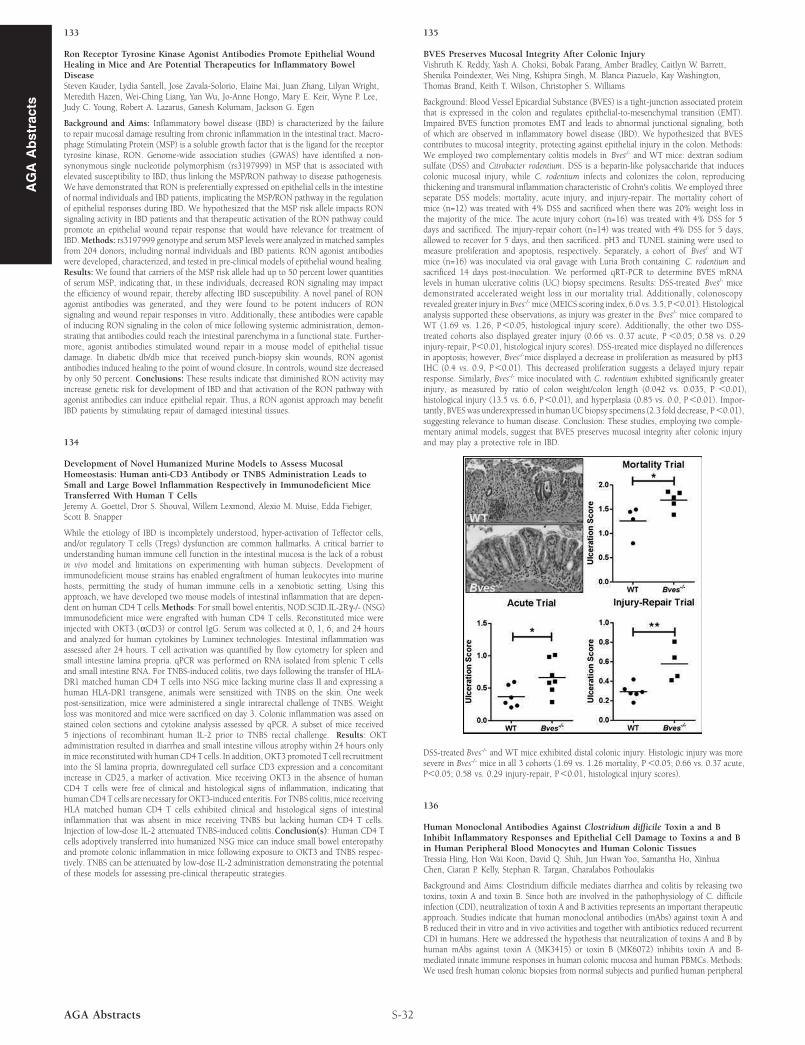
BVES Preserves Mucosal Integrity After Colonic InjuryVishruth K. Reddy, Yash A. Choksi, Bobak Parang, Amber Bradley, Caitlyn W. Barrett,Shenika Poindexter, Wei Ning, Kshipra Singh, M. Blanca Piazuelo, Kay Washington,Thomas Brand, Keith T. Wilson, Christopher S. Williams

Background: Blood Vessel Epicardial Substance (BVES) is a tight-junction associated proteinthat is expressed in the colon and regulates epithelial-to-mesenchymal transition (EMT).Impaired BVES function promotes EMT and leads to abnormal junctional signaling, bothof which are observed in inflammatory bowel disease (IBD). We hypothesized that BVEScontributes to mucosal integrity, protecting against epithelial injury in the colon. Methods:We employed two complementary colitis models in Bves-/- and WT mice: dextran sodiumsulfate (DSS) and Citrobacter rodentium. DSS is a heparin-like polysaccharide that inducescolonic mucosal injury, while C. rodentium infects and colonizes the colon, reproducingthickening and transmural inflammation characteristic of Crohn's colitis. We employed threeseparate DSS models: mortality, acute injury, and injury-repair. The mortality cohort ofmice (n=12) was treated with 4% DSS and sacrificed when there was 20% weight loss inthe majority of the mice. The acute injury cohort (n=16) was treated with 4% DSS for 5days and sacrificed. The injury-repair cohort (n=14) was treated with 4% DSS for 5 days,allowed to recover for 5 days, and then sacrificed. pH3 and TUNEL staining were used tomeasure proliferation and apoptosis, respectively. Separately, a cohort of Bves-/- and WTmice (n=16) was inoculated via oral gavage with Luria Broth containing C. rodentium andsacrificed 14 days post-inoculation. We performed qRT-PCR to determine BVES mRNAlevels in human ulcerative colitis (UC) biopsy specimens. Results: DSS-treated Bves-/- micedemonstrated accelerated weight loss in our mortality trial. Additionally, colonoscopyrevealed greater injury in Bves-/-mice (MEICS scoring index, 6.0 vs. 3.5, P,0.01). Histologicalanalysis supported these observations, as injury was greater in the Bves-/- mice compared toWT (1.69 vs. 1.26, P,0.05, histological injury score). Additionally, the other two DSS-treated cohorts also displayed greater injury (0.66 vs. 0.37 acute, P ,0.05; 0.58 vs. 0.29injury-repair, P,0.01, histological injury scores). DSS-treated mice displayed no differencesin apoptosis; however, Bves-/-mice displayed a decrease in proliferation as measured by pH3IHC (0.4 vs. 0.9, P,0.01). This decreased proliferation suggests a delayed injury repairresponse. Similarly, Bves-/- mice inoculated with C. rodentium exhibited significantly greaterinjury, as measured by ratio of colon weight/colon length (0.042 vs. 0.035, P ,0.01),histological injury (13.5 vs. 6.6, P,0.01), and hyperplasia (0.85 vs. 0.0, P,0.01). Impor-tantly, BVESwas underexpressed in humanUCbiopsy specimens (2.3 fold decrease, P,0.01),suggesting relevance to human disease. Conclusion: These studies, employing two comple-mentary animal models, suggest that BVES preserves mucosal integrity after colonic injuryand may play a protective role in IBD.

DSS-treated Bves-/- and WT mice exhibited distal colonic injury. Histologic injury was moresevere in Bves-/- mice in all 3 cohorts (1.69 vs. 1.26 mortality, P ,0.05; 0.66 vs. 0.37 acute,P,0.05; 0.58 vs. 0.29 injury-repair, P,0.01, histological injury scores).

136

Human Monoclonal Antibodies Against Clostridium difficile Toxin a and BInhibit Inflammatory Responses and Epithelial Cell Damage to Toxins a and Bin Human Peripheral Blood Monocytes and Human Colonic TissuesTressia Hing, Hon Wai Koon, David Q. Shih, Jun Hwan Yoo, Samantha Ho, XinhuaChen, Ciaran P. Kelly, Stephan R. Targan, Charalabos Pothoulakis

Background and Aims: Clostridium difficile mediates diarrhea and colitis by releasing twotoxins, toxin A and toxin B. Since both are involved in the pathophysiology of C. difficileinfection (CDI), neutralization of toxin A and B activities represents an important therapeuticapproach. Studies indicate that human monoclonal antibodies (mAbs) against toxin A andB reduced their in vitro and in vivo activities and together with antibiotics reduced recurrentCDI in humans. Here we addressed the hypothesis that neutralization of toxins A and B byhuman mAbs against toxin A (MK3415) or toxin B (MK6072) inhibits toxin A and B-mediated innate immune responses in human colonic mucosa and human PBMCs. Methods:We used fresh human colonic biopsies from normal subjects and purified human peripheral