13 integration tissue specific metabolism cpds v1
DESCRIPTION
tissue metabTRANSCRIPT
St. Luke’s College of Medicine – William H. Quasha Memorial
BIOCHEMISTRY
Lecture: 13 - Integration: Tissue Specific Metabolism Date: April 26,2016 Lecturer: Charina Paras-De Silva, PhD Checker: Kevin L. Trans Team: Miranda, Mirhan, Monteclaro
BLOCK 6
Page 1 of 4 XX - Integration: Tissue Specific Metabolism
Topic Outline
I. Objective II. Small Intestine III. Liver IV. Adipose Tissue V. Brain VI. Muscle
VII. Blood VIII. Kidney IX. Reference X. Quiz
PPT Book Audio Subhead’s Notes
I. Objective
To review the metabolic processes in major organ tissues and relate to their specialized functions. o Focus on small intestine, liver, brain, muscle, adipose
tissue and blood
Fig 1. Specialized metabolic functions of mammalian tissues
Review: Pancreas – secretes insulin and glucagon (crucial metabolic hormones) Liver – processes fats, carbohydrates, proteins from the diet
- Synthesizes and distributes lipid, ketone bodies, and glucose for other tissues
- Converts excess nitrogen to urea - Crucial for maintaining blood glucose level - Furnishes all other organs and tissues with an appropriate
mix of nutrients via the bloodstream
Portal vein – where the nutrients are transported
Small intestine – where digestion and absorption occurs Skeletal muscle – uses ATP for mechanical work
- allows directed motion Adipose tissue – stores and mobilizes the triglycerides, which serve as fuel throughout the body Lymphatic system – carries lipids from intestines to the liver Brain – transports ions to maintain membrane potential
- Integrated inputs from body and surroundings - Sends signals to other organs - Center of all things that needs to be done for our body - Requires a lot of ATP for neural signaling
II. Small Intestine
DIGESTION OF CARBOHYDRATES o (involves digestion of starch or other large carbohydrates) o Major monosaccharides as a result of digestion: D-
glucose, D-fructose, and D-galactose
DIGESTION OF PROTEINS o (occurs until the cytosol of the intestinal cells) o Most efficient digestion process o Digestion products, amino acids, are transported into the
hepatic portal vein.
DIGESTION OF FATS o Fatty acid chains that are less than 10 carbons just pass
through the cell across the membrane into the portal vein. o For long chain fatty acids (≥ 10), they become bound to
fatty acid binding protein transported in the
endoplasmic reticulum converted to triacylglycerol transported via chylomicrons into the lymphatic system adipose tissue
III. Liver
Maintenance of blood glucose levels
o Gluconeogenesis, metabolism of glucose, synthesis and
regulation of cholesterol, protein metabolism, synthesis of
plasma proteins, production of bile, breakdown of
haemoglobin, oxidation of drugs and alcohol, and
conjugation of bilirubin.
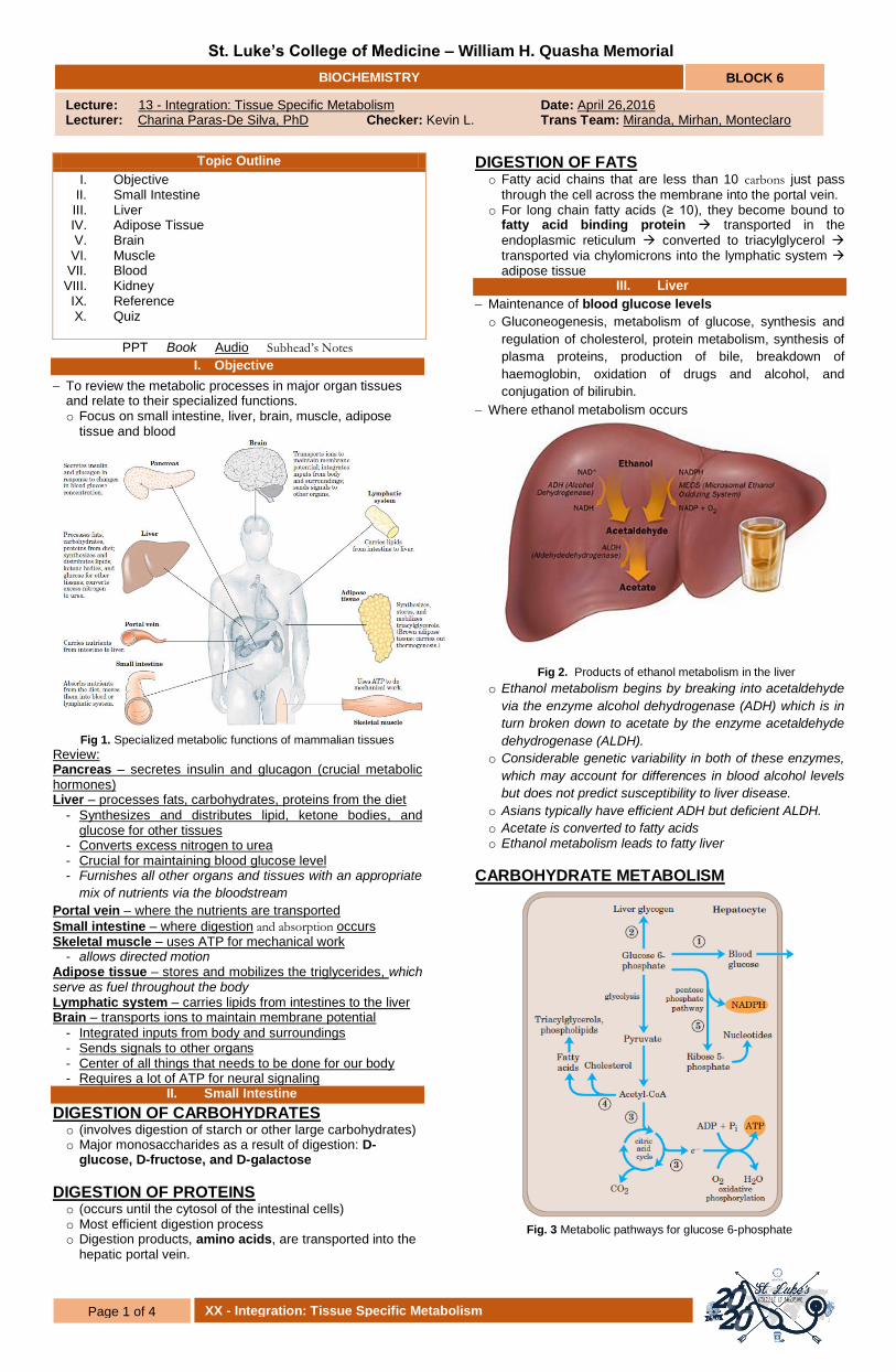
Where ethanol metabolism occurs
Fig 2. Products of ethanol metabolism in the liver
o Ethanol metabolism begins by breaking into acetaldehyde
via the enzyme alcohol dehydrogenase (ADH) which is in
turn broken down to acetate by the enzyme acetaldehyde
dehydrogenase (ALDH).
o Considerable genetic variability in both of these enzymes,
which may account for differences in blood alcohol levels
but does not predict susceptibility to liver disease.
o Asians typically have efficient ADH but deficient ALDH.
o Acetate is converted to fatty acids o Ethanol metabolism leads to fatty liver
CARBOHYDRATE METABOLISM
Fig. 3 Metabolic pathways for glucose 6-phosphate

Page 2 of 4 13 - Integration: Tissue Specific Metabolism
BIOCHEMISTRY BLOCK 6
1. Glucose 6-phosphate is dephosphorylated by glucose 6-phosphatase to yield free glucose, which is exported to replenish blood glucose. Export is the predominant pathway when glucose 6-phosphate is in limited supply 2. Glucose 6-phosphate not immediately needed to form blood glucose is converted to liver glycogen 3. The acetyl-CoA formed can be oxidized for energy production by the citric acid cycle (gives NADH, FADH), with ensuing electron transfer and oxidative phosphorylation, yielding ATP. (Normally, however, fatty acids are the preferred fuel for energy production in hepatocytes.) 4. Acetyl-CoA can also serve as the precursor of fatty acids, which are incorporated into TAGs and phospholipids, and of cholesterol. Much of the lipid synthesized in the liver is transported to other tissues by blood lipoproteins. 5. Alternatively, glucose 6-phosphate can enter the pentose phosphate pathway, yielding both reducing power (NADPH), needed for the biosynthesis of fatty acids and cholesterol, and D-ribose 5-phosphate, a precursor for nucleotide biosynthesis. NADPH is also an essential cofactor in the detoxification and elimination of many drugs and other xenobiotics metabolized in the liver. Table 1. Pathway of carbohydrates
NADPH from pentose phosphate pathway is needed for
glutathione metabolism and fatty acid synthesis
The alanine from glucose-alanine cycle comes from the muscle tissue.
Glucose-6-phospate is at the crossroads of carbohydrate
metabolism
AMINO ACID METABOLISM
Fig. 4 Metabolism of amino acids
1. Amino acids are precursors for protein synthesis. The liver is also the site of biosynthesis of most plasma proteins. 2. Alternatively, amino acids pass in the bloodstream to other organs, to be used in the synthesis of tissue proteins.
3. Other amino acids are precursors in the biosynthesis of nucleotides, hormones, and other nitrogenous compounds in the liver and other tissues. 4a. Amino acids not needed as biosynthetic precursors are transaminated or deaminated and degraded to yield pyruvate and citric acid cycle intermediates, with various fates; 4b. the ammonia released is converted to the excretory product urea. 5. Pyruvate can be converted to glucose and glycogen via gluconeogenesis, or 6. it can be converted to acetyl-CoA, which has several possible fates: 7. Oxidation via the citric acid cycle and 8. Oxidative phosphorylation to produce ATP, or 9. Conversion to lipids for storage. 10. Citric acid cycle intermediates can be siphoned off into glucose synthesis by gluconeogenesis. During the interval between meals, especially if prolonged, some muscle proteins are degraded to amino acids. These amino acids donate their amino groups (by transamination) to pyruvate, the product of glycolysis, to yield alanine, which 11 is transported to the liver and deaminated. (During exercise,
the muscle tissue can produce alanine and goes to the liver to be converted to glucose)
Table 2. Pathway of Amino Acid
Amino acid and nucleotide metabolism
Amino acid dehydrogenase: amino acids → acetyl-CoA, citric
acid cycle intermediates
Amino acid synthesis
Urea cycle: NH3 → urea
Glucose-alanine cycle: alanine → glucose
Nucleotide synthesis: amino acids → purines, pyrimidines
Hormone and neurotransmitter synthesis
LIPID METABOLISM
Fig. 5 Metabolism of Fatty Acids in the Liver
1. Some fatty acids are converted to liver lipids 2. Under most circumstances, fatty acids are the primary oxidative fuel in the liver. β-oxidation NADH and Acetyl-CoA 3&4. Acetyl-CoA is further oxidized via the citric acid cycle drive synthesis of ATP by oxidative phosphorylation 5. Excess Acetyl-CoA from β-oxidation is converted to ketone bodies Acetoacetate & β-hydroxybutyrate circulate in the blood to other tissues for use as fuel in the TCA cycle 6. Acetyl-CoA from fatty acids is used for biosynthesis of cholesterol, which is required for membrane synthesis, bile salts and steroid hormones 7. Fatty acids are converted to phospholipids and TAGs of plasma lipoproteins, which cary lipids to adipose tissue for storage as TAGs. 8. FFA become bound to serum albumin carried to heart and skeletal muscles absorb and oxidize FFA as major fuel
Carbohydrate catabolism
Glycogenolysis: glycogen → glucose 1-phosphate → blood glucose
Hexose entry into glycolysis: fructose, mannose, galactose → glucose 6-phosohate
Glycolysis: glucose → pyruvate
Pyruvate dehydrogenase reaction: pyruvate → acetyl-CoA
Lactic acid fermentation: glucose → lactate + 2 ATP
Pentose phosphate pathway: glucose 6-phosphate → pentose
phosphates + NADPH
Carbohydrate anabolism
Gluconeogenesis: citric acid cycle intermediates → glucose
Glucose-alanine cycle: glucose → pyruvate → alanine → glucose
Glycogen synthesis: glucose 6-phosphate → glucose 1-phosphate → glycogen

Page 3 of 4 13 - Integration: Tissue Specific Metabolism
BIOCHEMISTRY BLOCK 6
Table 3. Pathways of Fat Metabolism
STARVATION & DIABETES MELLITUS o Leads to overproduction of ketone bodies o There is increased lipolysis in diabetes mellitus &
starvation o During starvation, gluconeogenesis depletes citric acid
cycle intermediates, diverting Acetyl-CoA to ketone body formation
o In Diabetes insufficient insulin extrahepatic tissues cannot uptake glucose malonyl-CoA levels fall inhibition of carnitine acetyltransferase I is relieved FFA enter mitochondria to be degraded to acetyl-CoA acetyl-CoA accumulates ketone body formation
Fig 6. Ketoacidosis in Diabetes Mellitus & Starvation
ALTERED LIVER FUNCTION o Viral Hepatitis or Alcoholic Cirrhosis o Toxic NH4+ increases in blood, accumulation of
ammonia in CNS is a major reason for the coma in liver failure patients (liver encelopathy)
o Blood Urea Nitrogen (BUN) decreases due to decrease capacity to produce urea
o In outright liver failure, patients sometimes die of hypoglycaemia = blood glucose not maintained
IV. Adipose Tissue
Fig 7. Conversion of the fatty acid from the triacylglycerols of chylomicrons and VLDL to the TAG stored in adipose cells.
o Lipoprotein lipase (LPL) acts on triacylglycerol from VLDL
and chylomicron release FFA entry into adipose cell o Once FFA is inside adipose cell, it is activated and
converted to triacylglycerol, the storage form of fats. o Because adipose tissue lacks glycerol kinase: Glycerol
released from TAG goes back to the liver to be utilized for gluconeogenesis and lipogenesis
o Type I Diabetes Mellitus: Low insulin no activation of LPL FFA cannot enter adipose cell high triglyceride in the bloodstream
However, patient is still thin because they cannot store the fats in adipose
FASTING STATE o Adipose tissue provides FFA during fasting, triggered by
low insulin & high glucagon o Low insulin & high glucagon stimulates cAMP production
which stimulates protein kinase A activity o Protein kinase A activates hormone sensitive lipase o Hormone sensitive lipase converts TAG to FFA + Glycerol
Fig 8. Adipose tissue metabolism in fasting state
V. Brain
Normally uses glucose as fuel
The brain needs energy to think, conduct nerve impulses, and synthesize neurotransmitters
Metabolism accounts to almost 20% of O2 utilization by the
body at rest
Because the brain contains very little glycogen, it is constantly dependent on incoming glucose from the blood
Fig 9. Energy sources of the brain
Utilizes a lot of ATP from glucose During fasting to starvation state, the cells use
hydroxybutyrate, which is a ketone body that can pass
through the blood-brain barrier Hydroxybutyrate is formed from fatty acids in the liver o The brain oxidizes hydroxybutarate via acetyl-Coa during
prolonged fasting or starvation, after glycogen has been depleted, because it allows the brain to use body fat as an energy source
Cannot utilize fatty acids
Fat catabolism
β Oxidation of fatty acids → acetyl CoA
Oxidation of ketone bodies: β-hydroxybutyrate → acetyl-CoA → CO2 citric acid cycle
Fat anabolism
Fatty acid synthesis: acetyl-CoA → fatty acids
Triacylglycerol synthesis: acetyl-CoA → fatty acids → triacylglycerol
Ketone body formation: acetyl-CoA → acetoacetate, β-hydroxybutyrate
Cholesterol and cholesteryl synthesis: acetyl-CoA → cholesterol → cholesteryl esters
Phospholipid synthesis: fatty acids → phospholipids

Page 4 of 4 13 - Integration: Tissue Specific Metabolism
BIOCHEMISTRY BLOCK 6
Neurons oxidize glucose by glycolysis and the citric acid cycle
Energy is required to create and maintain an electrical potential across the neuronal plasma membrane.
VI. Muscle
What are major sources of fuel for skeletal muscles? o At rest?
Glycogen and glucose in the fed state, fatty acids in the fasting state
More muscle mass will utilize more fatty acids at rest
These are oxidized and degraded to yield acetyl-CoA, which enters the citric acid cycle for oxidation to CO. The ensuing transfer of electrons
provides the energy for ATP synthesis by oxidative phosphorylation.
o During an exercise?
ATP, Phosphagen, glycogen With enough oxygen, oxidative metabolism is used
Fatty acids, amino acids, glucose Fatty acids (prolonged exercise); phosphagen and anaerobic
metabolism (short-term exercise)
How many ATPs are produced from anaerobic glycolyisis from Glucose-1-P from glycogenolysis? o 3 ATPs
Fig 10. Energy Sources of Muscle Contraction
Skeletal muscle can use free fatty acids, ketone bodies, or glucose as fuel, depending on the degree of muscular activity
Heart Muscle o Continuously active in a regular rhythm of contraction and
relaxation o It has a completely aerobic metabolism at all times;
mitochondria more abundant than skeletal muscles o Major fuel is fatty acids but may utilize glucose and ketone
bodies from blood o Since mainly aerobic, deprivation of O2 when blood and
vessels are blocked can cause the region of the heart muscle to die like in myocardial infarction
o Don’t want anaerobic glycolysis, want oxidative metabolism
o The fuel is only glucose (to lactate)
VII. Blood
Regulate and integrate activities of different organs by providing acting as a medium for transport of nutrients, fuel, hormones, and metabolites
Delivers oxygen to various tissues o But energy of RBC is derived from anaerobic glycolysis
due to lack of mitochondria
Transports waste products from various tissue to kidneys for excretion o Tightly Regulated
Na+, K
+, Ca
2+, glucose
o Blood also carries hormonal signals from one tissue to another. In its role as signal carrier, the circulatory system
resembles the nervous system; both regulate and integrate the activities of different organs.
o Maintaining the normal concentration of glucose in the blood is therefore a very high priority
VIII. Kidney
Excrete substance via urine
Urea produced by urea cycle from liver
Uric acid from purine degredation
Creatinine from creatinine phosphate o Daily creatinine excretion is constant which depends on
muscle mass
NH4+, H2SO4 and phosphoric acid
Ketone bodies during ketoacidosis
Lactic acid during lactic acidosis
During starvation the kidney become an important site of gluconeogenesis
IX. References
Lehninger, Principles of Biochemistry [5th ed]
Mark’s Basic Medical Biochemistry 2nd ed.
Chapter 17 Integration of Energy Metabolism in Basic Concepts in Biochemistry – A students survival guide
Doc De Silva’s PPT
X. Quiz
1. NADPH from pentose phosphate pathway is needed for fatty acid: A. Oxidation B. Reduction C. Synthesis D. None of the above 2. The alanine from glucose-alanine cycle comes from the: A. Adipose tissue B. Brain C. Small intestine D. Muscle tissue 3. Which of the following substances causes coma in liver failure? A. Ethanol B. Ammonium C. Urea D. Pyruvate 4. Lipoprotein lipase is activated by ________, while hormone sensitive lipase is activated by ___________ A. Insulin, Glucagon B. Glucose, Amino Acids C. Lipoproteins, Hormones D. VLDL, Hormones
5. The ketone body, beta-hydroxybutarate, is one of the energy sources of the _______ A. kidney B. liver C. brain D. blood 6. ________ regulates and integrates activities of different organs by providing acting as a medium for transport of nutrients, fuel, hormones and metabolites A. kidney B. liver C. brain D. blood
1C 2D 3B 4A 5C 6D