11 dalk
TRANSCRIPT
INTRODUCTION
Deep anterior lamellar keratoplasty (DALK) is asurgical technique for cornea replacement, which thehealthy host corneal endothelium is preserved.Keratoconus patients are those who benefit most froma successful DALK procedure, once the endothelium ofthese patients is usually normal and should therefore bemaintained.
Several studies comparing visual outcomes betweenpenetrating keratoplasty (PK) and DALK have shownsimilar results1-4. Initially, DALK was performed usingmanual corneal dissection5. The greatest limitation ofearly DALK techniques was that they left variableamounts of residual stroma. This provided a scaffoldfor vascular ingrowth or led to variable amounts ofpostoperative scarring in the interface due to theremaining vessels. Also, the uneven or irregular dissec-tion plane could impair visual acuity. The main disad-vantage of DALK is the significant learning curve tomaster the technique. Moreover, it is time-consuming
procedure, especially when the stroma has to be manu-ally separated from the Descemet´s membrane.
Anwar and Teichmann’s6 big-bubble technique ofinjecting air into the corneal stroma to isolate Descemet’smembrane markedly improved DALK outcomes.Removing the overlying stroma completely created a cleargraft interface without irregularities. Visual outcomes areexcellent, and postoperative interface problems from vas-cularization of the recipient corneal bed are minimal.
A variant of the big-bubble technique in deep ante-rior lamellar keratoplasty (DALK) assisted by the fem-tosecond laser has been described, with good andreproducible results7,8.
The present technique allows a deep and smoothseparation of corneal stroma from Descemet´s mem-brane, by an interlamela cleavage plane created by anylon wire.
SURGICAL TECHNIQUE
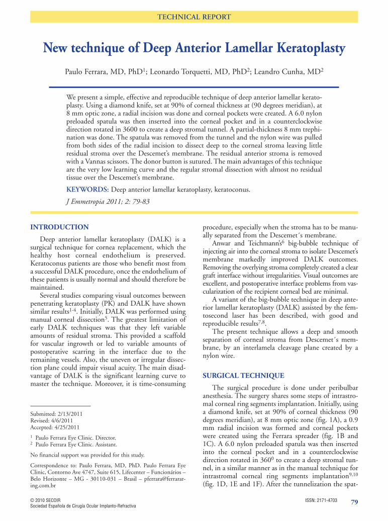
The surgical procedure is done under peribulbaranesthesia. The surgery shares some steps of intrastro-mal corneal ring segments implantation. Initially, usinga diamond knife, set at 90% of corneal thickness (90degrees meridian), at 8 mm optic zone (fig. 1A), a 0.9mm radial incision was formed and corneal pocketswere created using the Ferrara spreader (fig. 1B and1C). A 6.0 nylon preloaded spatula was then insertedinto the corneal pocket and in a counterclockwisedirection rotated in 3600 to create a deep stromal tun-nel, in a similar manner as in the manual technique forintrastromal corneal ring segments implantation9,10
(fig. 1D, 1E and 1F). After the tunnelization the spat-
79
TECHNICAL REPORT
New technique of Deep Anterior Lamellar Keratoplasty
Paulo Ferrara, MD, PhD1; Leonardo Torquetti, MD, PhD2; Leandro Cunha, MD2
We present a simple, effective and reproducible technique of deep anterior lamellar kerato-plasty. Using a diamond knife, set at 90% of corneal thickness at (90 degrees meridian), at8 mm optic zone, a radial incision was done and corneal pockets were created. A 6.0 nylonpreloaded spatula was then inserted into the corneal pocket and in a counterclockwisedirection rotated in 3600 to create a deep stromal tunnel. A partial-thickness 8 mm trephi-nation was done. The spatula was removed from the tunnel and the nylon wire was pulledfrom both sides of the radial incision to dissect deep to the corneal stroma leaving littleresidual stroma over the Descemet’s membrane. The residual anterior stroma is removedwith a Vannas scissors. The donor button is sutured. The main advantages of this techniqueare the very low learning curve and the regular stromal dissection with almost no residualtissue over the Descemet’s membrane.
KEYWORDS: Deep anterior lamellar keratoplasty, keratoconus.
J Emmetropia 2011; 2: 79-83
Submitted: 2/13/2011Revised: 4/6/2011Accepted: 4/25/2011
1 Paulo Ferrara Eye Clinic. Director.2 Paulo Ferrara Eye Clinic. Assistant.
No financial support was provided for this study.
Correspondence to: Paulo Ferrara, MD, PhD. Paulo Ferrara EyeClinic, Contorno Ave 4747, Suite 615, Lifecenter – Funcionários –Belo Horizonte – MG - 30110-031 – Brasil – [email protected]
© 2010 SECOIRSociedad Española de Cirugía Ocular Implanto-Refractiva
ISSN: 2171-4703

NEW TECHNIQUE OF DEEP ANTERIOR LAMELLAR KERATOPLASTY80
JOURNAL OF EMMETROPIA - VOL 2, APRIL-JUNE
Figure 1A.
Figure 1D.
Figure 1E. Figure 1F.
Figure 1B.
Figure 1C.
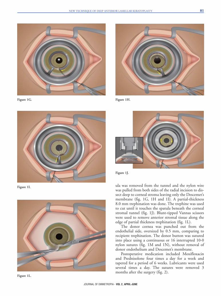
ula was removed from the tunnel and the nylon wirewas pulled from both sides of the radial incision to dis-sect deep to corneal stroma leaving only the Descemet’smembrane (fig. 1G, 1H and 1I). A partial-thickness8.0 mm trephination was done. The trephine was usedto cut until it touches the spatula beneath the cornealstromal tunnel (fig. 1J). Blunt-tipped Vannas scissorswere used to remove anterior stromal tissue along theedge of partial thickness trephination (fig. 1L).
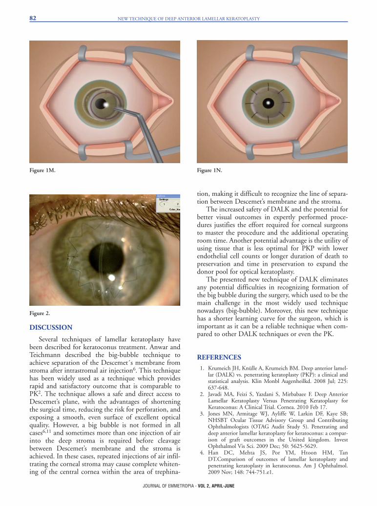
The donor cornea was punched out from theendothelial side, oversized by 0.5 mm, comparing torecipient trephination. The donor button was suturedinto place using a continuous or 16 interrupted 10-0nylon sutures (fig. 1M and 1N), without removal ofdonor endothelium and Descemet’s membrane.
Postoperative medication included Moxifloxacinand Prednisolone four times a day for a week andtapered for a period of 6 weeks. Lubricants were usedseveral times a day. The sutures were removed 3months after the surgery (fig. 2).
NEW TECHNIQUE OF DEEP ANTERIOR LAMELLAR KERATOPLASTY 81
JOURNAL OF EMMETROPIA - VOL 2, APRIL-JUNE
Figure 1G. Figure 1H.
Figure 1L.
Figure 1I.
Figure 1J.
DISCUSSION
Several techniques of lamellar keratoplasty havebeen described for keratoconus treatment. Anwar andTeichmann described the big-bubble technique toachieve separation of the Descemet´s membrane fromstroma after intrastromal air injection6. This techniquehas been widely used as a technique which providesrapid and satisfactory outcome that is comparable toPK2. The technique allows a safe and direct access toDescemet’s plane, with the advantages of shorteningthe surgical time, reducing the risk for perforation, andexposing a smooth, even surface of excellent opticalquality. However, a big bubble is not formed in allcases6,11 and sometimes more than one injection of airinto the deep stroma is required before cleavagebetween Descemet’s membrane and the stroma isachieved. In these cases, repeated injections of air infil-trating the corneal stroma may cause complete whiten-ing of the central cornea within the area of trephina-
tion, making it difficult to recognize the line of separa-tion between Descemet’s membrane and the stroma.
The increased safety of DALK and the potential forbetter visual outcomes in expertly performed proce-dures justifies the effort required for corneal surgeonsto master the procedure and the additional operatingroom time. Another potential advantage is the utility ofusing tissue that is less optimal for PKP with lowerendothelial cell counts or longer duration of death topreservation and time in preservation to expand thedonor pool for optical keratoplasty.
The presented new technique of DALK eliminatesany potential difficulties in recognizing formation ofthe big bubble during the surgery, which used to be themain challenge in the most widely used techniquenowadays (big-bubble). Moreover, this new techniquehas a shorter learning curve for the surgeon, which isimportant as it can be a reliable technique when com-pared to other DALK techniques or even the PK.
REFERENCES
1. Krumeich JH, Knülle A, Krumeich BM. Deep anterior lamel-lar (DALK) vs. penetrating keratoplasty (PKP): a clinical andstatistical analysis. Klin Monbl Augenheilkd. 2008 Jul; 225:637-648.
2. Javadi MA, Feizi S, Yazdani S, Mirbabaee F. Deep AnteriorLamellar Keratoplasty Versus Penetrating Keratoplasty forKeratoconus: A Clinical Trial. Cornea. 2010 Feb 17.
3. Jones MN, Armitage WJ, Ayliffe W, Larkin DF, Kaye SB;NHSBT Ocular Tissue Advisory Group and ContributingOphthalmologists (OTAG Audit Study 5). Penetrating anddeep anterior lamellar keratoplasty for keratoconus: a compar-ison of graft outcomes in the United kingdom. InvestOphthalmol Vis Sci. 2009 Dec; 50: 5625-5629.
4. Han DC, Mehta JS, Por YM, Htoon HM, TanDT.Comparison of outcomes of lamellar keratoplasty andpenetrating keratoplasty in keratoconus. Am J Ophthalmol.2009 Nov; 148: 744-751.e1.
NEW TECHNIQUE OF DEEP ANTERIOR LAMELLAR KERATOPLASTY82
JOURNAL OF EMMETROPIA - VOL 2, APRIL-JUNE
Figure 1M. Figure 1N.
Figure 2.
5. Malbran E. Lamellar keratoplasty in keratoconus. IntOphthalmol Clin 1966; 6: 99-109.
6. Anwar M, Teichmann KD. Big-bubble technique to bareDescemet´s membrane in anterior lamellar keratoplasty. JCataract Refract Surg 2002; 28: 398-403.
7. Chamberlain W, Cabezas M. Femtosecond-assisted deep ante-rior lamellar keratoplasty using big-bubble technique in acornea with 16 radial keratotomy incisions. Cornea. 2011 Feb;30: 233-6.
8. Buzzonetti L, Laborante A, Petrocelli G. Standardized big-bubble technique in deep anterior lamellar keratoplasty assist-ed by the femtosecond laser.J Cataract Refract Surg. 2010 Oct;36(10): 1631-6.
9. Colin J, Cochener B, Savary G, et al. Correcting keratoconuswith intracorneal rings. J Cataract Refract Surg. 2000; 26:1117-1122.
10. Siganos D, Ferrara P, Chatzinikolas K, et al. Ferrara intrastro-mal corneal rings for the correction of keratoconus. J CataractRefract Surg 2002; 28: 1947-1951.
11. Fogla R, Padmanabhan P. Results of deep lamellar keratoplas-ty using the big bubble technique in patients with kerato-conus. Am J Ophthalmol 2006; 141: 254-259.
NEW TECHNIQUE OF DEEP ANTERIOR LAMELLAR KERATOPLASTY 83
JOURNAL OF EMMETROPIA - VOL 2, APRIL-JUNE
First author:Paulo Ferrara, MD, PhD
Paulo Ferrara Eye Clinic, Brasil