1 temporal production of the signaling lipid phosphatidic acid by
TRANSCRIPT

1
Temporal production of the signaling lipid phosphatidic acid by phospholipase D2 determines な the output of ERK signaling in cancer cells に
Feng Zhanga, Ziqing Wang
a, Maryia Lu
a, Yoshiya Yonekubo
a, Xiao Liang
ab, Yueqiang Zhang
a, ぬ
Ping Wua, Yong Zhou
a, Sergio Grinstein
c, John F. Hancock
a and Guangwei Du
a, # ね
aDepartment of Integrative Biology and Pharmacology, University of Texas Health Science Center の
at Houston, 6431 Fannin St, Houston, TX 77030, United States. は bShanghai Institute of Digestive Disease, Shanghai Renji Hospital, Shanghai Jiao Tong University ば
School of Medicine, 145 Shan-dong Zhong Rd, Shanghai 200001, China ぱ cDivision of Cell Biology, Hospital for Sick Children ひ 555 University Ave., Toronto, M5G 1X8, Canada など
Running title: Phosphatidic acid regulation of ERK signaling なな なに #To whom correspondence should be addressed: Guangwei Du, Department of Integrative なぬ
Biology and Pharmacology, University of Texas Health Science Center at Houston, なね 6431 Fannin St, Houston, TX 77030, United States. Tel: (713)500-7055; FAX: (713)500-7444; なの Email: [email protected] なは なば Abbreviations: DAG (diacylglycerol), DGK (diacylglycerol kinase), EGF (Epidermal growth なぱ factor), ERK (extracellular signal-regulated kinases), FIPI (5-fluoro-2-indolyl des-なひ chlorohalopemide), FLIM-FRET (fluorescence lifetime imaging microscopy-Förster resonance にど energy transfer), GEF (guanine exchange factor), PA (phosphatidic acid), PASS (Phosphatidic Acid にな biosensor with Superior Sensitivity), PI(4,5)P2 (phosphatidylinositol 4,5-bisphosphate), PI(3,4,5)P2 にに (phosphatidylinositol 3,4,5-bisphosphate), PLD (phospholipase D), PM (plasma membrane), PMA にぬ (phorbol-12-myristate-13-acetate). にね
MCB Accepts, published online ahead of print on 28 October 2013Mol. Cell. Biol. doi:10.1128/MCB.00987-13Copyright © 2013, American Society for Microbiology. All Rights Reserved.
on April 3, 2018 by guest
http://mcb.asm
.org/D
ownloaded from

2
Abstract にの The Ras-extracellular signal-regulated kinases (ERK) cascade is an important signaling には
module in cells. One regulator of the Ras-ERK cascade is phosphatidic acid (PA) generated by にば phospholipase D (PLD) and diacylglycerol kinase (DGK). Using a newly developed PA biosensor, にぱ PASS (Phosphatidic Acid biosensor with Superior Sensitivity), we found that PA was generated にひ sequentially by PLD and DGK in epidermal growth factor (EGF)-stimulated HCC1806 breast ぬど cancer cells. Inhibition of PLD2, one of the two PLD members, was sufficient to eliminate most of ぬな the PA production, whereas inhibition of DGK decreased PA production only at the later stages of ぬに EGF stimulation, suggesting that PLD2 is prior to DGK activation. The temporal production of PA ぬぬ by PLD2 is important for the nuclear activation of ERK. While inhibition of both PLD and DGK ぬね had no effect on the overall ERK activity, inhibition of PLD2, but not PLD1 or DGK, blocked the ぬの nuclear ERK activity in several cancer cell lines. The decrease of active ERK in the nucleus ぬは inhibited the activation of Elk1, an ERK nuclear target, leading to decreased proliferation of ぬば HCC1806 cells. Together, these findings reveal that PA production by PLD2 determines the output ぬぱ of ERK in cancer cell growth factor signaling. ぬひ
ねど Key words: ERK, PASS, phosphatidic acid, phospholipase D, PLD2. ねな
on April 3, 2018 by guest
http://mcb.asm
.org/D
ownloaded from

3
Introduction ねに Phosphatidic acid (PA) has attracted increasing attention in recent years due to its roles as a ねぬ
signaling molecule and as a central intermediate in the synthesis of membrane lipids (1-3). PA can ねね be produced by multiple enzymes, including two well-known families of enzymes: phospholipase D ねの (PLD) and diacylglycerol kinase (DGK) (4-7). In mammalian cells, there are two PLD family ねは members, PLD1 and PLD2, which differ strikingly in subcellular localization and function (5,7). ねば The mammalian DGK family consists of 10 members, classified into five different subtypes ねぱ characterized by different regulatory domains (6). It has been proposed that activation of distinct ねひ PA-generating enzymes at different times and in different subcellular compartments determines the のど specific cellular functions of PA, including cell proliferation, survival and migration (1,5). のな
One of the most important intracellular signaling pathways involves the cascade of Ras, Raf, のに MEK, and the extracellular signal-regulated kinases 1 and 2 (ERK1/2, referred to as ERK
herein) のぬ
(8,9). Activated ERK can either remain in the cytoplasm or translocate to the nucleus, where it のね phosphorylates and activates a number of proteins that control proliferation, differentiation, のの survival, apoptosis and development (8-10). The precise outcome of stimulating the Ras-ERK のは cascade depends on the duration, strength, and localization of the signals (8,10,11). It has been のば reported that PA is involved in the regulation of the Ras-ERK pathway in fibroblasts and のぱ lymphocytes (4,12-14). However, the mechanisms whereby PA regulates the Ras-ERK cascade のひ appear to be very distinct in different cell types. Moreover, it remains unknown how growth factors はど activate different PA-generating enzymes, i.e., PLD and DGK, and whether PA generated from はな different sources regulates the Ras-ERK cascade in the same manner. Importantly, signaling by はに growth factors such as epidermal growth factor receptor (EGFR) and the Ras-ERK cascade is はぬ frequently upregulated in many types of cancer (15,16). Interestingly, the PA-generating enzymes, はね PLD and DGK, have also been reported to be critical for proliferation, migration and survival of はの
on April 3, 2018 by guest
http://mcb.asm
.org/D
ownloaded from

4
cancer cells (6,7,17). It is not clear how and why dysregulation of the Ras-ERK cascade by PA はは contributes to cancer initiation and progression. はば
To study the functions of PA, it is critical to faithfully monitor its spatiotemporal はぱ production. Traditionally, PA levels have been measured using biochemical methods such as thin はひ layer chromatography (TLC) and high-performance liquid chromatography (18). In recent years, ばど identification and quantification of various lipids, including PA, have become more simple and ばな sensitive with substantially improved mass spectrometry analyses (19,20). However, all these ばに biochemical techniques measure only the total cellular PA level, and cannot reveal the intracellular ばぬ locations of PA production. In addition, when PA is measured by biochemical methods, the ばね relatively high level of PA on the surface of the ER, where it is used as a precursor for the synthesis ばの of phospholipids and triglycerides (TAG) (3,21), may mask the changes of the comparatively less ばは abundant PA generated during signaling at the plasma membrane and other intracellular organelles. ばば As an alternative method, changes in phospholipid levels can be detected by using fluorescently-ばぱ tagged protein domains that bind specifically to certain lipids. For example, PH domains from PLCh ばひ and AKT have been used widely to monitor phosphatidylinositol 4,5-bisphosphate (PI(4,5)P2) and ぱど phosphatidylinositol 3,4,5-trisphosphate (PI(3,4,5)P3), respectively (18,22). Such reagents have ぱな greatly advanced our understanding of the dynamics and functions of phosphatidylinositides. ぱに However, despite great interest (23), we still lack a PA biosensor with the specificity and sensitivity ぱぬ comparable to those of the phosphatidylinositide probes. ぱね
In the present study, we report the development of a specific and sensitive PA biosensor. ぱの Using this new tool, we demonstrate that PA production is differentially controlled by PLD and ぱは DGK in epidermal growth factor (EGF) signaling, and that PA generated by PLD2 is critical for the ぱば nuclear activity of ERK and proliferation in cancer cells. Our findings reveal that PLD2-generated ぱぱ PA determines the signaling output of ERK. ぱひ
on April 3, 2018 by guest
http://mcb.asm
.org/D
ownloaded from

5
ひど Experimental procedures ひな
General reagents and antibodies –g-tubulin, Flag, phospho- and total ERK1/2 antibodies, ひに Phosphatidylserine (PS), and the PLD inhibitor 5-Fluoro-2-indolyl des-chlorohalopemide (FIPI) ひぬ (PLDi) were from Sigma-Aldrich (St. Louis, MO) (24). The inhibitors for PLD1/2 (VU0155056), ひね PLD1 (PLD1i, VU0359595), and PLD2 (PLD2i, VU0285655-1) were from Avanti Polar Lipids ひの (Alabaster, AL) (25). The DGK inhibitor R59022 (DGKi) was from Calbiochem (La Jolla, CA). ひは EGFR, phospho- and total Elk1, phospho- and total Fra1, total c-Fos and phospho-Rsk antibodies, ひば were from Cell Signaling Technology (Danvers, MA). Mouse GST, phospho-c-Fos and PLD1 ひぱ rabbit monoclonal antibodies were from Abcam (Cambridge, MA). PLD2 rabbit polyclonal ひひ antibody was kindly provided by Dr. Y. Banno (Gifu University of Tokyo, Gifu, Japan). Goat anti-などど mouse and anti-rabbit IgGs conjugated with Alexa 680 were from Invitrogen. Goat anti-mouse and などな anti-rabbit IgGs conjugated with IRDye 800CW were from Rockland Immunochemicals などに (Gilbertsville, PA). 1,2-Dilauroyl-sn-glycero-3-phosphate (DLPA) was from Echelon (Salt Lake などぬ City, UT). などね
Construction of plasmids – To generate EGFP-PASS, a nuclear export sequence (NES) derived などの from protein kinase A inhibitor (PKI)-α伊 (26) was added between EGFP and Spo20-PABD cloned in などは pEGFP-C1 (27,28). PASS tagged with monomeric GFP or RFP (mGFP or mRFP) was generated by などば replacing EGFP in the EGFP-PASS with mGFP or mRFP using Age I and BsrG I sites. Different などぱ tags did not change the localization of PASS. All PASS constructs used in the current study are などひ tagged with monomeric GFP and RFP. GST-PASS-His was generated by inserting PASS and a pair ななど of oligos encoding 6xHis into the BamH I site of pGEX-4T1 from GE Healthcare Biosciences ななな (Pittsburgh, PA). The 4E mutant was generated by site-directed mutagenesis using the QuikChange ななに kit from Agilent Technologies (Santa Clara, CA). The lentiviral vectors carrying GFP-PASS and ななぬ
on April 3, 2018 by guest
http://mcb.asm
.org/D
ownloaded from

6
RFP-PASS were subcloned into the Nhe I and BamH I sites of pCDH-CMV-MCS and pCDH-ななね CMV-MCS-EF1-Puro from System Biosciences (Mountain View, CA), respectively. The shRNAs ななの for PLD1 and PLD2 characterized in the previous publications (12,28-31) were subcloned into the ななは lentiviral vector pLKO.1. ななば
Bacterial expression, purification of recombinant PASS, and liposome pulldown assay - ななぱ Expression and purification of GST-PASS-His using Ni
2+-chelate chromatography from Qiagen ななひ
(Valencia, CA) were as described before (32). All purified recombinant proteins used in our なにど experiments are >95% pure, as judged by Coomassie Blue stained SDS-PAGE (polyacrylamide gel なにな electrophoresis) gels. The purified proteins were used within one week. The preparation of なにに liposomes has been previously described (32,33). POPC (phosphatidylcholine), DOPE なにぬ (Phosphatidylethanolamine), and POPA (phosphatidic acid) were from Avanti Polar Lipids, なにね phosphatidylinositol 4,5-bisphosphate (PI(4,5)P2) and phosphatidylinositol 3,4,5-trisphosphate (PI(3, なにの 4,5)P3) were purchased from Matreya (Pleasant Gap, PA). なには
Cell culture and treatment – Cell culture media were from Thermo Fisher (Waltham, MA). なにば Sera were from Invitrogen (Carlsbad, CA). All cell lines were from ATCC (Manassas, VA). CHO なにぱ cells were grown in Ham's F12 medium. HCC1806 and HCC827 cells were grown in RPMI-1640 なにひ medium. MDA-MB-468 and A431 cells were grown in Dulbecco's Modified Eagle's Medium. BT-なぬど 20 cells were grown in Eagle's Minimum Essential Medium. All media were supplemented with なぬな 10% fetal bovine serum. For inhibitor treatment, cells were pre-incubated with dimethyl sulfoxide なぬに (DMSO) or inhibitors for 1 hr. Transient transfection in CHO cells was done using the deacylated なぬぬ polyethylenimine reagent (PEI) (34). Unless specified, cells were serum-starved overnight and then なぬね stimulated with 100 nM PMA, 100 たM PA (DLPA), 100 たM PS, or 100 ng/ml EGF for the なぬの indicated time. For cell proliferation measurements, cells were seeded in 6-well plates in RPMI-なぬは
on April 3, 2018 by guest
http://mcb.asm
.org/D
ownloaded from

7
1640 media containing 0.1 % FBS and 5 ng/ml EGF in the presence of DMSO or inhibitors, or in なぬば the presence of control and PLD shRNAs. Viable cells were measured by trypan blue exclusion. なぬぱ
Lentivirus production and transduction - The delivery of expression constructs in HCC1806 なぬひ cells was through lentiviral infection. Viruses were generated in TLA-293T cells from Thermo なねど Fisher by co-transfecting four plasmids including the lentiviral vector, pMDLg/pRRE, pRSV-Rev なねな and pMD2.G using Lipofectamine and Plus reagent from Invitrogen. At 48 hours and 72 hours post-なねに transfection, virus-containing supernatants were collected for infection. The cells were used for なねぬ experiments 2-3 days postinfection. なねね
Western blot analysis – Fluorescently labeled secondary antibodies were used for Western blot, なねの and detected by the LI-COR Odyssey Infrared Imaging System from LI-COR Biotechnology なねは (Lincoln, NE). Band intensity was quantified using the program supplied by the manufacturer. The なねば bands on Western blots were defined by identical region of interest, and measured as integrated なねぱ pixel intensity for all samples. The background was determined by measuring the average pixel なねひ intensity of a user-defined area. なのど
Confocal microscopy – All images were captured with a 60X oil-immersion objective on a なのな Nikon A1 confocal microscope (Melville, NY). Fields with moderate cell confluence were selected なのに for measurement. For visualizing GFP and RFP fluorescence of PASS, cells grown on coverslips なのぬ were transiently transfected (CHO cells) or infected with lentiviruses (HCC1806 cells), and then なのね directly fixed in 4% paraformaldehyde in PBS for 10 minutes at room temperature. After several なのの PBS washes, the coverslips were mounted onto slides using 4% propyl gallate mounting solution. なのは The fluorescent images of GFP and RFP of random fields were collected. For each treatment, at なのば least five fields, including more than 50 cells, were quantified. Cells with saturated exposure were なのぱ not used for quantification. The change in the plasma membrane localization was quantified using なのひ the line pixel intensity histogram in NIH Image J as described previously (22). A line was randomly なはど
on April 3, 2018 by guest
http://mcb.asm
.org/D
ownloaded from

8
drawn to span a large area of the cytoplasm, but not the nucleus. The pixel intensity of the two most なはな outside peaks represented the PASS on the plasma membrane, while the average value between なはに represented that in the cytoplasm. なはぬ
For pERK staining, cells were stained with a pERK1/2 antibody/Alexa 594-conjugated goat なはね anti-mouse secondary antibody and DAPI after cold methanol fixation at -20°C for 10 minutes. なはの After defining the nuclei with DAPI staining, the cytoplasmic and nuclear areas were manually なはは marked, and then measured for the fluorescent intensity of pERK staining using NIH Image J. The なはば change of pERK localization was represented as the ratio of pERK in the nucleus and cytoplasm. なはぱ
Statistics – All statistical differences were evaluated between control and each of the treatments なはひ using two-tailed student’s t-test. All data are shown as mean ± SD. なばど なばな Results なばに Development of a sensitive PA biosensor なばぬ
To study the regulation and functions of PA in distinct cellular compartments, it is important なばね to precisely monitor the spatial and temporal production of PA. Currently, the most widely used PA なばの biosensor is the PA-binding domain derived from a yeast protein Spo20 (Spo20-PABD) (23,28,35). なばは However, green fluorescent protein (GFP)-tagged Spo20-PABD only responded to strong stimuli なばば such as phorbol-12-myristate-13-acetate (PMA) (24) and depolarizing concentrations of potassium なばぱ (35), or some cellular processes such as cell replating (28). We were unable to detect changes of PA なばひ levels in cells stimulated with ligands for membrane receptors using this PA biosensor. Even in なぱど cells stimulated with PMA, a potent activator of PLD activity (36,37), the membrane localization of なぱな GFP-Spo20-PABD was barely visible in most cells unless the images were overexposed. These なぱに results seem contradictory to the original reports that Spo20-PABD binds to PA with exceptional なぱぬ
on April 3, 2018 by guest
http://mcb.asm
.org/D
ownloaded from

9
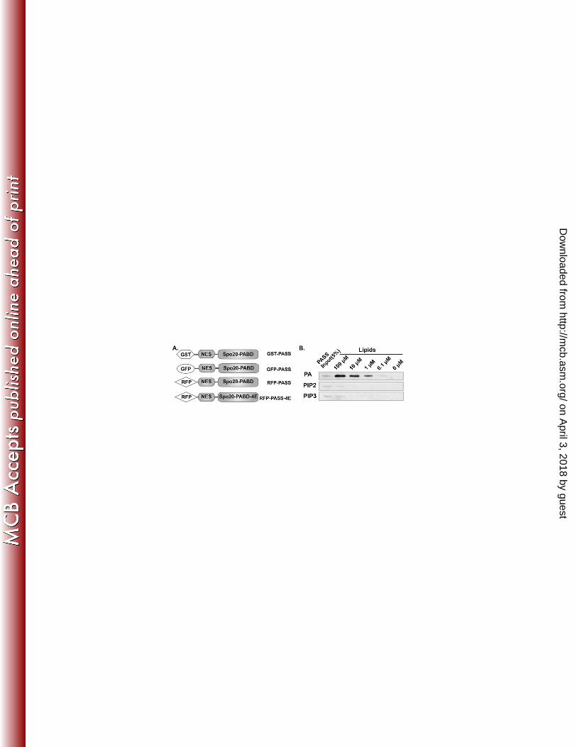
sensitivity and specificity in vitro (23,38,39). Since GFP-Spo20-PABD predominantly localizes to なぱね the nucleus, we reason that the nuclear localization prevents it from accessing PA molecules on the なぱの plasma membrane or other cytoplasmic organelles. To improve the sensitivity of Spo20-PABD, we なぱは added a nuclear export sequence (NES) derived from protein kinase A inhibitor-α (PKI-α) 伊 (26) to なぱば the N-terminus of the Spo20-PABD (Fig. 1A), and named the resulting construct PASS なぱぱ (Phosphatidic Acid biosensor with Superior Sensitivity) due to its exceptional sensitivity to detect なぱひ the cellular PA levels (further demonstrated in Figs. 2 and 3). We first tested whether the addition of なひど NES affected the binding specificity of PA in vitro using recombinant PASS protein purified from なひな E. coli and synthetic liposomes. As controls, we also included liposomes containing PI(4,5)P2 or なひに PI(3,4,5)P3, since the original Spo20-PABD also showed some binding to these lipids (23,38). As なひぬ expected, we found that PASS specifically bound to PA-containing liposomes in a concentration-なひね dependent manner (Fig. 1B). In sharp contrast, PASS bound to PI(4,5)P2- or PI(3,4,5)P3-containing なひの liposomes with much lower affinity (Fig. 1B). These results are consistent with the previous finding なひは that Spo20-PABD binds to PA selectively, and confirm that addition of the NES does not change なひば the PA-binding specificity of PASS. なひぱ
We next tested whether PASS can be used to measure PA levels in intact CHO cells when なひひ fused with either a green fluorescent protein (GFP) or red fluorescent protein (RFP). PLD activity is にどど very low-to-undetectable
in unstimulated (basal) CHO cells, but can be elevated by
stimulation with にどな
PMA or by overexpression of PLD2 (24,28,40). As observed before, the original GFP-Spo20-にどに PABD mainly localized to the nucleus in both basal and PMA-stimulated conditions ((28,35) Fig. にどぬ 2A). Two other PA biosensors, GFP-Raf1-PABD (27,41) and GFP-Sos1-PH (12), localized to the にどね cytoplasm in basal conditions, and only partially translocated to membranes in PMA-stimulated にどの conditions (Fig 2A). In addition, GFP-Raf1-PABD often aggregated in some cells (Fig 2A), and にどは induced cell toxicity when it was expressed for more than two days (not shown). In contrast, both にどば
on April 3, 2018 by guest
http://mcb.asm
.org/D
ownloaded from

10
GFP- and RFP-tagged PASS localized diffusely in the cytoplasm under nonstimulatory conditions, にどぱ but translocated to the plasma membrane and endosome-like vesicles upon PMA stimulation (Fig. にどひ 2B) when PLD was activated (29,33,42). Furthermore, the plasma membrane translocation of PASS になど induced by PMA was blocked when PLD activity was inhibited by FIPI (24) (Fig. 2B). The になな mutation of four lysine residues to glutamic acid (K66E, K68E, R71E, and K73E, called 4E になに hereinafter) on the original Spo20-PABD disrupted its binding to PA (38). We generated the same になぬ 4E mutations in RFP-PASS. As expected, PMA stimulation caused the plasma membrane になね translocation of the wild-type GFP-PASS, but not that of the PA-binding deficient mutant RFP-になの PASS-4E (Fig. 2C). Another way to increase the cellular PA level is through overexpression of になは PLD2, which has a high basal activity (42,43). As before, GFP-PASS, but not the RFP-PASS-4E になば mutant, was recruited to the plasma membranes in CHO cells stably overexpressing mouse PLD2 になぱ (29) (Fig. 2D). In contrast, PASS localized diffusely in the cytoplasm, and only translocated to the になひ plasma membrane and intracellular vesicles upon PMA treatment in CHO cells stably ににど overexpressing human PLD1 (Fig 2D), which has a low basal activity and is activated by external ににな stimulation (30,36). The increased PASS localization on intracellular vesicles in PLD1-ににに overexpressing cells suggests the activation of PLD1 on these organelles, where the majority of ににぬ PLD1 is localized (7,33). Finally, we tested whether addition of exogenous PA to culture medium ににね was able to recruit GFP-PASS to the plasma membrane; PA can be rapidly incorporated into the ににの plasma membrane and can induce cellular responses (12,44,45). As expected, exogenously added にには PA stimulated translocation of GFP- and RFP-PASS to the plasma membrane, but failed to induce ににば the translocation of RFP-PASS-4E (Fig. 2E). In contrast, external addition of another negatively ににぱ charged phospholipid, phosphatidylserine (PS), did not cause recruitment of GFP- and RFP-PASS ににひ to the plasma membrane (Fig. 2E), further supporting the specificity of this biosensor. にぬど にぬな
on April 3, 2018 by guest
http://mcb.asm
.org/D
ownloaded from

11
EGF regulation of temporal PA production in HCC1806 breast cancer cells にぬに Our ultimate goal was to study PA dynamics during the course of growth factor stimulation. にぬぬ
Due to their poor sensitivity, we were not able to observe consistent membrane translocation of にぬね several previously generated PA biosensors, i.e., GFP-Raf1-PABD, GFP-SOS-PH and GFP-Spo20-にぬの PABD, in cells stimulated with EGF or other growth factors. Having established the specificity and にぬは improved sensitivity of PASS, we used this probe to examinutee growth factor signaling in cancer にぬば cells. We initially used HCC1806 cells, a good experimental model for EGF signaling and にぬぱ aggressive triple-negative breast cancer (46,47). GFP-PASS diffusely localized in the cytoplasm of にぬひ serum-starved HCC1806 cells, but quickly translocated to the plasma membrane within 1 minute of にねど EGF stimulation (Fig. 3A and 3B), reaching maximal levels at 3 minutes. Most of the GFP-PASS にねな returned to the cytoplasm after 30 minutes (Fig. 3A and 3B). To our knowledge, PASS is the only にねに probe capable of detecting such dynamic changes in PA subcellular distribution in response to にねぬ physiological stimuli like growth factors. にねね
Two major sources of PA are phosphatidylcholine and diacylglycerol (DAG), which are にねの used as the substrates by PLD and DGK, respectively. To determine the source of PA in response to にねは EGF, we treated cells with PLD or DGK inhibitors. Interestingly, PLD and DGK inhibitors showed にねば different effects on the EGF-dependent translocation of GFP-PASS. The PLD inhibitor FIPI almost にねぱ completely abolished the translocation of GFP-PASS to the membrane (Fig. 3A and 3B). In にねひ contrast, the DGK inhibitor R59022 only blocked the late (≥ 3minutes), but not the early (1 minute) にのど component of GFP-PASS translocation (Fig. 3A and 3B). These results suggest that upon EGF にのな stimulation, PA is sequentially generated by PLD and DGK. To determine the PLD isoform にのに responsible for PA generation in response to EGF, we examined the localization of PASS in にのぬ HCC1806 cells infected with lentiviruses carrying either control luciferase (Luc) small hairpin にのね RNAs (shRNAs) or shRNA targeting PLD1 or PLD2 (12,28,29), or treated with PLD isoform-にのの
on April 3, 2018 by guest
http://mcb.asm
.org/D
ownloaded from

12
specific inhibitors (25). Only PLD2 shRNA or inhibitor, but not the control, PLD1 shRNA or にのは inhibitor, blocked the plasma membrane translocation of PASS triggered by EGF stimulation (Fig. にのば 3C-E). In addition, PLD2 was mainly localized on the plasma membrane in HCC1806 cells before にのぱ and after EGF stimulation, whereas PLD1 was mainly localized to perinuclear vesicles (Fig. 3F). にのひ Taken together, these results suggest that PLD2 is the predominant source of PA. にはど にはな PLD2-generated PA is important for EGF-triggered nuclear ERK activation にはに
Since PA regulates the Ras-ERK cascade in fibroblasts and lymphocytes (4,12-14), we にはぬ investigated whether PA generated from PLD and DGK also regulates ERK activation in cancer にはね cells. ERK activity (as measured by phosphorylated ERK1/2, pERK) was low in non-stimulated にはの HCC1806 cells, and was not changed by treatments with the PLD and DGK inhibitors (Fig. 4A and にはは 4B). EGF stimulation triggered a rapid increase in pERK levels, which reached a maximum at 3 にはば minutes and was maintained for more than 2 hours. However, inhibition of either PLD or DGK did にはぱ not affect the formation of pERK level in EGF-stimulated HCC1806 cells, as measured by Western にはひ blot analysis (Fig. 4A and 4B). Downregulation of the expression of PLD isoforms using shRNAs にばど also failed to affect ERK phosphorylation (Fig. 4C and 4D). We next assessed ERK activation and にばな localization by monitoring pERK in the nucleus and cytoplasm using quantitative fluorescence にばに microscopy. The staining of activated ERK (pERK) in untreated cells was already strong in the にばぬ cytoplasm 3 minutes after EGF stimulation, and by 6 minutes activated ERK became abundant in にばね the nucleus (Fig. 4E and 4F). Addition of PLD and DGK inhibitors did not affect ERK activation or にばの alter its kinetics of appearance in the cytoplasm. However, the PLD inhibitor significantly blocked にばは the appearance of nuclear pERK staining after 3 minutes of EGF stimulation. In contrast, the DGK にばば inhibitor only slightly decreased the nuclear pERK staining at all the time points tested (Fig. 4E and にばぱ 4F). にばひ
on April 3, 2018 by guest
http://mcb.asm
.org/D
ownloaded from

13
We then examined which PLD isoform was responsible for regulating nuclear ERK activity にぱど using shRNAs. While expression of Luc and PLD1 shRNAs did not change the nuclear and にぱな cytoplasmic distribution of pERK in cells stimulated with EGF, PLD2 shRNA inhibited the にぱに appearance of nuclear pERK staining (Fig. 4G and 4H), as found in cells treated with pan-PLD にぱぬ inhibitor. To further confirm that PA is responsible for PLD2 regulation of nuclear ERK activity, にぱね we treated HCC1806 cells with both PLD2 inhibitor and different concentrations of PA. Addition of にぱの exogenous PA rescued the decrease in pERK staining in a dose-dependent manner (Fig. 4I and 4J). にぱは These results suggest that PLD2 activity is required for the nuclear translocation of activated ERK. にぱば Activations of EGFR on the plasma membrane and endosomes lead to selective activation of ERK にぱぱ on different subcellular compartments or distinct biological activities (8,10,48,49). Previous studies にぱひ in fibroblasts found that overexpression of catalytically inactive mutants for PLD1 and PLD2 にひど blocked the activation of ERK by inhibiting the endocytosis of EGFR (50). To determine if PLD2 にひな regulates ERK activation in the nucleus through modulating EGFR endocytosis in HCC1806 cells, にひに we also examined the localization of EGFR in the presence and absence of PLD2 inhibitor. PLD2 にひぬ inhibitor treatment did not seem to affect the localization of EGFR before and after EGF stimulation にひね (Fig 4K). This result, together with the dominant plasma membrane localization of PLD2 and PASS にひの (Fig 3), suggest that PLD2 regulation of the nuclear ERK activity is independent of EGFR にひは endocytosis. にひば
We then addressed the question whether PLD2 also controls the activation of ERK in the にひぱ nucleus in other cancer cells treated with EGF. Both PLD2 inhibitor and shRNAs significantly にひひ blocked the nuclear staining of pERK in all four cancer cell lines we tested: HCC827 (lung cancer), ぬどど MDA-MB-468 and BT-20 (breast cancer), and A431 (skin cancer) (Fig. 5), suggesting that PLD2 ぬどな regulation of nuclear ERK activity is a universal phenomenon in cancer cells. ぬどに
ぬどぬ
on April 3, 2018 by guest
http://mcb.asm
.org/D
ownloaded from

14
Inhibition of PLD2 suppressed the activity of Elk1 and cell proliferation in HCC1806 cells ぬどね After translocation into the nucleus, active ERK phosphorylates several proteins involved in ぬどの
transcriptional regulation. One of the best studied ERK targets in the nucleus is Elk1 (11,51). ぬどは Consistent with the decrease in pERK in the nucleus, we found that the PLD inhibitor caused a ぬどば decrease in Elk1 phosphorylation, whereas the DGK inhibitor did not (Fig. 6A and 6B). Moreover, ぬどぱ only PLD2 shRNA but not PLD1 shRNA, inhibited Elk1 phosphorylation (Fig. 6C and 6D), ぬどひ supporting our findings that PLD2 is specifically required for ERK activity in the nucleus in ぬなど HCC1806 cells. We also checked the phosphorylation of other ERK substrates. Without ぬなな phosphorylation, c-Fos and Fra1, the other two nuclear substrates of ERK, undergo rapid ぬなに degradation; c-Fos and Fra1 are stabilized when phosphorylated by ERK (52-54). As expected, EGF ぬなぬ stimulation increased both phosphorylation and total protein levels of c-Fos and Fra1, which were ぬなね inhibited by PLD but not DGK inhibitor treatment (Fig. 6E and 6F). In contrast, inhibition of PLD ぬなの and DGK had no effect on phosphorylation and total protein levels of a cytoplasmic substrate of ぬなは ERK, Rsk (8,11) (Fig. 6E and 6F). ぬなば
Phosphorylation of ERK nuclear targets increases their affinity for the serum response ぬなぱ factor, and thus enhances transcription of proliferation-promoting (16,51). The observation that ぬなひ PLD2 inhibition decreased the activity of Elk1, c-Fos and Fra1, implies that PLD2 activity is ぬにど required for cell proliferation. To test this hypothesis, we examined cell proliferation in HCC1806 ぬにな cells in the presence of inhibitors or shRNAs. As expected, the rate of cell proliferation ぬにに corresponded to the phosphorylation and protein levels of Elk1, c-Fos and Fra1. Cells treated with a ぬにぬ PLD1 inhibitor, DGK inhibitor, or expressing PLD1 shRNA showed similar proliferation rates as ぬにね the controls (Fig. 7). However, cells treated with the PLD2 inhibitor or expressing PLD2 shRNA ぬにの displayed marked decrease in cell proliferation (Fig. 7). These results suggest that PLD2 regulates ぬには cell proliferation through modulating the activity of nuclear ERK and its nuclear substrates in ぬにば
on April 3, 2018 by guest
http://mcb.asm
.org/D
ownloaded from

15
HCC1806 breast cancer cells. ぬにぱ ぬにひ Discussion ぬぬど The new biosensor PASS offers a useful tool to study phospholipid signaling ぬぬな
The ability to precisely monitor the dynamics of PA in different subcellular locations is ぬぬに important for understanding the role of PA signaling in cells. We and others have developed several ぬぬぬ PA biosensors, i.e., the PA-binding region of Raf-1 (Raf1-PABD) (41) and Spo20-PABD (28,35), ぬぬね the Sos1 PH domain (12), and the Pii FRET biosensors derived from Dock2 and Sos1-PH (55). ぬぬの However, compared to the phosphoinositide probes, none of these PA biosensors has been widely ぬぬは used, because they either lack sufficient sensitivity, require a special microscopy setup, or only ぬぬば monitor PA production in certain membrane compartments. The PA biosensor PASS developed in ぬぬぱ the current study has circumvented these problems. The specificity of PASS as a PA biosensor has ぬぬひ been validated by a series of in vitro and in vivo experiments: (1) it bound to PA with high affinity ぬねど and specificity, as shown by a liposome-pull-down assay using the purified recombinant protein; (2) ぬねな increasing cellular PA levels by PMA stimulation, exogenous addition of PA, or by overexpression ぬねに of PLD2 promoted the plasma membrane binding of PASS tagged with either GFP or RFP; (3) ぬねぬ association of PASS with the membrane was abolished by treatment with PLD inhibitors and ぬねね expression of PLD2 shRNA; (4) point mutations that disrupt PA binding in vitro inhibited the ぬねの recruitment of PASS to the membrane. ぬねは
The most significant improvement of PASS is its exceptional sensitivity. We have attempted ぬねば to use several previously developed PA biosensors, i.e. Raf1-PABD, Spo20-PABD and Sos1-PH, to ぬねぱ monitor PA levels in various cells. However, only a small fraction of these biosensors, if any, ぬねひ translocated to membranes upon PMA (Fig 2A) and growth factor stimulation. PASS is by far the ぬのど only PA biosensor that quickly responds to stimulation and binds to the plasma membrane of the ぬのな
on April 3, 2018 by guest
http://mcb.asm
.org/D
ownloaded from

16
various cell types that we have tested. In addition, GFP- Raf1-PABD also aggregates in cells and ぬのに causes cell toxicity. The binding of Sos1-PH to both PI(4,5)P2 and PA also limits its use as a PA-ぬのぬ specific biosensor (12). To increase PA detection, previous PA biosensors derived from Spo20-ぬのね PABD contain tandem repeats of Spo20-PABD (35,56). We find that PASS described in our current ぬのの work (contain one copy of Spo20-PABD fused to NES) responds to stimulation equally well or ぬのは better while having lower background membrane binding. In addition, we also observed that PASS ぬのば labeling of some intracellular vesicles, presumably endosomes, are further increased by PLD1 ぬのぱ overexpression in PMA-stimulated cells (Fig. 2). These results support the previous findings that ぬのひ PLD and DGK function on both the plasma membrane and endosomes (4-7). Moreover, a tandem ぬはど repeat version of PASS labels also the endoplasmic reticulum in macrophages, HeLa, and HEK293 ぬはな cells (57). This is particularly interesting since PASS is the only PA biosensor currently known to ぬはに label the PA pool at the endoplasmic reticulum, which is the site of phospholipid and triglyceride ぬはぬ synthesis (3). Finally, when paired with specific membrane markers, the new biosensor may also be ぬはね used for fluorescence lifetime imaging microscopy-Förster resonance energy transfer (FLIM-FRET) ぬはの or FRET analysis to examine the production of PA in different membrane domains. Compared to ぬはは the Pii FRET sensors (55), the pairing of PASS with an independent membrane marker should ぬはば provide a more reliable assessment of the sites of PA production. It is noteworthy that, to increase ぬはぱ their sensitivity, the Pii biosensors are targeted to the plasma membrane through the C-terminus of ぬはひ K-Ras (55). Since the C-terminus of K-Ras only resides in a subdomain of the plasma membrane ぬばど (58,59), the Pii biosensors cannot, in principle, monitor the PA generated in other plasma membrane ぬばな domains or in intracellular organelles. ぬばに ぬばぬ Differential activation and functions of PLD and DGK in growth factor signaling in cancer ぬばね cells ぬばの
on April 3, 2018 by guest
http://mcb.asm
.org/D
ownloaded from

17
In mammalian cells, there are two PLD family members and ten members in the DGK ぬばは family (4-7). It has been unclear how different PA-generating enzymes contribute to PA production ぬばば upon activation of a particular signaling pathway, and whether different sources of PA perform the ぬばぱ same functions. An important finding from our experiments is that EGF triggers a sequential ぬばひ activation of PLD and DGK in distinct membrane nanodomains. Moreover, our results also suggest ぬぱど that DGK activation may be dependent on PLD activity. It is likely that, as an early event in EGF ぬぱな signaling, PLD2 activation is required for the activation of DGK or for the production of DAG, the ぬぱに substrate of DGK. More work is still needed to further investigate the crosstalk between PLD and ぬぱぬ DGK. These include monitoring the production of both PA and DAG using PASS and DAG ぬぱね biosensors, and examining the spatiotemporal activation of their downstream signaling pathways, in ぬぱの cells where PLD or DGK is inhibited individually, or simultaneously. In combination with small ぬぱは molecule inhibitors and RNA interference, the newly developed PASS should be a useful tool in ぬぱば determining the regulation of individual PA-generating enzymes and of their cellular functions in ぬぱぱ different physiological and pathological conditions. ぬぱひ
ぬひど PLD and DGK differentially regulate Ras-ERK signaling in cancer cells ぬひな
Various mechanisms are thought to ensure the specific outcomes of Ras-ERK signaling (60). ぬひに However, little is known about how and why the activation of the Ras-ERK components in separate ぬひぬ cellular compartments might be regulated. Previous studies have suggested that the site where Ras ぬひね is activated dictates the nature of the downstream signals and of subsequent biological outputs ぬひの elicited (60-62). For example, Ras activated on the plasma membrane led to the translocation of ぬひは activated ERK to the nucleus, whereas active ERK was restricted to the Golgi when Ras was ぬひば activated in that compartment (63). These findings are also consistent with the observation that Ras ぬひぱ activated on the Golgi was fully dispensable for proliferation and transformation (61). Interestingly, ぬひひ
on April 3, 2018 by guest
http://mcb.asm
.org/D
ownloaded from

18
we found that PLD2 activity is important for the translocation of activated ERK to the nucleus in ねどど cancer cells upon growth factor stimulation. In this regard, it is important to note that at the early ねどな stages of EGF stimulation PA is generated by PLD2, but not by DGK. Growth factor activation of ねどに ERK on the plasma membrane requires the binding of Raf, MEK and ERK to the scaffold protein ねどぬ KSR1 (9,64). However, only activated MEK and ERK transport to the nucleus (9,64), suggesting ねどね that disassembly of the Raf/MEK/ERK/KSR1 complex is prerequisite to pERK nuclear ねどの translocation. It is therefore conceivable that the stability of the Raf/MEK/ERK/KSR1 complex ねどは determines the cytoplasmic and nuclear destination of the pERK, a mechanism similar to those used ねどば to retain pERK on the endosomes by く-arrestin-2 and on the Golgi by another scaffold protein Sef ねどぱ (63,64). Interestingly, PA binds to several components of the ERK cascade on the plasma ねどひ membrane, such as Sos1, Raf1 and KSR1 (12,41,65). It is likely that PLD2-generated PA ねなど association to Raf1, KSR1, or other scaffold proteins, potentially weakens their binding to MEK ねなな and ERK, therefore allowing MEK/ERK nuclear translocation in cancer cells. ねなに
There are clear differences between the mechanisms through which PLD2-generated PA ねなぬ regulates the Ras-ERK cascade in cancer cells and other cell types. In EGF-stimulated fibroblasts, ねなね PLD2-generated PA directly regulates the activity of total Ras and ERK levels though Sos1, a Ras ねなの guanine exchange factor (12), or Raf1 (41). In lymphocyte function-associated antigen-1-stimulated ねなは lymphocytes, PLD2-generated PA activates Ras indirectly when being converted to DAG by PA-ねなば phosphatases, which then activates a different Ras guanine exchange factor–RasGRP1 (14). Our ねなぱ findings that the precise control of PA production by PLD2 is critical for the delivery of active ERK ねなひ to the nucleus of cancer cells contribute a mechanistic insight into many previous observations. It is ねにど possible that different expression levels of MAPK components, such as Sos1, Raf1 and scaffold ねにな proteins, and PA-generating enzymes, such as PLD and DGK, in different cell types, may affect the ねにに duration, magnitude, and compartmentalization of ERK, leading to different biological outcomes. ねにぬ
on April 3, 2018 by guest
http://mcb.asm
.org/D
ownloaded from

19
PLD expression and activity are elevated in a variety of human cancer tissues (66-71) and human ねにね cancer cell lines (72-76). Interestingly, high levels of PLD2 activity also correlate with more ねにの aggressive cancer phenotypes (77). Moreover, it has been proposed that PLD2 could be a “driver” ねには in breast tumorigenesis, based on the relatively high rate of somatic mutation of PLD2 found in a ねにば sequencing analysis of human breast cancer patients (78). Finally, consistent with elevated PLD ねにぱ expression and activity, PLD has been reported to be important for cell proliferation, survival and ねにひ migration in many cancer cell lines (79,80). It is therefore important to clarify how upregulation of ねぬど PLD2 expression and activity may “rewire” the MAPK pathway in cancer cells. Further ねぬな understanding the unique role of PLD2 in cancer cell signaling may help us design strategies for the ねぬに treatment of human malignancies. ねぬぬ
In summary, the present studies demonstrate that the new PA biosensor PASS is capable of ねぬね recognizing PA with unprecedented specificity and sensitivity, which allows us to monitor the ねぬの spatiotemporal changes of PA at the single-cell level. Analysis of cellular PA with this biosensor ねぬは revealed that EGF stimulation activates PLD2 and DGK sequentially. Because the spatiotemporal ねぬば production of PA by PLD2 is important for the nuclear ERK activity and proliferation, the use of ねぬぱ this novel probe should facilitate our research into the biology of both normal and cancerous cells. ねぬひ ねねど Acknowledgements ねねな This work was supported by the U.S. Army Medical Research and Materiel Command W81XWH-ねねに 10-1-0624 (to GD), and the National Institutes of Health Grant GM066717 (to JH). ねねぬ
on April 3, 2018 by guest
http://mcb.asm
.org/D
ownloaded from

20
References ねねね 1. Wang, X., Devaiah, S. P., Zhang, W., and Welti, R. (2006) Signaling functions of ねねの
phosphatidic acid. Prog Lipid Res 45, 250-278 ねねは 2. Hancock, J. F. (2007) PA promoted to manager. Nat. Cell. Biol. 9, 615-617 ねねば 3. Carman, G. M., and Han, G. S. (2011) Regulation of phospholipid synthesis in the yeast ねねぱ
Saccharomyces cerevisiae. Annu Rev Biochem 80, 859-883 ねねひ 4. Zhang, Y., and Du, G. (2009) Phosphatidic acid signaling regulation of Ras superfamily of ねのど
small guanosine triphosphatases. Biochim Biophys Acta 1791, 850-855 ねのな 5. Selvy, P. E., Lavieri, R. R., Lindsley, C. W., and Brown, H. A. (2011) Phospholipase D: ねのに
enzymology, functionality, and chemical modulation. Chemical reviews 111, 6064-6119 ねのぬ 6. Shulga, Y. V., Topham, M. K., and Epand, R. M. (2011) Regulation and functions of ねのね
diacylglycerol kinases. Chemical reviews 111, 6186-6208 ねのの 7. Peng, X., and Frohman, M. A. (2012) Mammalian phospholipase D physiological and ねのは
pathological roles. Acta Physiol (Oxf) 204, 219-226 ねのば 8. Murphy, L. O., and Blenis, J. (2006) MAPK signal specificity: the right place at the right ねのぱ
time. Trends Biochem. Sci. 31, 268-275 ねのひ 9. McKay, M. M., and Morrison, D. K. (2007) Integrating signals from RTKs to ERK/MAPK. ねはど
Oncogene 26, 3113-3121 ねはな 10. Ebisuya, M., Kondoh, K., and Nishida, E. (2005) The duration, magnitude and ねはに
compartmentalization of ERK MAP kinase activity: mechanisms for providing signaling ねはぬ specificity. J. Cell. Sci. 118, 2997-3002 ねはね
11. Mebratu, Y., and Tesfaigzi, Y. (2009) How ERK1/2 activation controls cell proliferation and ねはの cell death: Is subcellular localization the answer? Cell Cycle 8, 1168-1175 ねはは
on April 3, 2018 by guest
http://mcb.asm
.org/D
ownloaded from

21
12. Zhao, C., Du, G., Skowronek, K., Frohman, M. A., and Bar-Sagi, D. (2007) Phospholipase ねはば D2-generated phosphatidic acid couples EGFR stimulation to Ras activation by Sos. Nat. ねはぱ Cell. Biol. 9, 706-712 ねはひ
13. Rizzo, M. A., Shome, K., Vasudevan, C., Stolz, D. B., Sung, T.-C., Frohman, M. A., ねばど Watkins, S. C., and Romero, G. (1999) Phospholipase D and its product, phosphatidic acid, ねばな mediate agonist-dependent Raf-1 translocation to the plasma membrane and the activation of ねばに the MAP kinase pathway. J. Biol. Chem. 274, 1131-1139 ねばぬ
14. Mor, A., Campi, G., Du, G., Zheng, Y., Foster, D. A., Dustin, M. L., and Philips, M. R. ねばね (2007) The lymphocyte function-associated antigen-1 receptor costimulates plasma ねばの membrane Ras via phospholipase D2. Nat. Cell. Biol. 9, 713-719 ねばは
15. Dhillon, A. S., Hagan, S., Rath, O., and Kolch, W. (2007) MAP kinase signalling pathways ねばば in cancer. Oncogene 26, 3279-3290 ねばぱ
16. Roberts, P. J., and Der, C. J. (2007) Targeting the Raf-MEK-ERK mitogen-activated protein ねばひ kinase cascade for the treatment of cancer. Oncogene 26, 3291-3310 ねぱど
17. Park, J. B., Lee, C. S., Jang, J. H., Ghim, J., Kim, Y. J., You, S., Hwang, D., Suh, P. G., and ねぱな Ryu, S. H. (2012) Phospholipase signalling networks in cancer. Nat Rev Cancer 12, 782-792 ねぱに
18. Rusten, T. E., and Stenmark, H. (2006) Analyzing phosphoinositides and their interacting ねぱぬ proteins. Nat Methods 3, 251-258 ねぱね
19. Ivanova, P. T., Milne, S. B., Myers, D. S., and Brown, H. A. (2009) Lipidomics: a mass ねぱの spectrometry based systems level analysis of cellular lipids. Current opinion in chemical ねぱは biology 13, 526-531 ねぱば
20. Oliveira, T. G., Chan, R. B., Tian, H., Laredo, M., Shui, G., Staniszewski, A., Zhang, H., ねぱぱ Wang, L., Kim, T. W., Duff, K. E., Wenk, M. R., Arancio, O., and Di Paolo, G. (2010) ねぱひ
on April 3, 2018 by guest
http://mcb.asm
.org/D
ownloaded from

22
Phospholipase d2 ablation ameliorates Alzheimer's disease-linked synaptic dysfunction and ねひど cognitive deficits. J. Neurosci. 30, 16419-16428 ねひな
21. Athenstaedt, K., and Daum, G. (1999) Phosphatidic acid, a key intermediate in lipid ねひに metabolism. Eur. J. Biochem. 266, 1-16 ねひぬ
22. Balla, T., and Varnai, P. (2002) Visualizing cellular phosphoinositide pools with GFP-fused ねひね protein-modules. Sci STKE 2002, pl3 ねひの
23. Kassas, N., Tryoen-Toth, P., Corrotte, M., Thahouly, T., Bader, M. F., Grant, N. J., and ねひは Vitale, N. (2012) Genetically encoded probes for phosphatidic acid. Methods in cell biology ねひば 108, 445-459 ねひぱ
24. Su, W., Yeku, O., Olepu, S., Genna, A., Park, J. S., Ren, H., Du, G., Gelb, M. H., Morris, A. ねひひ J., and Frohman, M. A. (2009) 5-Fluoro-2-indolyl des-chlorohalopemide (FIPI), a のどど phospholipase D pharmacological inhibitor that alters cell spreading and inhibits のどな chemotaxis. Mol. Pharmacol. 75, 437-446 のどに
25. Scott, S. A., Selvy, P. E., Buck, J. R., Cho, H. P., Criswell, T. L., Thomas, A. L., Armstrong, のどぬ M. D., Arteaga, C. L., Lindsley, C. W., and Brown, H. A. (2009) Design of isoform-のどね selective phospholipase D inhibitors that modulate cancer cell invasiveness. Nat Chem Biol のどの 5, 108-117 のどは
26. Henderson, B. R., and Eleftheriou, A. (2000) A comparison of the activity, sequence のどば specificity, and CRM1-dependence of different nuclear export signals. Exp. Cell. Res. 256, のどぱ 213-224 のどひ
27. Corrotte, M., Chasserot-Golaz, S., Huang, P., Du, G., Ktistakis, N. T., Frohman, M. A., のなど Vitale, N., Bader, M. F., and Grant, N. J. (2006) Dynamics and function of phospholipase D のなな and phosphatidic acid during phagocytosis. Traffic 7, 365-377 のなに
on April 3, 2018 by guest
http://mcb.asm
.org/D
ownloaded from

23
28. Du, G., and Frohman, M. A. (2009) A lipid-signaled myosin phosphatase surge disperses のなぬ cortical contractile force early in cell spreading. Mol. Biol. Cell. 20, 200-208 のなね
29. Du, G., Huang, P., Liang, B. T., and Frohman, M. A. (2004) Phospholipase D2 localizes to のなの the plasma membrane and regulates angiotensin II receptor endocytosis. Mol. Biol. Cell. 15, のなは 1024-1030 のなば
30. Su, W., Chardin, P., Yamazaki, M., Kanaho, Y., and Du, G. (2006) RhoA-mediated のなぱ Phospholipase D1 signaling is not required for the formation of stress fibers and focal のなひ adhesions. Cell Signal 18, 469-478 のにど
31. Mor, A., Wynne, J. P., Ahearn, I. M., Dustin, M. L., Du, G., and Philips, M. R. (2009) のにな Phospholipase D1 regulates lymphocyte adhesion via upregulation of Rap1 at the plasma のにに membrane. Mol. Cell. Biol. 29, 3297-3306 のにぬ
32. Roach, A. N., Wang, Z., Wu, P., Zhang, F., Chan, R. B., Yonekubo, Y., Di Paolo, G., Gorfe, のにね A. A., and Du, G. (2012) Phosphatidic acid regulation of PIPKI is critical for actin のにの cytoskeletal reorganization. J Lipid Res 53, 2598-2609 のには
33. Du, G., Altshuller, Y. M., Vitale, N., Huang, P., Chasserot-Golaz, S., Morris, A. J., Bader, のにば M. F., and Frohman, M. A. (2003) Regulation of phospholipase D1 subcellular cycling のにぱ through coordination of multiple membrane association motifs. J. Cell. Biol. 162, 305-315 のにひ
34. Thomas, M., Lu, J. J., Ge, Q., Zhang, C., Chen, J., and Klibanov, A. M. (2005) Full のぬど deacylation of polyethylenimine dramatically boosts its gene delivery efficiency and のぬな specificity to mouse lung. Proc. Nat. Acad. Sci. USA 102, 5679-5684 のぬに
35. Zeniou-Meyer, M., Zabari, N., Ashery, U., Chasserot-Golaz, S., Haeberle, A. M., Demais, のぬぬ V., Bailly, Y., Gottfried, I., Nakanishi, H., Neiman, A. M., Du, G., Frohman, M. A., Bader, のぬね M. F., and Vitale, N. (2007) Phospholipase D1 production of phosphatidic acid at the plasma のぬの
on April 3, 2018 by guest
http://mcb.asm
.org/D
ownloaded from

24
membrane promotes exocytosis of large dense-core granules at a late stage. J. Biol. Chem. のぬは 282, 21746-21757 のぬば
36. Xiao, N., Du, G., and Frohman, M. A. (2005) Peroxiredoxin II functions as a signal のぬぱ terminator for H2O2-activated phospholipase D1. Febs J 272, 3929-3937 のぬひ
37. Du, G., Morris, A. J., Sciorra, V. A., and Frohman, M. A. (2002) G-protein-coupled receptor のねど regulation of phospholipase D. Methods Enzymol. 345, 265-274 のねな
38. Nakanishi, H., de los Santos, P., and Neiman, A. M. (2004) Positive and negative regulation のねに of a SNARE protein by control of intracellular localization. Mol. Biol. Cell. 15, 1802-1815 のねぬ
39. Liu, S., Wilson, K. A., Rice-Stitt, T., Neiman, A. M., and McNew, J. A. (2007) In vitro のねね fusion catalyzed by the sporulation-specific t-SNARE light-chain Spo20p is stimulated by のねの phosphatidic acid. Traffic 8, 1630-1643 のねは
40. Du, G., Altshuller, Y. M., Kim, Y., Han, J. M., Ryu, S. H., Morris, A. J., and Frohman, M. のねば A. (2000) Dual requirement for rho and protein kinase C in direct activation of のねぱ phospholipase D1 through G protein-coupled receptor signaling. Mol. Biol. Cell. 11, 4359-のねひ 4368 ののど
41. Rizzo, M. A., Shome, K., Watkins, S. C., and Romero, G. (2000) The recruitment of Raf-1 ののな to membranes is mediated by direct interaction with phosphatidic acid and is independent of ののに association with Ras. J. Biol. Chem. 275, 23911-23918 ののぬ
42. Jenkins, G. M., and Frohman, M. A. (2005) Phospholipase D: a lipid centric review. Cell. ののね Mol. Life. Sci. 62, 2305-2316 ののの
43. Colley, W. C., Sung, T. C., Roll, R., Jenco, J., Hammond, S. M., Altshuller, Y., Bar-Sagi, ののは D., Morris, A. J., and Frohman, M. A. (1997) Phospholipase D2, a distinct phospholipase D ののば isoform with novel regulatory properties that provokes cytoskeletal reorganization. Curr. ののぱ Biol. 7, 191-201 ののひ
on April 3, 2018 by guest
http://mcb.asm
.org/D
ownloaded from

25
44. Fang, Y., Vilella-Bach, M., Bachmann, R., Flanigan, A., and Chen, J. (2001) Phosphatidic のはど acid-mediated mitogenic activation of mTOR signaling. Science 294, 1942-1945 のはな
45. Nishikimi, A., Fukuhara, H., Su, W., Hongu, T., Takasuga, S., Mihara, H., Cao, Q., のはに Sanematsu, F., Kanai, M., Hasegawa, H., Tanaka, Y., Shibasaki, M., Kanaho, Y., Sasaki, T., のはぬ Frohman, M. A., and Fukui, Y. (2009) Sequential regulation of DOCK2 dynamics by two のはね phospholipids during neutrophil chemotaxis. Science 324, 384-387 のはの
46. Volk-Draper, L. D., Rajput, S., Hall, K. L., Wilber, A., and Ran, S. (2012) Novel model for のはは basaloid triple-negative breast cancer: behavior in vivo and response to therapy. Neoplasia のはば (New York, N.Y 14, 926-942 のはぱ
47. Lehmann, B. D., Bauer, J. A., Chen, X., Sanders, M. E., Chakravarthy, A. B., Shyr, Y., and のはひ Pietenpol, J. A. (2011) Identification of human triple-negative breast cancer subtypes and のばど preclinical models for selection of targeted therapies. J. Clin. Invest. 121, 2750-2767 のばな
48. Kranenburg, O., Verlaan, I., and Moolenaar, W. H. (1999) Dynamin is required for the のばに activation of mitogen-activated protein (MAP) kinase by MAP kinase kinase. J. Biol. Chem. のばぬ 274, 35301-35304 のばね
49. Vieira, A. V., Lamaze, C., and Schmid, S. L. (1996) Control of EGF receptor signaling by のばの clathrin-mediated endocytosis. Science 274, 2086-2089 のばは
50. Shen, Y., Xu, L., and Foster, D. A. (2001) Role for phospholipase D in receptor-mediated のばば endocytosis. Mol. Cell. Biol. 21, 595-602 のばぱ
51. Whitmarsh, A. J., Shore, P., Sharrocks, A. D., and Davis, R. J. (1995) Integration of MAP のばひ kinase signal transduction pathways at the serum response element. Science 269, 403-407 のぱど
52. Murphy, L. O., Smith, S., Chen, R. H., Fingar, D. C., and Blenis, J. (2002) Molecular のぱな interpretation of ERK signal duration by immediate early gene products. Nat. Cell. Biol. 4, のぱに 556-564 のぱぬ
on April 3, 2018 by guest
http://mcb.asm
.org/D
ownloaded from
26
53. Okazaki, K., and Sagata, N. (1995) The Mos/MAP kinase pathway stabilizes c-Fos by のぱね phosphorylation and augments its transforming activity in NIH 3T3 cells. EMBO J. 14, のぱの 5048-5059 のぱは
54. Basbous, J., Chalbos, D., Hipskind, R., Jariel-Encontre, I., and Piechaczyk, M. (2007) のぱば Ubiquitin-independent proteasomal degradation of Fra-1 is antagonized by Erk1/2 pathway-のぱぱ mediated phosphorylation of a unique C-terminal destabilizer. Mol. Cell. Biol. 27, 3936-のぱひ 3950 のひど
55. Nishioka, T., Frohman, M. A., Matsuda, M., and Kiyokawa, E. (2010) Heterogeneity of のひな phosphatidic acid levels and distribution at the plasma membrane in living cells as のひに visualized by a Foster resonance energy transfer (FRET) biosensor. J. Biol. Chem. 285, のひぬ 35979-35987 のひね
56. Ariotti, N., Liang, H., Xu, Y., Zhang, Y., Yonekubo, Y., Inder, K., Du, G., Parton, R. G., のひの Hancock, J. F., and Plowman, S. J. (2010) Epidermal growth factor receptor activation のひは remodels the plasma membrane lipid environment to induce nanocluster formation. Mol. のひば Cell. Biol. 30, 3795-3804 のひぱ
57. Bohdanowicz, M., Schlam, D., Hermansson, M., Rizzuti, D., Fairn, G. D., Ueyama, T., のひひ Somerharju, P., Du, G., and Grinstein, S. (2013) Phosphatidic acid is required for the はどど constitutive ruffling and macropinocytosis of phagocytes. Mol. Biol. Cell. 24, 1700-1712 はどな
58. Hancock, J. F. (2006) Lipid rafts: contentious only from simplistic standpoints. Nat Rev Mol はどに Cell Biol 7, 456-462 はどぬ
59. Harding, A. S., and Hancock, J. F. (2008) Using plasma membrane nanoclusters to build はどね better signaling circuits. Trends Cell Biol 18, 364-371 はどの
60. Prior, I. A., and Hancock, J. F. (2012) Ras trafficking, localization and compartmentalized はどは signalling. Semin Cell Dev Biol 23, 145-153 はどば
on April 3, 2018 by guest
http://mcb.asm
.org/D
ownloaded from

27
61. Matallanas, D., Sanz-Moreno, V., Arozarena, I., Calvo, F., Agudo-Ibanez, L., Santos, E., はどぱ Berciano, M. T., and Crespo, P. (2006) Distinct utilization of effectors and biological はどひ outcomes resulting from site-specific Ras activation: Ras functions in lipid rafts and Golgi はなど complex are dispensable for proliferation and transformation. Mol. Cell. Biol. 26, 100-116 はなな
62. Cheng, C. M., Li, H., Gasman, S., Huang, J., Schiff, R., and Chang, E. C. (2011) はなに Compartmentalized Ras proteins transform NIH 3T3 cells with different efficiencies. Mol. はなぬ Cell. Biol. 31, 983-997 はなね
63. Torii, S., Kusakabe, M., Yamamoto, T., Maekawa, M., and Nishida, E. (2004) Sef is a はなの spatial regulator for Ras/MAP kinase signaling. Dev Cell 7, 33-44 はなは
64. Kolch, W. (2005) Coordinating ERK/MAPK signalling through scaffolds and inhibitors. Nat はなば Rev Mol Cell Biol 6, 827-837 はなぱ
65. Kraft, C. A., Garrido, J. L., Fluharty, E., Leiva-Vega, L., and Romero, G. (2008) Role of はなひ phosphatidic acid in the coupling of the ERK cascade. J. Biol. Chem. 283, 36636-36645 はにど
66. Noh, D., Ahn, S., Lee, R., Park, I., Kim, J., Suh, P., Ryu, S., Lee, K., and Han, J. (2000) はにな Overexpression of phospholipase D1 in human breast cancer tissues. Cancer Lett 161, 207-はにに 214 はにぬ
67. Uchida, N., Okamura, S., and Kuwano, H. (1999) Phospholipase D activity in human gastric はにね carcinoma. Anticancer Res 19, 671-675 はにの
68. Yamada, Y., Hamajima, N., Kato, T., Iwata, H., Yamamura, Y., Shinoda, M., Suyama, M., はには Mitsudomi, T., Tajima, K., Kusakabe, S., Yoshida, H., Banno, Y., Akao, Y., Tanaka, M., はにば and Nozawa, Y. (2003) Association of a polymorphism of the phospholipase D2 gene with はにぱ the prevalence of colorectal cancer. J Mol Med 81, 126-131 はにひ
on April 3, 2018 by guest
http://mcb.asm
.org/D
ownloaded from

28
69. Zhao, Y., Ehara, H., Akao, Y., Shamoto, M., Nakagawa, Y., Banno, Y., Deguchi, T., Ohishi, はぬど N., Yagi, K., and Nozawa, Y. (2000) Increased activity and intranuclear expression of はぬな phospholipase D2 in human renal cancer. Biochem. Biophys. Res. Comm. 278, 140-143 はぬに
70. Buchanan, F. G., McReynolds, M., Couvillon, A., Kam, Y., Holla, V. R., Dubois, R. N., and はぬぬ Exton, J. H. (2005) Requirement of phospholipase D1 activity in H-RasV12-induced はぬね transformation. Proc. Nat. Acad. Sci. USA 102, 1638-1642 はぬの
71. Saito, M., Iwadate, M., Higashimoto, M., Ono, K., Takebayashi, Y., and Takenoshita, S. はぬは (2007) Expression of phospholipase D2 in human colorectal carcinoma. Oncol Rep 18, はぬば 1329-1334 はぬぱ
72. Zheng, Y., Rodrik, V., Toschi, A., Shi, M., Hui, L., Shen, Y., and Foster, D. A. (2006) はぬひ Phospholipase D couples survival and migration signals in stress response of human cancer はねど cells. J. Biol. Chem. 281, 15862-15868 はねな
73. Iorio, E., Mezzanzanica, D., Alberti, P., Spadaro, F., Ramoni, C., D'Ascenzo, S., はねに Millimaggi, D., Pavan, A., Dolo, V., Canevari, S., and Podo, F. (2005) Alterations of はねぬ choline phospholipid metabolism in ovarian tumor progression. Cancer Res. 65, 9369-9376 はねね
74. Gadir, N., Lee, E., Garcia, A., Toschi, A., and Foster, D. A. (2007) Suppression of TGF-beta はねの signaling by phospholipase D. Cell Cycle 6, 2840-2845 はねは
75. Shi, M., Zheng, Y., Garcia, A., Xu, L., and Foster, D. A. (2007) Phospholipase D provides a はねば survival signal in human cancer cells with activated H-Ras or K-Ras. Cancer Lett 258, 268-はねぱ 275 はねひ
76. Chen, Y., Zheng, Y., and Foster, D. A. (2003) Phospholipase D confers rapamycin はのど resistance in human breast cancer cells. Oncogene 22, 3937-3942 はのな
on April 3, 2018 by guest
http://mcb.asm
.org/D
ownloaded from

29
77. Fiucci, G., Czarny, M., Lavie, Y., Zhao, D., Berse, B., Blusztajn, J. K., and Liscovitch, M. はのに (2000) Changes in phospholipase D isoform activity and expression in multidrug-resistant はのぬ human cancer cells. Int J Cancer 85, 882-888 はのね
78. Wood, L. D., Parsons, D. W., Jones, S., Lin, J., Sjoblom, T., Leary, R. J., Shen, D., Boca, S. はのの M., Barber, T., Ptak, J., Silliman, N., Szabo, S., Dezso, Z., Ustyanksky, V., Nikolskaya, T., はのは Nikolsky, Y., Karchin, R., Wilson, P. A., Kaminker, J. S., Zhang, Z., Croshaw, R., Willis, J., はのば Dawson, D., Shipitsin, M., Willson, J. K., Sukumar, S., Polyak, K., Park, B. H., はのぱ Pethiyagoda, C. L., Pant, P. V., Ballinger, D. G., Sparks, A. B., Hartigan, J., Smith, D. R., はのひ Suh, E., Papadopoulos, N., Buckhaults, P., Markowitz, S. D., Parmigiani, G., Kinzler, K. ははど W., Velculescu, V. E., and Vogelstein, B. (2007) The genomic landscapes of human breast ははな and colorectal cancers. Science 318, 1108-1113 ははに
79. Foster, D. A. (2009) Phosphatidic acid signaling to mTOR: signals for the survival of human ははぬ cancer cells. Biochim Biophys Acta 1791, 949-955 ははね
80. Rudge, S. A., and Wakelam, M. J. (2009) Inter-regulatory dynamics of phospholipase D and ははの the actin cytoskeleton. Biochim Biophys Acta 1791, 856-861 ははは
ははば ははぱ
on April 3, 2018 by guest
http://mcb.asm
.org/D
ownloaded from

30
Figure Legends ははひ Fig 1. Design of a new phosphatidic acid (PA) biosensor. (A) Schematic of the design of new PA はばど biosensors. To improve the PA accessibility, a nuclear export sequence (NES) derived from protein はばな kinase A inhibitor was added to either the N-terminus of Spo20-PABD, which was derived from はばに yeast t-SNEAR Spo20 protein. The new NES-Spo20-PABD is named PASS (Phosphatidic Acid はばぬ biosensor with Superior Sensitivity). PASS-4E is a mutant that four lysine residues were mutated to はばね glutamic acid (K66E, K68E, R71E, and K73E), which disrupted the PA binding of the original はばの Spo20-PABD (38). (B) PASS specifically binds to PA. Purified GST-PASS-His was mixed with はばは sucrose-loaded liposomes containing PA, phosphatidylinositol 4,5-bisphosphate (PI(4,5)P2), and はばば phosphatidylinositol 3,4,5-bisphosphate (PI(3,4,5)P3). After centrifugation, the pellets bound to はばぱ liposomes were analyzed by Western blot using a GST antibody. はばひ はぱど Fig 2. PASS specifically monitors the changes in cellular PA levels. (A) Minimal membrane はぱな localization of previous PA biosensors (GFP-Spo20-PABD, GFP-Sos1-PH and Raf1-PABD) in はぱに PMA-stimulated CHO cells. The original GFP-Spo20-PABD was dominantly localized in the はぱぬ nucleus, and its plasma membrane localization was only observed in PMA-stimulated cells when はぱね the images were overexposed. GFP-Raf1-PABD aggregates in some cells. (B) The majority of GFP- はぱの and RFP-PASS is translocated to the plasma membrane upon PMA stimulation, which can be はぱは inhibited by the PLD inhibitor FIPI (PLDi, 0.75たM). (C) PMA stimulation recruited GFP-PASS but はぱば not RFP-PASS-4E to the plasma membrane. (D) Increased PLD activity recruited PASS to はぱぱ membranes. Overexpression of PLD2 recruited GFP-PASS but not RFP-PASS-4E to the plasma はぱひ membrane in the same cells. Overexpression of PLD1 increased the localization of PASS on はひど intracellular vesicles upon PMA stimulation (compared to B and C). (E) Delivery of synthetic PA はひな (100 たM), but not PS (100 たM), recruited PASS to the plasma membrane. (F) Quantification of the はひに
on April 3, 2018 by guest
http://mcb.asm
.org/D
ownloaded from

31
fluorescent intensity of PASS at the plasma membrane relative to that of the cytoplasm in the はひぬ experiment in B-E. N=3. *, p < 0.001 versus GFP-PASS. はひね はひの Fig 3. Temporal production of PA by PLD and DGK in EGF-stimulated HCC1806 breast cancer はひは cells. (A) Different effects of PLD and DGK inhibition on EGF-promoted plasma membrane はひば translocation of GFP-PASS. Cells were pre-incubated with either DMSO, PLD inhibitor FIPI はひぱ (PLDi, 0.75 たM) or DGK inhibitor (DGKi, 10 たM) for 1 hr, and then stimulated with EGF for the はひひ indicated time. (B) Quantification of the fluorescent intensity of GFP-PASS at the plasma ばどど membrane relative to that of the cytoplasm in the same experiment in A. N=3. *, p < 0.01; **, p < ばどな 0.001 versus DMSO treatment at the same time points. (C) PLD2 is responsible for the generation ばどに of PA on the plasma membrane. HCC1806 cells expressing GFP-PASS were infected with ばどぬ lentiviruses carrying luciferase (Luc), PLD1 and PLD2 shRNAs, and stimulated with EGF for 3 ばどね min. (D) Western blot analysis of PLD1 and PLD2 knockdown. (E) Quantification of the ばどの fluorescent intensity of GFP-PASS at the plasma membrane relative to that of the cytoplasm in the ばどは same experiment in C and those treated with PLD1 and PLD2 inhibitor (PLD1i, 5 たM; PLD2i, 5 ばどば たM). N=3. **, p < 0.001 versus Luc shRNA or DMSO treatment. (F) PLD1 and PLD2 subcellular ばどぱ localization in HCC1806 cells. Cells expressing Flag-PLD1 or Flag-PLD2 stimulated with or ばどひ without EGF were stained with an anti-Flag antibody. ばなど
ばなな Fig 4. PLD2 modulates the nuclear activity of ERK in EGF-stimulated cancer cells. (A) Inhibition ばなに of either PLD (PLDi, 0.75 たM) or DGK (DGKi, 10 たM) has no effect on the global phosphorylation ばなぬ of ERK in HCC1806 cells. Serum-starved cells were pre-incubated with inhibitors, and then ばなね stimulated with EGF for the indicated time. Phospho-ERK1/2 (pERK) and total ERK1/2 were ばなの detected on the same membranes by LI-COR Odyssey infrared imaging system using a mouse ばなは
on April 3, 2018 by guest
http://mcb.asm
.org/D
ownloaded from

32
monoclonal antibody and a rabbit polyclonal antibody, respectively. (B) Quantification of the ばなば relative pixel intensity of pERK levels in the same experiment in A. pERK levels were normalized ばなぱ by total ERK of the same samples. N=3. (C) Downregulation of PLD1 and PLD2 by shRNAs does ばなひ not affect the global activation of ERK. Serum-starved cells expressing luciferase (Luc), PLD1 or ばにど PLD2, were stimulated with EGF for the indicated time. Phospho-ERK1/2 (pERK) and total ばにな ERK1/2 were detected as described in A. (D) Quantification of the relative pixel intensity of pERK ばにに levels in the same experiment in C. pERK levels were normalized by total ERK of the same ばにぬ samples. N=3. (E) PLD inhibitor decreases the nuclear translocation of pERK. Cells grown on ばにね coverslips were treated, stimulated for 0, 3, 6, 15, 30 and 60 min, as described in A, and stained for ばにの a pERK antibody (only the images at 6 min of EGF stimulation are shown). (F) Quantification of ばには the fluorescent intensity of pERK in the nucleus relative to that of the cytoplasm in C. N=3. *, p ばにば <0.001 versus DMSO treatment at the same time points. (G) PLD2 knockdown decreases the ばにぱ nuclear translocation of pERK. Cells expressing shRNAs were grown on coverslips, stimulated with ばにひ EGF for 6 min, and stained for a pERK antibody. (H) Quantification of the fluorescent intensity of ばぬど pERK in the nucleus relative to that of the cytoplasm in E. N=3. *, p <0.001 versus Luc shRNA. (I) ばぬな Exogenous PA rescues reduced nuclear pERK staining caused by PLD2 inhibitor (5たM) in a dose-ばぬに dependent manner. Serum-starved cells were pre-treated by exogenous PA in indicated ばぬぬ concentrations 20 min before EGF stimulation. (J) Quantification of the fluorescent intensity of ばぬね pERK in the nucleus relative to that of the cytoplasm in I. N=3. *, p <0.01, **, p <0.001 versus cells ばぬの without PA addition. (K) PLD2 inhibition does not affect EGFR internalization. 1806 cells pre-ばぬは treated with DMSO or PLD2 inhibitor (5たM) were stimulated with EGF for 0, 3, 6, 15, and 30 min, ばぬば and then stained for EGFR antibody. There is no difference in the localization of EGFR on the ばぬぱ plasma membrane and endosomes. Only results from 0 and 15 min are shown. ばぬひ ばねど
on April 3, 2018 by guest
http://mcb.asm
.org/D
ownloaded from

33
Fig 5. PLD2 regulation of the nuclear ERK activity is a common mechanism of modulating EGF ばねな signaling pathway in cancer cells. (A) Serum-starved cancer cells were pre-incubated with either ばねに DMSO or PLD2 inhibitor (5たM), and then stimulated with EGF. The images shown were from the ばねぬ time points of EGF stimulation at which pERK reached to the maximal levels, which varied in ばねね different cancer cell lines, e.g., 6 min for HCC827, 6 min for MDA-MB-468, 3 min for A-431, and ばねの 3 min for BT-20. (C) Cancer cells expressing luciferase (Luc) control and PLD2 shRNAs were ばねは stimulated with EGF as those in A. (B, D) Quantification of the fluorescent intensity of pERK in the ばねば nucleus relative to that of the cytoplasm in A and C. N=3. *, p <0.001 versus DMSO or Luc shRNA ばねぱ control in the same cell lines. ばねひ ばのど Fig 6. Inhibition of PLD2 decreases the phosphorylation and protein levels of ERK nuclear ばのな substrates. Serum-starved HCC1806 cells pre-treated with inhibitors (A, E) or expressing shRNAs ばのに (C) were stimulated with EGF for the indicated time. Total cell lysates were collected for Western ばのぬ blot analyses of indicated proteins. (B, D, F) Quantification of the relative pixel intensity of ばのね phosphorylated and total proteins in the same experiment in A, C and E. pElk1 levels were ばのの normalized by total Elk of the same samples. Phosphorylated and total levels of other proteins were ばのは normalized by g-tubulin of the same samples. PLDi, 0.75 たM; DGKi, 10 たM. N=3. *, p<0.01 versus ばのば DMSO or Luc shRNA treatment at the same time points. ばのぱ
ばのひ Fig 7. Inhibition of PLD2 decreases the proliferation of HCC1806 cells. (A) Inhibitor treatment. ばはど (B) PLD1 and PLD2 knockdown by shRNA expression. Cells were seeded in 6-well plates in ばはな RPMI-1640 media containing 0.1 % FBS and 5 ng/ml EGF in the presence of DMSO or inhibitors ばはに (PLD1i, 5 たM; PLD2i, 5 たM; DGKi, 10 たM), or in the presence of control and PLD shRNAs. ばはぬ
on April 3, 2018 by guest
http://mcb.asm
.org/D
ownloaded from

34
Viable cells were measured by trypan blue exclusion. N=3. *, p < 0.05; **, p < 0.01; #, p < 0.001; ばはね versus DMSO treatment or Luc shRNA at the same time points. ばはの
on April 3, 2018 by guest
http://mcb.asm
.org/D
ownloaded from