a major lipid raft protein raftlin modulates t cell … · a major lipid raft protein raftlin...
TRANSCRIPT

A Major Lipid Raft Protein Raftlin Modulates T Cell ReceptorSignaling and Enhances Th17-Mediated Autoimmune Responses1
Kazuko Saeki,* Satoru Fukuyama,*† Toranoshin Ayada,* Mako Nakaya,* Daisuke Aki,*Giichi Takaesu,* Toshikatsu Hanada,* Yumiko Matsumura,* Takashi Kobayashi,*Ryusuke Nakagawa,‡ and Akihiko Yoshimura2‡
The membrane microdomains known as lipid rafts have been shown to act as platforms for the initiation of various receptorsignals. Through proteomic analysis, we have identified a novel protein termed Raftlin (raft-linking protein) as a majorprotein in lipid rafts. To determine the physiological and immunological functions of Raftlin in mammals, we generatedRaftlin-deficient mice, as well as Raftlin-transgenic (Tg) mice. Although Raftlin was originally identified in B cells, weobserve no severe abnormalities in the B cells of these mice, presumably due to a high expression of Raftlin-homologue(Raftlin-2). T cells, in contrast, expressed a substantial amount of Raftlin but no Raftlin-2. In Raftlin-deficient mice, Tcell-dependent Ab production was reduced, and experimental autoimmune encephalomyelitis, a Th17-dependent autoim-mune disease model, was ameliorated. In Raftlin-Tg mice, in contrast, Ab production was enhanced and experimentalautoimmune encephalomyelitis was more severe. Cytokine production, especially that of IL-17, was reduced in Raftlin-deficient T cells, while it was enhanced in Raftlin-Tg T cells. We found that these changes were associated with the strengthof the TCR-mediated signals. Importantly, localization of Lck protein in the lipid rafts was enhanced by Raftlin overex-pression and reduced by Raftlin deficiency. These data indicate that Raftlin modulates TCR signals and is necessary for thefine-tuning of T cell-mediated immune responses. The Journal of Immunology, 2009, 182: 5929 –5937.
B CR engagement by Ags leads to the activation of certainintracellular signaling molecules, which results in theproliferation of B cells (1) and the promotion of Ag-
specific Ab secretion. TCR engagement by APC leads to the ac-tivation of other intracellular signaling molecules, which results inthe proliferation of T cells and the secretion of various kinds ofcytokines. These events are thought to occur in microdomains ofthe plasma membrane known as lipid rafts (2, 3).
Lipid rafts are rich in cholesterol and sphingolipids (4) and areinsoluble in certain nonionic detergents such as Triton X-100 (5, 6)because of the closely packed structure. Certain lipid-modifiedproteins, such as the GPI-linked proteins, dually acylated Src fam-ily kinases, and isoprenylated small G proteins, are enriched inlipid rafts on steady state cells. In addition, some transmembraneproteins, such as TCR and BCR, associate with lipid rafts follow-ing stimulation by multivalent ligands (7, 8). Cellular activation
causes small lipid rafts to group into clusters, and large lipid raftsare thought to sustain efficient signal transduction (9). Several re-ports suggest another possibility, however: that lipid rafts are nec-essary for the termination of signaling by accumulating negativeregulators such as Csk and Cbp (10). The precise role of lipid raftsin lymphoid cells is still unclear.
To clarify the function of lipid rafts, we used a strategy in whichwe first identified raft-resident proteins comprehensively by pro-teomic analysis, then generated knockout cells lacking the genesencoding each of these proteins. We characterized a novel majorraft protein called Raftlin (raft-linking protein), which we found inRaji B cells. Raftlin is localized in lipid rafts by fatty acylation ofN-terminal Gly2 and Cys3, as are the Src family kinases. Disrup-tion of the Raftlin gene in DT40 B cells resulted in a markedreduction in the quantity of lipid raft components, including Lyn,and a reduction in BCR-mediated tyrosine phosphorylation andcalcium mobilization (11). However, the role of Raftlin in mam-malian lymphoid cells and in in vivo immune responses has notbeen clarified.
To investigate the role of Raftlin in mice, we generated Raftlin-deficient mice as well as Raftlin-transgenic (Tg)3 mice that expressRaftlin excessively in B cells and T cells. We could not find anyobvious difference in BCR signaling between wild-type (WT) andRaftlin-deficient B cells, but T cell-mediated immune responseswere impaired in Raftlin-deficient mice. In Raftlin-Tg T cells, incontrast, TCR signaling was potentiated, Ab production was en-hanced, and experimental autoimmune encephalomyelitis (EAE)
*Division of Molecular and Cellular Immunology, Medical Institute of Bioregulation,Kyushu University, Fukuoka, Japan; †Research Institute for Diseases of the Chest,Graduate School of Medical Science, Kyushu University, Fukuoka, Japan; and ‡De-partment of Microbiology and Immunology, Keio University School of Medicine,Tokyo, Japan
Received for publication August 13, 2008. Accepted for publication March 1, 2009.
The costs of publication of this article were defrayed in part by the payment of pagecharges. This article must therefore be hereby marked advertisement in accordancewith 18 U.S.C. Section 1734 solely to indicate this fact.1 This study was supported by special Grants-in-Aid from the Ministry of Education,Culture, Sports, Science, and Technology of Japan, the Program for Promotion ofFundamental Studies in Health Sciences of the National Institute of Biomedical In-novation (NIBIO), Research Fellowships of the Japan Society for the Promotion ofScience (to K.S.), the Takeda Science Foundation, the Kato Memorial Trust for Nam-byo Research, the Mitsubishi Pharma Research Foundation, the Naito Foundation,Astellas Foundation for Research on Metabolic Disorders, the Japan Intractable Dis-ease Research Foundation, the Suzuken Memorial Foundation, the Yakult BioscienceResearch Foundation and the Princess Takamatsu Cancer Research Fund.2 Address correspondence and reprint requests to Dr. Akihiko Yoshimura, Depart-ment of Microbiology and Immunology, Keio University School of Medicine, 35Shinanomachi, Shinjuku-ku, Tokyo, Japan. E-mail address: [email protected]
3 Abbreviations used in this paper: Tg, transgenic; Raftlin, raft-linking protein; WT,wild type; EAE, experimental autoimmune encephalomyelitis; DC, dendritic cell;BMDC, bone-marrow-derived DC; MOG, myelin oligodendrocyte glycoprotein;iTreg, inducible regulatory T cell; TNP, trinitrophenyl; KLH, keyhole limpet hemo-cyanin; EGFP, enhanced GFP.
Copyright © 2009 by The American Association of Immunologists, Inc. 0022-1767/09/$2.00
The Journal of Immunology
www.jimmunol.org/cgi/doi/10.4049/jimmunol.0802672

was more severe. These data indicate that Raftlin positivelyregulates TCR signaling, and that excessive Raftlin expressionmight be involved in the development or progression of T cell-mediated immune diseases.
Materials and MethodsMice
To generate Raftlin-deficient mice, we constructed a targeting vector byreplacing the genomic fragment containing the second exon that corre-sponds to Raftlin amino acid residues 1–48 with a PGK-NEO cassette. Thecassette was flanked by 4.4 and 2.3 kb of genomic sequences on the 5� and3� sides, respectively. The PGK-TK and the MC1-DTA genes were in-serted for negative selection. Homologous recombination in murine em-bryonic stem cells was performed as described elsewhere (12) and wasconfirmed by Southern blot analysis. The chimeric mice were backcrossedto C57BL/6J mice at least six times.
To generate Raftlin-Tg mice that expressed Raftlin excessively in B andT cells, we constructed an Lck- E� Raftlin transgene. Murine RaftlincDNA was inserted into the p1026x vector, which consists of the murinelck proximal promoter, the Ig intronic H chain enhancer E�, and the humannontranslatable growth hormone gene cassette; the fragments containingthe transgene were injected into the zygote pronuclei of C57BL/6J mice.
All experiments using mice were approved by and performed accordingto the guidelines of the Animal Ethics Committee of Kyushu University,Fukuoka, Japan.
Abs and cells
Anti-murine Raftlin Abs were obtained by immunizing rabbits with bacteriallyexpressed GST fusion protein containing full-length murine Raftlin. We ob-tained anti-STAT5b, anti-ERK2, and anti- I�B� Abs from Santa Cruz Bio-technology; anti-pERK, anti-p I�B�, anti-pAKT, and anti-AKT from Cell Sig-naling Technology; anti-actin Ab and HRP-conjugated cholera toxin B subunitfrom Sigma-Aldrich; 4G10, an anti-phosphotyrosine Ab, from Upstate Bio-technology; anti-Lck Ab from Transduction Laboratories; and anti-CD28, bi-otinylated, and fluorochrom-conjugated Abs from eBioscience. Anti-CD3 Abwas purified from serum free culture supernatant of hybridoma (145-2C11)using Protein G Sepharose 4FF (GE Healthcare). Murine splenic T cells, mu-rine splenic B cells, and the murine EL4 T cell-line were cultured in RPMI1640 supplemented with 10% FCS, 50 �M 2-ME, penicillin, and streptomy-cin. Splenic T cells were prepared using biotinylated anti-CD4 Ab (CD4�
cells) or biotinylated anti-Thy1.2 Ab (Thy1.2� cells), streptavidin MACS mi-crobeads, and MACS MS columns (Miltenyi Biotec) according to the manu-facturer’s protocol. Splenic naive CD4� T cells were prepared by the negativeselection method using biotinylated anti-Ter119, anti-DX5, anti-CD8�, anti-CD11b, anti-CD11c, anti-B220, anti-CD25 Abs, streptavidin MACS mi-crobeads, and MACS LD columns. Splenic B cells were prepared as describedelsewhere (13). Bone-marrow-derived dendritic cells (BMDCs) were preparedfrom bone marrow suspension from murine femurs and tibias. Bone marrowcells were differentiated for 7 days using RPMI 1640 supplemented with 10%FCS, 50 �M 2-ME, penicillin, streptomycin, and the culture supernatant fromJ558L cells transfected with the murine GM-CSF gene.
Ag-specific Ab production assay
For the T cell-independent response, mice were immunized i.p. with 10�g of trinitrophenyl (TNP)-Ficoll (Biosearch Technologies). Mice werebled before immunization and on day 7 after immunization. The titersof Ag-specific Abs in these sera were analyzed using ELISA platescoated with TNP-BSA (Biosearch Technologies) and ELISA quantita-tion kits (Bethyl Laboratories). For the T cell-dependent response, micewere immunized s.c. with 100 �g of DNP-keyhole limpet hemocyanin(KLH) (LSL Co.) in CFA on day 0 and were boosted i.p. with 50 �g ofDNP-KLH in saline on day 21. Mice were bled on days 7 and 28 afterimmunization. The titers of Ag-specific Abs in these sera were analyzedusing ELISA plates coated with DNP-BSA (LSL) and ELISA quanti-tation kits. One month after the latest immunization, the splenocytes(1 � 106 cells/well) from these mice were cultured with or without 10�g/ml DNP-KLH for 2.5 days in a 96-well plate, and the amounts ofIFN-� contained in the supernatants were determined using an ELISAkit (eBioscience).
B cell proliferation assay
Splenic B cells (2 � 105 cells/well) were cultured with or without 10 �g/mlanti-IgM Abs for 2 days in a 96-well plate, the final 20 h of the incubationbeing performed in the presence of BrdU. The incorporation of BrdU wasevaluated using a Cell Proliferation ELISA, BrdU kit (Roche).
OVA-induced asthma model
Mice were sensitized with i.p. injections of 20 �g of OVA (Sigma-Aldrich)plus 2.25 mg of aluminum hydroxide (Pierce) on days 1 and 14. On days26–28, mice received an aerosol challenge containing 1% OVA for 20min/day. On day 30, 36 h after the last aerosol challenge, mice were ven-tilated to measure airway hyperresponsiveness to acetylcholine aerosol asdescribed elsewhere (14). Airway opening pressure was measured with adifferential pressure transducer and continuously recorded. Stepwise in-creases in the acetylcholine dose were given with an ultrasonic nebulizer.
EAE induction
Myelin oligodendrocyte glycoprotein (MOG)35–55 peptide (MEVGWYRSPFSRVVHLYRNGK) (BEX) was used to induce EAE. In brief, micewere injected s.c. with 200 �g of MOG peptide in 100 �l of PBS emul-sified in 100 �l of CFA that was further enriched with 5 mg/ml Mycobac-terium tuberculosis (H37Ra; Difco/BD Pharmingen). In addition, 500 ng ofpertussis toxin (Calbiochem) was injected i.p. on days 0 and 2. Paralysiswas evaluated according to the following scores: 0 � no disease, 1 � taillimpness, 2 � hind limb weakness, 3 � hind limb paralysis, 4 � forelimbweakness, 5 � quadriplegia, 6 � death. We also recorded relative weight,meaning the ratio of that day’s weight to the weight measured on day 2.After 11 days, splenocytes (5 � 105 cells/well) from these mice werecultured with MOG peptide at the indicated concentrations in a 96-wellplate and the amounts of IL-17 in the culture supernatants were measuredwith an ELISA kit (eBioscience).
Cytokine production assay
Splenic CD4� cells (4 � 105 cells/well for Raftlin-deficient mice and 2 �105 cells/well for Raftlin-Tg mice) were stimulated with various concen-trations of plate-bound anti-CD3 Ab for 4 days in a 24-well plate. SplenicOTII CD4� cells (1 � 105 cells/well) were cocultured with BMDCs (2 �104 cells/well) in the presence of 20 �g/ml OVA and 10 �g/ml M. tuber-culosis for 6 days in a 96-well plate. The amounts of IL-17, IFN-�, andIL-4 in the culture supernatants were measured with ELISA kits. For theinduction of Th17 differentiation, splenic naive CD4� T cells (5 � 105
cells/well) were cultured with various concentrations of plate-bound anti-CD3 Ab, 10 �g/ml anti-IFN-� Ab, 10 �g/ml anti-IL-4 Ab, 1 ng/ml humanTGF-� (R&D Systems), and 20 ng/ml human IL-6 (R&D Systems) in theabsence or presence of 0.5 �g/ml anti-CD28 Ab for 4.5 days in a 24-wellplate, and restimulated with 50 ng/ml PMA and 1 �g/ml ionomycin for 4 hin the presence of brefeldin A, a protein transport inhibitor (eBioscience).These cells were stained with PerCP-conjugated anti-CD4 Ab and allo-phycocyanin-conjugated anti-IL17 Ab using a fixation and permeabiliza-tion kit (eBioscience).
Microscopic observation of T cells
Anti-CD3 and anti-CD28 Abs immobilized on beads and OVA323–339 pep-tide (ISQAVHAAHAEINEAGR) (Sigma-Aldrich) presented on BMDCswere prepared for the EL4 T cell and splenic OTII CD4� T cell-stimula-tions, respectively. The mixture of anti-CD3 and anti-CD28 Abs was im-mobilized with NHS-activated Sepharose 4FF (GE Healthcare) accordingto the manufacturer’s protocol. Immature BMDCs that were cultured inmedium containing GM-CSF for 7 days were incubated with 1 �g/ml OVApeptide at 37°C for 3 h and activated by 10 ng/ml LPS overnight. Thesemature BMDCs were washed with the medium and immobilized on glass-bottom dishes coated with anti-Fc�R Ab. To express murine Raftlin-en-hanced GFP (EGFP) and murine CD3 �-DsRed monomer in T cells, murineRaftlin and murine CD3 � cDNAs were cloned into pEGFP-N1 andpDsRed Monomer-N1 vectors (Clontech Laboratories), respectively. Be-fore the transfection, primary splenic OTII CD4� T cells were maintainedin medium supplemented with 1 �g/ml soluble anti-CD28 Ab on platesprecoated with 2 �g/ml anti-CD3 Ab for 1 day. EL4 T cells and splenicOTII CD4� T cells were transfected by these plasmids using the Amaxaelectroporation system (Amaxa Biosystems). After 16–24 h of electropo-ration, these T cells were observed using the DeltaVision RT system (Ap-plied Precision). EL4 T cells and splenic OTII CD4� T cells expressingRaftlin-EGFP and CD3 �-DsRed monomer were incubated with the anti-CD3 and anti-CD28 Ab-beads and the BMDCs presenting OVA peptide,respectively, at 37°C for 30 min to observe the immune synapse.
Measurement of the intracellular Ca2� mobilization
Splenic CD4� T cells were incubated with HBSS/0.5% BSA containing 3�M Indo-1/AM (Molecular Probes) (1 � 107 cells/ml) for 45 min at 37°Cand washed with HBSS/0.5% BSA. One � 106 cells were resuspended in1 ml of HBSS/0.5% BSA and prewarmed for 15 min at 37°C. Cell sus-pension was stimulated with anti-CD3 Ab, and the Ca2� flux (Ca2�-bound
5930 RAFTLIN AND TCR SIGNALS

Indo-1 (FL5, 400 nm emission)/Ca2�-free Indo-1 (FL4, 510 nm emission))was monitored using a BD-LSR FACS machine equipped with a UV laserand CellQuest software (BD Biosciences).
Subcellular fractionation of splenic T cells
Splenic Thy1.2� cells (6 � 107 cells or 4 � 107 cells) were lysed in 150�l of Triton X-100 lysis buffer (50 mM Tris-HCl (pH 8.0), 150 mM NaCl,20 mM NaF, 1 mM Na3VO4, 0.1% Triton X-100, 5 mM 2-ME, 5% glyc-erol, and protease inhibitor mixture) and the lysate was mixed with anequal volume of 80% sucrose in buffer A (50 mM Tris-HCl (pH 7.4), 50mM NaCl, 10 mM MgCl2, 1 mM Na3VO4, and protease inhibitor mixture).The mixture was transferred to a centrifuge tube and sequentially overlaidwith 800 �l of 35% sucrose in buffer A and 300 �l of 5% sucrose in bufferA. After centrifugation at 100,000 � g at 4°C for 16 h, 135 �l fractionswere collected from the top of the tube.
ResultsGeneration of Raftlin-deficient and Raftlin-Tg mice
The tissue and cell distribution of murine Raftlin RNA was exam-ined by RT-PCR analysis (Fig. 1A). Compared with the other tis-
sues we examined, Raftlin mRNA was expressed abundantly in thespleen and thymus; it was detected in the various immune cells,such as B cells, T cells, dendritic cells (DCs), and macrophages.The expression of Raftlin-2 mRNA, in contrast, was restricted to Bcells in the spleen.
To examine the function of Raftlin in lymphocytes, we generatedmice lacking the Raftlin gene by homologous recombination tech-niques (Fig. 1B) and Tg mice expressing Raftlin excessively in T andB cells (Fig. 1E). Disruption of Raftlin in embryonic stem cells wasachieved by replacing exon 2 of the Raftlin gene with a PGK-NEOcassette. Successful recombination was confirmed by Southern blot-ting of murine livers (Fig. 1C), and the complete absence of theRaftlin protein in the Raftlin-deficient mice was confirmed by West-ern blotting of tissue extracts (Fig. 1D). The excessive expression ofRaftlin in the splenic T cells (Thy1.2�) and B cells (B220�) of ourRaftlin-Tg mice was also confirmed by Western blotting (Fig. 1F).The excessive expression of Raftlin did not affect either the number ofsplenocytes or the size of the spleen (data not shown).
FIGURE 1. Generation of Raftlin-defi-cient mice and Raftlin-Tg mice. A, RT-PCRanalysis of murine Raftlin, Raftlin-2, andcontrol G3PDH expression in total RNA ofmurine tissues and cells isolated fromspleen. B, Schematic representation of WTand mutant (KO) loci of the Raftlin gene to-gether with the targeting vector. The neomy-cin-resistance gene driven by a PGK pro-moter, the herpes simplex virus thymidinekinase gene driven by a PGK promoter, andthe gene coding the diphtheria toxin frag-ment A driven by an MC1 promoter are in-dicated by NEO, TK, and DTA, respec-tively. C, Representative Southern blotanalysis with HindIII-digested DNA. Thepositions of probes are shown in B. D, West-ern blotting of bone marrow, thymus, andspleen with anti-Raftlin Abs and control anti-STAT5b Abs. E, The construct of theLck-E� Raftlin transgene. The murine lckproximal promoter, the Ig intronic H chainenhancer, the murine Raftlin and hGH, re-spectively, cDNA, and the human nontrans-latable growth hormone gene are indicatedby Lck promoter, E�, Raftlin, and humannontranslatable growth hormone, respec-tively. F, Western blotting of thymus,spleen, B220� cells of spleen, and Thy1.2�
cells of spleen from WT and Tg mice withanti-Raftlin Abs and control anti-STAT5bAbs. G, Comparison of spleens and kidneysbetween WT and Raftlin-deficient mice. H,Number of splenocytes and body weights of12- to 16-wk-old mice. Each dot represents amouse, while bars represent the mean. �, p �0.05 by Student’s t test.
5931The Journal of Immunology
Raftlin-deficient offspring were born at the expected Mendelianratio from intercrosses of heterozygotes and were fertile. Thesedata indicate that Raftlin is not necessary for fertility and devel-opment. Adult Raftlin-deficient mice in the SPF condition ap-peared to be healthy and showed no apparent abnormalities in mostorgans; the one exception was their smaller spleens (Fig. 1G).
Raftlin-deficient mice had significantly fewer splenocytes thanWT mice (Fig. 1H), although their T cell development in the thy-mus and B cell development in bone marrow were both normal(data not shown). The ratio of B cells/T cells and that of CD4� Tcells/CD8� T cells in the spleens of Raftlin-deficient mice weresimilar to those of WT mice (data not shown).
Raftlin is involved in T cell-dependent but not Tcell-independent Ab production
To investigate the role of Raftlin in vivo, we first examined theinfluence of Raftlin-expression level on the induction of Ag-spe-cific humoral immune responses using a group of T cell-indepen-dent and T cell-dependent Ags (Fig. 2). The serum Ig levels of allisotypes examined in nonimmunized mice were the same in
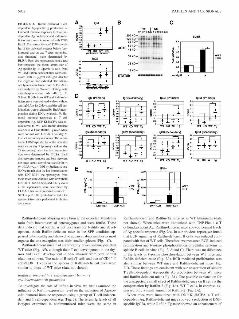
Raftlin-deficient and Raftlin-Tg mice as in WT littermates (datanot shown). When mice were immunized with TNP-Ficoll, a Tcell-independent Ag, Raftlin-deficient mice showed normal levelsof Ag-specific response (Fig. 2A). In our previous report, we foundthat BCR signaling of Raftlin-deficient B cells was reduced com-pared with that of WT cells. Therefore, we measured BCR-inducedproliferation and tyrosine phosphorylation of cellular proteins insplenic B cells in vitro (Fig. 2, B and C). There was no differencein the levels of tyrosine phosphorylation between WT mice andRaftlin-deficient mice (Fig. 2B). BCR-mediated proliferation wasalso similar between WT mice and Raftlin-deficient mice (Fig.2C). These findings are consistent with our observation of similarT cell-independent Ag-specific Ab production between WT miceand Raftlin-deficient mice (Fig. 2A). One possible explanation forthe unexpectedly small effect of Raftlin-deficiency on B cells is thecompensation by Raftlin-2 (Fig. 1A). WT T cells, in contrast, ex-pressed only a small amount of Raftlin-2 (Fig. 1A).
When mice were immunized with DNP-KLH/CFA, a T cell-dependent Ag, Raftlin-deficient mice showed a reduction of DNP-specific IgG2a, while Raftlin-Tg mice showed an enhancement of
FIGURE 2. Raftlin enhanced T celldependent Ag-specific Ig production. A,Humoral immune responses to T cell in-dependent Ag. Wild-type and Raftlin-de-ficient mice were immunized with TNP-Ficoll. The serum titers of TNP-specificIgs of the indicated isotypes before (pre-immune) and on day 7 after immuniza-tion (immune) were determined byELISA. Each dot represents a mouse andbars represent the mean serum titer ofAg-specific Ig. B, Splenic B cells fromWT and Raftlin-deficient mice were stim-ulated with 10 �g/ml anti-IgM Abs forthe length of time indicated. The whole-cell lysates were loaded onto SDS-PAGEand analyzed by Western blotting withanti-phosphotyrosine Ab (4G10). C,Splenic B cells from WT and Raftlin-de-ficient mice were cultured with or withoutanti-IgM Abs for 2 days, and the cell pro-liferations were evaluated by BrdU incor-poration during DNA synthesis. D, Hu-moral immune responses to T celldependent Ag. DNP-KLH/CFA was ad-ministered to WT and Raftlin-deficientmice or to WT and Raftlin-Tg mice. Micewere boosted with DNP-KLH on day 21to elicit secondary responses. The serumtiters of DNP-specific Igs of the indicatedisotypes on day 7 (primary) and on day28 (secondary) after the first immuniza-tion were determined by ELISA. Eachdot represents a mouse and bars representthe mean serum titer of Ag-specific Ig. �,p � 0.05; ��, p � 0.01 by Student’s t test.E, One month after the last immunizationwith DNP-KLH, the splenocytes fromthese mice were cultured with or withoutDNP-KLH for 2.5 days, and IFN-� levelsin the supernatants were determined byELISA. Data are represented as mean �STD. �, p � 0.05 by Student’s t test. Onerepresentative data performed triplicatesare shown.
5932 RAFTLIN AND TCR SIGNALS

DNP-specific IgG2a (Fig. 2D). To confirm whether T cell re-sponses were altered in Raftlin-deficient and Tg mice, we exam-ined IFN-� secretion from splenocytes. As expected, Raftlin-defi-cient splenocytes from mice immunized with DNP-KLH secretedless IFN-� than WT splenocytes after in vitro restimulation withDNP-KLH (Fig. 2E). In contrast, Raftlin-Tg splenocytes secretedmore IFN-� than WT splenocytes (Fig. 2E). In our Tg mice, theexogenous Raftlin level was lower in the B cells than in the T cells(Fig. 1F). This may account for the similar humoral immune re-sponses of Raftlin-Tg mice and WT mice to a T cell-independentAg (TNP-Ficoll) (data not shown). When a T cell-dependent Ag(DNP-KLH/CFA) was used, however, we clearly observed en-hanced humoral immune responses in Raftlin-Tg mice (Fig. 2D).
Raftlin was not involved in OVA-induced asthma, aTh2-mediated disease
To investigate the involvement of Raftlin in T cell dependent dis-eases, we examined OVA-induced asthma as a Th2-cell mediateddisease model. After mice were systemically sensitized to OVAand subjected to aerosolized OVA challenges, we measured theirairway responsiveness to acetylcholine aerosol using an invasivetechnique. Rates of airway hyperresponsiveness were the same inRaftlin-deficient mice, Raftlin-Tg mice, and WT mice (Fig. 3A).
Raftlin is deeply involved in EAE, a Th17-mediated disease
Next, we examined MOG-induced EAE to investigate the involve-ment of Raftlin in Th17 cell-mediated disease. We used Raftlin-deficient mice that were backcrossed at least nine times; even so,we still compared them with WT littermates to reduce the back-ground effects of the ES strain. WT mice exhibited characteristicsigns of EAE starting on day 7, whereas Raftlin-deficient micedeveloped less severe symptoms of EAE compared with WT mice(Fig. 3B, days 8–10). Raftlin-Tg mice, in contrast, initially devel-oped EAE symptoms similar to those seen in littermate WT mice,but after day 12, their condition was observed to be more severe;it also lasted longer, beyond day 21.
We then examined cytokine secretion from splenocytes inmice with EAE. Splenocytes were isolated from the spleens ofmice on day 11 and restimulated with MOG peptide. Restimu-lated splenocytes from Raftlin-deficient mice showed a reduc-tion in IL-17 production compared with those from WT mice,while those from Raftlin-Tg mice showed an increase in IL-17production (Fig. 3C). In addition, cytokine expression in thespinal cords on day 11 was examined by RT-PCR (Fig. 3D).Various kinds of cytokines were expressed at low levels in thespinal cords of Raftlin-deficient mice and at higher levels in thespinal cords of Raftlin-Tg mice. Histological examination re-vealed that the number of immune cells invading the spinalcords was increased in Raftlin-Tg mice and reduced in Raftlin-deficient mice (data not shown). These data indicate that ex-cessive Raftlin expression leads to excessive IL-17 production,which deteriorates Th17-mediated autoimmune disease.
Raftlin modulates TCR-mediated cytokine production
Next, we measured TCR-mediated T cell responses in vitro. Pro-liferation of Raftlin-deficient T cells was not altered, while that ofRaftlin-Tg T cells was slightly enhanced (data not shown). Thencytokines secreted from CD4� T cells were measured after stim-ulation with plate-bound anti-CD3 Ab in various concentrations(Fig. 4, A and B). The CD4� T cells of Raftlin-deficient miceresponded less efficiently to the anti-CD3 Ab for IFN-� and IL-4secretion than WT cells did; they needed a higher concentration ofthe Ab to secrete the same amount of cytokines (Fig. 4A). Thedifference between WT and Raftlin-deficient T cells in the secre-
tion of IL-17 was more obvious than that in the secretion of IFN-�or of IL-4. IL-17 levels were always lower in Raftlin-deficient Tcells than in WT T cells at any anti-CD3 concentrations (Fig. 4A).In contrast, the CD4� T cells of Raftlin-Tg mice responded moresensitively to the anti-CD3 Ab (Fig. 4B). Again, the most obviousdifference between WT and Raftlin-Tg T cells in terms of cytokinesecretion was in the secretion of IL-17. We also investigated thewhether there was a difference in the cell survivability betweenWT and Raftlin-deficient T cells or WT and Raftlin-Tg T cellsunder stimulation by anti-CD3 Ab with or without anti-CD28 Ab.We observed no differences in the cell death between WT andRaftlin-deficient T cells or between WT and Raftlin-Tg T cellsunder either set of conditions (supplemental Fig. 1).4
Next, we examined Ag-specific T cells responses by using OTIItransgenic mice that have MHC class II restricted OVA323–339-specific TCR transgene (Fig. 4, C and D). Raftlin-deficient orRaftlin-Tg mice were crossed with OTII-Tg mice, and CD4� Tcells were stimulated with OVA-loaded BMDCs. As we mighthave expected given the results obtained through anti-CD3 stim-ulation, the amount of IL-17 secreted from Raftlin-deficient OTIICD4� T cells was less than that secreted from WT T cells (Fig.4C), while that secreted from Raftlin-Tg OTII CD4� T cells wasgreater than that secreted from WT T cells (Fig. 4D). IFN-� pro-duction, however, did not change markedly (Fig. 4, C and D), andIL-4 was not detectable under these conditions (data not shown).These data indicate that Raftlin has a more profound effect onIL-17 production than it has on IFN-� or IL-4 production. Th17differentiation seemed to require stronger TCR signaling than Th1or Th2 differentiation did (Fig. 4, A and B), suggesting that Raftlinis involved in determining the strength of TCR signaling.
To investigate the involvement of Raftlin in Th17 differentiationfrom naive T cells, naive CD4� T cells were differentiated to Th17with TGF-� and IL-6 in the absence or presence of anti-CD28 Ab(Fig. 4, E and F). Th17 differentiation from Raftlin-deficient naiveT cells was insufficient compared with that from WT naive T cells,especially under weak stimulation conditions (0.1–1 �g/ml anti-CD3 Ab without anti-CD28 Ab and 0.1 �g/ml anti-CD3 Ab withanti-CD28 Ab). In contrast, under strong TCR signaling condi-tions, there was little difference between WT and Raftlin-deficientT cells in terms of Th17 differentiation. These data suggest thatRaftlin is necessary for the enhancement of signals when theamount of Ag is limited. On the other hand, naive CD4� T cellsfrom Raftlin-deficient mice differentiate to Th1, Th2, and inducibleregulatory T cells (iTreg) normally (supplemental Fig. 2).
Raftlin affects TCR signaling
We identified Raftlin in lipid rafts fractions of B cells. To inves-tigate the intracellular localization of Raftlin in T cells, EL4 Tcells, and OTII CD4� T cells were transfected with CD3 �-DsRedMonomer and Raftlin-EGFP cDNAs. Raftlin-EGFP was uniformlylocalized in the plasma membrane without stimulation (data notshown). Immune synapse was formed by treating EL4 T cells withthe beads immobilizing anti-CD3 and CD28 Abs or by culturingOTII CD4� T cells with BMDCs presenting OVA323–339 peptide(Fig. 5A). CD3 �-DsRed Monomer assembled on the contact sidein these T cells at 30 min after stimulation. Raftlin-EGFP wasconcentrated at the same position, suggesting that Raftlin is accu-mulated in the immunological synapse of T cells.
Next, we investigated TCR-induced cellular protein phosphor-ylation by Western blotting (Fig. 5, B and C). Tyrosine phosphor-ylation of some proteins (for example, some bands stained with
4 The online version of this article contains supplementary material.
5933The Journal of Immunology

anti-phosphotyrosine Ab, 4G10, including 120 kDa, 60 kDa, and20 kDa) and phosphorylation of ERK2, I�B�, and AKT were re-duced in Raftlin-deficient T cells compared with WT T cells (Fig.5B). In Raftlin-Tg T cells, in contrast, these phosphorylations wereenhanced (Fig. 5C). We also investigated TCR-induced intracel-
lular Ca2� mobilization (Fig. 5, D and E). When the T cells werestimulated with a low concentration of anti-CD3 Ab, the intracel-lular Ca2� increase in Raftlin-deficient T cells was reduced andthat in Raftlin-Tg T cells was enhanced compared with that in WTlittermate T cells.
FIGURE 3. The level of Raftlinexpression affected Th17-dependentdisease. A, Effect of Raftlin expres-sion on asthmatic responses in anOVA-induced asthma as a Th2-de-pendent disease model. Mice weresensitized and aerosol challengedwith OVA. Airway responsivenesswas determined by the acetylcholine-dependent change in airway pressure.Data are represented as mean � SEM(n � 8). B, Effect of Raftlin expres-sion on autoimmune responses in aMOG-induced EAE as a Th17 depen-dent disease model. Raftlin-deficientmice and their littermate WT mice orRaftlin-Tg mice and their littermateWT mice were immunized with MOGpeptide in CFA and were adminis-tered Pertussis toxin on days 0 and 2(n � 8, 7, 6, and 6). Their clinicalscores and the relative body weights(body weight/body weight on day 2)were monitored every day. Data arerepresented as mean � SEM. �, p �0.05 by Student’s t test. C, On day 11after immunization, the splenocytesfrom these mice were re-stimulatedwith MOG peptide for 4 days, andIL-17 concentration in the culturedmedium was determined by ELISA.Data are represented as mean � STD.�, p � 0.05; ��, p � 0.01 by Stu-dent’s t test. D, RT-PCR was per-formed using total RNA of the spinalcodes from these mice on day 11 afterimmunization.
5934 RAFTLIN AND TCR SIGNALS

FIGURE 4. Raftlin enhanced the secretion of various cytokines from T cells. Splenic CD4� cells from WT and Raftlin-deficient mice (A) or splenicCD4� cells from WT and Raftlin-Tg mice (B) were stimulated with various concentrations of plate-bound anti-CD3 Ab for 4 days. The amounts of IL-17,IFN-�, and IL-4 in the culture supernatants were calculated using ELISA. Splenic CD4� cells from OTII-Tg and OTII-Tg/Raftlin-deficient mice (C) orsplenic CD4� cells from OTII-Tg and OTII-Tg/Raftlin-Tg mice (D) were cocultured with BMDCs in the presence of OVA for 6 days. The levels of IL-17and IFN-� in the culture supernatants were calculated using ELISA. Data are represented as mean � STD. �, p � 0.05; ���, p � 0.005 by Student’s t test.Naive CD4� T cells from WT and Raftlin-deficient mice were cultured under the Th17 conditions in the absence (E) or presence (F) of anti-CD28 Ab for4.5 days, and internal staining of IL-17 was applied. One representative data of performed triplicates are shown.
5935The Journal of Immunology
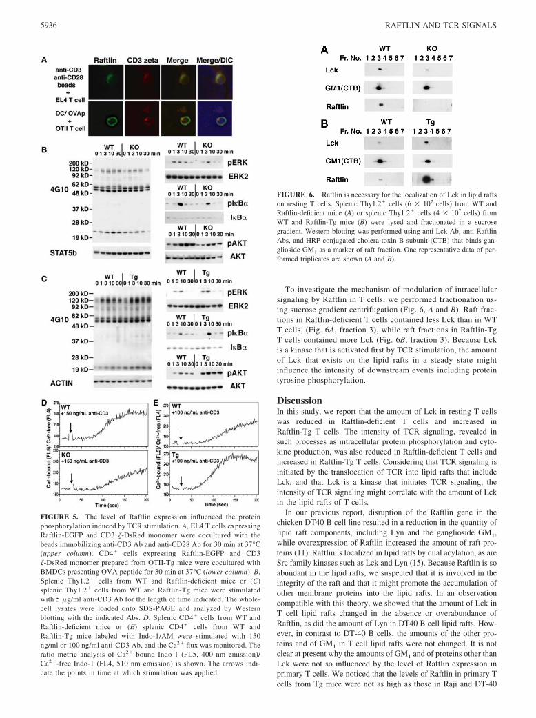
To investigate the mechanism of modulation of intracellularsignaling by Raftlin in T cells, we performed fractionation us-ing sucrose gradient centrifugation (Fig. 6, A and B). Raft frac-tions in Raftlin-deficient T cells contained less Lck than in WTT cells, (Fig. 6A, fraction 3), while raft fractions in Raftlin-TgT cells contained more Lck (Fig. 6B, fraction 3). Because Lckis a kinase that is activated first by TCR stimulation, the amountof Lck that exists on the lipid rafts in a steady state mightinfluence the intensity of downstream events including proteintyrosine phosphorylation.
DiscussionIn this study, we report that the amount of Lck in resting T cellswas reduced in Raftlin-deficient T cells and increased inRaftlin-Tg T cells. The intensity of TCR signaling, revealed insuch processes as intracellular protein phosphorylation and cyto-kine production, was also reduced in Raftlin-deficient T cells andincreased in Raftlin-Tg T cells. Considering that TCR signaling isinitiated by the translocation of TCR into lipid rafts that includeLck, and that Lck is a kinase that initiates TCR signaling, theintensity of TCR signaling might correlate with the amount of Lckin the lipid rafts of T cells.
In our previous report, disruption of the Raftlin gene in thechicken DT40 B cell line resulted in a reduction in the quantity oflipid raft components, including Lyn and the ganglioside GM1,while overexpression of Raftlin increased the amount of raft pro-teins (11). Raftlin is localized in lipid rafts by dual acylation, as areSrc family kinases such as Lck and Lyn (15). Because Raftlin is soabundant in the lipid rafts, we suspected that it is involved in theintegrity of the raft and that it might promote the accumulation ofother membrane proteins into the lipid rafts. In an observationcompatible with this theory, we showed that the amount of Lck inT cell lipid rafts changed in the absence or overabundance ofRaftlin, as did the amount of Lyn in DT40 B cell lipid rafts. How-ever, in contrast to DT-40 B cells, the amounts of the other pro-teins and of GM1 in T cell lipid rafts were not changed. It is notclear at present why the amounts of GM1 and of proteins other thanLck were not so influenced by the level of Raftlin expression inprimary T cells. We noticed that the levels of Raftlin in primary Tcells from Tg mice were not as high as those in Raji and DT-40
FIGURE 5. The level of Raftlin expression influenced the proteinphosphorylation induced by TCR stimulation. A, EL4 T cells expressingRaftlin-EGFP and CD3 �-DsRed monomer were cocultured with thebeads immobilizing anti-CD3 Ab and anti-CD28 Ab for 30 min at 37°C(upper column). CD4� cells expressing Raftlin-EGFP and CD3�-DsRed monomer prepared from OTII-Tg mice were cocultured withBMDCs presenting OVA peptide for 30 min at 37°C (lower column). B,Splenic Thy1.2� cells from WT and Raftlin-deficient mice or (C)splenic Thy1.2� cells from WT and Raftlin-Tg mice were stimulatedwith 5 �g/ml anti-CD3 Ab for the length of time indicated. The whole-cell lysates were loaded onto SDS-PAGE and analyzed by Westernblotting with the indicated Abs. D, Splenic CD4� cells from WT andRaftlin-deficient mice or (E) splenic CD4� cells from WT andRaftlin-Tg mice labeled with Indo-1/AM were stimulated with 150ng/ml or 100 ng/ml anti-CD3 Ab, and the Ca2� flux was monitored. Theratio metric analysis of Ca2�-bound Indo-1 (FL5, 400 nm emission)/Ca2�-free Indo-1 (FL4, 510 nm emission) is shown. The arrows indi-cate the points in time at which stimulation was applied.
FIGURE 6. Raftlin is necessary for the localization of Lck in lipid raftson resting T cells. Splenic Thy1.2� cells (6 � 107 cells) from WT andRaftlin-deficient mice (A) or splenic Thy1.2� cells (4 � 107 cells) fromWT and Raftlin-Tg mice (B) were lysed and fractionated in a sucrosegradient. Western blotting was performed using anti-Lck Ab, anti-RaftlinAbs, and HRP conjugated cholera toxin B subunit (CTB) that binds gan-glioside GM1 as a marker of raft fraction. One representative data of per-formed triplicates are shown (A and B).
5936 RAFTLIN AND TCR SIGNALS
cells. Therefore, on a physiological level, Raftlin may influence thelevels of specific raft proteins but not those of total raft proteins.
The remaining question is the biochemical role of Raftlin. Con-sidering that Raftlin is conserved among vertebrates and that it hastwo highly conserved domains, there is a possibility that Raftlininteracts with other molecules. To test this possibility, we havesearched for binding partners that were coprecipitated, and wehave identified two molecules: clathrin H chain and huntingtin-interacting protein 1 (K. Saeki and A. Yoshimura, unpublisheddata), both of which are involved in vesicle transport (16). Despitethis, the absence of Raftlin had no effect on either the amount ofTCR on the cell surface or the internalization rate of TCR afterstimulation. Further investigation is necessary to determine therole of Raftlin in vesicular transport.
In the present study, we investigated the cytokines produced byvarious concentrations of plate-bound anti-CD3 Ab. We noticedthat the intensity of Raftlin’s effect varies among cytokines; IL-17was most strongly affected by the amount of Raftlin, while IFN-�and IL-4 were modestly affected. IL-17 levels were affected byRaftlin when Ag-specific T cells were stimulated with Ag-peptideloaded DCs, while IFN-� levels were not affected. We also con-firmed that TGF-�-mediated Foxp3 induction was similar in WTand Raftlin-deficient T cells (data not shown). Therefore, Raftlinaffects mostly Th17, but does not affect Th1, Th2, or iTreg polar-ization. These data suggest that suitable intensity of TCR stimu-lation is necessary for the production of each cytokine: IL-4 issaturated in response to the weakest stimulation, and IL-17 is sat-urated in response to the strongest stimulation. We suspect that thefact that Th17 differentiation requires stronger TCR signaling thanother helper T cell lineages is the reason why Raftlin has such aprofound effect on IL-17 production, as seen in the comparisonbetween WT T cells and Raftlin-deficient T cells, over a broadrange. However, we cannot rule out the possibility that Raftlinis deeply involved in an unknown pathway that is specific toIL-17 production or to Th17 differentiation. One possibility isthe requirement of Raftlin for IL-2 signaling. IL-2 is known tosuppress Th17 but rather enhance proliferation of other subsets.However, we did not see much difference in the iTreg popula-tion, which is strictly dependent on IL-2; this makes it appearless likely that Raftlin affects the IL-2 signaling pathway. Fur-ther investigation may uncover a novel mechanism for modu-lation of Th17 differentiation.
In the in vivo Th17-disease model, Raftlin-deficient miceshowed less severe EAE and expressed smaller quantities of IL-6and IL-17 in their spinal cords than WT mice did. Because IL-6 isa cytokine that induces Th17 differentiation, there was a possibilitythat the reduced Th17 development in Raftlin-deficient mice wasalso due to reduced IL-6 expression. The mechanism behind sucha reduction in IL-6 levels in the Raftlin-deficient spinal cords is notclear at present, but Raftlin might have some function in mono-cytic cells, which causes IL-6 expression to be reduced in Raftlin-deficient spinal cords. To investigate this matter, we examinedLPS-induced IL-6 production from Raftlin-deficient BMDCs, butwe found no significant differences between WT and Raftlin-de-ficient DCs. Further study is necessary to clarify how Raftlin isinvolved in IL-6 production in the brain. Raftlin-Tg mice, in con-trast, exhibited more severe EAE and expressed larger quantities ofIFN-� as well as IL-17 in their spinal cords compared with WTmice. A prior studies (17, 18) have suggested that the ratio ofIL-17 to IFN-� determines whether disease pathology occurs in the
spine or in the brain, with increasing levels of IL-17 associatedwith disease in the brain. However, a more recent study has dem-onstrated that lesion location is mainly controlled by the brain’sresponse to IFN-� (19). Thus, there is a possibility that a higherTh1 induction in Raftlin-Tg mice is also involved in determiningdisease severity.
Taken together, our data suggest that Raftlin might modulate theamount of Lck in lipid rafts, regulate the intensity of TCR signal-ing, and control immune responses in vivo.
AcknowledgmentsWe thank M. Sasaki, T. Yoshioka, S. Muroi, Y. Yamada, and M. Ohtsu fortheir technical assistance and Y. Nishi for manuscript preparation.
DisclosuresThe authors have no financial conflict of interest.
References1. DeFranco, A. L. 1997. The complexity of signaling pathways activated by the
BCR. Curr. Opin. Immunol. 9: 296–308.2. Pierce, S. K. 2002. Lipid rafts and B-cell activation. Nat. Rev. Immunol. 2:
96–105.3. Kabouridis, P. S. 2006. Lipid rafts in T cell receptor signalling. Mol. Membr.
Biol. 23: 49–57.4. Fridriksson, E. K., P. A. Shipkova, E. D. Sheets, D. Holowka, B. Baird, and
F. W. McLafferty. 1999. Quantitative analysis of phospholipids in functionallyimportant membrane domains from RBL-2H3 mast cells using tandem high-resolution mass spectrometry. Biochemistry 38: 8056–8063.
5. Hooper, N. M. 1999. Detergent-insoluble glycosphingolipid/cholesterol-richmembrane domains, lipid rafts and caveolae (review). Mol. Membr. Biol. 16:145–156.
6. Brown, D. A., and E. London. 2000. Structure and function of sphingolipid- andcholesterol-rich membrane rafts. J. Biol. Chem. 275: 17221–17224.
7. Gupta, N., and A. L. DeFranco. 2003. Visualizing lipid raft dynamics and earlysignaling events during antigen receptor-mediated B-lymphocyte activation. Mol.Biol. Cell 14: 432–444.
8. Cheng, P. C., M. L. Dykstra, R. N. Mitchell, and S. K. Pierce. 1999. A role forlipid rafts in B cell antigen receptor signaling and antigen targeting. J. Exp. Med.190: 1549–1560.
9. Kusumi, A., I. Koyama-Honda, and K. Suzuki. 2004. Molecular dynamics andinteractions for creation of stimulation-induced stabilized rafts from small unsta-ble steady-state rafts. Traffic 5: 213–230.
10. Tani-ichi, S., K. Maruyama, N. Kondo, M. Nagafuku, K. Kabayama, J. Inokuchi,Y. Shimada, Y. Ohno-Iwashita, H. Yagita, S. Kawano, and A. Kosugi. 2005.Structure and function of lipid rafts in human activated T cells. Int. Immunol. 17:749–758.
11. Saeki, K., Y. Miura, D. Aki, T. Kurosaki, and A. Yoshimura. 2003. The B cell-specific major raft protein, Raftlin, is necessary for the integrity of lipid raft andBCR signal transduction. EMBO J. 22: 3015–3026.
12. Ohtsuka, S., S. Takaki, M. Iseki, K. Miyoshi, N. Nakagata, Y. Kataoka,N. Yoshida, K. Takatsu, and A. Yoshimura. 2002. SH2-B is required for bothmale and female reproduction. Mol. Cell Biol. 22: 3066–3077.
13. Miyamoto, A., K. Nakayama, H. Imaki, S. Hirose, Y. Jiang, M. Abe,T. Tsukiyama, H. Nagahama, S. Ohno, S. Hatakeyama, and K. I. Nakayama.2002. Increased proliferation of B cells and auto-immunity in mice lacking pro-tein kinase C�. Nature 416: 865–869.
14. Kibe, A., H. Inoue, S. Fukuyama, K. Machida, K. Matsumoto, H. Koto,T. Ikegami, H. Aizawa, and N. Hara. 2003. Differential regulation by glucocor-ticoid of interleukin-13-induced eosinophilia, hyperresponsiveness, and gobletcell hyperplasia in mouse airways. Am. J. Respir. Crit. Care Med. 167: 50–56.
15. Kosugi, A., F. Hayashi, D. R. Liddicoat, K. Yasuda, S. Saitoh, and T. Hamaoka.2001. A pivotal role of cysteine 3 of Lck tyrosine kinase for localization toglycolipid-enriched microdomains and T cell activation. Immunol. Lett. 76:133–138.
16. Hyun, T. S., and T. S. Ross. 2004. HIP1: trafficking roles and regulation oftumorigenesis. Trends Mol. Med. 10: 194–199.
17. Langrish, C. L., Y. Chen, W. M. Blumenschein, J. Mattson, B. Basham,J. D. Sedgwick, T. McClanahan, R. A. Kastelein, and D. J. Cua. 2005. IL-23drives a pathogenic T cell population that induces autoimmune inflammation.J. Exp. Med. 201: 233–240.
18. Komiyama, Y., S. Nakae, T. Matsuki, A. Nambu, H. Ishigame, S. Kakuta,K. Sudo, and Y. Iwakura. 2006. IL-17 plays an important role in the developmentof experimental autoimmune encephalomyelitis. J. Immunol. 177: 566–573.
19. Lees, J. R., P. T. Golumbek, J. Sim, D. Dorsey, and J. H. Russell. 2008. RegionalCNS responses to IFN-� determine lesion localization patterns during EAEpathogenesis. J. Exp. Med. 205: 2633–2642.
5937The Journal of Immunology