1 mosby items and derived items © 2010, 2007, 2003 by mosby, inc., an affiliate of elsevier inc....
TRANSCRIPT

1Mosby items and derived items © 2010, 2007, 2003 by Mosby, Inc., an affiliate of Elsevier Inc.
Chapter 6: Skin and Its Appendages
2Mosby items and derived items © 2010, 2007, 2003 by Mosby, Inc., an affiliate of Elsevier Inc.
INTRODUCTION
Skin (integument) is the body’s largest organ
Approximately 1.6 to 1.9 m2 in average-sized adult
Integumentary system describes the skin and its appendages: hair, nails, and skin glands

3Mosby items and derived items © 2010, 2007, 2003 by Mosby, Inc., an affiliate of Elsevier Inc.
STRUCTURE OF THE SKIN
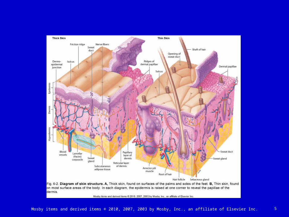
Skin classified as a cutaneous membrane Two primary layers: epidermis and dermis; joined
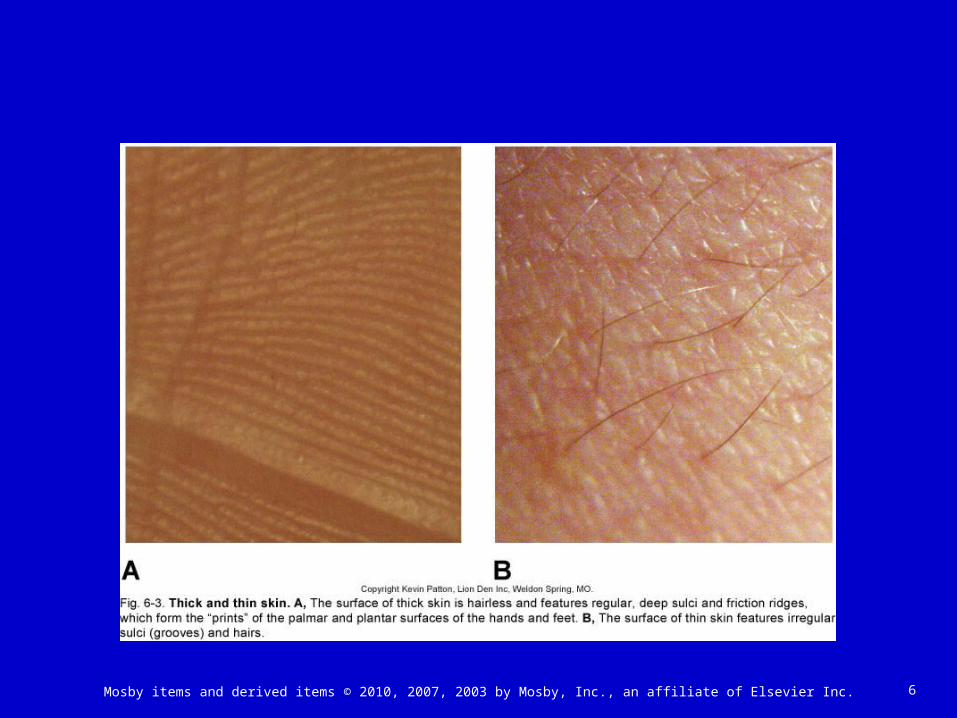
by dermoepidermal junction (Figures 6-1 and 6-2) Hypodermis lies beneath dermis Thin and thick skin (Figure 6-3)
Thin skin covers most of the body’s surface (1 to 3 mm thick)
Thick skin covers soles and palms (4 to 5 mm thick)

4Mosby items and derived items © 2010, 2007, 2003 by Mosby, Inc., an affiliate of Elsevier Inc.
5Mosby items and derived items © 2010, 2007, 2003 by Mosby, Inc., an affiliate of Elsevier Inc.
6Mosby items and derived items © 2010, 2007, 2003 by Mosby, Inc., an affiliate of Elsevier Inc.

7Mosby items and derived items © 2010, 2007, 2003 by Mosby, Inc., an affiliate of Elsevier Inc.
STRUCTURE OF THE SKIN: EPIDERMIS
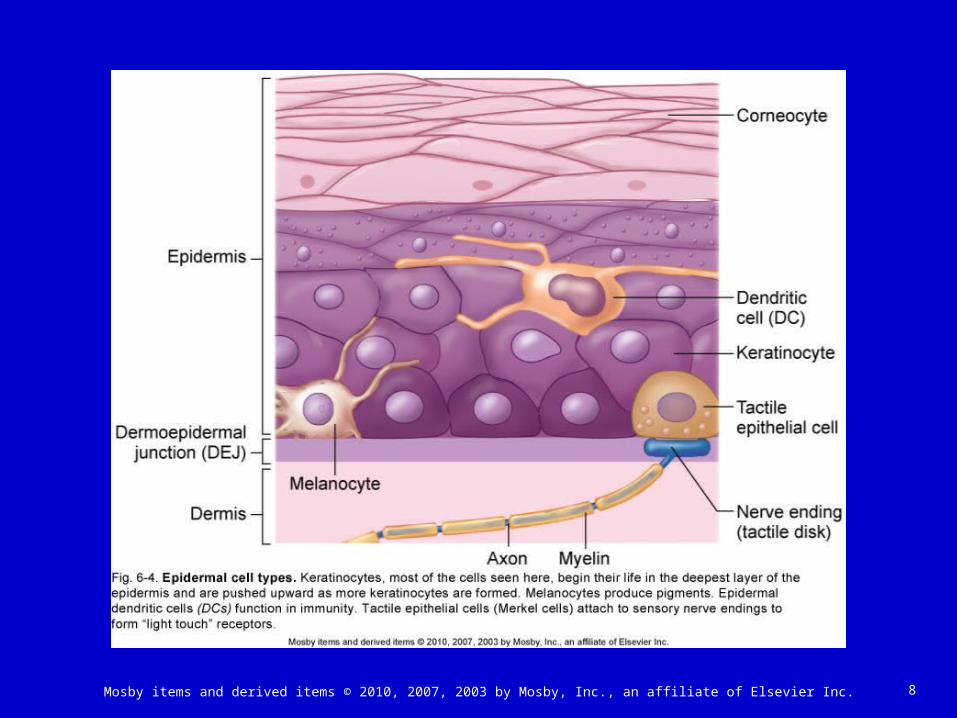
Epidermis Cell types (Figure 6-4)
• Keratinocytes: constitute more than 90% of cells present; principal structural element of the outer skin; sometimes called corneocytes after they are fully keratinized
• Melanocytes: pigment-producing cells (5% of the total); contribute to skin color and filter ultraviolet (UV) light
• Epidermal dendritic cells: branched antigen-presenting cells; play a role in immune response; also called Langerhans cells
• Tactile epithelial cells (Merkel cells): attach to sensory nerve endings to form “light touch” receptors
8Mosby items and derived items © 2010, 2007, 2003 by Mosby, Inc., an affiliate of Elsevier Inc.

9Mosby items and derived items © 2010, 2007, 2003 by Mosby, Inc., an affiliate of Elsevier Inc.
STRUCTURE OF THE SKIN: EPIDERMIS (cont.)
Cell layers• Stratum basale (base layer): single layer of
columnar cells; only these cells undergo mitosis and then migrate through the other layers until they are shed
• Stratum spinosum (spiny layer): cells arranged in eight to 10 layers with desmosomes that pull cells into spiny shapes; cells rich in RNA
• Stratum germinativum (growth layer): another name for stratum basale or stratum spinosum and stratum basale together

10Mosby items and derived items © 2010, 2007, 2003 by Mosby, Inc., an affiliate of Elsevier Inc.
STRUCTURE OF THE SKIN: EPIDERMIS (cont.)
• Stratum granulosum (granular layer): cells arranged in two to four layers and filled with keratohyalin granules; contains high levels of lysosomal enzymes
• Stratum lucidum (clear layer): cells filled with keratin precursor called eleidin; absent in thin skin
• Stratum corneum (horny layer): most superficial layer; dead cells filled with keratin (barrier area)

11Mosby items and derived items © 2010, 2007, 2003 by Mosby, Inc., an affiliate of Elsevier Inc.
STRUCTURE OF THE SKIN: EPIDERMIS (cont.)
Dermoepidermal junction A basement membrane with unique fibrous
elements and a polysaccharide gel “glue” the epidermis to the dermis below
The junction is a partial barrier to the passage of some cells and large molecules

12Mosby items and derived items © 2010, 2007, 2003 by Mosby, Inc., an affiliate of Elsevier Inc.
STRUCTURE OF THE SKIN: DERMIS
Dermis Sometimes called “true skin”—much thicker than the
epidermis and lies beneath it Gives strength to the skin Serves as a reservoir storage area for water and
electrolytes Contains various structures
• Arrector pili muscles and hair follicles (Figure 6-5)• Sensory receptors (Figure 6-6)• Sweat and sebaceous glands• Blood vessels
Rich vascular supply plays a critical role in temperature regulation

13Mosby items and derived items © 2010, 2007, 2003 by Mosby, Inc., an affiliate of Elsevier Inc.

14Mosby items and derived items © 2010, 2007, 2003 by Mosby, Inc., an affiliate of Elsevier Inc.

15Mosby items and derived items © 2010, 2007, 2003 by Mosby, Inc., an affiliate of Elsevier Inc.
STRUCTURE OF THE SKIN: DERMIS (cont.)
Layers of dermis• Papillary layer: composed of dermal papillae that
project into the epidermis; contains fine collagenous and elastic fibers and the dermoepidermal junction; forms a unique pattern that gives individual fingerprints
• Reticular layer: contains dense, interlacing white collagenous fibers and elastic fibers to make the skin tough yet stretchable; when processed from animal skin, produces leather
Dermal growth and repair• The dermis does not continually shed and
regenerate itself as does the epidermis

16Mosby items and derived items © 2010, 2007, 2003 by Mosby, Inc., an affiliate of Elsevier Inc.
17Mosby items and derived items © 2010, 2007, 2003 by Mosby, Inc., an affiliate of Elsevier Inc.
STRUCTURE OF THE SKIN: HYPODERMIS
Hypodermis Also called the subcutaneous layer or superficial fascia Located deep to the dermis; forms connection between
skin and other structures Not part of the skin

18Mosby items and derived items © 2010, 2007, 2003 by Mosby, Inc., an affiliate of Elsevier Inc.
SKIN COLOR
Melanin Basic determinant is quantity, type, and distribution of
melanin Types of melanin
• Eumelanin: group of dark brown, almost black, melanins• Pheomelanin: group of reddish and orange melanins
Melanin formed from tyrosine by melanocytes (Figure 6-8)
• Melanocytes release melanin in packets called melanosomes
• Melanosomes are ingested by surrounding keratinocytes and form a cap over the nucleus

19Mosby items and derived items © 2010, 2007, 2003 by Mosby, Inc., an affiliate of Elsevier Inc.
20Mosby items and derived items © 2010, 2007, 2003 by Mosby, Inc., an affiliate of Elsevier Inc.
SKIN COLOR (cont.)
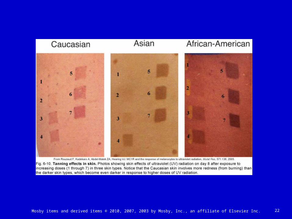
Albinism: congenital absence of melanin Process regulated by tyrosinase, exposure to sunlight
(UV radiation), and certain hormones, including melanocortins (adrenocorticotropic hormone, alpha melanocyte stimulating hormone) and endothelin-1 (Figures 6-9 and 6-10)
Cumulative effects of UV ray exposure may produce age spots (Figure 6-11)

21Mosby items and derived items © 2010, 2007, 2003 by Mosby, Inc., an affiliate of Elsevier Inc.
22Mosby items and derived items © 2010, 2007, 2003 by Mosby, Inc., an affiliate of Elsevier Inc.

23Mosby items and derived items © 2010, 2007, 2003 by Mosby, Inc., an affiliate of Elsevier Inc.

24Mosby items and derived items © 2010, 2007, 2003 by Mosby, Inc., an affiliate of Elsevier Inc.
SKIN COLOR: OTHER PIGMENTS
Other pigments Beta-carotene (group of yellowish pigments from food)
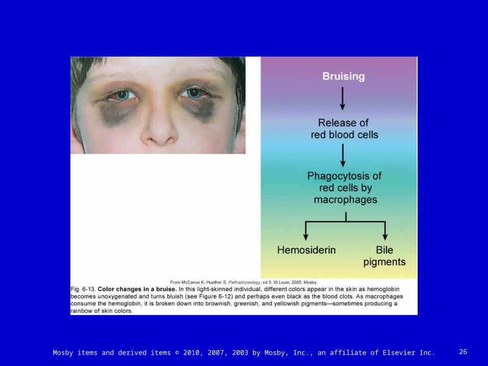
can also contribute to skin color Hemoglobin: color changes also occur as a result of
changes in blood flow• Redder skin color when blood flow to skin increases • Cyanosis: bluish color caused by darkening of hemoglobin
when it loses oxygen and gains carbon dioxide (Figure 6-12)
• Bruising can cause a rainbow of different colors to appear in the skin (Figure 6-13)
Other pigments: from cosmetics, tattoos, and bile pigments in jaundice (Box 6-4)

25Mosby items and derived items © 2010, 2007, 2003 by Mosby, Inc., an affiliate of Elsevier Inc.
26Mosby items and derived items © 2010, 2007, 2003 by Mosby, Inc., an affiliate of Elsevier Inc.

27Mosby items and derived items © 2010, 2007, 2003 by Mosby, Inc., an affiliate of Elsevier Inc.
FUNCTIONS OF THE SKIN
Protection (Table 6-2) Physical barrier to microorganisms Barrier to chemical hazards Reduces potential for mechanical trauma Prevents dehydration Protects from excess UV ray exposure (melanin
function)
28Mosby items and derived items © 2010, 2007, 2003 by Mosby, Inc., an affiliate of Elsevier Inc.
FUNCTIONS OF THE SKIN (cont.)
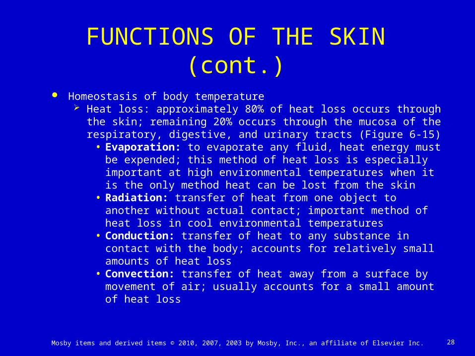
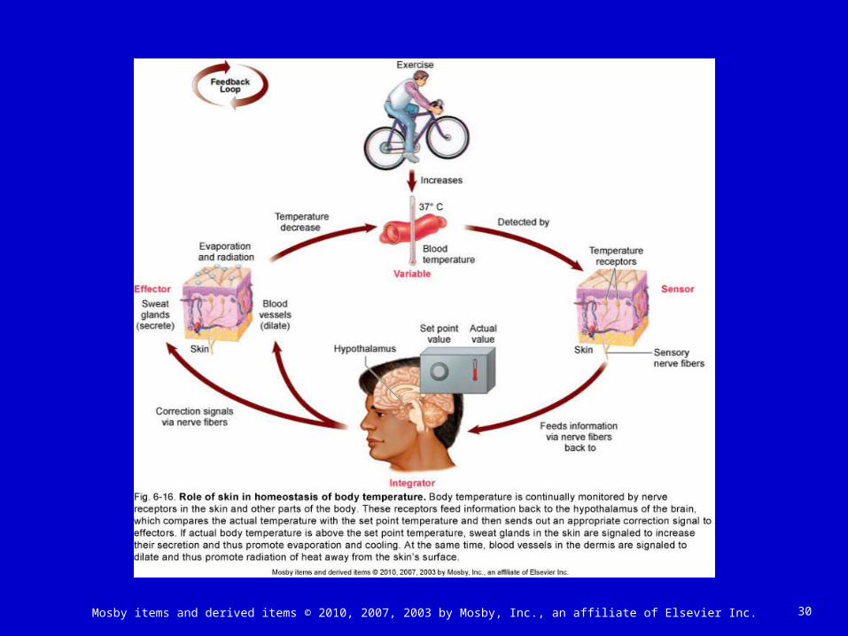
Homeostasis of body temperature Heat loss: approximately 80% of heat loss occurs through the skin;
remaining 20% occurs through the mucosa of the respiratory, digestive, and urinary tracts (Figure 6-15)
• Evaporation: to evaporate any fluid, heat energy must be expended; this method of heat loss is especially important at high environmental temperatures when it is the only method heat can be lost from the skin
• Radiation: transfer of heat from one object to another without actual contact; important method of heat loss in cool environmental temperatures
• Conduction: transfer of heat to any substance in contact with the body; accounts for relatively small amounts of heat loss
• Convection: transfer of heat away from a surface by movement of air; usually accounts for a small amount of heat loss

29Mosby items and derived items © 2010, 2007, 2003 by Mosby, Inc., an affiliate of Elsevier Inc.
30Mosby items and derived items © 2010, 2007, 2003 by Mosby, Inc., an affiliate of Elsevier Inc.

31Mosby items and derived items © 2010, 2007, 2003 by Mosby, Inc., an affiliate of Elsevier Inc.
APPENDAGES OF THE SKIN
Hair (Figure 6-17) Development of hair
• Distribution is over entire body except palms of hands and soles of feet and a few other small areas
• Fine and soft hair coat present before birth called lanugo• Coarse pubic and axillary hair that develops at puberty
called terminal hair• Hair follicles and hair develop from epidermis; mitosis of
cells of germinal matrix forms hairs• Root: part of hair embedded in follicle in dermis• Shaft: visible part of hair• Medulla: inner core of hair• Cortex: outer portion

32Mosby items and derived items © 2010, 2007, 2003 by Mosby, Inc., an affiliate of Elsevier Inc.
33Mosby items and derived items © 2010, 2007, 2003 by Mosby, Inc., an affiliate of Elsevier Inc.
APPENDAGES OF THE SKIN: HAIR (cont.)
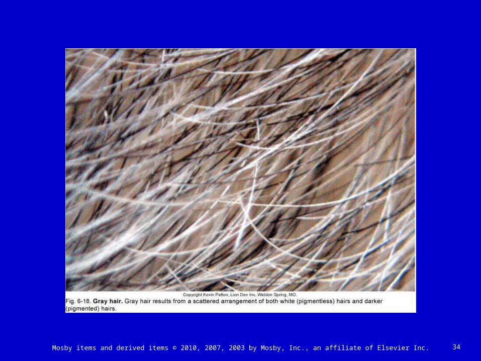
Appearance of hair• Color: result of different amounts, distribution, types
of melanin in cortex of hair (Figure 6-18)• Growth: growth and rest periods alternate; hair on
head averages 5 inches of growth per year• Sebaceous glands attach to and secrete sebum
(skin oil) into follicle• Male pattern baldness (androgenic alopecia) results
from combination of genetic tendency and male sex hormones (Figure 6-19)
34Mosby items and derived items © 2010, 2007, 2003 by Mosby, Inc., an affiliate of Elsevier Inc.

35Mosby items and derived items © 2010, 2007, 2003 by Mosby, Inc., an affiliate of Elsevier Inc.

36Mosby items and derived items © 2010, 2007, 2003 by Mosby, Inc., an affiliate of Elsevier Inc.
APPENDAGES OF THE SKIN (cont.)
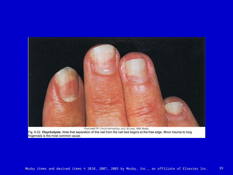
Nails (Figure 6-20) Consist of epidermal cells converted to hard keratin Nail body: visible part of each nail Root: part of nail in groove hidden by fold of skin, the cuticle Lunula: moon-shaped white area nearest root Nail bed: layer of epithelium under nail body; contains
abundant blood vessels• Appears pink under translucent nails• Nails may have pigmented streaks (Figure 6-21)• Separation of a nail from the nail bed is called onycholysis
(Figure 6-22) Growth: nails grow by mitosis of cells in stratum basale
beneath the lunula; average growth about 0.5 mm/week, or slightly over 1 inch per year

37Mosby items and derived items © 2010, 2007, 2003 by Mosby, Inc., an affiliate of Elsevier Inc.

38Mosby items and derived items © 2010, 2007, 2003 by Mosby, Inc., an affiliate of Elsevier Inc.
39Mosby items and derived items © 2010, 2007, 2003 by Mosby, Inc., an affiliate of Elsevier Inc.

40Mosby items and derived items © 2010, 2007, 2003 by Mosby, Inc., an affiliate of Elsevier Inc.
APPENDAGES OF THE SKIN (cont.)
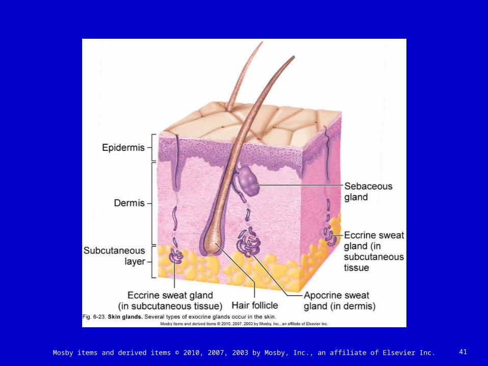
Skin glands (Figure 6-23) Two types of sweat glands
• Eccrine glands Most numerous sweat glands; quite small Distributed over total body surface with exception of a few small
areas Simple, coiled, tubular glands Function throughout life Secrete perspiration or sweat; eliminate wastes and help maintain a
constant core temperature• Apocrine glands
Located deep in subcutaneous layer Limited distribution: axilla, areola of breast, and around anus Large (often more than 5 mm in diameter) Simple, branched, tubular glands Begin to function at puberty Secretion shows cyclic changes in female with menstrual cycle
41Mosby items and derived items © 2010, 2007, 2003 by Mosby, Inc., an affiliate of Elsevier Inc.
42Mosby items and derived items © 2010, 2007, 2003 by Mosby, Inc., an affiliate of Elsevier Inc.
APPENDAGES OF THE SKIN: SKIN GLANDS (cont.)
Skin glands (cont.) Sebaceous glands
• Secrete sebum: oily substance that keeps hair and skin soft and pliant; prevents excessive water loss from skin
• Lipid components have antifungal activity• Simple, branched glands• Found in dermis except in palms and soles• Secretion increases in adolescence; may lead to formation of pimples
and blackheads Ceruminous glands
• Modified apocrine sweat glands• Simple, coiled, tubular glands• Empty contents into external ear canal alone or with sebaceous glands• Mixed secretions of sebaceous and ceruminous glands called
cerumen (wax)• Function of cerumen to protect area from dehydration; excess
secretion can cause blockage of ear canal and loss of hearing

43Mosby items and derived items © 2010, 2007, 2003 by Mosby, Inc., an affiliate of Elsevier Inc.

44Mosby items and derived items © 2010, 2007, 2003 by Mosby, Inc., an affiliate of Elsevier Inc.
THE BIG PICTURE: SKIN AND THE WHOLE BODY
Skin is a major component of the body’s structural framework
Skin defines the internal environment of the body
Primary functions are support and protection