1 molecular pathogenesis of renal cell carcinoma: a...
TRANSCRIPT

1
Molecular Pathogenesis of Renal Cell Carcinoma: A Review
Israel Gomy and Wilson Araújo Silva Jr. University of São Paulo, Medical School of Ribeirão Preto,
Genetics Department, Ribeirão Preto, Brazil
1. Introduction
Renal cell carcinomas (RCC) represent almost 90% of all kidney cancers and in about 2% of
cases there is a family history of RCC (McLaughlin et al, 1984). Despite their rare incidences,
Mendelian hereditary syndromes with RCC have provided important insights into the
molecular pathogenesis of this tumor. The cloning of susceptibility genes that are involved in
familial predisposition has offered entry points into the signaling pathways that are
deregulated in sporadic RCC. Sporadic RCC are extremely heterogeneous and are classified
into many histolological subtypes. The most frequent form is clear cell or conventional RCC,
accounting for approximately 75% of all cases, whereas the most common non-clear cell tumor
is papillary RCC (12% of cases), which is subdivided into types 1 and 2. Other subtypes
include chromophobe and oncocytomas, each of them occurring in 4% of patients, collecting-
duct (<1%) and rare forms or yet to be classified (< 2%). The correlation between
histopathological features and genetic alterations in RCC has been introduced in 1997 with the
Heidelberg classification (Kovacs et al, 1997). More recently, expression profiles through
microarrays have been done for many of the kidney tumor subtypes and provide the evidence
that their expression patterns reflect their histological classifications and demonstrate that
various renal tumor subtypes are genetically distinct entities (Higgins et al, 2003).
Generally, there is a good correlation between the genetic causes of familial RCC and their histopathological features as exemplified by the commonest form of hereditary RCC, von Hippel-Lindau (VHL) disease, which invariably presents a clear cell type. Moreover, germline mutations in the MET proto-oncogene cause type 1 papillary RCC, whereas type 2 is correlated with germline mutations in the fumarate hidratase gene, which cause hereditary leiomyomatosis. Birt-Hogg-Dubè syndrome is also characterized by susceptibility to RCC but with a mixed chromophobe-oncocytoma histopathology, and is associated with germline mutations in the BHD tumor suppressor gene.
All the genes identified so far, which are involved in the molecular pathogenesis of hereditary and sporadic RCC comprise a diverse set of complex biochemical and cell metabolism pathways, such as iron, energy, nutrient and oxygen-sensing (Linehan et al., 2010a). A plenty of biochemical and molecular studies of the numerous signalling pathways
www.intechopen.com

Emerging Research and Treatments in Renal Cell Carcinoma
4
disrupted in RCC have already provided reasonable translational approaches and clinical applications of target therapy RCC with promising results (Iliopoulos, 2006). The histopathological and molecular features of RCC are summarized in table 1.
Tumor type Locus Gene Pathway Syndrome
Clear cell
3p25 3p14 3p21 17p11
VHL FHIT RASSF1A BHD
VEGF TGF-┚ AMPK-mTOR
von Hippel-Lindau Familial clear cell RCC Birt-Hogg-Dube
Papillary type 1 type 2
7q31 1q42 7q31.1 9q34 16p13 1q25
MET FH FRA7G TSC1 TSC2 HRPT2
MET-HGF VEGF TGF-┚ mTOR mTOR
Hereditary papillary RCC Hereditary leiomatosis Tuberous sclerosis complex Tuberous sclerosis complex Hyperparathyroidism-jaw tumor
Chromophobe
17p11 BHD AMPK-mTOR
Birt-Hogg-Dube
Oncocytoma 17p11 9q34 16p13
BHD TSC1 TSC2
AMPK-mTOR mTOR mTOR
Birt-Hogg-Dube Tuberous complex Tuberous complex
Collecting duct carcinoma
-1q32,-6p,-8p, -9p,-13q,-19q32,-21q
unknown
unknown
none
Renal carcinoma associated with Xp11.2 translocation
1p34 1q21 17q23 17q25 3q23 Xq12
PSF-TFE3 PRCC-TFE3 CTLC-TFE3 ASPL-TFE3 ? NonO-TFE3
none
none
Mucinous tubular and spindle cell carcinoma
-8p,-9p,-11q,+12q,+16q+17,+20q
unknown
unknown
none
Table 1. Histopathological and genetic characteristics of RCC
www.intechopen.com

Molecular Pathogenesis of Renal Cell Carcinoma: A Review
5
2. The oxygen-sensing pathway: HIF-VHL interaction and clear-cell RCC pathogenesis
Hypoxia-inducible factors (HIF) are oxygen-sensitive basic helix–loop–helix transcription factors, which regulate biological processes that facilitate both oxygen delivery and cellular adaptation to oxygen deprivation. HIF is a heterodimer consisting of unstable a-subunits and stable constitutively expressed b-subunits. HIF-┙, together with HIF-┚ bind to hypoxia-response elements in gene promoters to regulate the expression of genes that are involved in energy metabolism, angiogenesis, erythropoiesis, iron metabolism, cell proliferation, apoptosis and other biological processes. HIF1-┙ and HIF2-┙ mediate transcription of a number of downstream genes thought to be important in cancer, including transforming growth factor alpha (TGF-┙), platelet-derived growth factor (PDGF), and vascular endothelial growth factor (VEGF) (Linehan et al., 2010a).
In clear-cell renal-cell carcinoma, HIF-┙ accumulates, resulting in the overexpression of proteins that are normally inducible with hypoxia, acting on neighboring vascular cells and promoting tumor angiogenesis. The augmented tumor vasculature provides additional nutrients and oxygen to promote the growth of tumor cells (Cohen & McGovern, 2005).
Germline mutations in the VHL gene are found in almost all families with VHL disease, an autosomal dominant condition characterized by a plenty of benign and malignant tumors (Figure 1). The most common cancer is clear cell RCC, which affects 25-30% of patients and is a major concern in disease morbity and mortality (Maher et al., 1990). Remarkably, up to
Fig. 1. von Hippel-Lindau disease predisposes to several tumors in brain, spinal cord, eyes, liver, pancreas, kidneys, testes.
www.intechopen.com
Emerging Research and Treatments in Renal Cell Carcinoma
6
91% of patients with sporadic clear cell RCC harbor biallelic VHL inactivation (Gnarra et al, 1994). Somatic mutations are found in approximately 50% of these tumors, whereas inactivation due to promoter hypermethylation has been identified in 10-20% of sporadic clear-cell RCC (Kim & Kaelin, 2004). Therefore, defects in the VHL gene are the major responsible for all cases of RCC.
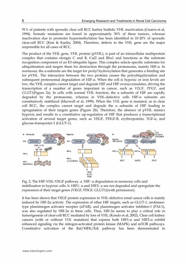
The product of the VHL gene, VHL protein (pVHL), is part of an intracellular multiprotein complex that contains elongin C and B, Cul2 and Rbx1 and functions as the substrate recognition component of an E3-ubiquitin ligase. This complex selects specific substrates for ubiquitination and targets them for destruction through the proteasome, mainly HIF-┙. In normoxia, the a-subunits are the target for prolyl hydroxylation that generates a binding site for pVHL. The interaction between the two proteins causes the polyubiquitynation and subsequent proteosomal degradation of HIF-┙. When the cell is hypoxic or iron levels are low, the VHL complex cannot target and degrade HIF and HIF overaccumulates, driving the transcription of a number of genes important in cancer, such as VEGF, PDGF, and GLUT1(Figure 2a). In cells with normal VHL function, the ┙ subunits of HIF are rapidly degraded by the proteasome, whereas in VHL-defective cells HIF-┙ subunits are constitutively stabilized (Maxwell et al, 1999). When the VHL gene is mutated, as in clear cell RCC, the complex cannot target and degrade the ┙ subunits of HIF leading to upregulation of their targets genes (Figure 2b). Therefore, the absence of pVHL mimics hypoxia and results in a constitutive up-regulation of HIF that produces a transcriptional activation of several target genes, such as VEGF, PDGF-B, erythropoietin, TGF-┙, and glucose-transporter-1 (GLUT-1).
Fig. 2. The HIF-VHL-VEGF pathway. a. HIF- ┙ degradation in normoxic cells and stabilization in hypoxic cells. b. HIF1- ┙ and HIF2- ┙ are not degraded and upregulate the expression of their target genes (VEGF, PDGF, GLUT1)(with permission).
It has been shown that VEGF protein expression in VHL-defective renal cancer cells is mainly induced by HIF-2┙ activity. The expression of other HIF targets, such as GLUT-1, urokinase-type plasminogen activator receptor (uPAR), and plasminogen activator inhibitor-1 (PAI-1), was also regulated by HIF-2a in these cells. Thus, HIF-2┙ seems to play a critical role in tumorigenesis of clear-cell RCC mediated by loss of VHL (Kondo et al, 2002). Clear cell kidney cancers (with or without VHL mutation) that express both HIF1-┙ and HIF2-┙ exhibit enhanced signaling via the mitogen-activated protein kinase (MAPK) and mTOR pathways. Constitutive activation of the Raf/MEK/Erk pathway has been demonstrated in
www.intechopen.com

Molecular Pathogenesis of Renal Cell Carcinoma: A Review
7
approximately 50% of RCC samples (ref.14. EJC). Conversely, clear cell tumors that express only HIF2-┙ have elevated activity of the oncogene c-myc (Gordan et al, 2008).
VHL substrates other than HIF have been reported, and although their exact role in kidney carcinogenesis has not been proved to be the same as HIF’s, it is possible that they may contribute directly or indirectly to VHL-deficient RCC and/or other solid tumors. VHL has been shown to interact with the RNA polymerase II subunits Rpb1, Rbp6, and Rpb7 at domains that present sequence and structural similarity to the HIF binding domain. Binding of these subunits to pVHL was shown to promote their ubiquitination and decrease transcriptional activity of Pol II (Kuznetsova et al, 2003).
Moreover, pVHL has been shown to influence the content of the extracellular matrix. Fibronectin coimmunoprecipitates with wild type but not mutant pVHL, and pVHL-deficient RCC cell lines fail to assemble extracellular fibronectin, most likely because of a defect in fibronectin maturation (Ohh et al, 1998).
Finally, it was recently shown that VHL is linked to the DNA damage response pathway. pVHL binds directly to p53, inhibits its mdm2-mediated ubiquitination, suppresses p53 nuclear export, and promotes its acetylation by p300 and overall p53 transcriptional activity. Reintroduction of pVHL in VHL-deficient RCC lines enhances p53-mediated G1 arrest and promotes their apoptotic response to genotoxic stress (Roe et al, 2006).
2.1 Therapeutic perspectives
The better understanding of VHL signaling pathway has provided the foundation for the development of therapeutic approaches that target this pathway in patients with advanced clear cell kidney cancer.
Most therapeutic agents currently approved to treat clear cell kidney cancer target either the downstream targets of HiF activity (such as the VEGF receptor) or inhibit mtorC1, which provides translational control of HiF1-┙ (Thomas et al., 2006). The responses to these agents observed in patients with advanced clear cell kidney cancer is proof of principle that targeting the VHL–HIF pathway can induce tumor regression in humans. However, these agents target only a small proportion of the downstream genes regulated by HIFs. An approach to target HIF transcriptional activity or translation of HIFs themselves—that is, to affect all the genes regulated by HIFs—could potentially provide more-effective therapy. Targeting the HIF2-┙ pathway is likely to be more successful than solely targeting the HIF1-┙ pathway. Efforts are currently underway to identify agents that down regulate HIF2-┙-induced gene expression (Linehan et al., 2010a). Tyrosine kinase receptors trigger activation of signal transduction pathways involved in cell proliferation and survival of RCCs (Patel et al, 2006). Growth factors, such as VEGF and TGF-a, might bind to the respective tyrosine kinase receptors (EGFR and VEGFR-1) that are expressed in renal cancer cells. Thus, these receptors could potentially be targets of small molecules inhibitors. Nevertheless, many chemotherapeutic and targeted tyrosine kinase approaches are limited by their toxic effects on non-tumor cells. An alternative therapy, might be done by targeting autophagy, a central component of the cell response to nutrient and energy deprivation (Turcotte et al, 2008). By this approach, only those cells with no VHL product would be affected, whereas cells with a wild-type copy of the VHL gene would not. Therefore, this would provide a less-toxic and more-effective form of therapy for patients with advanced renal cancer.
www.intechopen.com

Emerging Research and Treatments in Renal Cell Carcinoma
8
3. The MET-HGF pathway and type-1 papillary renal cancer
Papillary renal-cell carcinoma occurs in several familial syndromes. Hereditary papillary renal carcinoma (HPRC) is an autosomal dominant disorder associated with multifocal papillary renal-cell carcinoma with type 1 histological features and is caused by germline activating mutations in the MET proto-oncogene (Schmidt et al, 1997) (Figure 3).
Fig. 3. Hereditary Papillary Renal Carcinoma leads to multiple (even thousands) of papillary tumors. (with permission)
The MET proto-oncogene encodes a receptor tyrosine kinase that is physiologically activated by hepatocyte growth factor (HGF). Binding of HGF to the extracellular portion of the MET receptor triggers autophosphorylation of critical tyrosines in the intracellular tyrosine kinase domain, thus activating a downstream signaling cascade. Activation of the MET/HGF signaling pathway has been shown to be involved in a number of biological activities including cell proliferation, cell motility, branching morphogenesis and epithelial-mesenchymal transition (Boccaccio & Comoglio, 2006).
Mutations of the MET proto-oncogene were detected in germline of all HPRC patients but only in a small subset of cases with sporadic type 1- papillary RCC (Schmidt et al, 1999). Thus, the pathogenesis of hereditary papillary renal carcinoma is usually different from that of sporadic papillary RCC. Tumorigenesis of most sporadic papillary RCCs are not be induced only by MET mutations, but mostly by MET and chromosome 7-related genes dosage. Since the frequency of MET mutations in sporadic papillary RCCs is low, it is possible that other genes might be involved in this form of renal cancer. In this respect, mutations of the KIT proto-oncogene have been described in 68% of papillary RCC patients (Lin et al, 2004). However, the role of KIT in the pathogenesis of this disease needs to be confirmed by functional studies. Furthermore, nutrient-stimulated HGF–MET signaling induces phosphorylation of serine–threonine protein kinase 11 (STK11or LKB1) through the RAS–ERK pathway, implicating MET in the LKB1–AMPK–mTOR nutrient and energy sensing pathway (Figure 4).
www.intechopen.com

Molecular Pathogenesis of Renal Cell Carcinoma: A Review
9
Fig. 4. HGF-MET pathway. Crosslinks between other energy, iron and oxygen-sensing pathways. (with permission)
Of particular interest for renal carcinogenesis is the observation that MET and VHL signaling pathways intersect via pVHL-mediated regulation of HIF function. HIF stabilization through hypoxia or loss of VHL function results in transcriptional upregulation, and therefore promotion of the transforming potential of the c-MET receptor (Pennacchietti et al, 2003). This crosstalk between VHL and c-MET pathways may explain why clear-cell and papillary histologies often coexist in the same tumor (Iliopoulos et al, 2006).
3.1 Therapeutic perspectives
The therapeutic implications of inhibiting c-MET signaling are promising, as activating mutations or copy number–overexpression of c-MET underlies a significant subset of type 1 papillary RCC.
A clinical trial is currently underway to determine the effect of foretinib, a kinase inhibitor of both MET and VEGF receptors, in patients with either hereditary or sporadic papillary kidney cancer. Early evidence shows the efficacy of this agent in patients with germline mutations in the tyrosine kinase domain of MET (Srinivasan et al, 2009). Response to such an agent with activity against MET might be seen in tumors that are characterized by a
www.intechopen.com

Emerging Research and Treatments in Renal Cell Carcinoma
10
mutation in the tyrosine kinase domain of MET; this drug might also have some activity in tumors that have MET amplification (Linehan et al., 2010b).
4. The mitochondrial metabolic pathway: The FH gene and type-2 papillary renal cancer and the SDH gene
Germline mutations of the FH gene, which encodes mitochondrial fumarate hydratase, have been detected in most individuals with hereditary leiomyomatosis renal cell carcinoma (HLRCC), who are susceptible to develop cutaneous and uterine leiomyomas, uterine leiomyosarcoma and, most often solitary and unilateral renal tumors, which may present different histological patterns, particularly type-2 papillary RCC (Tomlinson et al, 2002) (Figure 5).
Fig. 5. Hereditary Leiomyomatosis Renal Cell Carcinoma predisposes to type-2 papillary RCC (left), uterine (middle) and cutaneous (right) leiomyomas (with permission).
FH is an enzyme component of the mitochondrial tricarboxylic acid or Krebs cycle, which
has an important role in energy metabolism. In patients with FH deficient cells, it has been
proposed that the inactivation of this enzyme might lead to a hypoxic environment that can
favour renal carcinogenesis. FH inactivating mutations increase fumarate levels, and
consequently the concentration of the fumarate precursor succinate. The high levels of
succinate in the cytoplasm lead to stabilization of HIF-1┙ subunits and transcriptional
upregulation of hypoxia-inducible genes, such as VEGF and GLUT1 (which encodes the
glucose transporter type 1) (Figure 6). Thus, these factors are critically important to increase
vasculature and glucose transport in RCC cells, thereby contributing to the highly
aggressive nature of HLRCC-associated renal tumors (Iliopoulos et al, 2006).
The inhibition of HIF-1┙ by the increased levels of fumarate provides a VHL-independent
mechanism for dysregulation of HIF degradation in FH-deficient HLRCC-associated kidney
cancers. FH-deficient kidney cancer cell lines are glucose-dependent and have significantly
impaired oxidative phosphorylation. The glucose-mediated generation of cellular reactive
oxygen species in an FH-deficient kidney cancer cell line results in stabilization of HIF1-┙
(Sudarshan et al, 2009). The impaired oxidative phosphorylation in FH-deficient kidney
cancer results in a nearly total dependence on glycolysis for energy production.
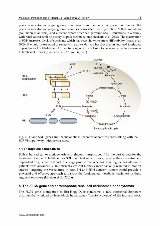
Succinate dehydrogenase (SDH) is another Krebs cycle enzyme gene that has been
associated with the development of familial tumors. Familial paraganglioma/
pheochromocytoma kindreds have been found to have germline mutations in the
mitochondrial complex II genes SDHB, SDHC, and SDHD. Renal carcinoma, along with
www.intechopen.com
Molecular Pathogenesis of Renal Cell Carcinoma: A Review
11
pheochromocytoma/paraganglioma, has been found to be a component of the familial
pheochromocytoma/paraganglioma complex associated with germline SDHB mutations
(Neumann et al, 2004), and a recent report described germline SDHB mutations in a family
with renal cancer with no history of pheochromocytoma (Ricketts et al, 2008). The inactivation
of SDH increases levels of succinate, which has been shown to affect HIF stability (Isaacs et al,
2005). It would be expected to severely impair oxidative phosphorylation and lead to glucose
dependence of SDH-deficient kidney tumors, which are likely to be as sensitive to glucose as
FH-deficient tumors (Linehan et al., 2010a) (Figure 6).
Fig. 6. FH and SDH genes and the metabolic mitochondrial pathway crosslinking with the HIF-VHL pathway (with permission).
4.1 Therapeutic perspectives
Both enhanced tumor angiogenesis and glucose transport could be the best targets for the
treatment of either FH-deficient or SDH-deficient renal tumors, because they are extremely
dependent on glucose transport for energy production. Whereas targeting the vasculature in
patients with advanced VHL-deficient clear cell kidney cancer has only resulted in modest
success, targeting the vasculature in both FH and SDH-deficient tumors could provide a
powerful and effective approach to disrupt the fundamental metabolic machinery of these
aggressive cancers (Linehan et al., 2010a).
5. The FLCN gene and chromophobe renal cell carcinomas-oncocytomas
The FLCN gene is impaired in Birt-Hogg-Dubè syndrome, a rare autosomal dominant disorder characterized by hair-follicle hamartomas (fibrofolliculomas) of the face and neck,
www.intechopen.com

Emerging Research and Treatments in Renal Cell Carcinoma
12
spontaneous pneumotorax, lung cysts and RCCs, particularly chromophobe RCC (33%), hybrid oncocytic renal cell carcinoma (50%), clear-cell RCC (9%) and oncocytomas (5%) (De Luca et al, 2008) (Figure 7).
Fig. 7. Birt-Hogg-Dubè syndrome. Fibrofolliculomas on the face and bilateral RCC (with permission).
The FLCN gene encodes a protein called folliculin, which has no significant homology to any
known human protein, although it is highly conserved across species (Nickerson et al, 2002).
All described germline FLCN mutations are insertions, deletions, nonsense and splice-site
mutations that predict truncation of the protein (Schmidt et al, 2005). In addition, somatic
mutations in the second copy of BHD or loss of heterozygosity at the FLCN locus were
observed in 70% of patients with germline mutations, thus suggesting FLCN to play a role as
a tumor suppressor gene (Vocke et al, 2005).
In sporadic cases, somatic mutations are rare, but promoter methylation has been observed, indicating the involvement of FLCN in sporadic RCC tumorigenesis (Khoo et al, 2003).The function of folliculin has not been completely elucidated yet, but recent findings have showed that folliculin interacts with FNIP1, which binds to the 5’ AMP-activated protein kinase (AMPK), an energy sensor that negatively regulates mammalian target of rapamycin (mTOR) (Baba et al, 2006). When folliculin functions normally, the FLCN–FNIP1–FNIP2 complex binds to AMPK so FLCN is phosphorylated by a rapamycin-sensitive kinase (mTORC1). Conversely, when folliculin is deficient, AKT, mTORC1 and mTORC2 are activated, thus stimulating tumorigenesis (Figure 8). These findings suggest a potential role of folliculin in regulating the activation of this cell survival pathway.
www.intechopen.com

Molecular Pathogenesis of Renal Cell Carcinoma: A Review
13
The research in murine models of Birt–Hogg–Dube syndrome would suggest that rapamycin analogs such as sirolimus might be potential therapeutic agents against renal tumors (Linehan et al, 2010a).
Fig. 8. FLCN interacts with AMPK-mTOR pathway. FLCN deficiency allows activation of proto-oncogenes (red circles) (with permission).
6. The AMPK-mTOR nutrient and energy sensing pathway and Tuberous Sclerosis Complex
Germline mutations in either TSC1 or TSC2 genes lead to the Tuberous Sclerosis Complex,
an autosomal dominant genodermatosis characterized by multiple hamartomatous lesions
that affect the skin, retina, brain, lungs and kidneys (Figure 9).
Eighty percent of children with TSC have renal lesions including benign angiomyolipomas
(70%), cysts (20%), and oncocytoma (<1%) (Ewalt et al, 1998). Renal angiomyolipomas are
most often multiple and bilateral, but rarely transform to a malignant tumor (<1%). The
cumulative renal cancer incidence is 2.2%–4.4% (4,5) and the average age at diagnosis is 28
years, with occasional early childhood cases (Washecka et al, 1991). The renal abnormalities
in TSC are unusual in patients that develop epithelial lesions, such as cysts, oncocytomas
and clear cell, papillary, or chromophobe carcinomas as well as mesenchymal lesions
(angiomyolipomas), suggesting that TSC genes regulate early differentiation and
proliferation of renal precursor cells (Henske, 2004).
www.intechopen.com

Emerging Research and Treatments in Renal Cell Carcinoma
14
The TSC1 gene encodes hamartin and TSC2 encodes tuberin. Both proteins form a heterodimer, which interacts with many cellular pathways, including the AMPK-mTOR nutrient and energy sensing pathway. TSC1-TSC2 acts as a GTPase-activating protein toward rheb, a ras-family GTPase that activates mMTORC1. GTPase activity of the TSC1–TSC2 complex on rheb results in inhibition of mTOR activity. TSC1-deficient and TSC2-deficient tumors exhibit increased phosphorylation of p70s6 kinase, s6 ribosomal protein and 4e-BP1, downstream effectors of mtorC1 activation, and readouts for initiation of mRNA translation and protein synthesis (Crino et al, 2006). Lack of TSC1–TSC2 inhibition of mTOR would presumably also result in HIF accumulation through increased HIF mRNA translation by activated mTORC1 (Figure 10).
Fig. 9. Tuberous Sclerosis Complex. Hamartomatous lesions on skin (top left), hypomelanotic macules (top right), cortical tubers (bottom left) and angiomyolipomas (bottom right).
www.intechopen.com

Molecular Pathogenesis of Renal Cell Carcinoma: A Review
15
Fig. 10. TSC1-TSC2 tumor suppressor complex interacts with AMPK-mTOR nutrient and energy sensing pathway (with permission).
6.1 Therapeutic perspectives
Currently, there are a number of trials that evaluate the role of sirolimus in patients with Tuberous Sclerosis Complex. This agent has been demonstrated to cause the regression of angiomyolipomas, through the inhibition of the mTOR signaling pathway. Nevertheless, after the treatment was stopped, most renal tumors tended to growth again. In spite of that, this study provided the grounds for a molecular approach to the treatment of renal tumors associated with the TSC1–TSC2 pathway (Linehan et al., 2010b).
7. Collecting-duct carcinomas
Collecting duct carcinomas (CDC) are uncommon and aggressive tumors thought to arise from cells of the distal nephron.
Their genetic defects have not been completely elucidated; however, they have shown cytogenetic and molecular alterations different from other renal tumors (Kennedy et al, 1990).
Nonetheless, some molecular findings suggest that these cancers are heterogeneous and can exhibit features similar to more common types of RCC as well as to urothelial carcinoma.
Genetic studies have shown monosomy of chromosomes 1, 6, 14, 15 and 22 and frequent allelic loss of 1q, 6p, 8p, 13q and 21q. Monosomy of 8p has been associated with high stage and aggressive behavior and might be responsible for the poor prognosis of CDC (Fuzesi et al, 1992). Loss of heterozygosity (LOH) of 3p is rarely detectable in these tumors, although VHL allelic loss has been occasionally reported. Moreover, some studies have found 9p LOH in half of CDC cases, whereas other studies have not. Mutations of the RB gene and LOH of 13q have also been observed in some CDC but its role in the pathogenesis of CDC needs to be clarified (Fogt et al, 1998).
www.intechopen.com
Emerging Research and Treatments in Renal Cell Carcinoma
16
8. Tubulocystic carcinomas
The molecular pathogenesis of tubulocystic carcinoma, a rare renal tumor composed of
tubular and cystic structures, remains poorly understood.
The genomic defects of tubulocystic carcinoma are similar to those of papillary RCC, as it
often exhibits trisomy of chromosome 17. It has been hypothesized that it may represent a
lowgrade collecting duct carcinoma of the kidney despite the lack of enough citogenetic and
molecular evidence, such as trisomy of 7, monosomy of 1, 6, 14, 15, and 22, allelic loss on 1q,
6p, 8p, 13q, and 21q, which are often found in CDC (Yang et al, 2008).
9. Renal medullary carcinomas
Renal medullary carcinoma is considered as an aggressive variant of collecting duct
carcinoma, a rare and rapidly growing tumor of the medulla of kidneys. This tumor is
frequently seen in young male with African ancestry and carriers of sickle cell trait. In a
study of nine tumors through comparative genomic hybridization (CGH), eight depicted no
changes and only one presented monosomy of chromosome 22 (Swartz et al, 2002).
10. TFE translocation carcinomas family
Accordingly with the WHO classification of renal tumors (Eble et al, 2004), an entity recently
defined by chromosomal translocations involving the Xp11.2 region is responsible for about
one third of renal carcinomas in children and young adults. These cancers resemble clear
cell RCC and seems to have a benign evolution, even with metastasis, while others may
behave aggressively. They are also known as the TFE translocation carcinoma family
(MTTCF) (Tomlinson et al, 1991).
All translocations result in the production of quimeric proteins containing the TFE3 product
, such as those from PRCC, ASPL, PSF, and NonO (p54nrb) genes. For instance, translocation
of Xp to chromosome 1 forms an in-frame fusion of the TFE3 gene on Xp11.2 to a novel gene
PRCC on chromosome 1(Camparo et al, 2008).
Because the normal TFE3 protein has a DNA-binding domain, fusion proteins composed of
this gene product and the ubiquitously expressed PRCC and ASPL proteins result in the
overexpression of an abnormal transcription factor that causes aberrant expression of
cellular genes. Another subset of renal tumors are associated with a translocation
t(6;11)(p21;q12) involving the transcription factor TFEB(Camparo et al, 2008).
Some gene expression profiling studies indicated a distinct subgroup of tumors. For
example, TRIM 63 glutathione S-transferase A1 and alanyl aminopeptidase are the main
differentially expressed genes for MTTCF (Camparo et al, 2008).
Therefore, the correct classification of these tumors may pose important prognostic and
therapeutical implications. For instance, tumors with the ASPL-TFE3 translocation
particularly present at an advanced stage associated with lymph node metastases. In
addition, some tumors with PRCC-TFE3 fusion have been shown to lack a normal mitotic
checkpoint control, which may turn them more sensitive to chemotherapeutic agents that
target microtubules, such as vincristine and paclitaxel (Lopez-Beltran et al, 2010).
www.intechopen.com
Molecular Pathogenesis of Renal Cell Carcinoma: A Review
17
11. Mucinous tubular and spindle renal cell carcinomas
These are rare and morphologically distinctive tumors, although they share some features of
type I papillary RCC.
The main molecular differences between them have been studied through CGH expression
microarrays. Two studies found multiple genetic abnormalities in all cases, such as losses of
chromosomes 1, 4, 6, 8, 9,13, 14, 15, 18 and 22. The major differential diagnosis is papillary
RCC with solid growth, whose gains of chromosomes 7 and 17 and losses of chromosome Y
are typical, but lack in mucinous tubular and spindle RCC (Rakozy et al, 2002).
12. Renal cell carcinomas with sarcomatoid transformation
Sarcomatoid transformation has been seen in all of the common types of RCC: clear cell,
papillary, chromophobe and collecting duct, but its specific molecular mechanisms remain
poorly understood.
It is hypothesized that the sarcomatoid components of RCCs represent areas of
dedifferentiation, and it seems that the genomic changes associated with a specific type of
RCC should be conserved within the dedifferentiated sarcomatoid RCC component. In a
recent study, the allelic loss profile between clear cell and sarcomatoid components of RCCs
was compared and showed the same pattern of nonrandom X-chromosome inactivation in
most cases. The results suggested that both clear cell and sarcomatoid components of RCCs
are derived from the same progenitor cell. Some other studies suggest a link between
mutation of the TP53 tumor suppressor gene and sarcomatoid morphology. Major
differential diagnosis is poorly differentiated urothelial carcinoma of the renal pelvis, which
presents peculiar genetic profiles, specifically, gains of chromosome 3, 7, 17 and losses of
9p21 (Lopez-Beltran et al, 2010).
13. Acquired cystic disease-associated renal tumors
Interestingly, in kidneys that have developed acquired cystic disease and, consequently,
resulting in end-stage renal disease, two distinctive types of renal neoplasm have been
found to occur: ‘acquired cystic disease-associated RCC’ and ‘clear cell papillary RCC’
(Cossu-Rocca et al, 2010).
Acquired cystic disease-associated RCCs are morphologically heterogeneous with abundant
eosinophilic cytoplasm and variably solid , cribriform, tubulocystic with papillary
architecture. Chromosomal aberrations have been found in these tumors in few studies,
such as gains of chromosomes 1, 2, 6, 7, 10 and 17 (Cossu-Rocca et al, 2010).
Clear cell papillary RCC presents with papillary structures proliferating within cystic
spaces, both lined by cells with clear cytoplasm. Through interphase FISH analysis, a study
found all tumors to lack the gains of chromosome 7 and loss of Y, which are peculiar for
papillary RCC and, in addition, there was no 3p deletion, which is typical of clear cell RCC.
The other major differential diagnoses are: tubulocystic carcinoma (trisomic for 17 but not
for 7), chromophobe RCC (multiple chromosomal losses) and oncocytoma (loss of
chromosome 1) (Lopez-Beltran et al, 2010).
www.intechopen.com

Emerging Research and Treatments in Renal Cell Carcinoma
18
14. Thyroid-like follicular renal cell carcinoma
Recently, there have been reports of an uncommon renal tumor, which had not been classified under a known subtype of RCC (Lopez-Beltran et al, 2010). It shows similar histology to thyroid follicular carcinoma and seems to affect more often women without previous lesions in the thyroid.
Chromosomal gains of 7q36, 8q24, 12, 16, 17p11-q11, 17q24, 19q, 20q13, 21q22.3, and Xp and losses of 1p36, 3, and 9q21-33 were identified through CGH. However, a recent report did not found any chromosomal alterations on CGH analysis. It is speculated that thyroid-like follicular RCC may represent a unique histological subtype of RCC of low malignant potential (Lopez-Beltran et al, 2010).
15. Conclusion
Currently, there is sufficient evidence that kidney cancer is essentially a disease of dysregulated cellular metabolism (Linehan et al, 2010a).
Germline mutations in each of the seven genes involved in inherited kidney cancer syndromes lead to the dysregulation of at least one metabolic pathway that is mediated by oxygen, iron, energy or nutrient sensing (Figure 11).
Fig. 11. The metabolic pathways mediated by the seven tumor suppressor genes mutated in hereditary cancer syndromes (with permission)
www.intechopen.com

Molecular Pathogenesis of Renal Cell Carcinoma: A Review
19
The shared consequence of VHL, FH and SDH mutations is the stabilization of HIFs through inactivation of the prolyl-hydroxilase domain, which leads to the transcriptional activation of genes that stimulate tumor growth, neovascularization, invasion and metastasis. HIF overexpression is also triggered by the upregulation of the mTOR pathway, either through inactivating mutations in the tumor suppressor genes TSC1,TSC2 and FLCN, or activating oncogenic “signalling pathways”.
By targeting HIF and its downstream genes, a first-line therapeutic approach to VHL-deficient renal tumors may be feasible and can also be applied to FH-deficient and SDH-deficient tumors. To date, novel agents focusing on the VHL pathway have been approved for the treatment of patients with advanced kidney cancer (Table 2) (Linehan et al, 2010b). Unfortunately, however, most of them occasionally present progression and rarely promote long-term complete responses.
In conclusion, the pursuit of a thorough understanding of the molecular pathogenesis and the intricate metabolic pathways of advanced renal cell carcinoma may provide the foundation for the development of novel approaches that might possibly increase the response and survival rates of patients with this extremely heterogeneous disease.
Histology Gene Drug
Clear cell VHL Sunitinib, sorafenib, bevacizumab, temsirolimus, everolimus, axitinib
Type 1 papillary
MET Foretinib
Type 2 papillary
FH Targeting VEGF
Chromophobe Oncocytoma Hibrid oncocytic
FLCN or BHD Rapamycin
Angiomyolipoma
TSC1, TSC2 Sirolimus
Table 2. Targeted therapies for the most common renal cell carcinomas
16. References
Baba, M.; Hong, S.; Sharma, N. (2006). Folliculin Encoded by the BHD Gene Interacts with a Binding Protein, FNIP1, and AMPK, and is Involved in AMPK and mTOR Signaling. Proc Natl Acad Sci U S A, Vol.103, pp. 15552–15557.
Boccaccio, C. & Comoglio, P. (2006). Invasive Growth: a MET-driven Genetic Programme for Cancer and Stem Cells. Nat Rev Cancer, Vol.6, pp. 637–645.
Camparo, P.; Vasiliu, V.; Molinie, V. (2008). Renal Translocation Carcinomas: Clinicopathologic, Immunohistochemical, and Gene Expression Profiling Analysis of 31 Cases with a Review of the Literature. Am. J.Surg. Pathol., Vol.32, pp. 656–670.
Cohen, H. & McGovern, F. (2005). Renal-Cell Carcinoma. N Engl J Med, Vol.353, pp. 2477-2490.
www.intechopen.com

Emerging Research and Treatments in Renal Cell Carcinoma
20
Cossu-Rocca, P.; Eble, J.; Zhang, S.; Martignoni, G.; Brunelli, M. & Cheng, L. (2010). Acquired Cystic Disease-associated Renal Tumors: an Immunohistochemical and Fluorescence in situ Hybridization Study. Mod. Pathol., Vol.19, pp.780–787.
Crino, P. B.; Nathanson, K. L. & Henske, E. P.(2006). The Tuberous Sclerosis Complex. N. Engl. J.Med. Vol.355, pp. 1345–1356.
De Luca, A.; Carotenuto, P.; D´Alessio, A.; Normanno, N. (2008). Molecular Biology of Renal-Cell Carcinoma. European Journal of Cancer, Vol.6, pp. 30-34.
Eble, J.; Sauter, G.; Epstein, J. & Sesterhenn, I. (2004). Pathology and Genetics. Tumors of the Urinary System and Male Genital Organs. IARC Press, Lyon, France.
Ewalt, D.; Sheffield, E.; Sparagana, S.; Delgado, M. & Roach, E. (1998). Renal Lesion Growth in Children with Tuberous Sclerosis Complex. J Urol., Vol.160, No.1, pp. 141–145.
Fogt, F.; Zhuang, Z.; Linehan, W. & Merino, M. (1998). Collecting Duct Carcinomas of the Kidney: a Comparative Loss of Heterozygosity Study with Clear Cell Renal Cell Carcinoma. Oncol. Rep., Vol.5, pp. 923–926.
Fuzesi, L.; Cober, M. & Mittermayer, C. (1992). Collecting Duct Carcinoma: Cytogenetic Characterization. Histopathology vol.21, pp. 155–160.
Gnarra, J.; Tory, K.; Weng, Y. (1994). Mutations of theVHL Tumor Suppressor Gene in Renal Carcinoma. Nat. Genet, Vol.7, pp. 85–90.
Gordan, J. D. et al. (2008). HIF-alpha Effects on c-Myc Distinguish Two Subtypes of Sporadic VHL-deficient Clear Cell Renal Carcinoma. Cancer Cell, Vol.14, pp. 435–446.
Henske, E. (2004). The genetic basis of kidney cancer: why is tuberous sclerosis complex often overlooked? Curr Mol Med. Vol. 4, No.8, pp. 825–831.
Higgins, J.; Shinghal, R.; Gill, H. (2003). Gene Expression Patterns in Renal Cell Carcinoma Assessed by Complementary DNA Microarray. Am J Pathol, Vol.162, pp. 925–932.
Iliopoulos O. (2006). Molecular Biology of Renal Cell Cancer and the Identification of Therapeutic Targets. J Clin Oncol., Vol.24, pp. 5593–5600.
Isaacs, J.; Jung, Y.; Mole, D. (2005). HIF Overexpression Correlates with Biallelic Loss of Fumarate Hydratase in Renal Cancer: Novel Role of Fumarate in Regulation of HIF Stability. Cancer Cell, Vol. 8, pp. 143–153.
Kennedy, S.; Merino, M.; Linehan, W.; Roberts, J.; Robertson, C. & Neumann, R.(1990). Collecting Duct Carcinoma of the Kidney. Hum. Pathol., Vol.21, pp. 449–456.
Khoo, S.; Kahnoski, K.; Sugimura, J. (2003). Inactivation of BHD in Sporadic Renal Tumors. Cancer Res, Vol.63, pp. 4583–4587.
Kim, W. & Kaelin, W. (2004). Role of VHL Gene Mutation in Human Cancer. J Clin Oncol, Vol.22, pp. 4991–5004.
Kondo, K., Klco, J., Nakamura, E., Lechpammer, M. & Kaelin, W. G. Jr. (2002). Inhibition of HIF is Necessary for Tumor Suppression by the von Hippel–Lindau Protein. Cancer Cell, Vol.1, pp. 237–246.
Kovacs G.; Akhtar, M.; Beckwith, B. (1997). The Heidelberg Classification of Renal Cell Tumours. J Pathol Vol.183, pp.131-133.
Kuznetsova, A.; Meller, J.; Schnell, P. (2003). Von Hippel-Lindau Protein Binds Hyperphosphorylated Large Subunit of RNA Polymerase II Through a Proline Hydroxylation Motif and Targets it for Ubiquitination. Proc Natl Acad Sci USA Vol.100, pp. 2706-2711.
Lin, Z.; Han, E.; Lee, E. (2004). A Distinct Expression Pattern and Point Mutation of c-kit in Papillary Renal Cell Carcinomas. Mod Pathol, Vol.17, pp. 611–616.
www.intechopen.com

Molecular Pathogenesis of Renal Cell Carcinoma: A Review
21
Linehan, W.; Srinivasan, R. & Schmidt, L. (2010a). The Genetic Basis of Kidney Cancer: a Metabolic Disease. Nature Reviews Urology, Vol.7, (May 2010), pp. 277-285.
Linehan, W.; Bratslavsky, G. ; Pinto, P. ; Schmidt, L. ; Neckers, L. ; Bottaro, D. & Srinivasan, R. (2010b). Molecular Diagnosis and Therapy of Kidney Cancer. Annual Review of Medicine, Vol.61, pp. 329-343.
Lopez-Beltran, A.; Montironi, R. ; Egevad, L. ; Caballero-Vargas, M. ; Scarpelli, M. ; Kirkali, Z. & Cheng, L. (2010). Genetic Profiles in Renal Tumors. Internation Journal of Urology, Vol.17, pp. 6-19.
Maher, E.; Yates, J.; Harries, R. (1990). Clinical Features and Natural History of von Hippel-Lindau Disease. Q.J.Med, Vol.77, pp. 1151-1163.
Maxwell, P.; Wiesener, M.; Chang, G. (1999). The Tumour Suppressor Protein VHL Targets Hypoxia-inducible Factors for Oxygen-dependent Proteolysis. Nature, Vol.399, pp. 271–275.
McLaughlin, J.; Mandel, J.; Blot, W.; Schuman, L.; Mehl, E. & Fraumeni, J. (1984). A Population-based Case-control Study of Renal Cell Carcinoma. J Natl Cancer Inst, Vol.72, pp. 275-284.
Neumann, H.; Pawlu, C.; Peczkowska, M. (2004). Distinct Clinical Features of Paraganglioma Syndromes Associated with SDHB and SDHD Gene Mutations. JAMA Vol.292, pp. 943–951.
Nickerson, M.; Warren, M.; Toro, J. (2002). Mutations in a Novel Gene Lead to Kidney Tumors, Lung Wall Defects, and Benign Tumors of the Hair Follicle in Patients with the Birt–Hogg–Dube Syndrome. Cancer Cell, Vol.2, pp. 157–164.
Ohh, M.; Yauch, R.; Lonergan, K. (1998). The von Hippel-Lindau Tumor Suppressor Protein is Required for Proper Assembly of an Extracellular Fibronectin Matrix. Mol Cell, Vol.1, pp. 959-968.
Patel, P.; Chadalavada, R.; Chaganti, R. (2006). Targeting von Hippel-Lindau Pathway in Renal Cell Carcinoma. Clin Cancer Res, Vol.12, pp. 7215–7220.
Pennacchietti, S.; Michieli, P.; Galluzzo M. (2003). Hypoxia Promotes Invasive Growth by Transcriptional Activation of the MET Protooncogene. Cancer Cell, Vol.3, pp. 347-361.
Rakozy, C.; Schmahl, G.; Bogner, S. & Storkel, S. (2002). Low-grade Tubular-mucinous Renal Neoplasms: Morphologic, Immunohistochemical, and genetic features. Mod. Pathol., Vol.15, pp. 1162–1171.
Ricketts, C.; Woodward, E.; Killick, P. (2008). Germline SDHB Mutations and Familial Renal Cell Carcinoma. J. Natl. Cancer Inst., Vol.100, pp. 1260–12662.
Roe, J.; Kim, H.; Lee, S. (2006). p53 Stabilization and Transactivation by a von Hippel-Lindau Protein. Mol Cell, Vol.22, pp. 395-405.
Schmidt, L.; Duh, F.; Chen, F. (1997). Germline and Somatic Mutations in the Tyrosine Kinase Domain of the MET Protooncogene in Papillary Renal Carcinomas. Nat Genet, Vol.16, pp. 68–73.
Schmidt, L.; Junker, K.; Nakaigawa, N. (1999). Novel Mutations of the MET Proto-oncogene in Papillary Renal Carcinomas. Oncogene, Vol.18, pp. 2343–2350.
Schmidt, L.; Nickerson, M.; Warren, M. (2005). Germline BHD Mutation Spectrum and Phenotype Analysis of a Large Cohort of Families with Birt–Hogg–Dube Syndrome. Am J Hum Genet, Vol.76, pp. 1023–1033.
www.intechopen.com

Emerging Research and Treatments in Renal Cell Carcinoma
22
Srinivasan, R.; Choueiri, T.; Vaishampayan, U. (2008). A Phase II Study of the Dual MET/VEGFR2 Inhibitor XL880 in Patients with Papillary Renal Carcinoma. J. Clin. Oncol. Vol.27 (Suppl), pp.15s.
Sudarshan, S.; Sourbier, C.; Kong, H. (2009). Fumarate Hydratase Deficiency in Renal Cancer Induces Glycolytic Addiction and HIF-1┙ Stabilization by Glucose-dependent Generation of Reactive Oxygen Species. Mol. Cell Biol. Vol.15, pp. 4080–4090.
Swartz, M.; Karth, J.; Schneider, D.; Rodriguez, R.; Beckwith, J. & Perlman, E. (2002). Renal Medullary Carcinoma: Clinical, Pathologic, Immunohistochemical, and Genetic Analysis with Pathogenetic Implications. Urology, Vol.60, pp. 1083–1089.
Thomas, G.; Tran, C.; Mellinghoff, I. (2006). Hypoxia-inducible Factor Determines Sensitivity to Inhibitors of mTOR in Kidney Cancer. Nat. Med., Vol.12, pp. 122–127.
Tomlinson, G.; Nisen, P.; Timmons, C. & Schneider, N. (1991). Cytogenetics of a Renal Cell Carcinoma in a 17-month-old Child. Evidence for Xp11.2 as a Recurring Breakpoint. Cancer Genet. Cytogenet., Vol.57, pp. 11–17.
Tomlinson, I.; Alam, N.; Rowan, A. (2002). Germline Mutations in FH Predispose to Dominantly Inherited Uterine Fibroids, Skin Leiomyomata and Papillary Renal Cell Cancer. Nat. Genet. Vol.30, pp. 406–410.
Turcotte, S. et al. (2008). A Molecule Targeting VHL-deficient Renal Cell Carcinoma that Induces Autophagy. Cancer Cell Vol.14, pp. 90–102.
Vocke, C.; Yang, Y.; Pavlovich, C. (2005). High Frequency of Somatic Frameshift BHD Gene Mutations in Birt–Hogg–Dube Associated Renal Tumors. J Natl Cancer Inst, Vol.97, pp. 931–935.
Washecka, R. & Hanna, M. (1991). Malignant Renal Tumors in Tuberous Sclerosis. Urology, Vol.37, No.4, pp. 340–343.
Yang, X.; Zhou, M.; Hes, O. (2008). Tubulocystic Carcinoma of the Kidney: Clinicopathologic and Molecular Characterization. Am. J. Surg. Pathol., Vol.32, pp. 177–187.
www.intechopen.com
Emerging Research and Treatments in Renal Cell CarcinomaEdited by Dr. Robert Amato
ISBN 978-953-51-0022-5Hard cover, 442 pagesPublisher InTechPublished online 03, February, 2012Published in print edition February, 2012
InTech EuropeUniversity Campus STeP Ri Slavka Krautzeka 83/A 51000 Rijeka, Croatia Phone: +385 (51) 770 447 Fax: +385 (51) 686 166www.intechopen.com
InTech ChinaUnit 405, Office Block, Hotel Equatorial Shanghai No.65, Yan An Road (West), Shanghai, 200040, China
Phone: +86-21-62489820 Fax: +86-21-62489821
The field of renal cell cancer has undergone a significant resurgence. This book summarizes up-to-dateresearch and innovative ideas for the future in this rapidly changing field, which encompasses medicine,surgery, radiation oncology, basic science, pathology, radiology, and supportive care. This book is aimed atthe clinician or scientist who has an interest in renal cell cancer, whether they are academic or nonacademic.The book covers tumor biology, molecular biology, surgery techniques, radiation therapy, personaltestimonies, and present and future treatments of the disease that are on the horizon. The goal was toproduce a textbook that would act as an authoritative source for scientists and clinicians and interpret the fieldfor trainees in surgery, medicine, radiation oncology, and pathology.
How to referenceIn order to correctly reference this scholarly work, feel free to copy and paste the following:
Israel Gomy and Wilson Araújo Silva Jr. (2012). Molecular Pathogenesis of Renal Cell Carcinoma: A Review,Emerging Research and Treatments in Renal Cell Carcinoma, Dr. Robert Amato (Ed.), ISBN: 978-953-51-0022-5, InTech, Available from: http://www.intechopen.com/books/emerging-research-and-treatments-in-renal-cell-carcinoma/molecular-pathogenesis-of-renal-cell-carcinoma-a-review

© 2012 The Author(s). Licensee IntechOpen. This is an open access articledistributed under the terms of the Creative Commons Attribution 3.0License, which permits unrestricted use, distribution, and reproduction inany medium, provided the original work is properly cited.