1. introduction - uni-halle.de · co-activators transcription pre-mrna dna enhansons cis-elements...
TRANSCRIPT
INTRODUCTION
1
1. INTRODUCTION
1.1. Regulation of transcription in vertebrates
Much has been learned about the regulation of the eukaryotic genes transcription
over the past three decades. Transcriptional regulation is the framework responsible for
a cell specification and development of a complex tissues and organs. Basically,
transcription is a polymerisation reaction of single nucleotides leading to mRNA. This
reaction is catalysed by polymerase I, II or III depending on DNA in the presence of
Mg2+
or Mn2+
ions. Transcription might be divided in three steps: initiation, elongation
and termination.
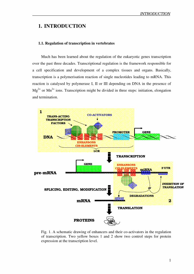
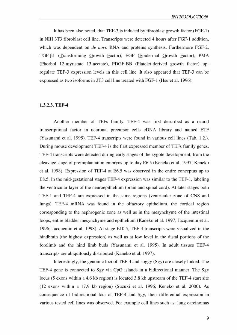
Fig. 1. A schematic drawing of enhancers and their co-activators in the regulation
of transcription. Two yellow boxes 1 and 2 show two control steps for protein
expression at the transcription level.
GENE
LCR
PROMOTER
ENHANSONS
CIS-ELEMENTS
TRANS-ACTING
TRANSCRIPTION
FACTORS
CO-ACTIVATORS
TRANSCRIPTION
GENE
pre-mRNA
DNA
ENHANSONS
CIS-ELEMENTS miRNA3’UTR
SPLICING, EDITING, MODIFICATION
mRNA
TRANSLATION
PROTEINS
1
2
INHIBITION OF
TRANSLATION
DEGRADATIONS
or
GENE
LCR
PROMOTER
ENHANSONS
CIS-ELEMENTS
TRANS-ACTING
TRANSCRIPTION
FACTORS
CO-ACTIVATORS
TRANSCRIPTION
GENE
pre-mRNA
DNA
ENHANSONS
CIS-ELEMENTS miRNA3’UTR
SPLICING, EDITING, MODIFICATION
mRNA
TRANSLATION
PROTEINS
1
2
INHIBITION OF
TRANSLATION
DEGRADATIONS
or
INTRODUCTION
2
From this point of view, transcription seems to be a simple enzymatic reaction.
However, the central of transcription requires many specific regulatory proteins, which
can interact with DNA via hydrogen and van der Waals bonds in the specific DNA
regions, so called promoters. At this point, it is assumed that transcription is regulated
by different bounding properties of proteins complexes to specific promoters. Such kind
of control of eukaryotic gene expression exists on Locus Control Regions (LCRs)
discovered first in the human β-globin locus. LCRs were defined as a cis-regulatory
elements and their ability to control tissue specific gene expression at physiological
levels (reviewed by Li et al. 2002). Extensive studies in various models of organisms
revealed a number of factors, which are responsible for the transcription control.
Heterogenity of those factors has suggested their specific role as a transcriptional gene
expression regulators including enhancers, silencers and has led to hypotheses about
detailed mechanism (reviewed by Lemon and Tjian, 2000; Ramji and Foka, 2002).
Recently a role of microRNA as trans-acting factors that exert their activity by
composition of cis-regulatory elements has been postulated (Hobert, 2005). It seems
that microRNA build a new discovered block (second checkpoint) controlling specific
gene expression (Fig. 1.).
Identification of different regulators keeping transcription machinery properly
working is a first step to understand how individual genes are turned on or off in cells
leading to their specification. It can also give an answer to the question how cells are
reprogrammed during differentiation, proliferation and how they can fulfil their specific
function in whole organisms. Another topic, which might be solved by a better
understanding of the transcriptional regulation, is lineage specification of pluripotent
stem cells, which can regenerate adult tissues (Heyworth et al. 2002; Weissman et al.
2001). Recently, transcriptional profiles of embryonic stem cells, neural stem cells and
hematopoietic stem cells of the bone morrow origin have been established (Ramalho-
Santos et al. 2002), indicating an enormous complexity of regulatory events in different
stem cell types.
Tight regulation of the transcription is the major process controlling gene
expression networks during embryogenesis in response to physiological and metabolic
changes to keep homeostasis. It also governs regenerations processes in adult tissues.
INTRODUCTION
3
1.2. Transcription regulation in skeletal and cardiac muscles
Transcriptional control in skeletal and cardiac muscles involves a cascade of
transcription factors, acting via different cis-regulatory elements. Three different major
groups of regulators have been described, namely bHLH, MADS and TEF families. All
members of those families can bind to the distinct regions of DNA, which are specific
and typical for each family. The properties of DNA binding result in the activation of
downstream targets genes.
It has been postulated, that skeletal muscles are controlled at the transcriptional
level by the Myogenic Regulatory Factors (MRFs) which belong to the bHLH family.
MRFs consist of four members: MyoD (Myod1), Myf-5, Myogenin and Myf-6 called
also MRF4 or herculin and all of them share a motif of a basic Helix-Loop-Helix
domain (bHLH). Myogenic bHLH members bind to the E-box site (CANNTG) within
DNA promoter regions to regulate transcription (reviewed by Emerson, 1990; Puri and
Sartorelli, 2000; Berkers and Tapscott, 2005). An E-box consensuses have been
identified in many muscles specific promoters like: Muscle Creatine Kinase (MCK)
(Buskin et al. 1989), cardiac α-actin (Sartorelli et al. 1992), cardiac Troponin T (cTNT)
(Iannello et al. 1991), cardiac Myosin Light Chain 2 (cMLC2) (Navankasattusas et al.
1992), β Myosin Heavy Chain (βMyHC) (Thompson et al. 1991; Kariya at al. 1994), α
Myosin Heavy Chain (αMyHC) (Gupta et al. 1994), cardiac Troponin C (cTNC)
(Parmeck at al. 1992), alpha-tropomyosin (Pasquet et al. 2006) and in skeletal α-actin
(MacLellan at al. 1994). It is assumed, that MyoD and Myf-5 play a role in the
specification of the muscle cell fate, whereas myogenin and MRF4 regulate the muscle
differentiation program (reviewed by Sabourin and Rudnicki, 2000; Buckingham,
2001). In addition two other subfamilies of bHLH have been identified: bHLH lucine
zipper, which consist Myc/Max/Mad transcriptional factors (Luscher, 2001) and the
bHLH-PAS subfamily (Crews, 1998). MRFs and bHLH leucine zipper subfamilies
recognise typical E-box whereas bHLH-PAS factors bind a DNA sequences which are
distinct from the prototypical E-box (Luscher, 2001).
MADS box transcription factors (MCM1-a yeast homolog, Agamous,
Deficiens,- plants homolog, Serum response factor) is the second major transcription
network which govern regulation of skeletal and cardiac muscles (Molketkin et al.
1995; Black and Olson 1998). It is also called myocyte enhancer factor-2 (MEF2). In
vertebrates, all known four members MEF2A, MEF2B, MEF2C and MEF2D posses a
INTRODUCTION
4
highly conserved 56 amino acids motif – so called MADS box. The MADS box is
responsible for the dimerisation of proteins containing this motif to create active
homodimmers as well as for specific binding to DNA A/T rich elements (Gossett et al.
1989). MEF2A, MEF2B and MEF2D are ubiquitously expressed in adult tissues, while
expression of MEF2C is enriched in the spleen and brain as well as in the skeletal and
cardiac muscles (Pollock et al. 1991; Yu et al. 1992; Martin et al. 1993; McDermott et
al. 1993; Breitbart et al. 1993). The expression pattern of MEF2 members has been also
described during mouse embryonic development. MEF2C was detected first at E7.5 in
the part of mesoderm, which forms the primitive heart tube. All others MEF2s start to
be expressed in the myocardium at E8.5. The earliest expression of MEF2C in the
embryonic skeletal muscle was detected at E9.0 in the rostral myotome while MEF2A
and MEF2D were found half a day later in the myotome (Edmondson et al. 1994).
Another member of MADS box proteins is Serum Response Factor (SRF), which
regulates transcription in cardiac, skeletal and smooth muscle cells by binding to the
CArG box found in several promoter regions (Norman et al. 1988; Miano, 2003). SRF
is the first known trans-acting factor, which regulates also muscle specific microRNA
by binding to cis-elements in the regulatory region of microRNA. Overexpression of
SRF can regulate specific microRNAs, which target the HAND2 transcription factor
during mouse development (Zhao et al. 2005).
1.3. Transcription Enhancer Factors (TEF) family
1.3.1. Evolution and structure of TEFs family
Transcription Enhancer Factor (TEF) is the last main family of transcription
regulators found in skeletal and cardiac muscles as well as in non muscles cells. All four
members of the TEF family (TEF-1, TEF-3, TEF-4, TEF-5) have very high homology
in so called TEA/ATTS DNA Binding Domain (DBD), (for nomenclature of TEFs see
Tab. 1.)
INTRODUCTION
5
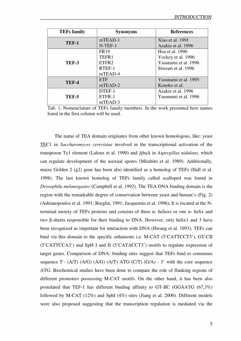
TEFs family Synonyms References
TEF-1 mTEAD-1
N-TEF-1
Xiao et al. 1991
Azakie et al. 1996
TEF-3
FR19
TEFR1
ETFR2
RTEF-1
mTEAD-4
Hsu et al. 1996
Yockey et al. 1996
Yasunami et al. 1996
Stweart et al. 1996
TEF-4 ETF
mTEAD-2
Yasunami et al. 1995
Keneko et al.
TEF-5
DTEF-1
ETFR-1
mTEAD-3
Azakie et al. 1996
Yasunami et al. 1996
Tab. 1. Nomenclature of TEFs family members. In the work presented here names
listed in the first column will be used.
The name of TEA domain originates from other known homologous, like: yeast
TEC1 in Saccharomyces cerevisiae involved in the transcriptional activation of the
transposon Ty1 element (Laloux et al. 1990) and AbaA in Aspergillus nidulans, which
can regulate development of the asexual spores (Mirabito et al. 1989). Additionally,
maize Golden 2 (g2) gene has been also identified as a homolog of TEFs (Hall et al.
1998). The last known homolog of TEFs family called scalloped was found in
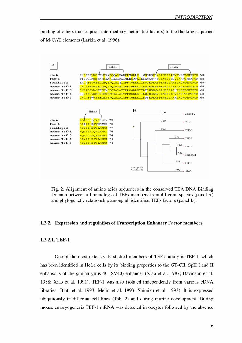
Drosophila melanogaster (Campbell et al. 1992). The TEA DNA binding domain is the
region with the remarkable degree of conservation between yeast and human’s (Fig. 2)
(Adrianopoulos et al. 1991; Burglin, 1991; Jacquemin et al. 1996). It is located at the N-
terminal moiety of TEFs proteins and consists of three α- helices or one α- helix and
two β-sheets responsible for their binding to DNA. However, only helix1 and 3 have
been recognized as important for interaction with DNA (Hwang et al. 1993). TEFs can
bind via this domain to the specific enhansons i.e. M-CAT (5’CATTCCT3’), GT-CII
(5’CATTCCA3`) and SpH I and II (5’CATACCT3’) motifs to regulate expression of
target genes. Comparison of DNA- binding sites suggest that TEFs bind to consensus
sequence 5`- (A/T) (A/G) (A/G) (A/T) ATG (C/T) (G/A) - 3` with the core sequence
ATG. Biochemical studies have been done to compare the role of flanking regions of
different promoters possessing M-CAT motifs. On the other hand, it has been also
postulated that TEF-1 has different binding affinity to GT-IIC (GGAATG (67,3%)
followed by M-CAT (12%) and SphI (4%) sites (Jiang et al. 2000). Different models
were also proposed suggesting that the transcription regulation is mediated via the
INTRODUCTION
6
binding of others transcription intermediary factors (co-factors) to the flanking sequence
of M-CAT elements (Larkin et al. 1996).
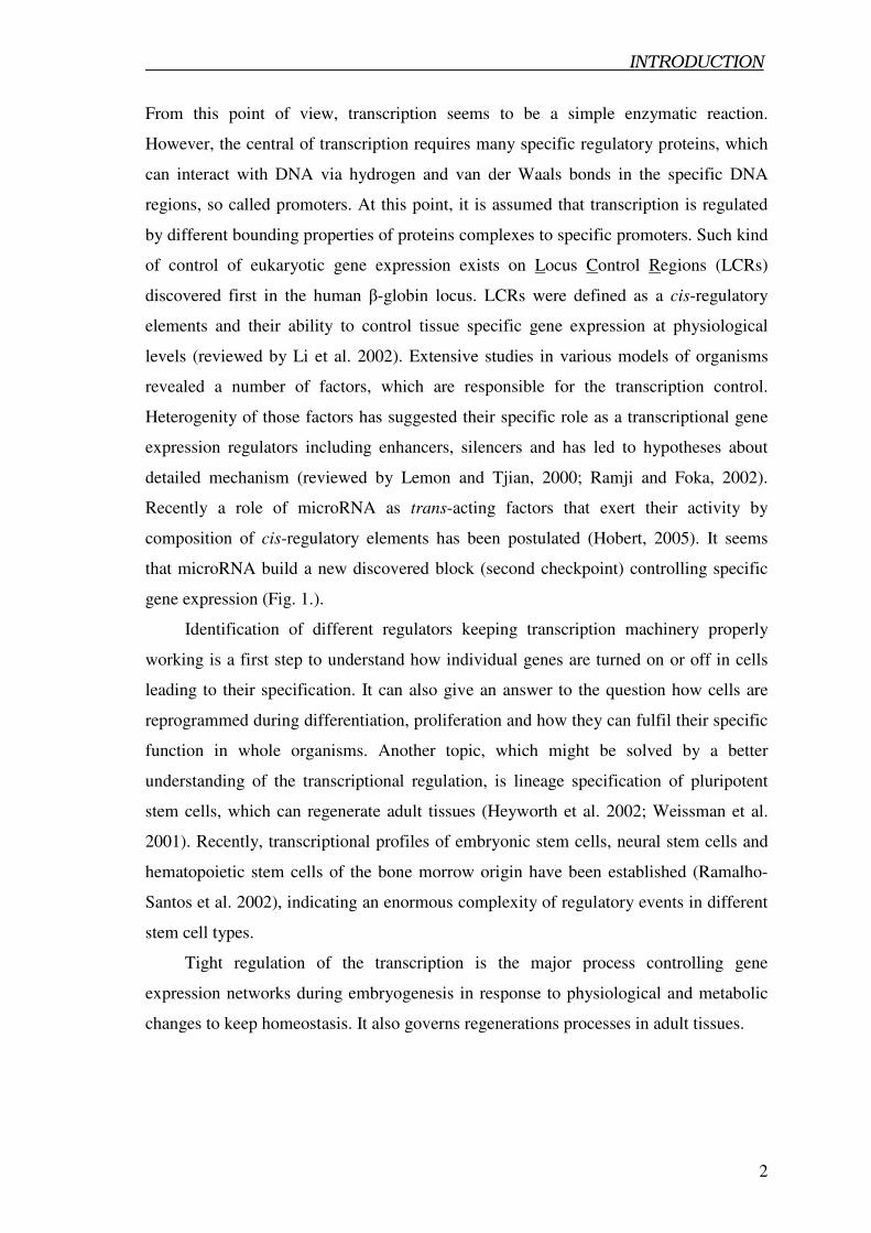
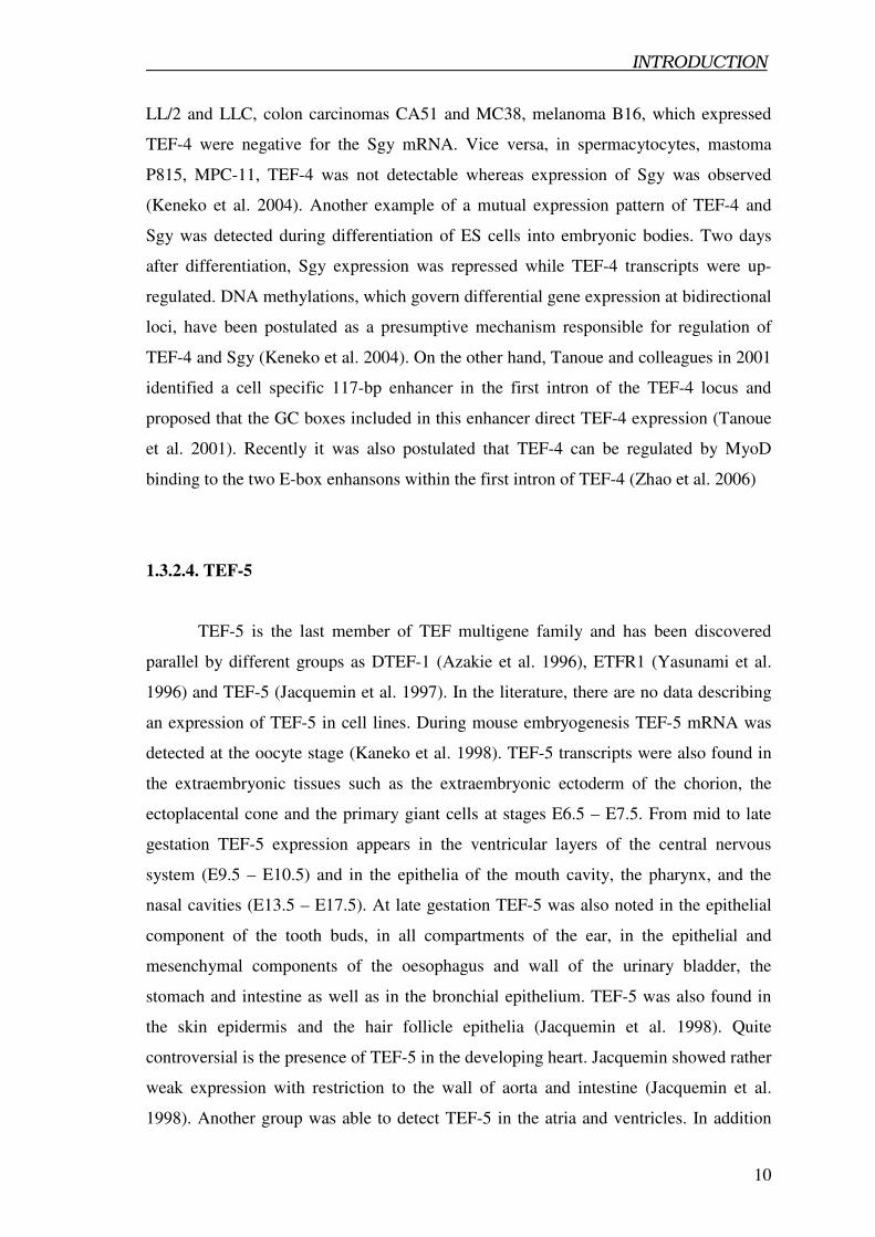
Fig. 2. Alignment of amino acids sequences in the conserved TEA DNA Binding
Domain between all homologs of TEFs members from different species (panel A)
and phylogenetic relationship among all identified TEFs factors (panel B).
1.3.2. Expression and regulation of Transcription Enhancer Factor members
1.3.2.1. TEF-1
One of the most extensively studied members of TEFs family is TEF-1, which
has been identified in HeLa cells by its binding properties to the GT-CII, SpH I and II
enhansons of the simian virus 40 (SV40) enhancer (Xiao et al. 1987; Davidson et al.
1988; Xiao et al. 1991). TEF-1 was also isolated independently from various cDNA
libraries (Blatt et al. 1993; Melin et al. 1993; Shimizu et al. 1993). It is expressed
ubiquitously in different cell lines (Tab. 2) and during murine development. During
mouse embryogenesis TEF-1 mRNA was detected in oocytes followed by the absence
INTRODUCTION
7
of its transcripts up to E8.0 (Kaneko et al. 1998). On the other hand, continues β-
galactosidase activity in the TEF-1+/-
embryo was observed from the oocytes stage up to
later stages of mouse development (Chen et al. 1994). Another study, based on whole
mount in situ hybridization revealed TEF-1 transcripts in decidual cells and in the
distinct extra-embryonic regions e.g. in the ectoplacental cone at E6.5 (pregastrula or
egg cylinder stage) (Jacquemin et al. 1996; Jacquemin et al. 1998). From E8.5, TEF-1 is
detected in the entire embryo and between E9.5 and E10.5 it shows restricted expression
in specific structures such as: the ventricular layer of the neuroepithelium, the spinal
cord in the developing brain and the myocardium at E10.5. At later stages (E13.5 -
E18.5) TEF-1 mRNA was found in various facial and axial muscles, in the
differentiating myocardium, in the forebrain ventricles as well as in the olfactory and
respiratory regions of nasal epithelium. Additionally, TEF-1 was uniformly detected in
the metanephros of the developing kidney and adrenal gland. Pronounced expression
was observed in the internal layer of the urinary bladder epithelium and in the external
layer of the mesenchyme. Furthermore, TEF-1 is localized in the labyrinthine of the
chorioallantoic placenta (Jacquemin et al. 1996; Jacquemin et al. 1998). However, there
is no clear evidence that TEF-1 is expressed in somites during embryogenesis. In adult
tissues TEF-1 is detectable at high level in the: kidney, lungs, skeletal muscles and heart
and at low level in the brain and liver (Shimizu et al. 1993). Another report showed a
ubiquitous TEF-1 distribution by β-galactosidase staining of TEF-1+/-
heterozygous
mice carrying LacZ cassette (Chen et al. 1994). As TEF-1 was identified in the
developing myocardium during mouse embryogenesis and its transcripts are enriched in
adult heart, TEF-1 was used in many reports as a specific marker of adult
cardiomyocytes. In contrast, TEF-1 was also found in a subpopulation of the Sca-1+ and
Sca-1- non-cardiomyocytes isolated from the adult heart. In this respect TEF-1 can not
be described as a specific marker of cardiomyocytes (Oh et al. 2003).
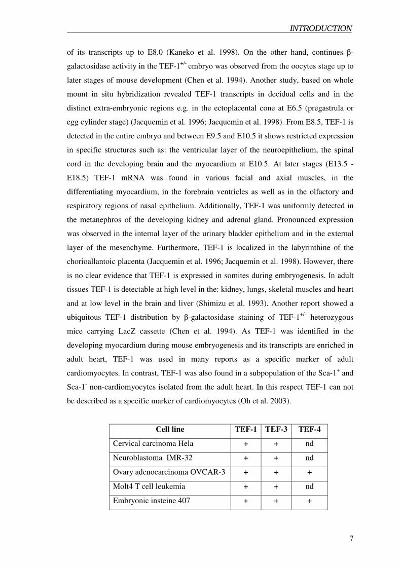
Cell line TEF-1 TEF-3 TEF-4
Cervical carcinoma Hela + + nd
Neuroblastoma IMR-32 + + nd
Ovary adenocarcinoma OVCAR-3 + + +
Molt4 T cell leukemia + + nd
Embryonic insteine 407 + + +
INTRODUCTION
8
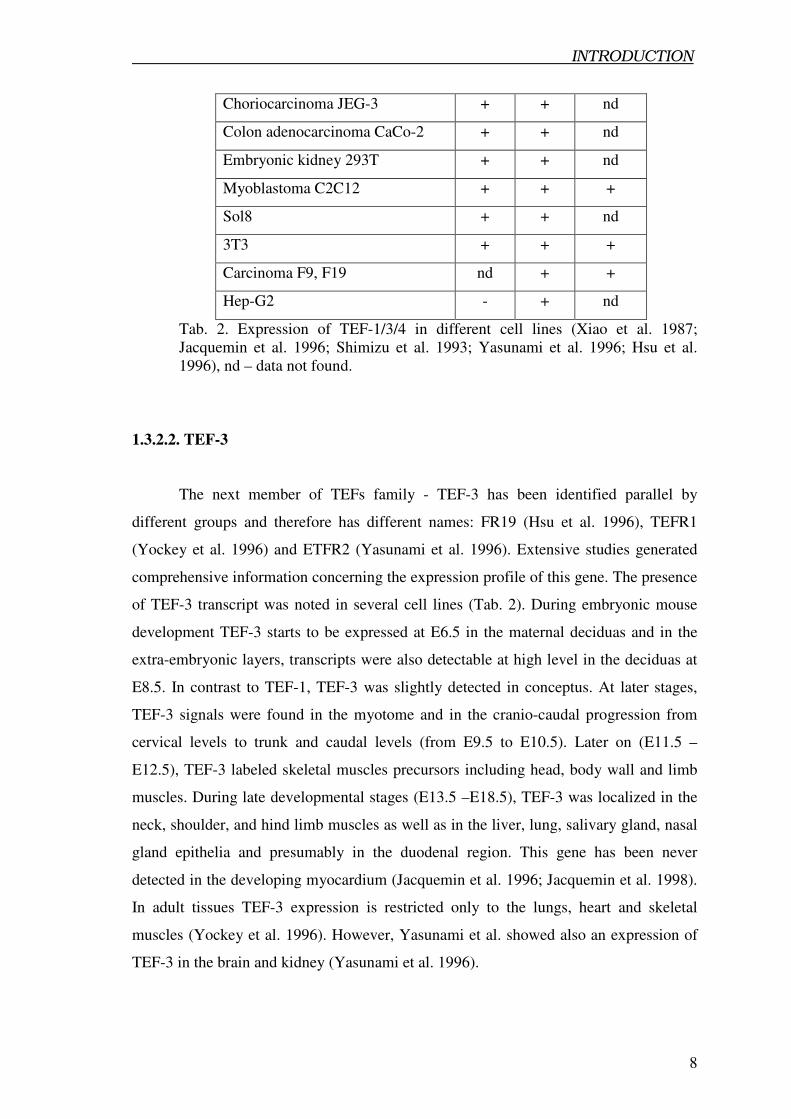
Choriocarcinoma JEG-3 + + nd
Colon adenocarcinoma CaCo-2 + + nd
Embryonic kidney 293T + + nd
Myoblastoma C2C12 + + +
Sol8 + + nd
3T3 + + +
Carcinoma F9, F19 nd + +
Hep-G2 - + nd
Tab. 2. Expression of TEF-1/3/4 in different cell lines (Xiao et al. 1987;
Jacquemin et al. 1996; Shimizu et al. 1993; Yasunami et al. 1996; Hsu et al.
1996), nd – data not found.
1.3.2.2. TEF-3
The next member of TEFs family - TEF-3 has been identified parallel by
different groups and therefore has different names: FR19 (Hsu et al. 1996), TEFR1
(Yockey et al. 1996) and ETFR2 (Yasunami et al. 1996). Extensive studies generated
comprehensive information concerning the expression profile of this gene. The presence
of TEF-3 transcript was noted in several cell lines (Tab. 2). During embryonic mouse
development TEF-3 starts to be expressed at E6.5 in the maternal deciduas and in the
extra-embryonic layers, transcripts were also detectable at high level in the deciduas at
E8.5. In contrast to TEF-1, TEF-3 was slightly detected in conceptus. At later stages,
TEF-3 signals were found in the myotome and in the cranio-caudal progression from
cervical levels to trunk and caudal levels (from E9.5 to E10.5). Later on (E11.5 –
E12.5), TEF-3 labeled skeletal muscles precursors including head, body wall and limb
muscles. During late developmental stages (E13.5 –E18.5), TEF-3 was localized in the
neck, shoulder, and hind limb muscles as well as in the liver, lung, salivary gland, nasal
gland epithelia and presumably in the duodenal region. This gene has been never
detected in the developing myocardium (Jacquemin et al. 1996; Jacquemin et al. 1998).
In adult tissues TEF-3 expression is restricted only to the lungs, heart and skeletal
muscles (Yockey et al. 1996). However, Yasunami et al. showed also an expression of
TEF-3 in the brain and kidney (Yasunami et al. 1996).
INTRODUCTION
9
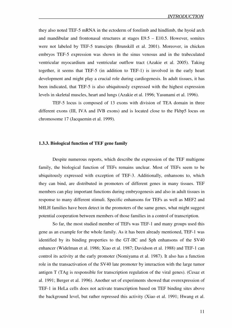
It has been also noted, that TEF-3 is induced by fibroblast growth factor (FGF-1)
in NIH 3T3 fibroblast cell line. Transcripts were detected 4 hours after FGF-1 addition,
which was dependent on de novo RNA and proteins synthesis. Furthermore FGF-2,
TGF-β1 (Transforming Growth Factor), EGF (Epidermal Growth Factor), PMA
(Phorbol 12-myristate 13-acetate), PDGF-BB (Platelet-derived growth factor) up-
regulate TEF-3 expression levels in this cell line. It also appeared that TEF-3 can be
expressed as two isoforms in 3T3 cell line treated with FGF-1 (Hsu et al. 1996).
1.3.2.3. TEF-4
Another member of TEFs family, TEF-4 was first described as a neural
transcriptional factor in neuronal precursor cells cDNA library and named ETF
(Yasunami et al. 1995). TEF-4 transcripts were found in various cell lines (Tab. 1.2.).
During mouse development TEF-4 is the first expressed member of TEFs family genes.
TEF-4 transcripts were detected during early stages of the zygote development, from the
cleavage stage of preimplantation embryos up to day E6.5 (Keneko et al. 1997; Keneko
et al. 1998). Expression of TEF-4 at E6.5 was observed in the entire conceptus up to
E8.5. In the mid-gestational stages TEF-4 expression was similar to the TEF-1, labeling
the ventricular layer of the neuroepithelium (brain and spinal cord). At later stages both
TEF-1 and TEF-4 are expressed in the same regions (ventricular zone of CNS and
lungs). TEF-4 mRNA was found in the olfactory epithelium, the cortical region
corresponding to the nephrogenic zone as well as in the mesynchyme of the intestinal
loops, entire bladder mesynchyme and epithelium (Kaneko et al. 1997; Jacquemin et al.
1996; Jacquemin et al. 1998). At stage E10.5, TEF-4 transcripts were visualized in the
hindbrain (the highest expression) as well as at low level in the distal portions of the
forelimb and the hind limb buds (Yasunami et al. 1995). In adult tissues TEF-4
transcripts are ubiquitously distributed (Kaneko et al. 1997).
Interestingly, the genomic loci of TEF-4 and soggy (Sgy) are closely linked. The
TEF-4 gene is connected to Sgy via CpG islands in a bidirectional manner. The Sgy
locus (5 exons within a 4,6 kb region) is located 3.8 kb upstream of the TEF-4 start site
(12 exons within a 17,9 kb region) (Suzuki et al. 1996; Keneko et al. 2000). As
consequence of bidirectional loci of TEF-4 and Sgy, their differential expression in
various tested cell lines was observed. For example cell lines such as: lung carcinomas
INTRODUCTION
10
LL/2 and LLC, colon carcinomas CA51 and MC38, melanoma B16, which expressed
TEF-4 were negative for the Sgy mRNA. Vice versa, in spermacytocytes, mastoma
P815, MPC-11, TEF-4 was not detectable whereas expression of Sgy was observed
(Keneko et al. 2004). Another example of a mutual expression pattern of TEF-4 and
Sgy was detected during differentiation of ES cells into embryonic bodies. Two days
after differentiation, Sgy expression was repressed while TEF-4 transcripts were up-
regulated. DNA methylations, which govern differential gene expression at bidirectional
loci, have been postulated as a presumptive mechanism responsible for regulation of
TEF-4 and Sgy (Keneko et al. 2004). On the other hand, Tanoue and colleagues in 2001
identified a cell specific 117-bp enhancer in the first intron of the TEF-4 locus and
proposed that the GC boxes included in this enhancer direct TEF-4 expression (Tanoue
et al. 2001). Recently it was also postulated that TEF-4 can be regulated by MyoD
binding to the two E-box enhansons within the first intron of TEF-4 (Zhao et al. 2006)
1.3.2.4. TEF-5
TEF-5 is the last member of TEF multigene family and has been discovered
parallel by different groups as DTEF-1 (Azakie et al. 1996), ETFR1 (Yasunami et al.
1996) and TEF-5 (Jacquemin et al. 1997). In the literature, there are no data describing
an expression of TEF-5 in cell lines. During mouse embryogenesis TEF-5 mRNA was
detected at the oocyte stage (Kaneko et al. 1998). TEF-5 transcripts were also found in
the extraembryonic tissues such as the extraembryonic ectoderm of the chorion, the
ectoplacental cone and the primary giant cells at stages E6.5 – E7.5. From mid to late
gestation TEF-5 expression appears in the ventricular layers of the central nervous
system (E9.5 – E10.5) and in the epithelia of the mouth cavity, the pharynx, and the
nasal cavities (E13.5 – E17.5). At late gestation TEF-5 was also noted in the epithelial
component of the tooth buds, in all compartments of the ear, in the epithelial and
mesenchymal components of the oesophagus and wall of the urinary bladder, the
stomach and intestine as well as in the bronchial epithelium. TEF-5 was also found in
the skin epidermis and the hair follicle epithelia (Jacquemin et al. 1998). Quite
controversial is the presence of TEF-5 in the developing heart. Jacquemin showed rather
weak expression with restriction to the wall of aorta and intestine (Jacquemin et al.
1998). Another group was able to detect TEF-5 in the atria and ventricles. In addition
INTRODUCTION
11
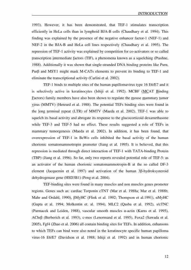
they also noted TEF-5 mRNA in the ectoderm of forelimb and hindlimb, the hyoid arch
and mandibular and frontonasal structures at stages E9.5 – E10.5. However, somites
were not labeled by TEF-5 transcipts (Brunskill et al. 2001). Moreover, in chicken
embryos TEF-5 expression was shown in the sinus venosus and in the trabeculated
ventricular myocardium and ventricular outflow tract (Azakie et al. 2005). Taking
together, it seems that TEF-5 (in addition to TEF-1) is involved in the early heart
development and might play a crucial role during cardiogenesis. In adult tissues, it has
been indicated, that TEF-5 is also ubiquitously expressed with the highest expression
levels in skeletal muscles, heart and lungs (Azakie et al. 1996; Yasunami et al. 1996).
TEF-5 locus is composed of 13 exons with division of TEA domain in three
different exons (III, IVA and IVB exons) and is located close to the Fkbp5 locus on
chromosome 17 (Jacquemin et al. 1999).
1.3.3. Biological function of TEF gene family
Despite numerous reports, which describe the expression of the TEF multigene
family, the biological function of TEFs remains unclear. Most of TEFs seem to be
ubiquitously expressed with exception of TEF-3. Additionally, enhansons to, which
they can bind, are distributed in promoters of different genes in many tissues. TEF
members can play important functions during embryogenesis and also in adult tissues in
response to many different stimuli. Specific enhansons for TEFs as well as MEF2 and
bHLH families have been detect in the promoters of the same genes, what might suggest
potential cooperation between members of those families in a control of transcription.
So far, the most studied member of TEFs was TEF-1 and many groups used this
gene as an example for the whole family. As it has been already mentioned, TEF-1 was
identified by its binding properties to the GT-IIC and Sph enhansons of the SV40
enhancer (Widelman et al. 1986; Xiao et al. 1987; Davidson et al. 1988) and TEF-1 can
control its activity at the early promoter (Nomiyama et al. 1987). It also has a function
role in the transactivation of the SV40 late promoter by interaction with the large tumor
antigen T (TAg is responsible for transcription regulation of the viral genes). (Cesaz et
al. 1991; Berger et al. 1996). Another set of experiments showed that overexpression of
TEF-1 in HeLa cells does not activate transcription based on TEF binding sites above
the background level, but rather repressed this activity (Xiao et al. 1991; Hwang et al.
INTRODUCTION
12
1993). However, it has been demonstrated, that TEF-1 stimulates transcription
efficiently in HeLa cells than in lymphoid BJA-B cells (Chaudhary et al. 1994). This
finding was explained by the presence of the negative enhancer factor-1 (NEF-1) and
NEF-2 in the BJA-B and HeLa cell lines respectively (Chaudhary et al. 1995). The
repression of TEF-1 activity was explained by competition for co-activators or so called
transcription intermediate factors (TIF), a phenomena known as a squelching (Ptashne,
1988). Additionally it was shown that single-stranded DNA binding proteins like Purα,
Purβ and MSY1 might mask M-CATs elements to prevent its binding to TEF-1 and
eliminate the transcriptional activity (Carlini et al. 2002).
TEF-1 binds to multiple sites of the human papillomavirus type 16 E6/E7 and it
is selectively active in keratinocytes (Ishiji et al. 1992). MCBF (MCAT Binding
Factors) family members have also been shown to regulate the mouse mammary tumor
virus (MMTV) (Henrard et al. 1988). The potential TEFs binding sites were found in
the long terminal repeat (LTR) of MMTV (Maeda et al. 2002). TEF-1 was able to
squelch its basal activity and abrogate its response to the glucocorticoid dexamethasone
while TEF-3 and TEF-5 had no effect. These results suggested a role of TEFs in
mammary tumorgenesis (Maeda et al. 2002). In addition, it has been found, that
overexpression of TEF-1 in BeWo cells inhibited the basal activity of the human
chorionic somatomammotropin promoter (Jiang et al. 1995). It is believed, that this
repression is mediated through direct interaction of TEF-1 with TATA-binding Protein
(TBP) (Jiang et al. 1996). So far, only two reports revealed potential role of TEF-5: as
an activator of the human chorionic somatomammotropin-B at the so called DF-3
element (Jacquemin et al. 1997) and activation of the human 3β-hydroksysteroid
dehydrogenase gene (HSD3B1) (Peng et al. 2004).
TEF-binding sites were found in many muscles and non muscles genes promoter
regions. Genes such as: cardiac Torponin cTNT (Mar et al. 1988a; Mar et al. 1988b;
Mahr and Ordahl, 1990), βMyHC (Flink et al. 1992; Thompson et al.1991), αMyHC
(Gupta et al. 1994; Molkentin et. al. 1994), MLC2 (Qasba et al. 1992), s/cTNC
(Parmacek and Leiden, 1988), vascular smooth muscles α-actin (Karns et al. 1995),
AChrβ (Berberich et al. 1993), c-mos (Lenormand et al. 1995), Foxa2 (Sawada et al.
2005), Fgf4 (Zhao et al. 2006) all contain binding sites for TEFs. In addition, enhansons
to which TEFs can bind were also noted in the keratinocyte specific human papilloma
virus-16 E6/E7 (Davidson et al. 1988; Ishiji et al. 1992) and in human chorionic
INTRODUCTION
13
somatomammotropin gene also called placental lactogen–B gene (Jacquemin, et al.
1994a; Jacquemin, et al. 1994b).
Since cis-acting elements that bind TEFs were found in many different
promoters, various studies tried to establish a link between TEFs, MEF2 and bHLH
expression and postulated a cooperative binding of these proteins. Attempts were made
to identify potential interaction partners, jointly used by these proteins. Finally, several
groups screened for co-activators involved in gene specific regulation especially in
skeletal and cardiac muscles.
1.3.3.1. Role of TEFs in skeletal muscles
Since β-MyHC is expressed in adult slow skeletal muscles and in the ventricular
myocardium (Lompre et al. 1984), a large numbers of studies tried to identify elements,
which drive its differential expression. Another MyHC isoform, α-MyHC is expressed
in the atria and weakly in ventricular fibers (Bouvagnet et al. 1984; Gorza et al. 1984).
A detailed molecular analysis of the β-MyHC proximal promoter located several cis-
acting elements - distal and proximal M-CATs, A/T rich region and E-box site (Knotts
et al. 1994; Thomson et al. 1991). The ability of TEF-1 as well as other members of
TEFs family to bind these elements was proved (Flink et al. 1992; Shimizu et al. 1993;
Farrance et al. 1996). It has also been shown that β-MyHC can be induced in fast type
fibers by mechanical overload (MOV). Distal M-CAT elements contributed to the basal
expression of β-MyHC in the slow fiber by binding of a multiproteins complex
consisting of TEF-1, poly(ADP-ribose) polymerase (PARP) and Max bHLH proteins.
Furthermore, it was noted, that only TEF-1 can bind to the distal M-CAT element and
can physically interact with PARP and Max to form a functional complex. However,
such kind of regulation is not absolutely required for MOV responsiveness (Wiedenman
et al. 1996; Vyas et al. 1999; Vyas et al. 2001). The β-MyHC promoter region contains
also an additional enhanson A/T rich element that is necessary for its expression in slow
type muscles, which enhances MOV responsiveness. Surprisingly, it has been
described, that this element (which otherwise bind exclusively GATA and MEF2
transcription factors) binds also TEF-1 under the basal as well hypertophic conditions.
That means that TEFs family might contribute to the regulation of transcription in
cardiac and skeletal muscles by binding to cis-regulatory elements that differ from the
MCAT motif (Tsika et al. 2002; Karasseva et al. 2003). This finding also contradict data
INTRODUCTION
14
that TEF-1 and MEF2 physically interact to regulate specific gene expression (Maeda et
al. 2002). Another study published by Giger showed, that TEF-1 and Myogenin proteins
were significantly attenuated in the unloaded Soleus muscle in comparison to control,
which correspond to the transition of β-MyHC gene (Giger et al. 2004).
1.3.3.2. Regulation of transcription in cardiac muscles by TEFs
Since TEF-binding cis-elements are present in the enhansons of various
structural genes, they might also control transcription regulation in cardiac muscle cells.
Several groups described TEF-dependent gene regulation of cardiac myocytes, and it
seems that the cardiac transcription machinery is based on the direct interaction or
competition between MEF2, TEFs families and their co-activators. One of many
examples is the identification of cis-elements in the promoter region of αMyHC. Many
regulatory elements have been found within the promoter sequence such as: an element
specific for the erg-1 cellular oncogene, a thyroid regulatory element (TRE) by which
αMyHC can be regulated (Markham et al. 1990; Tsika et al. 1990) and an E-Box-M-
CAT (EM) hybrid motif (Gupta et al. 1991). The EM regulatory element is responsible
for the basal as well as cyclic AMP-inducible expression of the αMyHC gene. It has
been reported that TEF-1 and Max protein, which belongs to the basic helix-loop-helix
leucine zipper (bHLH LZ) subfamily, can trans-activate the αMyHC gene through their
binding to the EM motif. This effect seems to occur due to the synergistic cooperation
between both factors (Molkentin et al. 1994; Gupta et al. 1997). Moreover, TEF-1 can
interact with SRF (MADS box family) to activate the skeletal α-actin promoter in COS-
1 cells. The strong interaction between both proteins is mediated through the C-terminal
subdomain of the MADS box of SRF (204-224 amino acids) and the second and the
third α-helix of TEAD DBD domain of TEF-1. However, no data are available whether
other members of TEFs family can interact with SRF by the same structural elements
(Gupta et al. 2001). It was postulated, that the skeletal α-actin promoter can be regulated
by TEFs family members and SRF in response to stimuli like pressure overload, α1-
adrenergic agonists, FGF and TGF-β or interleukin-1β (MacLellan et al. 1994; Karns et
al. 1995; Patten et al. 1996), underscoring a potential role of TEFs in the regulation of
tissue homeostasis.
INTRODUCTION
15
M-CAT elements together with the TATA box, GC box in the proximal
promoter region as well as with A/T-rich MEF2 and GATA elements do also control the
cardiac troponin T activity (cTNT) (Mar et al. 1988; Mar et al. 1990; Iannello et al.
1991). The cTNT gene is expressed in cardiac and skeletal muscles during development
but at late embryonic stages is repressed in developing skeletal muscles with strong up-
regulation in cardiac tissue (Long et al. 1988). It has been noted that in cultured
myocytes, TEF-3 is not able to activate the cTNT promoter (Stewart et al. 1994). In
contrast to this observation TEF-5 can trans-activate cTNT promoter in a tissue specific
fashion, independently on A/T-rich, MEF2 or GATA elements. In contrast TEF-5 did
not activate the cTNT gene in transiently transfected embryonic skeletal muscles and
fibroblasts (Azakie et al. 2005).
Another gene that can be possibly regulated by TEFs is the muscle creatine
kinase (MCK). MCK is expressed in skeletal and cardiac muscles. Its promoter has
different enhansons and following elements were identified CArg, AP2, A/T-rich, right
and left E-boxes, MEF-2 sites as well as transcriptional regulatory element x (TREX) -
similar to the M-CAT (7/8 bases) (Buskin et al. 1989; Amacher et al. 1993; Fabre-Suver
et al. 1996). Due to MCK expression in both skeletal and cardiac muscles, the obvious
question was which elements were responsible for the heart and skeletal muscle
expression. Using sites specific mutation analysis it was shown that the right E-box and
TREX are necessary elements to regulate MCK expression in skeletal muscles, while
CArG and A/T-rich elements were responsible for the expression in the heart. Other
elements such as the AP2, MEF-2 sites and the left E-box are necessary in both skeletal
and cardiac muscles. Moreover, it was shown that the TrexBF factor, which binds to the
TREX element, is specific for skeletal muscle but distinct from TEF-1 (Amacher et al.
1993; Fabre-Suver et al. 1996).
In the β-myosin heavy chain promoter four M-CATs (additional two in
comparison to skeletal muscle) and GATA as well as NFAT binding elements were
identified (Simpson et al. 1991; McLean et al. 2003). Site direct mutagenesis showed
that one M-CAT is responsible for the basal activity of the promoter. Mutation of a
second M-CAT motif revealed decreasing activity of this promoter but only in response
to the α1-adrenergic agonist while mutation in both M-CATs generally decreased basal
activity of the promoter in response to pressure overload and leukaemia inhibitory
factor (Morimoto et al. 1999; Hasegawa et al. 1997). Moreover, a GATA binding
element within the βMyHC promoter seems to play a role in the transcriptional
INTRODUCTION
16
activation of this gene in response to the aortic constriction (Hasegawa et al. 1997). An
additional region between -71/+34 containing no consensus element and can also
mediate a pressure overload response (Wright et al. 2001). It was postulated that all four
M-CAT elements (by binding TEF factors) maintain the basal activity of βMyHC in
slow type muscles and the heart. NFAT sites within the βMyHC promoter can bind
NFAT factors in vitro but are not required for its basal activity in vivo (McLean et al.
2003).
Another studies showed that the α1A/C –adrenergic receptor promoter has
multiple M-CATs with binding properties for TEF-1, but only one is required for its
activity in cardiomyocytes and in response to β-adrenergic agonists’ (O’Connell et al.
2001). Another study presented that one from two M-CATs is responsible for activity of
vascular smooth muscle α-actin promoter (Swartz et al. 1998). The cardiac ankkyrin
repeat protein is another example of a gene under control of TEF transcription factors,
since MCATs were found within its promoter (Aihara et al. 2000). Biochemical data
supported the notion that MCATs binding sites (TEFs) are controlled by p38 and Rac1
–components of the stress activated MAPK pathway (Aihara et al. 2000; Ambrosino et
al. 2006). Additional, data revealed that both TEF-3 (activates skeletal muscle α-actin
and βMyHC) and TEF-5 (activates skeletal muscle α-actin) are targets for the α1-
adrenergic signalling pathway (Stewart et al. 1998; Ueyama et al. 2000; Maeda et al.
2002). TEF-3 has been also recognized as a positive regulator of the VEGF promoter
region (Vascular Endothelial Growth Factor) (Shie et al. 2004).
Despite of numerous studies, which tried to uncover functions of TEFs in vitro
in cell culture, only a little attempt was made to address the function of TEFs in vivo.
TEF-1 was disrupted by a retroviral gene trap insertion. TEF-1 mutant embryos die
between E10.5 and E11.5. The earliest phenotype appeared at E10.5, indicated by a pale
yolk sac and a dilated fourth ventricle in the brain. Later on, TEF-1 deficient embryos
showed a dilated heart, a slow heart beating rate as well as tissue edema. The thin
ventricular wall was accompanied by a reduced trabeculation. Hence, TEF-1 seems to
play an important role in the maturation of the embryonic heart but not during initiation
of the heart development. Surprisingly, no changes were reported in other tissues or
organs. According to the biochemical data, which suggest a stimulatory role of TEF-1 in
the expression of several sarcomeric genes, no significant changes were noted in cTNT,
cTNI and myosin expression (Chen et al. 1994). Similarly to TEF-1 knock out mice,
SRF null mice also did not show any changes in the expression of βMyHC, cardiac
INTRODUCTION
17
actin and slight changes in desmin, αMyHC and ANF expression was observed.
Surprisingly, mutant embryos lacking SRF showed a lower expression of transcription
factors like TEF-1, Nkx2.5, GATA4 as well as myocardin – the co-activator of SRF
(Parlakian et al. 2004). In contrast, another group showed that mice deficient in SRF are
characterized by a dramatic reduction in the expression of ANF, skeletal, cardiac and
smooth muscle α-actins as well as SM22α transcripts (Niu et al. 2005). In addition,
transgenic mice overexpressing TEF-3 develop progressive atria arrhythmias with
slower conduction velocities across the atria and the ventricular myocardium. The
conduction defect was concluded due to the dephosphorylation of connexins 40 and 43
with parallel up-regulation of the protein phosphatase 1β (Chen et al. 2004).
1.3.4. Co-activators of TEFs
Despite of many published data, the question remained open, how TEFs
specifically regulate transcription. So far, the main explanation of the mode of action
was based on the potential role of co-activators, which might modulate TEFs function,
acting as their positive or negative regulators. Such co-factors might bind directly to
TEFs family or to other proteins that might interact with TEFs (Majumder et al. 1997;
Roeder, 2005; McKenna et al. 2002).
1.3.4.1. YAP65 and TAZ as co-activators of TEFs
A potential and rather unexpected co-activator of TEFs is YAP65 (Yes
Associated Protein 65 kDa). YAP65 has been described previously as an interaction
partner of the Src/Yes protein kinase family (Sudol et al. 1994) and of the PDZ domain
protein EBP50 (Mohler et al. 1999). YAP65 has been identified as a potent trans-
activator comparable to herpes simplex virus VP16 (Yagi et al. 1999). It was observed,
that this co-activator can interact specifically with the carboxyl region of all members of
TEFs family by a newly identified TEAD protein binding domain in YAP65 (aa 32-
139) (Vassilev et al. 2001). The TEAD protein binding domain of YAP65 protein is
localized at the N-terminal end before 14-3-3 binding domain. The protein does also
contain two WW domains (called also RSp5) followed by proline rich motif (SH3-
INTRODUCTION
18
binding motif), and activation domain (Yagi et al. 1999; Kanai et al. 2000; Vassilev et
al. 2001). Interestingly, an overexpression of YAP65 strongly enhanced activity of
TEFs family in the lyphocytic MPC11 cell line, in which TEFs normally are not active.
Moreover, TEFs overexpression squelched YAP activity. YAP65 appeared to be
accumulated in the cytoplasm as a complex with cytoplasmatic localised protein called
14-3-3 whereas all TEFs members are located in the nucleus. Hence, it was postulated
that YAP65 might control TEFs specific activation of target genes in response to the
unknown stimuli (Vassilev et al. 2001). YAP65 seems to be a specific co-activator of
TEF-4. Both bind to enhansons in the Pax-3 promoter region, so called NCE2
resembling GT-IIC element and this complex is able to activate Pax-3 expression in
neural crest cells (Milewski et al. 2003).
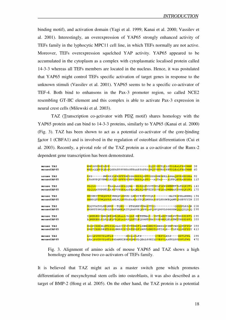
TAZ (Transcription co-activator with PDZ motif) shares homology with the
YAP65 protein and can bind to 14-3-3 proteins, similarly to YAP65 (Kanai et al. 2000)
(Fig. 3). TAZ has been shown to act as a potential co-activator of the core-binding
factor 1 (CBFA1) and is involved in the regulation of osteoblast differentiation (Cui et
al. 2003). Recently, a pivotal role of the TAZ protein as a co-activator of the Runx-2
dependent gene transcription has been demonstrated.
Fig. 3. Alignment of amino acids of mouse YAP65 and TAZ shows a high
homology among those two co-activators of TEFs family.
It is believed that TAZ might act as a master switch gene which promotes
differentiation of mesynchymal stem cells into osteoblasts, it was also described as a
target of BMP-2 (Hong et al. 2005). On the other hand, the TAZ protein is a potential
INTRODUCTION
19
co-activator of TEF-1 and/or TEF-3. In contrast, TEF-4 and TEF-5 are only weakly
activated by TAZ. Authors postulated that TAZ is a new co-activator of TEFs. TAZ can
interact via its N-terminal domain. Interaction with TAZ occurs by C-terminal domain
of TEFs (Mahoney et al. 2005).
1.3.4.2. p160 family as co-activators of TEFs
p160 is a member of the bHLH-PAS gene family, because of their highly
conserved N-terminal domain (basic helix-loop-helix/Per-Arnt-Sim) (reviewed by
Mckenna et al. 2005). This conserved region is typical for three members: SRC1, TIF2
and RAC3. The PAS subdomain is responsible for dimerisation to form active
homodimmers (Kewley et al. 2004). The strongest interaction was demonstrated
between SRC-1 and TEF-4, however interactions with others TEFs members are also
possible. SRC1 binds to TEFs via the bHLH-PAS domain and it was shown, that this
domain is necessary to enhance the transcriptional activation from TEF response
elements in transiently transfected cells. Moreover TIF2 and RAC3 are also potent co-
activators of TEFs. Furthermore, GRIP-1 a mouse homolog of TIF2 is an interaction
partner of MEF-2C as well as myogenin and binds them via the bHLH-PAS domain
(Chen et al. 2000; Belandia et al. 2000). In contrast, GRIP-1 acts as a co-repressor of
MyoD whereas SRC-1 and RAC3 are potent co-activators (Wu et al. 2005).
1.3.4.3. TONDU as co-activator of TEFs
TONDU is a human homolog of vestigial gene, which was identified in
Drosophila (Vaudin et al. 1999). Vestigial interacts with scalloped (a homolog of TEFs
family in fruits flay) and both proteins control target gene expression and promote the
wing formation. In Drosophila wing cells, vestigial interacts with scalloped via the
scalloped interaction domain (SID) (Simmonides et al. 1998, Paumard-Rigal et al. 1998;
Halder et al. 1998). It was postulated that binding of vestigial can modulate the
conformation of scalloped. Moreover, scalloped alone shows a different affinity to
enhansons than in a heterodimeric complex with vestigial (Vaudin et al. 1999; Halder et
al. 2001). Additionally, it was noted, that ectopic expression of vestigial induces a wing
INTRODUCTION
20
tissue overgrowth and activates wing specific genes (Kim et al. 1996; Paumard-Rigal et
al. 1998). On the other hand, the vestigial expression is controlled by different pathways
like decapentaplegic (dpp) a delta ligand of Notch, escargot and snail genes (Kim et al.
1997; Guss et al. 2001; Neumann et al. 1996; Celis, 1999). Interestingly, human
TONDU can rescue loss of vestigial function in Drosophila by forming transcriptional
active complex with scalloped (Vaudin et al. 1999). In summary, TONDU represents a
novel potential co-activator of TEFs family with the highest homology to vestigial in
Drosophila.
1.4. Aim of the studies
Transcription is a decisive process for proper function of all organisms. It requires
many transcription factors, enhancers and co-activators. Cooperation and synergism
between them play an important role in regulation of the transcription. However, to
fully understand mechanism controlling transcription it is necessary to identified all
components of this machinery including co-activators, which might modulate the role of
transcription factors. The primary aim of presented study was the identification and
functional characterization of vestigial and TONDU homologues in mammals. Further
aims of the current work were:
- to determine of the expression pattern of those homologous during mouse
development as well as in adult tissues.
- To investigate the role of vestigial/TONDU homologues in myogenesis
- To determinate their potency as transactivators.