1 host and environmental risk factors associated with...
TRANSCRIPT
1
Host and environmental risk factors associated with Cryptosporidium scophthalmi 1
(Apicomplexa) infection in cultured turbot, Psetta maxima (L.) (Pisces, Teleostei) 2
3
Pilar Alvarez-Pellitero1, Andrés Perez
2, A., M. Isabel Quiroga
3, M. José Redondo
1, Sonia 4
Vázquez3, Ana Riaza
4, Oswaldo Palenzuela
1, Ariadna Sitjà-Bobadilla
1, José M. Nieto
3 5
6
1Instituto de Acuicultura Torre de la Sal, Consejo Superior de Investigaciones Científicas, 7
12595 Ribera de Cabanes, Castellón, Spain 8
2Center for Animal Diseases Modeling and Surveillance, University of California in Davis, 9
One Shields Avenue, Davis, CA 95616, USA, and CONICET/Facultad de Ciencias 10
Veterinarias UNR, Ov. Lagos y Ruta 33, 2170, Casilda, Argentina 11
3Departamento de Ciencias Clínicas Veterinarias, Universidad de Santiago, Campus 12
Universitario, 27002 Lugo, Spain 13
4Stolt Sea Farm S.A., Lira, 15292 Carnota, La Coruña, Spain 14
15
16
Correspondence: Dr. P. Alvarez-Pellitero 17
Instituto de Acuicultura de Torre de la Sal, Consejo Superior de Investigaciones Científicas, 18
Torre de la Sal s/n, 12595 Ribera de Cabanes, Castellón, Spain 19
Phone: +34 964319500 20
Fax: +34 964319509 21
e-mail: [email protected] 22
23
2
Abstract 24
An epidemiological cohort study of Cryptosporidium scophthalmi in cultured turbot 25
Psettta maxima L. of Northwestern Spain was conducted along a four-year period. Four 26
different ongrowing cohorts were monitored monthly from introduction into the ongrowing 27
tanks (10-50 g) until reaching market size (400-1400 g). The association of host and 28
environmental factors with five categories of parasite abundance was assessed using a 29
multivariable regression framework. Epidemiological factors assessed here were water 30
temperature, weight, length, month of collection, season, age, origin, condition factor, water 31
filtration, and status to the myxozoan Enteromyxum scophthalmi infection. E. scophthalmi 32
was included into the analysis because it targets the same organ than C. scophthalmi and it 33
was prevalent in the studied population. The multivariable analysis demonstrated the 34
statistically significant association between several factors and parasite abundance. C. 35
scophthalmi abundance was associated (P<0.05) with age, condition factor, season, and status 36
to E. scophthalmi infection. Young animals, with poor condition factor, during spring or 37
summer, and not infected with the myxozoan were more likely to be highly infected by C. 38
scophthalmi. Inclusion of these four variables significantly (P<0.05) improved the model, 39
compared to the model that did not include any of these epidemiological factors. Increasing 40
levels of C. scophthalmi abundance were associated (P<0.01) with higher severity of C. 41
scophthalmi-compatible lesions. The frequency of distribution of C. scophthalmi abundance 42
was clearly right-skewed and fitted a negative binomial distribution, whereas the intensity of 43
infection fitted a Poisson distribution. The quantification of the variance-to-mean ratio 44
stratified by age demonstrated overdispersion for 8-16 months old fish, although this bivariate 45
association is likely affected by several other factors, as suggested by the results of the 46
multivariable analysis. The negative relation between C. scophthalmi abundance and status to 47
E. scophthalmi infection suggests differences in the transmission, onset, and course of both 48

3
infections. The coarse filtration used in some cohorts did not significantly affect the levels of 49
infection. C. scophthalmi was probably introduced into the ongrowing tanks mainly with 50
carrier fish, though the involvement of infective oocysts from the water supply cannot be 51
disregarded. Infection prevalence and mean intensity decreased with fish age and a seasonal 52
distribution was found. Results presented here will help to understand the epidemiology of C. 53
scophthalmi in turbot, to estimate the expected levels of infection associated to presence or 54
absence of epidemiological factors, and to quantify the impact that the disease may have on 55
susceptible turbot populations. The multivariable model used here is more powerful for 56
exploring associations in cooperative processes than the visual inspection of graphics and can 57
be easily extended to the assessment of epidemiological associations in other population and 58
parasitic diseases. 59
60
Key words: epidemiology, multivariable regression model, aquaculture, fish 61
62
4
1. Introduction 63
64
Parasites of the genus Cryptosporidium are intracellular protozoa that infect epithelial cells of 65
the gastrointestinal and respiratory tracts of a wide variety of vertebrates, including humans 66
(Fayer et al., 1997, 2000a; de Graaf et al., 1999; Dillingham et al., 2002; Tzipori and Ward, 67
2002; Fayer, 2004; Sunnotel et al., 2006). The characteristics of the host-parasite relationships 68
have been broadly assessed for members of this genus affecting economically important 69
mammalian and avian hosts. 70
The extensive information available for many members of the genus contrasts with the 71
rather limited knowledge on epidemiological characteristics of the infection caused by 72
Cryptosporidium spp. to poikiloterm and particularly piscine species. (Fayer et al., 1997). 73
Two Cryptosporidium species, referred to as C. molnari and C. scophthalmi, have recently 74
been reported from cultured fish. Infection by C. molnari was reported in gilthead sea bream 75
Sparus aurata L. and Mediterranean sea bass Dicentrarchus labrax L. and data on 76
epidemiology and pathological effects are available elsewhere (Alvarez-Pellitero and Sitjà-77
Bobadilla, 2002; Sitjà-Bobadilla et al., 2005). C. scophthalmi was described from turbot, 78
Psetta maxima (Alvarez-Pellitero et al., 2004). The different life-cycle stages, from 79
merozoites to oocysts, occur in the intestine, the target organ. Merogonial and gamogonial 80
stages occupy the typical extracytoplasmic position, whereas sporogonial stages are located 81
deep within the epithelium, as in other piscine cryptosporidioses. This turbot coccidian 82
produces a marked histopathological damage that is more evident in severely infected fishes 83
and that can lead to sloughing of epithelial cell remnants and oocysts, or even to detachment 84
of intestinal mucosa. 85
Epidemiological investigation is an important component in the process of 86
understanding infectious diseases in aquaculture (Georgiadis et al., 2001). Host-parasite 87
5
relationships at the population level are complex and many factors related to the microhabitat, 88
i.e., the host, and to the macrohabitat, i.e., the host environment, of the parasite must be 89
considered in order to understand the epidemiological factors associated to the prevalence of 90
and risk for diseases and syndromes (Arneberg et al., 1998; Poulin, 2000). In farmed hosts, 91
culture conditions and management practices are determinants of epidemiological factors, 92
such as host density, that are likely to affect the host-parasite relationship. Multivariable 93
regression is an approach commonly used in analytical epidemiology aimed at measuring the 94
individual and associated effects of multiple factors, forces, and predictors on the levels of 95
disease, while decreasing or eliminating the influence of confounders (Dohoo et al, 2003a) 96
Therefore, epidemiological investigation of disease-affected populations assessed in a 97
multivariable framework provides useful information on the nature and extent at which 98
certain factors or forces may influence the process of infection. This information can 99
ultimately be used to design strategies that are effective in controlling or preventing the 100
introduction of parasitic diseases into susceptible populations. 101
Turbot, Psetta maxima, is one of the main marine fishes farmed in Europe, and its 102
intensive cultivation is hampered by different parasites, including Cryptosporidium 103
scophthalmi. Incidental reports and recent routine surveys conducted in turbot cultures of 104
Galicia indicate that C. scophthalmi is prevalent in the region (authors’ unpublished 105
observations). However, to the best of the authors’ knowledge, no epidemiological assessment 106
of C. scophthalmi infection in P. maxima populations has been published and the importance 107
that this recently described parasite may have for the aquaculture industry is still unknown. 108
Here, results of an epidemiological assessment of C. scophthalmi infection in 109
susceptible populations of turbot from North Western Spain are presented. The study was 110
conducted along a four-year period, from pre-ongrowing to market size and using a cohort 111
study design. The association of host and environmental factors with parasite distribution and 112
6
levels of infection was assessed using a multivariable regression framework. Results 113
presented here will contribute to the understanding of the nature and extent at which 114
epidemiological factors are associated with the incidence of C. scophthalmi in naturally 115
infected populations of P. maxima and will ultimately help to design strategies to control and 116
prevent the spread of the disease. 117
118
2. Materials and methods 119
2.1. Fish 120
A parasitological cohort study was carried out in the ongrowing facilities of a turbot farm in 121
Galicia, Northwestern Spain. Fish were cultured in a land-based shore site with a pump flow-122
through seawater supply (37 ‰ salinity), in 600 liter round tanks at approximately 25 kg/m2 123
stocking density. Fish were kept in the hatchery until weighting 5 g with water filtered 124
through sequential cartridge filters up to 1 µm and UV-irradiated (25 J/m2 and 25-30 m
3/h 125
water flow). Fish were then transferred to nursery facilities (with coarse sand-filtered water 50 126
μm) until weighting about 10 g, when they were introduced into the ongrowing tanks. 127
2.2. Study design 128
A cohort study design was used to analyze data and samples collected from June 1997, 129
when the first cohort of turbot was introduced into the on-growing facilities, through 130
December 1999, when the last samples were collected. Data and samples were originally 131
collected, processed, and analyzed to identify factors associated with Enteromyxum 132
scophthalmi infection (Quiroga et al., 2006) and were recently re-analyzed with the objectives 133
described here. This type of design is referred to as a retrospective cohort study in classical 134
epidemiology (Dohoo et al, 2003b) and it has the advantage of reducing operative costs while 135
preserving the appropriateness of a prospective cohort design. The total number of studied 136
turbot specimens was 841, divided into six different cohorts (Table 1). Fish from cohorts A, B 137
7
and C, from the same broodstock, were hatched at the same farm in which fish were grown 138
(hatchery 1), whereas fish from cohort D came from another hatchery (hatchery 2) and 139
broodstock. To study the influence of water filtration, cohorts C and D were divided into two 140
cohorts, receiving filtered water (CF and DF) and unfiltered water (CUF and DUF), 141
respectively. Water filtration was carried out using a 50 m-pore sand filter. Temporal 142
changes in intensity and prevalence of infection were studied using the 841 fish of the four 143
cohorts A, B, C, D, whereas statistical analysis were only applied to 620 fish from cohorts C 144
and D for which complete records were available. The sampling scheme is summarized in 145
Table 1. 146
2.3. Sampling and histological processing 147
Turbot were sacrificed by chilling on ice and spinal cord severance or by overexposure 148
to the anaesthetic MS-222 (Sigma, St Louis MO, USA), and bled from the caudal vein before 149
the necropsies. Fish were weighed, measured, and necropsied. The digestive tract was excised 150
for fresh and histological examination for parasites. Samples of oesophagus; stomach; 151
anterior, middle and posterior parts of the intestine were fixed in 10% neutral buffered 152
formalin. 153
For the histological study, fixed tissue samples were embedded in paraffin or 154
Technovit-7100 resin (Kulzer, Heraeus). Paraffin sections (4-5 m) were routinely stained 155
with hematoxylin-eosin and toluidine-blue and resin sections (1-3 m) with toluidine blue. 156
Giemsa, Ziehl-Nielsen or PAS staining were occasionally employed to better visualize some 157
stages. 158
Fish were considered infected when the parasite was detected in histological sections 159
of intestine, which is the target organ of infection (occasional findings in the stomach were 160
registered, but only in fish parasitized in the intestine as well). The number of parasites (ps) 161
present in the microscope fields of at least two sections at 300x magnification was computed 162

8
and categorized using a semi-quantitative density scale from 1 to 4, where 1=1-10 ps per 163
field; 2=11-20 ps per field; 3=21-50 ps per field; and 4= >50 ps per field. A value of 0 was 164
used to indicate absence of ps, which denotes that the fish was free from infection. The 165
presence and severity of pathological lesions typical of cryptosporidiosis were also evaluated 166
in the sections and scored from 1 to 3, in which 1 (slight lesions) indicates few parasites, 167
mostly extracytoplasmic, and no evident associated lesions; 2 (moderate lesions) indicates 168
many parasites, some of them intraepithelial without detaching of epithelium; and 3 (severe 169
lesions) indicates many intraepithelial stages with detaching of epithelium and discrete 170
inflammatory reaction. 171
Throughout this manuscript, the categorized value of ps that included categories 1 to 4 172
was referred to as parasite intensity, whereas the term parasite abundance was preferred 173
when categories 0 to 4 were used, i.e., including non-infected fish. The percentage of 174
infected fish in a given cohort and sample was referred to as prevalence. Monthly prevalence 175
and mean intensity were calculated and analyzed for all sampled cohorts to evaluate the 176
temporal changes in infection (Table 2). The terms intensity, mean intensity, abundance and 177
prevalence were used as in Bush et al. (1997). 178
2.4. Data analysis 179
The nature and extent to which selected epidemiological factors were associated with 180
C. scophthalmi abundance was assessed in 620 fish from cohorts CF, CUF, DF, and DUF for 181
which complete records were available. Abundance of C. scophthalmi (Y) was grouped into 182
five categories (Y = 0, 1, 2, 3, 4), where Y=0 denoted absence of infection and Y=1 to 4 183
denoted gradually increasing intensities of infection (1 = slight, 4 = severe) (see above). The 184
association between abundance of C. scophthalmi infection and severity of C. scophthalmi-185
compatible lesions, which was coded from 0 to 3 (0=no lesions, 1=slight, 2=moderate, 186
3=severe), was quantified by computing the Jonckheere-Terpstra statistic. This test is a non-187
9
parametric extension of an ANOVA test, suitable for cases in which the categories of the 188
variable used to categorize the response, in this case, abundance, has been ordered in an 189
increasing order of magnitude, here, from 0 to 4. 190
A multivariable regression model for ordinal data (McCullagh, 1980) was used to 191
quantify the association between C. scophthalmi abundance and epidemiological factors 192
hypothesized to influence the presence of the parasite. The approach was aimed at identifying 193
the set of epidemiological factors that best described the C. scophthalmi infection, as 194
measured by the cumulative probability associated with the five categories of abundance (Y = 195
0, 1, 2, 3, 4). Because the distribution of Y was right-skewed, in other words, with high 196
probability posed in the category denoting absence of infection, cumulative probabilities (C) 197
were transformed using a function of the form −log[−log(C)]. 198
The 10 epidemiological factors assessed here were water temperature (ºC), weight (g), 199
length (cm), month of collection (where a value of one designated the month of inception of 200
the project and the largest value designated the last month when samples were collected), 201
season (summer, fall, winter, spring), age (months), origin (same hatchery and broodstock, 202
other hatchery and broodstock), condition factor (measured as the ratio between weight and 203
the third power of the length), the intestinal myxozoan Enteromyxum scophthalmi infection 204
(yes, no), and water filtration (yes, no). E. scophthalmi infection was included in the analysis 205
due to its enteric location and pathological concern. Epidemiological factors significantly 206
associated (P<0.05) with C. scophthalmi infection were retained in the final model. Intensity 207
of the association was quantified by computing the slope of the association (beta). Values of 208
beta > 0 indicated positive association (risk factors) and values of beta < 0 indicated negative 209
association (protective factors), whereas values of beta = 0 indicated absence of association. 210
Significance of the contribution of the epidemiological factors to the model was 211
assessed by computing a Chi-square test to compare the -2 log-likelihood values for the model 212
10
that did not include any epidemiological factor with the value estimated for the model that 213
included the epidemiological factors significantly (P<0.05) associated with the disease. A 214
value of P<0.05 was considered evidence that the inclusion of the selected variables improved 215
the model (McCullagh and Nelder, 1989). Goodness-of-fit of the model was assessed by 216
computing Pearson’s chi-square test to compare the values observed with those predicted by 217
the final model. A large P-value (P>0.05) was considered evidence of good model fitness. 218
Nagelkerke's coefficient of determination (R2) was computed to estimate the proportion of the 219
variation of Y that was associated with the epidemiological factors, with large R2 values, up 220
to a maximum of 1, indicating that most of the variation was explained by the model 221
(Nagelkerke, 1991). All tests were performed using SPSS version 15.0.1, SPSS Inc., Chicago 222
IL, 2006. 223
In order to study the dispersion pattern and the age-abundance profile of C. 224
scophthalmi, the variance-to-mean ratio (VMR) was calculated for the total sampled fish and 225
stratified by age. In addition, the frequency distribution of the parasite within the sampled 226
hosts was computed. 227
228
3. Results 229
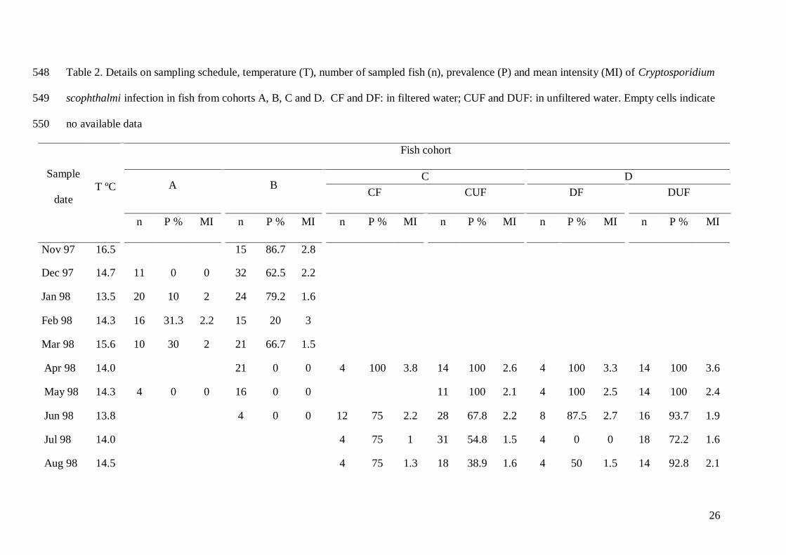
3.1. Temporal changes in infection 230
The temporal progression of infection was followed in the four studied cohorts (Table 2). Fish 231
from cohort A, introduced in the ongrowing tanks in June 97, were not found parasitized at 232
the first sampling in Autumn 97 (December), and showed low infection prevalence in Winter 233
98 (January to March). Conversely, fish from cohort B, introduced in October 97, showed 234
high prevalence and mean intensity of infection from the first sampling in Autumn 97 235
(November) and alos in Winter 98, whereas the parasite was not found in Spring 98. Fish 236
from cohorts C and D, both introduced into the on-growing tanks in April 98 showed high 237
11
prevalences (81-97.7 %) and mean intensities in Spring 98, just after introduction. Prevalence 238
and mean intensity decreased progressively in further samplings until Winter 99, whereas in 239
Spring 99 a moderate rise occurred in CF, DF and DUF fish. Prevalence and mean intensity 240
were higher in D than in C fish. No clear differences were detected between fish receiving 241
filtered or unfiltered water in C, whereas the parasite was more prevalent in DUF fish (Table 242
2). 243
3.2. Dispersion pattern 244
Two hundred and forty one (prevalence = 38.9 %) of the 620 fishes included in the 245
analysis were infected with C. scophthalmi. Parasite abundance approximated a negative 246
binomial distribution with parameters r = 1 and p = 0.5641, i.e., with 25% of the samples 247
having an intensity of infection larger or equal to 1 (Fig. 1). A Poisson distribution with 248
µ=1.98 best described the pattern of parasite intensity. 249
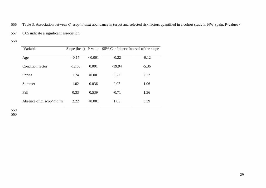
3.3. Epidemiological factors associated with C. scophthalmi abundance 250
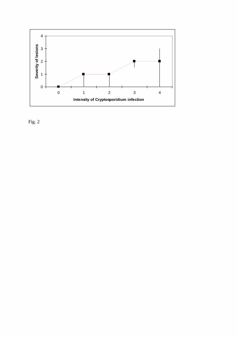
Abundance of C. scophthalmi infection was positively associated (Jonckheere-Terpstra 251
statistic = 16.524; P<0.01) with severity of C. scophthalmi-compatible lesions (Fig. 2). 252
Abundance of C. scophthalmi infection was associated (P<0.05) with age, condition factor, 253
season, and status to Enteromyxum scophthalmi infection, with the most abundant infections 254
observed in fish that were young, in poor condition, not infected by E. scophthalmi, and in 255
spring and summer (Table 3). 256
Inclusion of these four variables significantly (P<0.05) improved the model, compared 257
to the model that did not include any epidemiological factor. The model fitted the data 258
adequately (Pearson’s Chi-square = 2208, P = 0.820). Almost half of the variation in the 259
dependent variable was explained by the variables included in the model (R2
= 0.46). The 260
bivariate relation between the variables that fitted the final models and abundance of C. 261
scophthalmi infection has been graphed in Figs. 3-6. Fig. 7 depicts the age- and season-related 262
12
variation in C. scophthalmi prevalence, which helps to visualize the combined effect of two of 263
the epidemiological factors in the dynamics of the disease. Prevalence decreases siginificantly 264
with age and it was highest in spring and summer (P<0.05, Table 3). Therefore, prevalence in 265
>15 month old (m. o.) fish sampled in spring or summer was, in average, higher than the 266
prevalence in 10-15 m.o. fish sampled in fall or winter, but lower than the prevalence 267
observed in 4-9 m. o. fish sampled the previous spring and summer (Fig. 7). 268
The value of VMR varied with age. Values of VMR>1, indicating overdispersion were 269
obtained for 8-16 m. o. fish, whereas younger and older fish presented VMR < 1 (Fig. 8). 270
271
272
DISCUSSION 273
274
The influence of host and culture conditions on the epidemiology of Cryptosporidium 275
scophthalmi infection in turbot was assessed. The use of a multivariable analysis allowed for 276
the quantification of the extent at which certain risk factors were associated with C. 277
scophthalmi abundance and helped to establish their potential influence in cryptosporidiosis 278
onset and progress. This is the first time that the association between C. scophthalmi 279
abundance and factors hypothesized to influence the course of the disease is quantified in a 280
multivariable framework. Quantification of the strength of associations is important because it 281
leads to a better understanding of the relative importance that different factors and forces have 282
on the epidemiology of the disease. 283
Fish from cohorts B, C, and D were found highly infected from the first sampling, 284
shortly after introduction into the ongrowing system, which suggests that they were already 285
infected at the time of entrance. Furthermore, fish have been observed to be already infected 286
at hatchery and nursery facilities, being prevalence as high as 60-100 % in 3 m. o. (7-8 g) fish 287
(Alvarez-Pellitero et al., 1999, and authors unpublished results). Thus, transmission of C. 288
13
scophthalmi probably takes place through the water supply, as reported for C. molnari and 289
other cryptosporidia infections (Sitjà-Bobadilla et al., 2005; Brookes et al., 2004; Fayer, 290
2004). Filtration and UV irradiation of water in the hatchery and nursery did not prevent the 291
introduction of infective oocysts into the system. A similar treatment of water supply in the 292
hatchery did not prevent the infection by Cryptosporidium molnari in gilthead sea bream, 293
Sparus aurata (Sitjà-Bobadilla et al., 2005). UV irradiation (ranging between 20 and 120 294
mJ/cm2) and conventional filtration of tap water for human consumption have shown to be 295
unreliable for the removal of Cryptosporidium spp. (Fayer et al. 2000a; Betancourt and Rose, 296
2004). In addition to the water supply, the involvement of live food in the transmission of C. 297
scophthalmi in the larval stages should not be ruled out. Rotifers can ingest C. parvum 298
oocysts (Fayer et al., 2000b). Moreover, Artemia, which is a common live food used in 299
marine fish larval development, is capable of spreading Cryptosporidum oocysts and its 300
involvement in the transmission of cryptosporidiosis in cultured fish has been suggested 301
(Méndez-Hermida et al., 2006, 2007). 302
In the population analyzed here, C. scophthalmi seems to have been introduced into 303
the ongrowing tanks by carrier fish, as suggested by the positive disease status observed in 304
individuals sampled at the time of introduction. However, the involvement of infective 305
oocysts present in the water supply in ongrowing facilities cannot be disregarded, as 306
suggested by the prevalence increase observed in May in CF, DF and DUF fish. The coarse 307
filtration used in cohorts CF and DF did not significantly affect the levels of infection. 308
Transmission and dispersal of fish cryptosporidia are facilitated by the aquatic habitat and the 309
frequent releasing of fully sporulated oocysts with mucus casts or faeces. Although it has not 310
been demonstrated, horizontal transmission for C. scophthalmi is likely to occur, as in the 311
case of the piscine C. molnari (Sitjà-Bobadilla and Alvarez-Pellitero, 2003). In addition, 312
different aquatic organisms, such as filter feeding shellfish, could act as reservoirs for C. 313
14
scophthalmi, as it has been reported for other Crypstoporidium spp. (reviewed in King and 314
Monis, 2006; Fayer, 2004). 315
Analysis of our results shows a significant decrease in C. scophthalmi abundance with 316
fish age (beta = -0.17, Table 3). This result is in accordance with the observation that the 317
disease was less prevalent in fish from cohort A, which was examined from 10 to 17 months 318
of age, compared to fish from cohorts B, C and D, that were studied starting at 5 months of 319
age (Table 2). In cohorts C and D, prevalence reached 100 % in the first sampling, just after 320
introduction. In addition, a progressive decrease in infection prevalence and mean intensity 321
was detected in all cohorts along the study period. These results confirmed the age 322
distribution of Cryptosporidium spp. reported in other hosts. The piscine C. molnari showed 323
the highest prevalence levels in preongrowing and early ongrowing fish, with a trend to 324
decrease with fish weight (Sitjà-Bobadilla et al., 2005), and a similar trend occurs in other 325
piscine cryptosporidioses (reviewed in Sitjà-Bobadilla et al., 2005). Non-piscine 326
cryptosporidiosis show similar age distribution, as infections are usually high in neonates and 327
young, and less prevalent in adults, both in mammals and in poultry species (Atwill et al., 328
1999; de Graaf et al, 1999; Guselle et al., 2003; Thompson et al., 2005). In contrast with this 329
negative association of cryptosporidioses with age, the most commonly observed pattern for 330
metazoan parasites is the increase in prevalence and intensity with age or size of the host 331
(Zelmer and Arai, 1998; Poulin, 2000; Thomas, 2002). A non-linear infection pattern, with a 332
peak at a particular age and size of the host and a subsequent decrease in older animals, has 333
also been described for some fish protozoans and myxoporeans (Rintamäki et al., 1997; 334
Palenzuela et al., 1999; Gbankoto et al., 2003), including the turbot parasite Enteromyxum 335
scophthalmi (Quiroga et al., 2006). Such pattern was also found in the piscine C. molnari, and 336
it probably occurs in C. scophthalmi, as deduced from the high infection prevalence and 337
intensity found in larval fish (Alvarez-Pellitero et al., 1999 and authors’unpublished results). 338
15
In piscine cryptosporidioses, age-dependent differential exposure could be involved, since the 339
concentration of infective stages due to the use of recirculating systems or live food at the 340
hatchery could lead to a higher parasite load in these growing steps. This notwithstanding, the 341
decrease of infection prevalence and intensity in older individuals could be related to 342
decreased susceptibility due to acquired immunity, as it has been suggested in mammalian 343
and human cryptosporidioses (Current, 1989). 344
The frequency distribution of C. scophthalmi was right-skewed and fitted a negative 345
binomial distribution. Over-dispersion is the model described for the great majority of 346
parasites (Shaw et al., 1998). Here, the distribution of C. scophthalmi infection was over-347
dispersed for fish of intermediate age, as demonstrated by the quantification of the VMR 348
stratified by age. It must be considered, however, that the influence of several factors in the 349
distribution of infection abundance, such as condition factor, season, and infection with 350
Enteromyxum scophthalmi, may have affected the bivariate association between age and 351
VMR (Fig. 8). Duerr et al. (2003) suggested that interpretation of age-intensity profiles 352
derived from cooperating processes is, at least, difficult, due to the influence that other 353
epidemiological factors may impose on the distribution of infection. In the study here we have 354
overcome this problem by formulating a multivariable framework, which allowed for the 355
quantification of the association between factors and infection while adjusting by the presence 356
or absence of other factors significantly associated with the disease. Therefore, the approach 357
used here is more effective, informative, and powerful for exploring associations in 358
cooperative processes than the visual inspection of the graphics alone. Moreover, the adjusted 359
multivariable model may be used to predict the expected levels of C. scophthalmi abundance 360
in infected populations for given values of the epidemiological factors. This information may 361
be used, for example, to obtain estimates of the expected impact of the disease in populations 362
16
known to be infected and for which information on the epidemiological factors is known, but 363
for which it is not feasible to perform long term longitudinal studies as the one presented here. 364
A seasonal influence on the levels of C. scophthalmi infection was estimated here, 365
with maximum levels of prevalence, abundance, and intensity in spring and summer (Tables 2 366
and 3). The piscine C. molnari also exhibited maximum levels of infection in spring (Sitjà-367
Bobadilla et al., 2005). Seasonality occurs in natural infections by other fish coccidians, 368
which are generally more prevalent in spring (reviewed in Steinhagen and Davies, 2008). 369
Seasonal patterns have also been reported for non-piscine cryptosporidioses, being generally 370
predominant in warm seasons (de Graaf et al., 1999; Sturdee et al., 2003). Temperature is 371
probably one of the most important factors involved in cryptosporidia seasonality, although 372
other factors, such as infective status of fish at the introduction in the ongrowing tanks, 373
availability of infective stages, host density, or a combination of different processes may also 374
contribute to the observed seasonal pattern. 375
Increasing levels of C. scophthalmi infections were significantly associated with high 376
severity of Cryptosporidium lesions. In addition, abundance of infection was low in fish with 377
high condition factor. Such results point to a pathological significance of this infection, 378
though the effect in a decreased body condition as a contributor to possible mortalities, 379
remains to be demonstrated. 380
The negative association between the infection intensities of C. scophthalmi and E. 381
scophthalmi is probably due to differences in the transmission, onset and course of both 382
infections rather than to a true competition between both parasites. C. scophthalmi seemed to 383
be introduced into the ongrowing system mostly through carrier fish, and the infection 384
progressively decreased with fish age/weight. In contrast, fish are free from E. scophthalmi 385
infection when introduced in the ongrowing system, and the first infections are usually 386
detected at least three months after introduction. Culture conditions favour the transmission 387
17
and dispersion of this myxozoan, and thus infection levels progressively increase in growing 388
fish (Quiroga et al., 2006), with an opposite pattern to that observed for C. scophthalmi. 389
In conclusion, C. scophthalmi infection was confirmed in turbot cohorts from their 390
introduction into the ongrowing systems and abundance of infection in the fish was negatively 391
associated with age, concurrent infection with E. scophthalmi, and condition factor and 392
positively associated with spring and summer. The results presented here will help to 393
understand the epidemiology of C. scophthalmi in turbot, to produce estimates of expected 394
levels of infection in infected populations based on the presence or absence of risk factors, 395
and to quantify the impact that the disease may have on susceptible turbot populations of 396
Galicia. 397
398
Acknowledgements 399
Funding for this study was provided by the EU and the Spanish Ministerio de Ciencia y 400
Tecnología through research grants FEDER 1FD97-0679 and AGL2001-2241. Additional 401
support was obtained from Stolt Sea Farm S. A. We are thankful to J. Monfort from IATS, 402
and to M. C. Carreira Valle from the School of Veterinary Medicine of Lugo (University of 403
Santiago) for assistance in processing histological samples. 404
405
References 406
Alvarez-Pellitero, P., Sitjà-Bobadilla, A., 2002. Cryptosporidium molnari n. sp. (Apicomplexa: 407
Cryptosporidiidae) infecting two marine fish species, Sparus aurata L. and 408
Dicentrarchus labrax L. Int. J. Parasitol. 32, 1007-1021. 409
Alvarez-Pellitero, P., Redondo M.J., Sitjà-Bobadilla, A., Macías, A., Riaza, A., Padrós. F., 410
1999. Epidemiological study of an intestinal coccidiosis of cultured turbot 411
18
(Scophthalmus maximus L.). Fifth Int. Symp. Fish Parasites. Book of Abstracts, Ceské 412
Budejovice, Czech Republic, pp. 4. 413
Alvarez-Pellitero, P., Quiroga, M.I., Sitjà-Bobadilla, A., Redondo, M.J., Palenzuela, O., 414
Padrós, F., Vázquez, S., Nieto, J.M., 2004. Cryptosporidium scophthalmi n. sp. 415
(Apicomplexa: Cryptosporidiidae) from cultured turbot, Scophthalmus maximus L. 416
Light and electron microscope description and histopathological study. Dis. Aquat. 417
Organ. 62, 133-145. 418
Arneberg, P., Skorping, A., Grenfell, B., Read, A.F., 1998. Host densities as determinants of 419
abundance in parasite communities. Proc. R. Soc. Lond. B, 265, 1283-1289. 420
Atwill, E.R, Johnson, E., Klingborg, D.J., Veserat, G.M., Markegard, G., Jensen, W.A., Pratt, 421
D.W., Delmas, R.E., George, H.A., Forero, L.C., Philips, R.L., Barry, S.J., McDougald, 422
N.K., Gildersleeve, R.R., Frost, W.E., 1999. Age, geographic and temporal distribution 423
of fecal shedding of Cryptosporidium parvum oocysts in cow calf herds. Am. J. Vet. 424
Res. 60, 420-425. 425
Betancourt, W.Q. Rose, J.B., 2004. Drinking water treatment processes for removal of 426
Cryptosporidium and Giardia. Vet. Parasitol. 126, 219-234. 427
Brookes, J.D., Antenucci, J., Hipsey, M., Burch, M.D., Ashbolt, N. J., Ferguson, C., 2004. Fate 428
and transport of pathogens in lakes and reservoirs. Environ. Int. 30, 741-759. 429
Bush, A.O., Lafferty, K.D., Lotz, J.M, Shostak, A.W., 1997. Parasitology meets ecology on its 430
own terms: Margolis et al. revisited. J. Parasitol. 83, 575-583. 431
Current, W.L., 1989. Cryptosporidium spp. In: Walzer, P. D., Genta, R.A. (Eds.), Parasitic 432
infections in the compromised host. Marcel Deker, New York, pp. 281-341. 433
Dohoo I., Martin W., Stryhn H., 2003a. Linear regression. In: Dohoo, I., Martin, W., Stryhn, 434
H. (Eds), Veterinary epidemiologic research. Transcontinental Prince Edward Island. 435
Prince Edward Island, Canada. pp 273-316. 436

19
Dohoo I., Martin W., Stryhn H., 2003b. Cohort studies. In: Dohoo, I., Martin, W., Stryhn, H. 437
(Eds), Veterinary epidemiologic research. Transcontinental Prince Edward Island. 438
Prince Edward Island, Canada. pp: 151-162. 439
Duerr, H.P., Dietz, K. Eichner, M., 2003. On the interpretation of age-intensity profiles and 440
dispersion patterns in parasitological surveys. Parasitology 126, 87-101. 441
de Graaf, D.C., Vanopdenbosch, E., Ortega-Mora, L.M., Abbassi, H., Peeters, J.E., 1999. A 442
review of the importance of cryptosporidiosis in farm animals. Int. J. Parasitol. 29, 443
1269-1287. 444
Dillingham, R.A., Lima, A.A., Guerrant, R.L., 2002. Cryptosporidiosis: epidemiology and 445
impact. Microbes Infect. 4, 1059-1066. 446
Fayer, R., 2004. Cryptosporidium: a water-borne zoonotic parasite. Vet. Parasitol. 126, 37-56. 447
Fayer, R., Speer, C.A., Dubey, J.P. 1997. The general biology of Cryptosporidium. In: Fayer, 448
R. (Ed.), Cryptosporidium and cryptosporidiosis. CRC Press, Boca Ratón, pp. 1-41. 449
Fayer, R., Morgan, U., Upton, S.J. 2000a. Epidemiology of Cryptosporidium: transmission, 450
detection and identification. Int. J. Parasitol. 30, 1305-1322. 451
Fayer, R., Trout, J.M., Walsh, E., Cole, R. 2000b. Rotifers ingest oocysts of Cryptosporidium 452
parvum. J. Eukaryot. Microbiol. 47, 161-163. 453
Gbankoto, A., Pampoulie, C., Marques, A. Sakiti, G.N. and Dramane, K.L., 2003. Infection 454
patterns of Myxobolus heterospora in two tilapia species (Teleostei: Cichlidae) and its 455
potential effects. Dis. Aquat. Organ. 55, 125-131. 456
Georgiadis, M.P., Gardner, I.A., Hedrick, R.P., 2001. The role of epidemiology in the 457
prevention, diagnosis, and control of infectious diseases of fish. Prev. Vet. Med. 48, 458
287-302. 459
Guselle, N.J., Appelbee, A.J., Olson, M. E., 2003. Biology of Cryptosporidium parvum in pigs: 460
from weaning to market. Vet. Parasitol. 113, 7-18. 461

20
King, B.J., Monis, P.T., 2006. Critical processes affecting Cryptosporidium oocyst survival in 462
the environment. Parasitology 134, 1-15 463
McCullagh, P., 1980. Regression models for ordinal data. J. R. Stat. Soc. Ser. B 42:109-142. 464
McCullagh, P., Nelder, J.A., 1989. Generalized linear models, 2nd ed. London: Chapman & 465
Hall. 532 pp. 466
Méndez-Hermida, F., Gómez-Couso, H., Ares-Mazas, E., 2006. Artemia is capable of 467
spreading oocysts of Cryptosporidium and the cysts of Giardia. J. Eukaryot. Microbiol. 468
53, 432-434. 469
Méndez-Hermida, F., Gómez-Couso, H., Ares-Mazas, E., 2007. Possible involvement of 470
Artemia as a live diet in the transmission of cryptosporidiosis in cultured fish. Parasitol. 471
Res. 101, 823-827. 472
Nagelkerke, N. J. D., 1991. A note on the general definition of the coefficient of determination. 473
Biometrika, 78:691-692. 474
Palenzuela, O., Alvarez-Pellitero, P. Sitjà-Bobadilla, A. 1999. Glomerular disease associated to 475
Polysporoplasma sparis (Myxosporea: Bivalvulida) infections in the gilthead sea bream, 476
Sparus aurata, (Pisces; Teleostei): Aspects of the host-parasite relationship. Parasitology 477
118, 245-256. 478
Poulin, R. 2000. Variation in the intraspecific relationship between fish length and intensity of 479
parasitic infection: biological and statistical causes. J. Fish Biol. 56, 123-137. 480
Quiroga, M.I., Redondo, M.J, Sitjà-Bobadilla, A., Palenzuela, O., Riaza, A., Macías, A., 481
Vázquez, S., Perez, A., Nieto
, J. M, Alvarez-Pellitero, P., 2006. Risk factors associated 482
with Enteromyxum scophthalmi (Myxozoa) infection in cultured turbot, Scophthalmus 483
maximus (L.). Parasitology 133, 433-442. 484
Rintamäki-Kinnunen, P., Valtonen, T.E., 1997. Epizootiology of protozoans in farmed 485
salmonids at northern latitudes. Int. J. Parasitol. 27, 89-99. 486
21
Shaw, D.J., Grenfell, B.T. and Dobson, A.P., 1998. Patterns of macroparasite aggregation in 487
wildlife host populations. Parasitology 117, 597-610. 488
Sitjà-Bobadilla, A., Alvarez-Pellitero, P., 2003. Experimental transmission of Cryptosporidium 489
molnari (Apicomplexa: Coccidia) to gilthead sea bream (Sparus aurata L.) and 490
European sea bass (Dicenrarchus labrax L.). Parasitol. Res. 91, 209-214 491
Sitjà-Bobadilla, A., Padrós, F., Aguilera, C., Alvarez-Pellitero, P., 2005. Epidemiology of 492
Cryptosporidium molnari in Spanish gilthead sea bream (Sparus aurata L.) and 493
European sea bass (Dicentrarchus labrax L.) cultures: from hatchery to market size. 494
Appl. Environm. Microbiol. 71, 131-139. 495
Steinhagen D., Davies, A.J., 2008. Diseases caused by Apicomplexans. In: Eiras, J.C., Segner, 496
H., Wahli, T., Kapoor, B.G. (Eds.). Fish Diseases. Vol 1. Science Publishers, Enfield 497
(NH), pp. 517-567. 498
Sturdee, A.P., Bodley-Tickell, A.T., Archer, A., Chalmers, R.M., 2003. Long-term study of 499
Cryptosporidium prevalence on a lowland farm in the United Kingdom. Vet. Parasitol. 500
116, 97-113. 501
Sunnotel, O., Lowery, C. J., Moore, J.E., Dooley, J.S.G., Xiao, L., Millar, B.C., Rooney, P.J., 502
Snelling, W.J., 2006. Cryptosporidium. Lett. Appl. Microbiol. 43, 7-16. 503
Thomas, J.D., 2002. The ecology of fish parasites with particular reference to helminth 504
parasites and their salmonid fish hosts in Welsh rivers: A review of some of the central 505
questions. Adv. Parasitol. 52, 1-154, 506
Thompson, R.C.A., Olson, M.E., Zhu, G., Enomoto, S., Abrahamsen, M.S., Hijjawi, N.S., 507
2005. Cryptosporidium and Cryptosporidiosis. Adv. Parasitol. 59, 77-162. 508
Tzipori, S., Ward, H., 2002. Cryptosporidiosis: biology, pathogenesis and disease. Microbes 509
Infect. 4, 1047-1058 510
22
Zelmer, D.A., Arai, H.P., 1998. The contributions of host age and size to the aggregated 511
distribution of parasites in yellow perch, Perca flavescens, from Garner Lake, Alberta, 512
Can. J. Parasitol. 84, 24-28. 513
514
23
Figure legends 515
Fig. 1. Cryptosporidium scophthalmi abundance coded into five categories (0 = nil, 1 516
= slight, 4 = severe). Black lines indicate the proportion of fishes observed for a given 517
category of density. Grey bars depict the expected probability distribution using a negative 518
binomial distribution fitted with parameters r = 1 and p=0.564 519
Fig. 2. Association between abundance and severity of lesions due to Cryptosporidium 520
scophthalmi infection. Abundance and severity of lesions have been coded, respectively, into 521
five (0 = nil, 1 = slight, 4 = severe) and four (0 = no lesions, 3 = severe lesions) categories. 522
The black square and the upper and lower extremes of the black lines indicate, respectively, 523
the median value, first quartile and fourth quartile of the severity of lesions observed for a 524
given category of infection. 525
Fig. 3. Association between condition factor and Cryptosporidium scophthalmi abundance. 526
Abundance has been coded into five categories (0 = nil, 1 = slight, 4 = severe). The black 527
square and the upper and lower extremes of the black lines indicate, respectively, the mean 528
value, first quartile and fourth quartile of the condition factor observed for a given category of 529
abundance. 530
Fig. 4. Association between age (in months) and Cryptosporidium scophthalmi abundance. 531
See figure 3 for reference. 532
Fig. 5. Association between season and Cryptosporidium scophthalmi abundance. See figure 533
3 for reference. 534
Fig. 6. Proportion of Enteromyxum scophthalmi-infected fishes (dark bar) stratified by 535
Cryptosporidium scophtalmi abundance (0 = nil, 1 = slight, 4 = severe). 536
Fig. 7. Prevalence of Cryptosporidum scophthalmi infection in fish from cohorts C and D 537
stratified by age and season. 538
24
Fig. 8. Age-related variation of the variance-to-mean ratio (VMR) of the Cryptosporidium 539
scophthalmi abundance. Raw values (solid line) and three-month moving average (point line) 540
are shown. 541
542
25
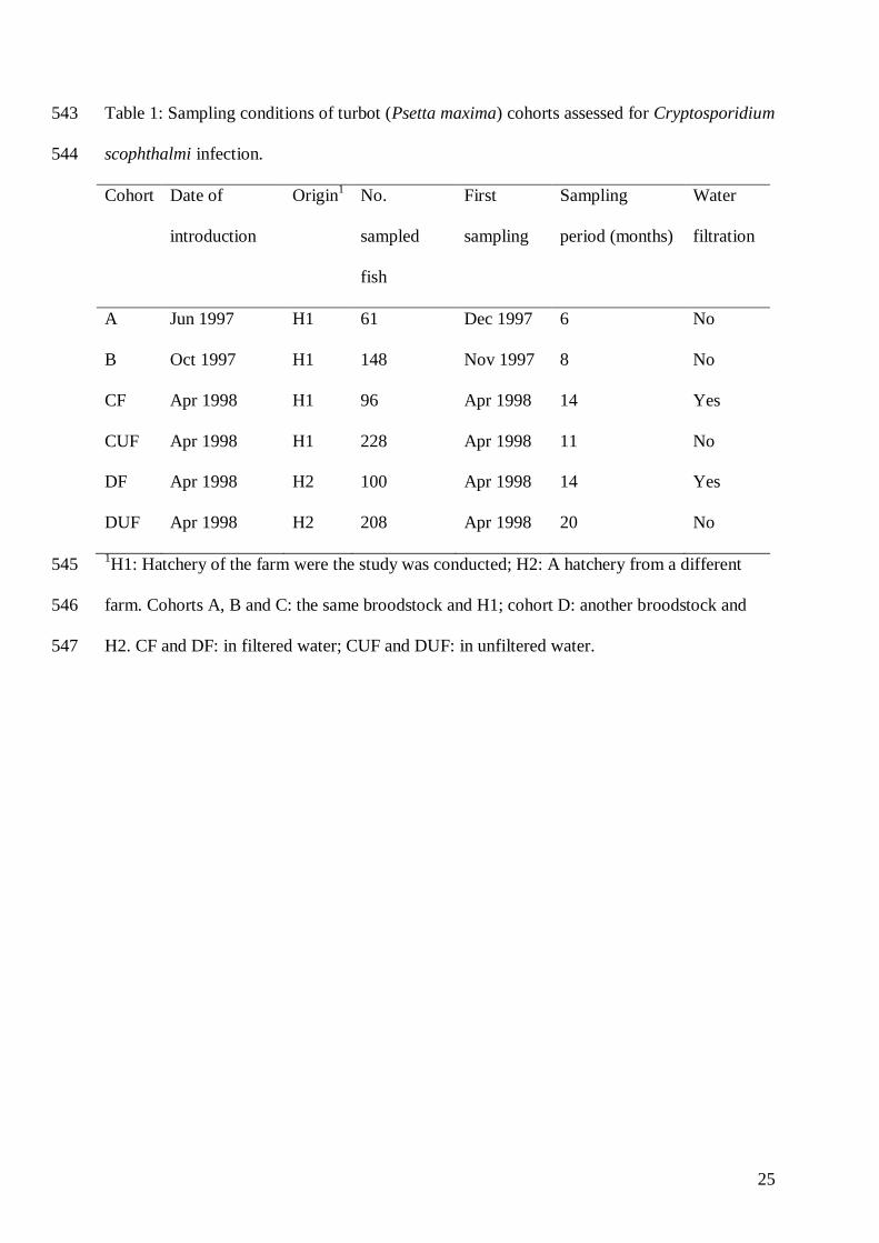
Table 1: Sampling conditions of turbot (Psetta maxima) cohorts assessed for Cryptosporidium 543
scophthalmi infection. 544
Cohort Date of
introduction
Origin1
No.
sampled
fish
First
sampling
Sampling
period (months)
Water
filtration
A Jun 1997 H1 61 Dec 1997 6 No
B Oct 1997 H1 148 Nov 1997 8 No
CF Apr 1998 H1 96 Apr 1998 14 Yes
CUF Apr 1998 H1 228 Apr 1998 11 No
DF Apr 1998 H2 100 Apr 1998 14 Yes
DUF Apr 1998 H2 208 Apr 1998 20 No
1H1: Hatchery of the farm were the study was conducted; H2: A hatchery from a different 545
farm. Cohorts A, B and C: the same broodstock and H1; cohort D: another broodstock and 546
H2. CF and DF: in filtered water; CUF and DUF: in unfiltered water.547
26
Table 2. Details on sampling schedule, temperature (T), number of sampled fish (n), prevalence (P) and mean intensity (MI) of Cryptosporidium 548
scophthalmi infection in fish from cohorts A, B, C and D. CF and DF: in filtered water; CUF and DUF: in unfiltered water. Empty cells indicate 549
no available data 550
Sample
date
T ºC
Fish cohort
A B C D
CF CUF DF DUF
n P % MI n P % MI n P % MI n P % MI n P % MI n P % MI
Nov 97 16.5 15 86.7 2.8
Dec 97 14.7 11 0 0 32 62.5 2.2
Jan 98 13.5 20 10 2 24 79.2 1.6
Feb 98 14.3 16 31.3 2.2 15 20 3
Mar 98 15.6 10 30 2 21 66.7 1.5
Apr 98 14.0 21 0 0 4 100 3.8 14 100 2.6 4 100 3.3 14 100 3.6
May 98 14.3 4 0 0 16 0 0 11 100 2.1 4 100 2.5 14 100 2.4
Jun 98 13.8 4 0 0 12 75 2.2 28 67.8 2.2 8 87.5 2.7 16 93.7 1.9
Jul 98 14.0 4 75 1 31 54.8 1.5 4 0 0 18 72.2 1.6
Aug 98 14.5 4 75 1.3 18 38.9 1.6 4 50 1.5 14 92.8 2.1

27
Sep 98 16.5 8 37.5 1.3 40 15 1.5 8 87.5 1.1 28 46.4 1.3
Oct 98 15.3 12 8.3 1 25 4 2 12 41.7 1 8 0 0
Nov 98 15.0 4 25 1 15 0 0 4 0 0 4 0 0
Dec 98 10.7 12 0 0 12 0 0 12 8.3 1 12 50 1
Jan 99 11.4 8 12.5 1 18 0 0 8 0 0 8 12.5 1
Feb 99 12.8 8 0 0 16 6.3 2 8 0 0 8 12.5 2
Mar 99 12.6 4 25 1 4 0 0 8 12.5 1
Apr 99 13.0 8 25 1 8 0 0 8 25 1
May 99 15.2 8 62.5 1 12 33.3 1 8 50 1
Jun 99 15.1 4 0 0
Jul 99 14.9 8 25 2
Aug 99 16.3 4 50 2
Sept 99 16.1 8 0 0
Nov 99 16.5 8 0 0
Dec 99 12.0 8 0 0
551
28
Table 2. Details on sampling schedule, temperature (T), number of sampled fish (n), prevalence (P) and mean intensity (MI) of Cryptosporidium 552
scophthalmi infection in fish from cohorts A, B, C and D. CF and DF: in filtered water; CUF and DUF: in unfiltered water. Empty cells indicate 553
no available data 554
Sample date T ºC
Fish cohort
A B C D
CF CUF DF DUF
n P % MI n P % MI n P % MI n P % MI n P % MI n P % MI
Autumn 97 16.5- 14.7
-13.5
1
1
0 0 4
7
70.
2
2.
4
Winter 98 13.5-15.6 4
6
21.
7
2.
1
6
0
60 1.
7
Spring 98 14.0-13.8 4 0 0 4
1
0 0 1
6
81.3 2.
7
5
3
83 2.
3
1
6
93.
8
2.
8
4
4
97.
7
2.
6 Summer 98 14.0-16.5 1
6
56.2
5
1.
5
8
9
33.
7
1.
5
1
6
56.
3
1.
2
6
0
65 1.
7 Autumn 98 15.3-10.7 2
8
7.1 1 5
2
1.9 2 2
8
21.
4
1 2
4
25 1
Winter 99 11.4-12.6 2
0
10 1 3
4
2.9 2 2
0
0 0 2
4
12.
5
1.
3 Spring 99 13.0-15.1 1
6
43.8 1 1
2
20 1 2
0
30 1
Summer 99 14.9-16.1 2
0
20 2
Autumn 99 16.5-12 1
6
0 0
555
29
Table 3. Association between C. scophthalmi abundance in turbot and selected risk factors quantified in a cohort study in NW Spain. P-values < 556
0.05 indicate a significant association. 557
558
Variable Slope (beta) P-value 95% Confidence Interval of the slope
Age -0.17 <0.001 -0.22 -0.12
Condition factor -12.65 0.001 -19.94 -5.36
Spring 1.74 <0.001 0.77 2.72
Summer 1.02 0.036 0.07 1.96
Fall 0.33 0.539 -0.71 1.36
Absence of E. scophthalmi 2.22 <0.001 1.05 3.39
559
560
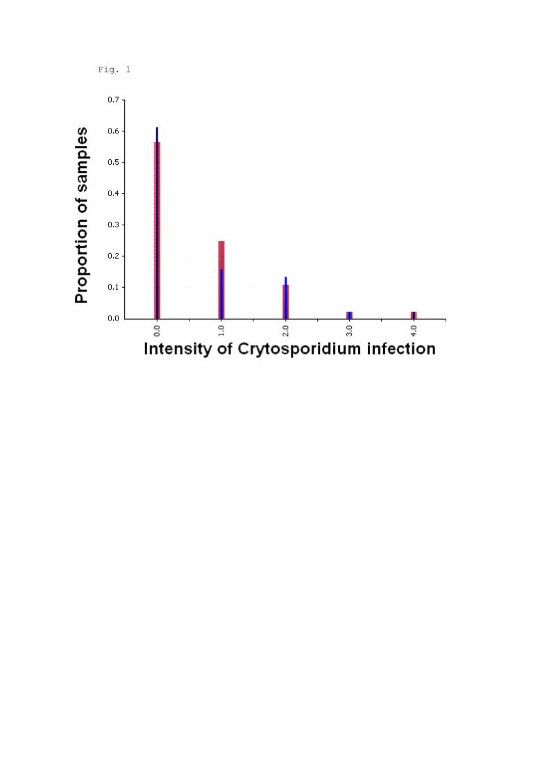
Fig. 1
0
1
2
3
4
0 1 2 3 4
Intensity of Cryptosporidium infection
Severi
ty o
f le
sio
ns
Fig. 2
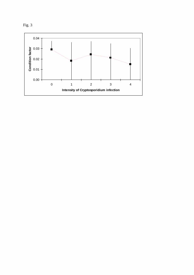
Fig. 3
0.00
0.01
0.02
0.03
0.04
0 1 2 3 4
Intensity of Cryptosporidium infection
Co
nd
itio
n f
acto
r

Fig. 4
0
1
2
3
4
5
6
7
8
9
10
0 1 2 3 4
Intensity of Cryptosporidium infection
Ag
e (
mo
nth
s)
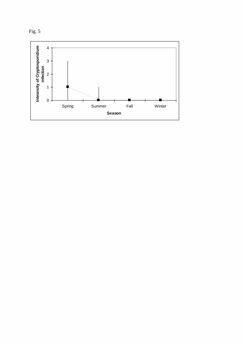
Fig. 5
0
1
2
3
4
Spring Summer Fall Winter
Season
Inte
sn
sit
y o
f C
ryp
tosp
ori
diu
m
infe
cti
on
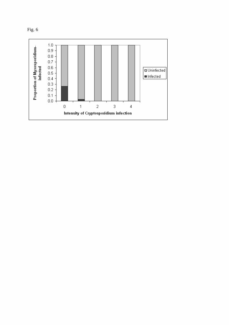
Fig. 6
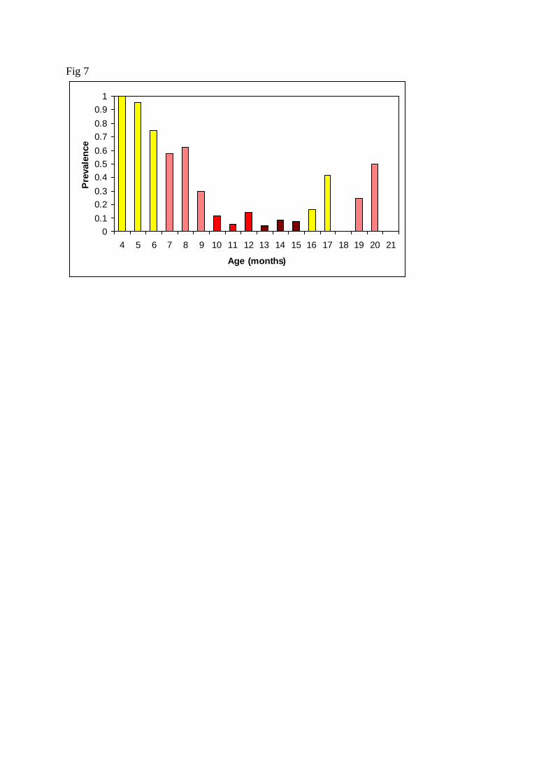
Fig 7
0
0.1
0.2
0.3
0.4
0.5
0.6
0.7
0.8
0.9
1
4 5 6 7 8 9 10 11 12 13 14 15 16 17 18 19 20 21
Age (months)
Pre
vale
nce
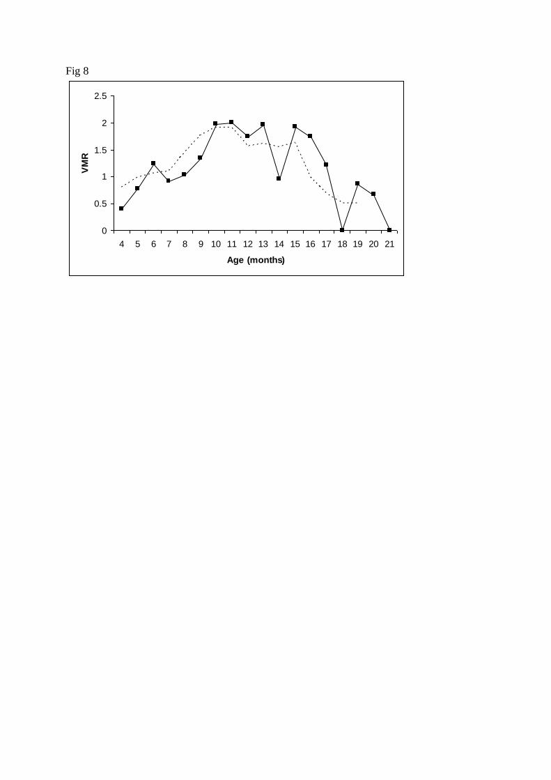
Fig 8
0
0.5
1
1.5
2
2.5
4 5 6 7 8 9 10 11 12 13 14 15 16 17 18 19 20 21
Age (months)
VM
R