1 comparative methods for the quantification of …
TRANSCRIPT
1
COMPARATIVE METHODS FOR THE QUANTIFICATION OF POST-IMA MYOCARDIAL DAMAGE
INTRODUCTIONThe presence and correct definition of the extent of the infarcted area of the heart has been one of the main short- and long-term prognostic elements in the natural history of acute myocardial infarction. Given the impossibility of immediately performing imaging methods such as SPECT (Single-Photon Emission Computed Tomography) and MRI (Magnetic Resonance Imaging), especially since these methods have only been optimized so much in recent years that they give fast and reliable results, an attempt was made to improve diagnostics by quantifying immediately available diagnostic enzymes in the bloodstream that could be weighed in relation to possible myocardial damage.
MATERIALS E METHODS According to the most recent update of the redefinition of acute myocardial infarction (Fourth universal definition of myocardial infarction 2018, FUDMI), the term IMA should be used when there is evidence of “myocardial damage” with myocardial necrosis, in a consistent clinical situation. with acute myocardial ischemia. The latter can be documented on the basis of the patient’s symptoms, the presence of characteristic electrocardiographic changes or by cardiac imaging evidence of loss of vital myocardial tissue. “Myocardial damage” is defined as a troponin (cTn) elevation at least once above the upper 99th percentile of the reference population; at this concentration the methods must exhibit an analytical imprecision ≤10% (expressed as coefficient of variation, CV). Cardiac troponins are specific isoforms of the heart. Among the markers of myocardial cytonecrosis (CK-MB enzyme, troponin I, troponin T and myoglobin) it is definitively acquired that the hs-cTn data is
quantitatively correlated to the size of the IMA and its prognosis. Some research assays reach as low as 0.001 ng / mL (1 pg / mL) therefore highly sensitive even to damage to a single myocardiocyte. Any cellular damage that alters the integrity of the cardiomyocyte cell membrane can cause cTn (both I and T) to be released into the circulation. * Many specific studies have shown that the hs-cTnT concentration is directly related to the size of the infarct and the extent of coronary lesions, in both STEMI and NSTEMI patients. The same group demonstrated that hs-cTnI also has a significant correlation with the infarct area [99]: the value of hs-cTn at 24 h correlates with the size of the infarction at least as well as creatine kinase-MB at 48 h and more faster than conventional CT. The advent of cTn in clinical practice suggested its use as a laboratory method also for the estimation of the size of the infarcted area, on the basis of the release kinetics by the myocardial cell (a more rapid release of the cytosolic fraction, followed by that later and prolonged due to necrosis) and relative independence from reperfusion, especially with regard to the first phase of release.However, the different troponin concentrations in different cardiac areas and the complexity of the kinetic models slowed the introduction of cTn as an indicator of the size of the infarct. Ingrained habits added reasons to the continued use of the CK-MB enzyme. Again in the Redefinition of IMA [2], the determination of the extent of the heart attack, the absence of universally accepted criteria, is referred to the degree of increase in “biomarkers”, understood in a generic sense. In this context, imaging diagnostics could represent a useful second-level method following the laboratory investigations carried out in the first hours following the symptoms of IMA.
Maiello Vincenzo1, Iacono Maria2, Cangiano Valerio3, Calligari Dalila4, Scarfato Emma5, Stile Stefania6,Cecere Claudio7, Frasca Carmine8
1TSRM presso Centro IGEA Sant’Antimo, Napoli 2Dott.ssa in Biologia 3TSRM presso AO dei Colli – Ospedale Monaldi – UOC Radiodiagnostica Napoli 4TSRM presso Centro IGEA Sant’Antimo, Napoli 5TSRM presso centro Mavis 6TSRM presso U.O Radiologia, Ospedale Infermi Rimini- AUSL Romagna7TSRM presso S.C. Radiologia generale AORN Santobono-Pausillipon, Napoli8TSRM presso UOC di Radiodiagnostica CTO - Ospedali dei Colli, Napoli
KEYWORDS: IMA, troponin, Cardiac-MRI, SPECT
ABSTRACT This work aims to make a comparison between laboratory methods, especially by evaluating the role of troponin dosage and diagnostic imaging methods, specifically SPECT and cardiac magnetic resonance in the post-IMA evaluation. In both methods, we want to understand which one is able to be more predictive in terms of quantifying the extension of the infarcted area. Given the high sensitivity of troponin, even a very small damage can result from this examination, which SPECT or MRI cannot do. It must be said, however, that the initial high sensitivity troponin is not predictive of how much ischemic damage will be but is only useful for prognosis. The objective of the study was to determine the feasibility and diagnostic accuracy of an RMC stress protocol with the combined assessment of regional myocardial perfusion and contractility in the recognition of patients with significant coronary atherosclerotic disease leading to AMI, correlating the results with what would have been the study in SPECT.
RESE
ARCH
ART
ICLE
| TEC
NIC
O S
AN
ITA
RIO
JOURNAL OF ADVANCED HEALTH CARE (ISSN 2612-1344) - 2021 - VOLUME3 - ISSUE ON LINE
In the following years, several comparative studies have shown that troponin is a good indicator, possibly superior to CK-MB for estimating the size of the infarct.In particular, a study of 65 patients with IMA, a single measurement of troponin T obtained 72 hours after admission of coronary care units significantly correlated with the CK-MB peak in estimating the size of the infarct (r = 0 , 76, P <0.001), using single photon emission computed tomography (SPECT) as the “gold standard”. Unlike CK-MB, troponin T’s ability to predict infarct size was independent of coronary reperfusion. On the basis of these and other similar data, it could be seen that cTn correlated better than CK-MB with the determinations of Nuclear Medicine and Magnetic Resonance of the size of the infarct area and suggested its exclusive use.
Although the study of ischemic myocardial pathology in the world of diagnostic imaging still currently sees SPECT as a reference diagnostic technique, Nuclear Magnetic Resonance used in the Cardiovascular field is certainly one of the most suitable specialist applications for the study of IMA. Only recently has it been possible to partially overcome technical impediments that limited its clinical use. Compared to the other diagnostic imaging techniques available (Echocardiography, Chest X-ray, Catheterization, CT, SPECT, PET), MRI is a complete method which has numerous advantages;1) It does not use ionizing radiation2) It is a multiplanar and multiparametric method,3) It is not invasive4) It allows very large FOVs while still guaranteeing
good spatial resolution and high contrast5) It allows static, dynamic, angiographic, and
above all tissue perfusion studies.Similarly to SPECT, also in MRI it is possible to study myocardial perfusion, with results that are completely
comparable, if not better, to those obtained with nuclear medicine techniques, without exposure of the patient to ionizing radiation, and with timing of the execution of the very short examination compared to those of nuclear medicine.Stress CMR perfusion study has a high value in evaluating significant coronary artery disease. It is a valid alternative to SPECT and allows to evaluate signal intensity defects during the first passage of contrast medium injected as an intravenous bolus. The evaluation is carried out both at rest and after intravenous injection of Dipyridamole, the latter being used to induce a stress phase in a pharmacological way.
DISCUSSION
Comparison between myocardial perfusional SPECT and cardio-MRI in ischemic heart disease.Over the last few years, studies conducted with cardiac magnetic resonance imaging (cardio-MRI) from stress have reported good values of feasibility and accuracy in the characterization of patients with known or suspected ischemic heart disease.This issue is addressed because today cardiovascular diseases are still the leading cause of death. Diagnostic imaging offers two non-invasive methods: myocardial perfusion SPECT and Cardio-MR Perfusion. They are two imaging techniques that have a completely different method of execution but with a single purpose, namely the diagnosis of a perfusion defect. Ischemic heart disease is caused by a reduction in the supply of oxygen to the heart muscle. The major etiological factor is atherosclerosis, an accumulation of lipids within the lumen of the coronary arteries. This atherosclerotic plaque is made up of a percentage of collagen and a portion of the lipid nucleus, called “core”. The prevalence of one of these two components affects the stability of the plaque.
2
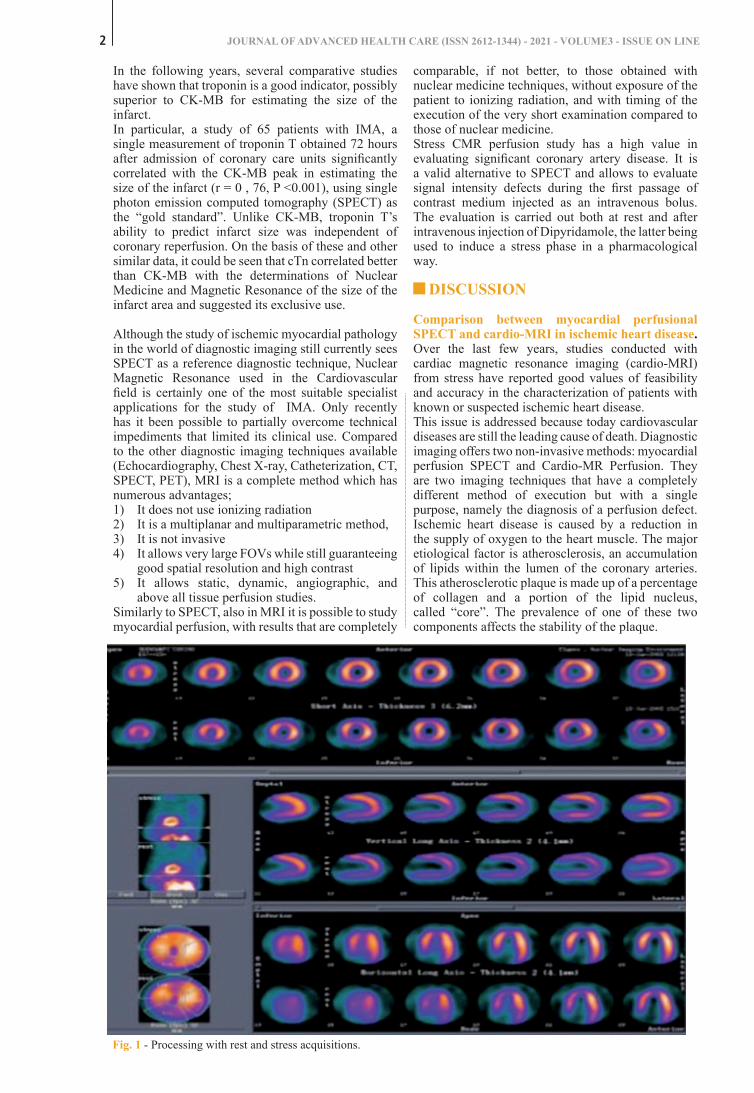
Fig. 1 - Processing with rest and stress acquisitions.
evaluate the function of the organ. Another substantial difference with all the other radiological methods and that the radiation is emitted by the patient, the gamma camera has a purely detector purpose. In particular, the SPECT allows the conduct of a three-dimensional study thanks to the rotation of the detector during the execution of the survey. Myocardial perfusion SPECT is performed with Sestamibi or Tertofosmin, labeled with technetium-99-mb, therefore the collimators used will be LEGP or LEHR. Acquisitions take place in conditions of rest and stress in Gated mode.
Fig. 1 - Processing with rest and stress acquisitions
The dosage for rest is 296-370 Mbq while that for stress is 888-1110 Mbq. This technique is called split dose because it involves the use of multiples of three for the injection of stress with respect to the rest (Fig. 1). The heads of the gamma cameras are arranged at 90 ° and start from the right anterior oblique 45 ° up to the left oblique at 135 °. The processing is performed on the different cardiac axes which are: Short Axis, Long Vertical Axis and Long Horizontal Axis (Fig. 2).
Fig. 2 - The short axis is shown in box A, the long horizontal axis in box B and the long vertical axis in box C

COMPARATIVE METHODS FOR THE QUANTIFICATION OF POST-IMA MYOCARDIAL DAMAGE 3
Myocardial perfusion SPECT: study techniques, equipment and radiopharmaceuticalsSPECT is a nuclear medicine technique that is based on the injection of a radiopharmaceutical intravenously, allowing the acquisition of a scintigraphic map through an equipment, the gamma camera. What characterizes nuclear medicine is the ability to carry out studies that are in vain to evaluate the function of the organ. Another substantial difference with all the other radiological methods and that the radiation is emitted by the patient, the gamma camera has a purely detector purpose. In particular, the SPECT allows the conduct of a three-dimensional study thanks to the rotation of the detector during the execution of the survey. Myocardial perfusion SPECT is performed with Sestamibi or Tertofosmin, labeled with technetium-99-mb, therefore the collimators used will be LEGP or LEHR. Acquisitions take place in conditions of rest and stress in Gated mode.The dosage for rest is 296-370 Mbq while that for stress is 888-1110 Mbq. This technique is called split dose because it involves the use of multiples of three
for the injection of stress with respect to the rest (Fig. 1). The heads of the gamma cameras are arranged at 90 ° and start from the right anterior oblique 45 ° up to the left oblique at 135 °. The processing is performed on the different cardiac axes which are: Short Axis, Long Vertical Axis and Long Horizontal Axis (Fig. 2).
Also important is the polar map (Fig. 3), or bull’s eye, which allows the visualization of all the walls of the left ventricle in a single image.
In order to formulate a diagnosis of coronary heart disease, it is necessary to highlight the different modes of presentation of ischemia and heart attack. In ischemia there is a reduced perfusion in the acquisition under effort with recovery in that of rest (Fig. 4). In the presence of a heart attack, persistent hypoperfusion occurs in both images. Based on the area affected by less fixation, it is possible to understand which coronary artery is affected by the stenosis. Obviously, the size of the stenosis is directly proportional to the extent of the damage.
Fig. 2 - The short axis is shown in box A, the long horizontal axis in box B and the long vertical axis in box C.
Fig. 3 - Polar map with study of stress and rest compared with the relative difference. The chromatic gradations of the box below highlight the different degrees of uptake.
Fig. 4 - Difference in uptake between ischemia and infarction.
evaluate the function of the organ. Another substantial difference with all the other radiological methods and that the radiation is emitted by the patient, the gamma camera has a purely detector purpose. In particular, the SPECT allows the conduct of a three-dimensional study thanks to the rotation of the detector during the execution of the survey. Myocardial perfusion SPECT is performed with Sestamibi or Tertofosmin, labeled with technetium-99-mb, therefore the collimators used will be LEGP or LEHR. Acquisitions take place in conditions of rest and stress in Gated mode.
Fig. 1 - Processing with rest and stress acquisitions
The dosage for rest is 296-370 Mbq while that for stress is 888-1110 Mbq. This technique is called split dose because it involves the use of multiples of three for the injection of stress with respect to the rest (Fig. 1). The heads of the gamma cameras are arranged at 90 ° and start from the right anterior oblique 45 ° up to the left oblique at 135 °. The processing is performed on the different cardiac axes which are: Short Axis, Long Vertical Axis and Long Horizontal Axis (Fig. 2).
Fig. 2 - The short axis is shown in box A, the long horizontal axis in box B and the long vertical axis in box C
Also important is the polar map (Fig. 3), or bull's eye, which allows the visualization of all the walls of the left ventricle in a single image.
Fig. 3 - Polar map with study of stress and rest compared with the relative difference. The chromatic gradations of the box below highlight the different degrees of uptake.
In order to formulate a diagnosis of coronary heart disease, it is necessary to highlight the different modes of presentation of ischemia and heart attack. In ischemia there is a reduced perfusion in the acquisition under effort with recovery in that of rest (Fig. 4). In the presence of a heart attack, persistent hypoperfusion occurs in both images. Based on the area affected by less fixation, it is possible to understand which coronary artery is affected by the stenosis. Obviously, the size of the stenosis is directly proportional to the extent of the damage.
Fig. 4 - Difference in uptake between ischemia and infarction
Ischemic heart disease analysis in MN and RM imaging
RM exploits the properties of the magnetic field and the rotation of hydrogen protons for the acquisition of images on the axial, coronal and sagittal study planes. It is necessary that the magnet is high field (at least 1.5T), and, according to the study carried out, RF coils are applied. For cardiology

Ischemic heart disease analysis in MN and RM imagingRM exploits the properties of the magnetic field and the rotation of hydrogen protons for the acquisition of images on the axial, coronal and sagittal study planes. It is necessary that the magnet is high field (at least 1.5T), and, according to the study carried out, RF coils are applied. For cardiology studies the coils can be dedicated or phased-array multichannel. The contrast medium used is based on gadolinium, a paramagnetic substance, always combined with a chelating agent, such as DTPA. To perform cardio RM it is mandatory
that the patient is equipped with electrodes for cardio-synchronization and respiratory trigger. We proceed with the surveys on the conventional planes and then orient the cuts along the cardiac axes (Fig. 5). From the coronal, the axial is obtained as well, proceeding with the long vertical axis (or two chambers), the long horizontal axis (or 4 chambers) and the short axis. The sequences used are the Dark blood (Fig. 6-7), they are fast and ultra-fast spin echoes (FSE and TSE) and are typically morphological.There are T2 STIR sequences, used for the characterization of the edamentous component and
4 JOURNAL OF ADVANCED HEALTH CARE (ISSN 2612-1344) - 2021 - VOLUME3 - ISSUE ON LINE
Fig. 5 - A coronal survey, B pure axial, C vertical long axis (2 chambers), D horizontal long axis (4 chambers), E short axis.
Fig. 6 - Dark Blood sequences.
studies the coils can be dedicated or phased-array multichannel. The contrast medium used is based on gadolinium, a paramagnetic substance, always combined with a chelating agent, such as DTPA. To perform cardio RM it is mandatory that the patient is equipped with electrodes for cardio-synchronization and respiratory trigger. We proceed with the surveys on the conventional planes and then orient the cuts along the cardiac axes (Fig. 5). From the coronal, the axial is obtained as well, proceeding with the long vertical axis (or two chambers), the long horizontal axis (or 4 chambers) and the short axis.
Fig. 5 - A coronal survey, B pure axial, C vertical long axis (2 chambers), D horizontal long axis (4 chambers), E short axis
The sequences used are the Dark blood (Fig. 6-7), they are fast and ultra-fast spin echoes (FSE and TSE) and are typically morphological.
Fig. 6 - Dark Blood sequences
There are T2 STIR sequences, used for the characterization of the edamentous component and therefore allow the discrimination between acute and chronic infarction.
Fig. 7 - T2 STIR for visualization of edema
The bright blood (Fig. 8) are gradient echoes for the study of myocardial movement and contractility and are acquired in Cine.

5
therefore allow the discrimination between acute and chronic infarction.The bright blood (Fig. 8) are gradient echoes for the study of myocardial movement and contractility and are acquired in Cine.The perfusion sequences (Fig. 9) are performed during the administration of the MDC. They are weighed in T1 and acquired on the short axis. They highlight any hypoperfusion of the stenotic tract.The most important are the LATE GADOLINIUM ENHACEMENT (LGE), gradient echoes with an inversion pulse. They are based on the principle that gadolinium remains for a longer time inside the necrotic tissue (Fig. 10), so it has a slower wash out than healthy tissue. The necrotic component is highlighted with a hyperintensity after some time from the injection of the MDC.Cardio-synchronization is a technique common to both methods because, in the case of MRI, it eliminates movement artifacts by coordinating the radiofrequency pulse with diastole, in nuclear medicine the synchronization with the heartbeat is used in order to divide the RR interval in different
temporal fragments (bins) in order to reconstruct the image in any phase of the cardiac cycle. Analyzing the advantages of SPECT, it is possible to see that it is a well-established and easily executable method, there is the possibility of comparison with databases of healthy people and there are no anaphylactic reactions to radiopharmaceuticals. In cardio-MRI, however, there is no use of ionizing radiation, there is excellent spatial and contrast resolution and shorter acquisition times. Among the disadvantages of SPECT there is certainly the use of ionizing radiation, so it cannot be performed in pregnant and breastfeeding patients, it has poor spatial resolution and long acquisition times. MRI cannot be performed in all those patients who have non-magneto-compatible metal implants (prostheses, stents, pacemakers, etc.), renal insufficiency, allergic reactions to MDC and claustrophobia. Arrhythmias are common contraindications to both methods. From some studies that have been taken into consideration, it was possible to establish an equalization of the two methods in terms of accuracy of diagnosis and specificity, with an advantage of MRI in terms of sensitivity. Below are some images in which the
COMPARATIVE METHODS FOR THE QUANTIFICATION OF POST-IMA MYOCARDIAL DAMAGE
Fig. 7 - T2 STIR for visualization of edema.
Fig. 8 - Bright Blood sequences.
Fig. 6 - Dark Blood sequences
There are T2 STIR sequences, used for the characterization of the edamentous component and therefore allow the discrimination between acute and chronic infarction.
Fig. 7 - T2 STIR for visualization of edema
The bright blood (Fig. 8) are gradient echoes for the study of myocardial movement and contractility and are acquired in Cine.
Fig. 8 - Bright Blood sequences
The perfusion sequences (Fig. 9) are performed during the administration of the MDC. They are weighed in T1 and acquired on the short axis. They highlight any hypoperfusion of the stenotic tract.
Fig. 9 - Perfusion sequences
The most important are the LATE GADOLINIUM ENHACEMENT (LGE), gradient echoes with an inversion pulse. They are based on the principle that gadolinium remains for a longer time inside the necrotic tissue (Fig. 10), so it has a slower wash out than healthy tissue. The necrotic component is highlighted with a hyperintensity after some time from the injection of the MDC.

6
acquisitions with SPECT and MRI are compared in which the diagnosis is confirmed by both methods (Fig. 11).In the short axis of the cardio-MRI, as shown in Figure 11-A, the arrow indicates hypoperfusion induced by pharmacological stress at the level of the anterior septum, the diagnosis confirmed in SPECT during acquisition under stress. It is an inducible ischemia because there is recovery at rest. In the SPECT acquisition indicated in Figure 11-B it is possible
to highlight a heart attack myocardial infarctions (because in both conditions there is hypocaptation, so there has been no recovery because the tissue is necrotic. Cardio-MRI corresponds to the diagnosis of lower wall infarction.Also in this case, SPECT and CARDIORM agree on the diagnosis of coronary heart disease (Fig. 12). Both images show a perfusion defect of modest extent and severity. In cardio-MRI it is possible to evaluate the reduction of flow thanks to the LGE sequence where
JOURNAL OF ADVANCED HEALTH CARE (ISSN 2612-1344) - 2021 - VOLUME3 - ISSUE ON LINE
Fig. 8 - Bright Blood sequences
The perfusion sequences (Fig. 9) are performed during the administration of the MDC. They are weighed in T1 and acquired on the short axis. They highlight any hypoperfusion of the stenotic tract.
Fig. 9 - Perfusion sequences
The most important are the LATE GADOLINIUM ENHACEMENT (LGE), gradient echoes with an inversion pulse. They are based on the principle that gadolinium remains for a longer time inside the necrotic tissue (Fig. 10), so it has a slower wash out than healthy tissue. The necrotic component is highlighted with a hyperintensity after some time from the injection of the MDC.
Fig. 9 - Perfusion sequences.
Fig. 10 - LGE sequences showing necrotic tissue.Fig. 10 - LGE sequences showing necrotic tissue
Cardio-synchronization is a technique common to both methods because, in the case of MRI, it eliminates movement artifacts by coordinating the radiofrequency pulse with diastole, in nuclear medicine the synchronization with the heartbeat is used in order to divide the RR interval in different temporal fragments (bins) in order to reconstruct the image in any phase of the cardiac cycle. Analyzing the advantages of SPECT, it is possible to see that it is a well-established and easily executable method, there is the possibility of comparison with databases of healthy people and there are no anaphylactic reactions to radiopharmaceuticals. In cardio-MRI, however, there is no use of ionizing radiation, there is excellent spatial and contrast resolution and shorter acquisition times. Among the disadvantages of SPECT there is certainly the use of ionizing radiation, so it cannot be performed in pregnant and breastfeeding patients, it has poor spatial resolution and long acquisition times. MRI cannot be performed in all those patients who have non-magneto-compatible metal implants (prostheses, stents, pacemakers, etc.), renal insufficiency, allergic reactions to MDC and claustrophobia. Arrhythmias are common contraindications to both methods. From some studies that have been taken into consideration, it was possible to establish an equalization of the two methods in terms of accuracy of diagnosis and specificity, with an advantage of MRI in terms of sensitivity. Below are some images in which the acquisitions with SPECT and MRI are compared in which the diagnosis is confirmed by both methods (Fig. 11).

COMPARATIVE METHODS FOR THE QUANTIFICATION OF POST-IMA MYOCARDIAL DAMAGE 7
Fig. 11 - SPECT and Cardio-MRI studies.
Fig. 12 - Comparison between the two methods.
Fig. 11 - SPECT and Cardio-MRI studies
In the short axis of the cardio-MRI, as shown in Figure 11-A, the arrow indicates hypoperfusion induced by pharmacological stress at the level of the anterior septum, the diagnosis confirmed in SPECT during acquisition under stress. It is an inducible ischemia because there is recovery at rest. In the SPECT acquisition indicated in Figure 11-B it is possible to highlight a heart attack myocardial infarctions ( because in both conditions there is hypocaptation, so there has been no recovery because the tissue is necrotic. Cardio-MRI corresponds to the diagnosis of lower wall infarction.
Fig. 12 - Comparison between the two methods
Also in this case, SPECT and CARDIORM agree on the diagnosis of coronary heart disease (Fig. 12). Both images show a perfusion defect of modest extent and severity. In cardio-MRI it is possible to evaluate the reduction of flow thanks to the LGE sequence where hyperintensity is an index of pathological tissue; in SPECT perfusion, on the other hand, the hypoaflux is found with a reduced uptake.
Conclusions
High-sensitivity troponin (hs-cTn) remains the gold standard for the detection of heart damage and is the first predictive method for a second step in identifying the area of AMI necrosis in subjects in a clinical situation consistent with ischemia. acute myocardial. Its serial measurement certainly correlates with heart damage: the greater the damage, the higher the concentration of troponin released in the blood. Given the high sensitivity of the test, even a very small damage can result from this examination, which SPECT or MRI cannot do. It must be said, however, that the initial high sensitivity troponin is not predictive of how much ischemic damage will be but is only useful for prognosis.This work exposed a cardiac magnetic resonance imaging protocol during stress with Adenosine or Dipyridamole (RMC stress) which may include sequential image acquisition for the evaluation of perfusion and for the study of myocardial contractility.

JOURNAL OF ADVANCED HEALTH CARE (ISSN 2612-1344) - 2021 - VOLUME3 - ISSUE ON LINE8
hyperintensity is an index of pathological tissue; in SPECT perfusion, on the other hand, the hypoaflux is found with a reduced uptake.
CONCLUSIONSHigh-sensitivity troponin (hs-cTn) remains the gold standard for the detection of heart damage and is the first predictive method for a second step in identifying the area of AMI necrosis in subjects in a clinical situation consistent with ischemia. acute myocardial. Its serial measurement certainly correlates with heart damage: the greater the damage, the higher the concentration of troponin released in the blood. Given the high sensitivity of the test, even a very small damage can result from this examination, which SPECT or MRI cannot do. It must be said, however, that the initial high sensitivity troponin is not predictive of how much ischemic damage will be but is only useful for prognosis.This work exposed a cardiac magnetic resonance imaging protocol during stress with Adenosine or Dipyridamole (RMC stress) which may include
sequential image acquisition for the evaluation of perfusion and for the study of myocardial contractility.The objective of the study was to determine the feasibility and diagnostic accuracy of an RMC stress protocol with the combined evaluation of regional myocardial perfusion and contractility in the recognition of patients with significant coronary atherosclerotic disease that led to AMI. MRI confirms, even in the cardiology field, a complete diagnostic method; in the study of first transit myocardial perfusion, offering the undoubted advantage of not using ionizing radiation and having a sensitivity and specificity that are completely comparable with other available methods (SPECT, PET), if not even superior.Therefore, having been able to observe the efficiency of the RMC stress protocol which provides for the evaluation of both perfusion and contractile function during infusion of Adenosine or Dipyridamole, it can be applied in the identification of patients with coronary atherosclerotic disease with good feasibility and diagnostic performance values.
REFERENCES
1) Thygesen K, Alpert JS, White HD; Joint ESC/ACCF/AHA/WHF Task Force for the Redefinition of MyocardialInfarction. Universal definition of myocardial infarction. Eur Heart J 2007;28:2525-38.
2) Thygesen K, Alpert JS, Jaffe AS, Simoons ML, Chaitman BR, White HD; Writing Group on the Joint ESC/ACCF/AHA/WHF Task Force for the Universal Definition of Myocardial Infarction. Third universal definition of myocardialinfarction. J Am Coll Cardiol 2012;60:1581-98.
3) Lynch KL, Wu AHB. Implementation of high-sensitivity cardiactroponin into clinical practice. MLO-online2020. [Internet] Disponibile alla pagina: https://www.mlo-online.com/continuing-education/article/21121636/implementation-of-highsensitivity-cardiac-troponin-into-clinical-practice [citato 11 marzo 2020]
4) Puelacher C, Lurati Buse G, Seeberger D, Sazgary L, Marbot S, Lampart A et al. BASEL-PMI Investigators.Perioperative Myocardial Injury After Noncardiac Surgery: ncidence, Mortality, and Characterization. Circulation.2018;137:1221–32.
5) Boden H., Ahmed T.A., Velders M.A., and al. Peak and fixed-time high-sensitive troponin for prediction of infarctsize, impaired left ventricular function, and adverse outcomes in patients with first ST-segment elevation myocardialinfarction receiving percutaneous coronary intervention Am J Cardiol 2013;111:1387-93.
6) Wu AHB. Is there a continual role for serum creatine kinase and myoglobin testing in the era of high sensitivitytroponin assays? Frontiers in Laboratory Medicine 2018;2:1-4.
7) Panteghini M, Cuccia C, Bonetti G, Giubbini R, Pagani F, Bonini E. Single-point cardiac troponin T at coronary careunit discharge after myocardial infarction correlates with infarct size and ejection fraction. Clin Chem. 2002;48:1432–6.
8) Andrew G. Elkington, Peter D. Gatehouse, Timothy M. Cannel, James C. Moon, Sanjay K. Prasad, David N. Flrmin,Dudley J. Pennell: Comparison of Hybrid Echo-Planar Imaging and FLASH Myocardial Perfusion CardiovascularMR Imaging; Radiolgy 2005, 235, 237-243.
9) Lombardi M., Bartolozzi C.: Risonanza Magnetica del Cuore e dei Vasi, Spriger, 2004.
10) A. Vanzulli, P. Torricelli: Manuale di RM per Tsrm (Poletto) 2018
11) L. Natale, F. De Cobelli: Risonanza magnetica cardiaca (springer) 2010
12) www.giornaledicardiologia.it
13) Norbert Wilke, Michael Jerosh-Herold, Ying Wang, Yimei Huang, Betsy V. Christensen, Arthur E. Stillman, KamilUgurbil, Kenneth Mc Donald, Robert F. Wilson: Myocardial Perfusion Reserve: Assessment with Multisection,Quantitative, First Pass MR Imaging; Radiology 1997, 204, 373-384.
14) Wolfang Utz, Thoralf Niendorf, Ralf Wassmuth, Daniel Messroghli, Rainer Dietz, Jeanette Schulz-Menger: ContrastDose Relation in First Pass Myocardial MR Perfusion Imaging; Journal of Magnetic Resonance 25, 1131-1135, 2007.
15) W.E. Kyriakos, L.P. Panych, D.F. Kacher, C.F. Westin, S.M. Bao, R.V. Mulkern, F.A. Jolesz, Sensitivity profiles froman array of coils for encoding and reconstruction in parallel (SPACE RIP). Magn. Reson. Med.
16) Glenn S. Slavin, Steven D. Wolff, Sandeep N. Gupta, Thomas K.F. Foo: First Pass Myocardial Perfusion MR ImagingWith Interleaved Notched Saturation: Feasibility Study; Radiology 2001, 219, 258-263.