1. appropriateness of gastroscopy: dyspepsiabib_10421.p001/ref.pdf580 endoscopy 1999; 31 f....
TRANSCRIPT

1. Appropriateness of Gastroscopy: Dyspepsia1
F. Froehl ich*, M. Bochud** , J.-J. Gonvers* , R.W. Dubo i s*** , J.-P. Vader**, V Wie t l i sbach*** , B. B u r n a n d * *
* Policlinique Medicale Universitaire, Lausanne, Switzerland **Institut Universitaire de Medecine Sociale et Preventive, Lausanne, Switzerland
*** Protocare Sciences, Santa Monica, USA
Introduction
Dyspepsia is a very common set of clinical symptoms. Clear-cut scientific evidence of the effectiveness of diagnostic schemes is unavailable for most clinical situations related to dyspepsia. For this and other reasons, practice patterns vary widely. The development of explicit detailed criteria of appropriateness of use of endoscopy is an attempt to produce best available evidence (based on a validated panel process and expert j udgmen t ) where better evidence is lacking, with the aim to assist the clinician in daily decision making.
In November 1998, a mult idiscipl inary European expert panel convened in Lausanne, Switzerland, to discuss and develop criteria for the appropriate use of gastrointestinal endoscopy, a widely-used procedure, regarded as highly accurate and safe. The R A N D appropriateness method was chosen for this purpose, because it a l lows the development of appropriateness criteria based on published evidence and supplemented by explicit expert opinion. A detailed description of the R A N D appropriateness method, including the literature search process [1], and of the whole process, as well as the global results of the panel [2], are published as separate articles in this issue of the Journal . The literature review was based on a systematic search of Medl ine , Embase and the Cochrane Library conducted up to the end of 1997 and completed with some key articles published in 1998. Updating and revision of the literature review is currently ongoing.
This article presents a literature review on dyspepsia , that was provided to the panelists to study and comment prior to the panel meet ing to support their ratings of appropriateness of use of upper gastrointestinal endoscopy. This article furthermore presents an overview of the main panel results
related to dyspepsia and a summary of published evidence and panel-based appropriateness criteria.
1 . Literature Review
Definition of Dyspepsia
Evaluation of dyspepsia is reportedly the most frequent indication for upper endoscopy referrals [3]. Al though commonly used by cl inicians, the term dyspepsia has not been uniformly defined, thus compl ica t ing the critical review of the literature relating dyspepsia to patient ou tcome. To permit standardisation of te rminology and a better understanding of dyspepsia, a 1988 working group [4] established the following classification: Dyspepsia is either organic (that is, due to specific lesions such as peptic ulcer, esophagit is , gastric carc inoma or other pathologies) or non-organic (upper abdominal discomfort for which no focal lesion is responsible) . Four dyspepsia sub-groups were defined, based on predominant symptoms and potential etiologies [4 ,5 ] : ulcer-l ike, reflux-like, dysmoti l i ty- l ike, non-specif ic . An international working par ty consensus [6] defined dyspepsia as episodic or persistent abdominal symptoms , often related to food intake, which patients or physicians believe to be due to disorders of the proximal port ion of the digestive tract. At the Maastr icht consensus conference in 1997, a workshop on dyspepsia [7] adopted the following definition of dyspepsia: pain or discomfort in the upper abdomen, including nausea, vomit ing, early satiety, epigastric fullness and regurgitation but not heartburn or dysphagia.
The development of a reliable tool providing a global measurement scale for severity of dyspepsia is a difficult task, due to the difficulty in defining dyspepsia uniformly. The Glasgow dyspepsia severity score, a global measurement scale for dyspepsia , seems to be a valid, reproducible tool but no definition of dyspepsia is given and no distinct ion is m a d e between the different forms of dyspepsia [8].
Endoscopy 1999; 31(8) : 579-595 C Georg Thieme Verlag Stuttgart • New York ISSN 0013-726X
The European Panel on Appropriateness of Gastrointestinal Endoscopy (EPAGE, Lausanne. Switzerland)
Dow
nloa
ded
by: B
iblio
thèq
ue C
anto
nale
et U
nive
rsita
ire. C
opyr
ight
ed m
ater
ial.

580 Endoscopy 1999; 31 F. Froehlich et al
Table 1 Prevalence of endoscopic changes in dyspeptic patients
Heikkinen 1995 Sobala 1991 Patel 1994 Vaira 1 997 Vaira 1997 Mansi 1993 n (%) n (%) n (%) n (%) n (%) n (%)
Normal 254 (63) 169 (58) 75 (63) 807 (25) 359 (23) 630 (28)
Gastritis/erosions 0 (0 ) 16(5 ) 0 (0 ) 1214 (38) 757 (47) 783 (35)
Duodenitis 9 (2 ) 17(6 ) 12 (10) 430 (1 3) 214 (13) 328 (14)
Gastric ulcer 17 (4) 17 (6) 4 ( 3 ) 1 1 9 (4) 55 (3) 35 (2)
Duodenal ulcer 34 (9) 25 (8 ) 15 (13) 474 (1 5) 1 55 (10) 1 10 (5)
Gastric cancer/malignancy 9 (2) 5 (2 ) 1 (D 24 (1 ) 12 (1) 45 (2)
Esophagitis 59 (1 5) 44 (1 5) 12 (10) NA NA 295 (13)
Other diagnoses 18 (5) 0 (0 ) 0 (0 ) 1 1 9 (4) 49 (3) 27 (1)
Total 400 293 / 79 3187 7607 2253
NA: not assessed.
A severity quest ionnaire of the eight most frequently occurr ing and most severe symptoms of dyspepsia has recently been validated for research purposes [9].
Symptom pattern has a poor predict ive value for the underlying cause of dyspepsia (see sub-chapter 1.4), and we have thus elected to group patients with upper abdominal symptoms as defined above, using the term "dyspeps ia" in the indication matr ix . This summary specifically refers to uncompl ica ted dyspept ic symptoms and the average-risk patient. Patients wi th weight loss, anemia, evidence of gastrointestinal bleeding, obstruct ion, dysphagia or odynophagia, immunodef ic iency or other systemic illnesses are not considered to be typical patients in the context of the summary which follows. Fur thermore , patients present ing with isolated hear tburn or regurgitat ion are discussed in a separate article on G E R D in this issue of the Journal [10] .
Occurrence of Dyspepsia
The prevalence of dyspeptic symptoms in the general population is es t imated to be 14 to 41 % [7 ,11 - 1 3 ] , with geographical differences in the prevalence of dyspepsia , for example between Sweden ( 1 9 % ) and England ( 4 1 % ) [14]. Populat ion surveys suggest that about 25 % of patients with dyspepsia seek medical at tention [12 ,15] . The prevalence of dyspepsia is characterised by an important turnover when measur ing onset and disappearance rates [11]. John-sen et al. examined the associat ion between dyspeptic symptoms and endoscopic and histological d iagnoses . With the except ions of peptic ulcer disease and endoscopic duodenitis, they found no associat ion of clinical value [16].
Etiology of Dyspepsia
A m o n g random dyspeptic patients , endoscopy is considered normal in 25 to 7 6 % [ 1 7 - 2 5 ] . Table 1 shows the prevalence of endoscopic changes in dyspept ic patients (combined results of five European prospect ive studies including 7,853 pat ients) .
Many gastroenterologists and pathologists have come to realise that endoscopic appearances frequently do not predict histological alterations. Gastric biopsy is therefore an essential part of routine endoscopic examination regardless of the macroscopic appearance of the mucosa [26].
Increasing age is related to higher frequency of organic disease in dyspeptic patients [ 1 7 , 2 0 , 2 1 , 2 7 - 3 0 ] , Cancer is rarely found in patients below 45 years of age. Table 2 illustrates the aggregate results of three studies [17 ,20 .21] .
Hel icobacter status has a significant influence on the prevalence of organic disease at endoscopy in patients with dyspepsia. Most gastric and duodenal ulcers, and most gastric cancers are thus associated with a positive Helicobacter status; erosive and non-erosive gastritis as well as duodenitis are significantly more frequent in Helicobacter-po-sitive than in Helicobacter-negat ive patients, whereas the frequency of esophagit is does not seem to be different between the two groups. Table 3 shows the prevalence of organic disease in dyspeptic patients with respect to HP status (combined results [ 1 7 - 1 9 ] , including a total of 1.964 patients).
Predictive Value of Symptoms for Organic Diagnosis in Dyspepsia
The classification of ulcer-like, reflux-like, dysmotilitv-like, non-specif ic symptoms was first formally tested b \ Talley [23]. In a prospect ive evaluation of 820 outpatients referred for endoscopy, 31 % of patients fitted into more than one historical dyspepsia subgroup, and 27 had nonspecific symptoms that could not be classified. Symptom?, alone were not found to be sensitive in differentiating patients with organic disease from patients with non-organic symptoms . These findings were confi rmed in other studies [ 2 1 , 3 1 - 3 3 ] . Dysmoti l i ty- l ike dyspepsia was found to result more often in a negative endoscopy [21]. There was no predict ive value as regards the patients ' predictions of their own diagnoses [32]. In a simulation study of three dyspeptic symptom complexes performed with general pract i t ioners, it was recently found that there is a consider-
Dow
nloa
ded
by: B
iblio
thèq
ue C
anto
nale
et U
nive
rsita
ire. C
opyr
ight
ed m
ater
ial.

1. Appropriateness of Gastroscopy: Dyspepsia Endoscopy 1999; 31 581
Table 2 Prevalence of endoscopic changes in dyspeptic patients, by age category
Heikkinen 1995 < 45 % > 4 5 %
Mansi < 4 0
1993 % > 4 0 %
Vaira < 4 5
1997 % > 4 5 %
Normal 66 73 188 61 1 52 37 478 26 613 29 553 21
Gastritis/erosions 0 0 0 0 80 19 703 38 788 38 1 183 44
Duodenitis 1 1 8 3 75 18 253 14 290 14 354 1 3
Gastric ulcer 1 1 16 5 1 1 3 24 1 45 2 129 5
Duodenal ulcer 3 3 31 10 51 12 59 3 280 13 349 13
Gastric cancer/ 0 0 9 3 0 0 45 2 2 0 34 1 malignancy
Other diagnoses 20 22 57 18 42 10 280 15 78 4 90 3
Total 97 7 00 309 700 47 7 7 00 1842 7 00 2096 100 2692 7 00
Table 3 Prevalence of organic disease in dyspeptic patients wi th respect to HP status [ 1 7 - 1 9 ]
Total HP pos % pos Total HP neg % neg Total %
Normal 279 22 324 47 603 31
Gastritis/erosions 522 41 251 36 773 39
Oesophagitis 27 2 29 4 56 3
Duodenitis 179 14 64 9 243 12
Gastric ulcer 58 5 18 3 76 4
Duodenal ulcer 187 15 8 1 195 10
Gastric cancer/malignancy 16 1 2 0 18 1
Total 7268 7 00 696 7 00 7 964 7 00
able variation in the reliability with which different symptoms are reported [34], which may partially explain the inability of conventional history-taking to identify the cause of dyspepsia.
Appropriate prel iminary screening of patients with acute dyspepsia can separate a group at low risk w h o will require investigations only if their symptoms do not resolve from a group at high risk requiring urgent outpatient consultat ions [35]. Numans et al. found that pain on an empty s tomach, absence of pain after a meal , together with age, sex, information on former dyspeptic diseases, medicat ion and smoking could predict peptic ulcer with an A U C (area under the curve) of 0.78 [36]. In the study of Muris et al. higher age, male sex, pain at night, relief by antacids or food and previous history of peptic ulcer disease were identified as predictors of organic cause for abdominal symptoms [37].
Helicobacter pylori in Dyspepsia
Prevalence/Incidence of Helicobacter pylori
Helicobacter pylori is found in 10 to 5 2 % of asymptomat ic individuals [ 8 , 3 8 - 4 2 ] . The prevalence of H P increases with age but is not correlated with gender [ 3 9 - 4 1 ] . With the advent of improved living condit ions, the incidence of HP infection has probably decreased over the generat ions [43,44] .
Relat ionship between Hel icobacter pylori and Peptic Ulcer Disease (PUD)
Various investigators have documented H. pylori infection in 90 to 1 0 0 % of patients with duodenal ulcers and 70 to 9 0 % of patients with gastric ulcers [ 4 5 - 4 7 ] . In patients with duodenal ulcers, Hel icobacter eradication results in long-lasting remission. At one year, ulcers had recurred in 2 % of antibiotic-treated patients compared to 85 % of untreated patients [48]. H. pylori t rea tment has also been shown to be effective in prevent ing recurrence of gastric ulcers. One study documented a 2-year recurrence rate of 1 3 % in patients with gastric ulcers randomised to treatment with triple antibiotic therapy, compared to 7 4 % of the group treated with ranitidine only [45].
Relat ionship between Hel icobacter pylori and Non-Ulcer Dyspepsia ( N U D )
In contrast to gastroduodenal ulcer disease, gastric malignancy and proven gastritis, there is still a lack of convincing evidence of a causal relat ionship between Hel icobacter pylori and N U D [7 ,49] . Most studies thus did not report a significant difference in symptoms between Helico-bacter-posit ive and Hel icobacter-negat ive patients with non-ulcer dyspepsia [ 5 0 - 5 2 ] . A recent French consensus conference summar ised the results of 15 studies which attempted to establish a causal link between HP infection and dyspepsia [53]: the level of evidence for such an asso-
Dow
nloa
ded
by: B
iblio
thèq
ue C
anto
nale
et U
nive
rsita
ire. C
opyr
ight
ed m
ater
ial.
582 Endoscopy 1999; 31 F. Froehlich et al
ciation is poor. A recent meta-analysis of H P prevalence rates in N U D and asymptomat ic control patients indicates that prevalence is greater in patients with N U D than in the controls (difference 2 3 % ) [54] . The studies analysed were, however, he terogeneous and the definition of dyspepsia was not standardised, mak ing compar isons difficult.
Studies evaluating the impact of eradication t reatment in N U D have not yielded convincing results. Almost all studies showed major methodological flaws, including small sample size, lack of long-term follow-up and use of ill-defined ou tcome measures . Some of these studies have shown improvement of symptoms after eradication [ 5 5 -59] while others failed to show any such improvement [ 5 0 , 6 0 - 6 2 ] . In 1998, four placebo-control led randomised trials were reported in abstract form of which one showed improvement of symptoms after eradication t reatment [63] , whereas the three others did not [ 6 4 - 6 6 ] . In the posit ive English M R C trial [63], 2 1 % of the patients that had received eradicat ion t reatment were asymptomat ic after one year, compared to 7 % w h o received placebo t reatment . Al though this is statistically significant, the therapeutic gain was only 1 4 % , and the 7 % placebo rate found in this study is surprisingly low. If we compare these results wi th the Austral ian study [66], we see that the p lacebo response rates after one year were similar, 2 1 . 8 % versus 2 4 . 1 % , after eradication treatment .
It has to be r emembered that the Maastr icht r ecommendations support ing eradication t reatment in non-ulcer dyspepsia [7] contradict the N I H consensus [67] and the recommendat ions of the British Society of Gastroenterology [68] .
A systematic review of various drug t reatments in functional dyspepsia , summar is ing data for 3,978 patients from 52 trials, did not provide evidence of an effective t reatment for N U D [69].
Diagnosis of Hel icobacter pylori Infection
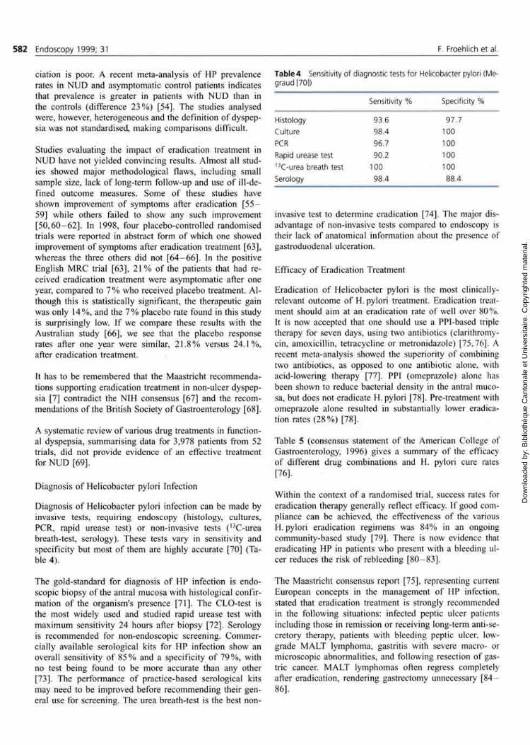
Diagnosis of Hel icobacter pylori infection can be made by invasive tests, requir ing endoscopy (histology, cultures, PCR, rapid urease test) or non-invasive tests ( l 3 C - u r e a breath-test , serology). These tests vary in sensitivity and specificity but mos t of them are highly accurate [70] (Table 4) .
The gold-s tandard for diagnosis of H P infection is endoscopic biopsy of the antral mucosa with histological confirmat ion of the organism's presence [71]. The CLO-tes t is the most widely used and studied rapid urease test with m a x i m u m sensitivity 24 hours after biopsy [72]. Serology is r ecommended for non-endoscopic screening. C o m m e r cially available serological kits for H P infection show an overall sensitivity of 8 5 % and a specificity of 7 9 % , wi th no test being found to be more accurate than any other [73]. The performance of pract ice-based serological kits may need to be improved before r ecommend ing their general use for screening. The urea breath-test is the best non-
Table 4 Sensitivity of diagnostic tests for Helicobacter pylori (Me-graud [70])
Sensitivity % Specificity %
Histology 93.6 97.7
Culture 98.4 100
PCR 96.7 100
Rapid urease test 90.2 100 1 3 C-urea breath test 100 100
Serology 98.4 88.4
invasive test to determine eradication [74]. The major disadvantage of non-invasive tests compared to endoscopy is their lack of anatomical information about the presence of gastroduodenal ulceration.
Efficacy of Eradication Treatment
Eradicat ion of Hel icobacter pylori is the most clinically-relevant ou tcome of H. pylori t reatment. Eradication treatment should aim at an eradication rate of well over 8 0 % . It is now accepted that one should use a PPI-based triple therapy for seven days, using two antibiotics (clarithromycin, amoxicil l in, tetracycline or metronidazole) [75 ,76] . A recent meta-analysis showed the superiority of combining two antibiotics, as opposed to one antibiotic alone, with acid-lowering therapy [77]. PPI (omeprazole) alone has been shown to reduce bacterial density in the antral mucosa, but does not eradicate H. pylori [78]. Pre-treatment with omeprazo le alone resulted in substantially lower eradicat ion rates ( 2 8 % ) [78].
Table 5 (consensus s tatement of the American College of Gastroenterology, 1996) gives a summary of the efficacy of different drug combinat ions and H. pylori cure rates [76].
Within the context of a randomised trial, success rates for eradicat ion therapy generally reflect efficacy. If good compliance can be achieved, the effectiveness of the various H. pylori eradicat ion regimens was 8 4 % in an ongoing communi ty-based study [79]. There is now evidence that eradicat ing H P in pat ients who present with a bleeding ulcer reduces the risk of rebleeding [ 8 0 - 8 3 ] .
The Maastr icht consensus report [75], representing current European concepts in the management of HP infection, stated that eradication treatment is strongly recommended in the following situations: infected peptic ulcer patients including those in remission or receiving long-term anti-secretory therapy, patients with bleeding peptic ulcer, low-grade M A L T lymphoma, gastritis with severe macro- or microscopic abnormal i t ies , and following resection of gastric cancer. M A L T lymphomas often regress completely after eradication, rendering gastrectomy unnecessary [ 8 4 -86] .
Dow
nloa
ded
by: B
iblio
thèq
ue C
anto
nale
et U
nive
rsita
ire. C
opyr
ight
ed m
ater
ial.

1. Appropriateness o f Gastroscopy: Dyspepsia Endoscopy 1999; 31 583
Table 5 Cure rates of various Helicobacter pylori eradication regimens (Soil [76])
Drug combination (duration) HP cure rates ( 9 5 % CI)
MOC (1 wk) 8 7 - 9 1
AOC (1 wk) 8 6 - 9 1
MOA (1 wk) 7 7 - 8 3
BMT (1 wk) 8 6 - 9 0
BMT (2 wk) 8 8 - 9 0
BMT + 0 ( 1 wk) 9 4 - 9 8
BMA (1 wk) 7 5 - 8 1
BMA (2 wk) 8 0 - 8 6
Legends: A: amoxicil l in, B: b ismuth, C: clar i thromycin, M: metronidazole, T: tetracycline, 0 : omeprazole
Side-effects, usually mild, affect 1 0 % of pat ients receiving triple therapy including bismuth [87]. Omeprazole-based triple therapy was better tolerated than b ismuth-based therapy in a randomised controlled trial [88].
Recurrence after Eradication Treatment
Ulcer recurrence is significantly less c o m m o n a m o n g H. pylori-cured patients versus uncured patients ( 6 % vs . 6 7 % for duodenal ulcer, 4 % vs. 5 9 % for gastric ulcer) [89]. The follow-up t ime in these studies ranged from six to 33 months . In a recently-published prospect ive long-term follow-up study [90] in non-NSAIDs users with endoscopically confi rmed ulcer healing and eradicated H. pylori, no ulcer recurrence was detected over a period of up to 9.8 years.
Confirmation of the success of Hel icobacter eradicat ion is generally considered necessary in patients with persist ing [80] or relapsing symptoms [87] after eradication therapy. Symptoms alone may not allow to dist inguish between ulcer recurrence and reflux esophagit is as a substantial ( 1 0 % ) proportion of duodenal ulcer patients developed reflux esophagitis after H. pylori eradication [91]. Endoscopy therefore seems indicated in these cases. The annual reinfection rate after successful eradication therapy is low (1 .2%) [92].
Impact of Endoscopy on Patient Outcome
There is only limited direct evidence of endoscopic impact on outcome in patients with dyspepsia; that is, studies comparing the ou tcome of patients with dyspeptic symptoms who either did or did not undergo diagnost ic endoscopy. Three studies merit discussion here. The first study [93] randomised dyspeptic patients to empirical H 2 -b locker therapy or endoscopy/upper GI series prior to prescript ion of H 2 -blockers . By the end of six months , H 2 -b locker use in both groups was similar (11 vs. 8 .7%) . Equal numbers of patients in each group were asymptomat ic (42.5 vs. 39 .5%) . The second study [94] compared initial upper GI radiography to antacids and reassurance. After six months
of fol low-up, there were no significant differences in symptoms , disability, satisfaction or quali ty of life scores between the two groups. The third study [95] compared prompt endoscopy with H 2 -b locker therapy. In contrast to the two other studies, this trial [95] showed better ou tcome (less work loss, less drug use) and lower costs in the group randomised to prompt endoscopy. Two-thirds of the patients initially randomised to empirical t rea tment were finally endoscoped at one year. In all three studies, Helicobacter pylori infection was not assessed.
An alternative approach to establishing the efficacy of endoscopy in patients with dyspepsia is to examine population trends. The first study [96] examined rates of peptic ulcer-related mortality, hospital isation, surgery, physician visits, work loss and disabili ty re t i rements in the US from 1977 to 1986. All these factors decl ined over t ime. The t ime-scale trends described were attr ibuted to several factors, including the introduction of H 2 -b locke r therapy, reduction in smoking and possible changes due to the increasing use of endoscopy. The second study [97] retrospectively reviewed the use of endoscopy compared to peptic ulcer mortal i ty be tween 1979 and 1989. Al though the util isation of endoscopy rose from 21.7 to 25.6 procedures per t h o u s a n d the mortal i ty rate for pept ic ulcer disease increased by 4 % in w o m e n whi le remaining stable in men. Death certification rates from peptic ulcer decl ined over the four decades in both sexes [98] . The main determinant of this is bel ieved to be the introduction of H 2 - recep to r antagonists in the late 1970s, but other factors, such as therapeutic endoscopy, may also have played a role. A populat ion-based study [99] deal ing with diagnosis , t rea tment and prognosis of gastric cancer showed that endoscopy is progressively becoming the only viable diagnost ic tool. These changes in diagnost ic strategy were , however, associated with less remarkable t rends in t reatment and stage at diagnosis , thus failing to demonst ra te an important contribution by endoscopy to improving ou tcome of gastric cancer. In summary, populat ion studies and studies compar ing outcome before and after introduction of endoscopy have generally not shown conclusively that the introduction of endoscopy substantially affected patient ou tcome.
Clinical Practice: Management Strategies in Dyspepsia
When developing appropria teness criteria for gastrointestinal endoscopy, the st i l l -unanswered quest ion of how diagnosis and treatment of this condit ion should best be managed is crucial . Considerable confusion may exist in the literature as pr imary care physicians use the term "dyspepsia" in general to describe a complex of symptoms referable to the upper digestive tract, whereas specialists (gastroenterologists) often refer to this te rm once endoscopy is negative ( i .e . non-ulcer dyspepsia) . Increasing costs, efforts to contain costs, endoscopic workload and long wait ing lists do not allow endoscopy to be offered to every dyspeptic patient a l though there is evidence that symptoms show a poor predict ive value for endoscopic diagnoses [23,
Dow
nloa
ded
by: B
iblio
thèq
ue C
anto
nale
et U
nive
rsita
ire. C
opyr
ight
ed m
ater
ial.

584 Endoscopy 1999; 31 F. Froehlich et al.
31] , and that a " n o r m a l " result may substantially reduce work loss and medical care consumpt ion [24].
From a conceptual standpoint, endoscopy can be restricted to certain pat ients either based on the response to empirical therapy or based on criteria such as age, H P status, intake of N S A I D s or warning symptoms . Both approaches will be discussed briefly.
Decis ion to Endoscope Based on the Response to Empirical Therapy
In 1985, the Amer ican College of Physicians issued a practice guidel ine for dyspepsia [100], which was also adopted some years later by the Amer ican Society for Gastrointestinal Endoscopy [101]. Al though not based on a clinical trial, this consensus s ta tement r ecommended empirical anti-secretory therapy in all patients with uncomplicated dyspepsia, reserving a diagnost ic upper GI endoscopy for those patients w h o did not respond to therapy or whose symptoms recurred on cessation of t reatment . This recommendat ion was based on observat ions that a precise anatomical diagnosis did not impact on the choice of t reatment for most of the diseases associated with dyspeptic symptoms at that t ime. Fur thermore , it was hoped that empirical t reatment would improve case selection for organic d iagnoses at endoscopy. The role of empirical t reatment as a decision tool for deciding on the use of endoscopy has been quest ioned. Bytzer [95] has shown that case selection of organic diagnoses is not rel iably enhanced by empirical t reatment as only 60 % of ulcer pat ients could be identified with this strategy. In addit ion, empirical therapy proved to be more expensive due to higher work loss and drug consumption. Fur thermore , empirical t reatment pos tpones rather than e l iminates the need for endoscopy [102] as dyspeptic symptoms recur and two-thirds of patients randomised to empirical t rea tment were thus finally endoscoped after one year [95] . Empirical t reatment may also lead to an er roneous diagnosis of functional dyspepsia in patients with endoscopic lesions w h o have not experienced symptom relief but have undergone comple te heal ing of the lesion (e .g . ulcer) because the relat ionship between symptoms and ulcer heal ing is not conclusive [102]. Empir ical therapy therefore proved to be a weak selection criterion for endoscopy.
Decision to Endoscope Based on Specific Patient-Related Characterist ics
Sobala [19] assessed a pol icy of screening dyspeptic patients before endoscopy using a strategy based on Hel icobacter status and use of non-steroidal ant i - inf lammatory drugs. He used three criteria to identify patients expected to show a low yield from diagnost ic endoscopy: 1) age < 4 5 years; 2) negat ive H. pylori test, and 3) no history of N S A I D s use. The screening criteria were applied retrospectively in 842 pat ients with known histological H. pylori status, and prospectively to 293 pat ients referred for d iagnostic endoscopy. Overal l , this screening strategy would have
reduced endoscopy workload by 2 3 . 3 % and would have had a sensitivity rate for detection of peptic ulcer of 9 7 . 4 % . N o peptic ulcer or mal ignant disease was missed in the patients studied prospectively, but six out of 192 peptic ulcers in the histology ( i .e . , retrospective) group would have been missed. In another study [103], 52 subjec ts aged 45 or less were screened by HP serology. All 27 w h o were sero-negative had no ulcer disease while seven out of 25 sero-posit ive patients had ulcer disease. Screening would have avoided 3 5 % of endoscopies in these patients while miss ing 1 3 % of patients with endoscopic findings (esophagit is and gastritis). In a further study [18], 183 dyspeptic patients aged < 4 5 were screened by a his tory-taking of sinister symptoms and regular use of N S A I D s , together with serological testing for H. pylori. Endoscopy was performed in 113 patients, of w h o m 90 were sero-posit ive, 14 had sinister symptoms, and nine had used N S A I D s regularly. The remaining 70 patients w h o were H. pylori sero-negative had no sinister symptoms and had not taken N S A I D s , did not undergo endoscopy but received symptomat ic t reatment . Of these patients, only three were re-referred after screening for endoscopy. Thus. 67 ( 3 7 % ) endoscopies were avoided. When the non-endos-coped screening-negat ive patients were compared with the cohort of endoscoped screening-negative patients, there was no difference be tween the groups in terms of symptom severity. Medicat ion use was , however, significantly less in those pat ients who did not undergo endoscopy [18]. This study indicates that a screening based on H. pylori serology, a history of sinister symptoms (e. g. weight loss, hemorrhage) or a his tory of N S A I D s use was beneficial in dyspeptic pat ients . Thir ty-seven percent of endoscopies were avoided, and drug usage was reduced without disadvantaging those patients not endoscoped.
The above-ment ioned studies all took place in patients referred for endoscopy. Two randomized studies, published as abstracts in 1998, prospectively compared a "test and treat" strategy ( i .e . , H. pylori-posit ive patients with dyspepsia received eradication therapy without endoscopy) with p rompt endoscopy in pr imary care. In the first study [104] which included 500 pat ients , no difference between the two groups was found with respect to rate of symptom-free days, severity of symptoms or number of sick leave days after one year follow-up. However, the prompt endoscopy group resulted in higher patient satisfaction whereas the "test and treat" group was , not surprisingly, associated with a significant (63 % ) reduct ion in endoscopic work load. Patients with a larm symptoms were excluded from the study, and pat ients taking N S A I D s were automatical ly endoscoped. The cost-effectiveness of a "test and treat" strategy, compared to prompt endoscopy, was confi rmed in another randomized control led trial [105]. However, none of these studies directly compared a "test and treat" strategy with a "test and scope" strategy (i. e., H. pylori-positive patients with dyspepsia are routinely endoscoped) in pr imary care. This might yield different results, in as much that the cost advantage of a "test and treat" strategy may be less evident
Dow
nloa
ded
by: B
iblio
thèq
ue C
anto
nale
et U
nive
rsita
ire. C
opyr
ight
ed m
ater
ial.

1. Appropriateness of Gastroscopy: Dyspepsia Endoscopy 1999; 31 585
and the problem of overtreatment with eradication therapy (see below) would be avoided.
The question of whether patients testing posit ive for Helicobacter pylori should be endoscoped ("test and scope") or treated ("test and treat") cont inues to be hotly debated, with indirect evidence coming from several decis ion analyses. The first decision analysis [106] in HP-posi t ive patients with dyspepsia concluded that initial anti-H. pylori therapy is the most cost-effective management strategy. Results were not substantially affected by varying the degree of H. pylori eradication, by the side-effects of antibiotics, or the range of symptoms in curing H. pylori infection. Endoscopy-related costs would need to be reduced by 9 6 % before the two strategies become equally cost-effective. Another decision analysis [107] concluded that eradication treatment is less costly than H 2 -b locke r therapy in patients under 45 years of age with dyspepsia . The model in this study used endoscopy to identify appropriate patients to receive eradication t reatment (patients with ulcer disease). When the initial cost of identifying appropriate patients for eradication treatment is added to the analysis, the cost savings of eradication treatment take almost eight years to accrue. Similar results were obtained in a third decision analysis [108]. A further decision analysis came to a different result. Direct medical charges in the first year after the onset of dyspepsia were compared be tween three strategies: prompt endoscopy, empirical therapy ( ^ - b l o c k ers) or testing for H. pylori [109]. Medical charges were-2162 US dollars for prompt endoscopy and 2122 US dollars for empirical therapy. Initial non-invasive testing for H. pylori cost less than prompt endoscopy if H. pylori-positive patients with dyspepsia received ant imicrobial therapy without endoscopy (that is, "test and treat" strategy) but would have cost more if patients with H. pylori were routinely endoscoped ("test and scope" strategy). The authors concluded that the choice of the opt imal management strategy was a " toss-up". Only very modest savings may result from practice guidelines that r ecommend empirical ant i -HP therapy in the management of patients with dyspepsia .
Table 6 "Test and scope" strategy
Pros Cons
Establishes clear diagnoses and allows biopsies
Allows exclusion of neoplasia
Reduces anxiety (patients' and physicians')
Avoids over-treatment of patients w h o wou ld not need eradication therapy (e. g. esophagitis).
Cost-effective if cost of endoscopy under 500 US dollars [110]
Cost of endoscopy (variable according to country)
Potential complications of upper GI endoscopy (rare)
Inconvenience of endoscopy (unpleasantness, pain, t ime, etc.)
Table 7 "Test and t reat" strategy
Pros Cons
Lowers the general HP prevalence and thus the future risk of gastric carcinoma and HP-related diseases
Dramatic reduction of endoscopic work- load wi th consequent cost savings
Allows management in primary care
Over-treatment of patients w h o do not need eradication therapy (e.g. esophagitis/ reflux disease, non-ulcer dyspepsia)
Potential deterioration of reflux symptoms
Missing of significant endoscopic diagnoses (e.g. gastric ulcer, neoplasia, Barrett's esophagus)
Development of resistance to antibiotics
Side-effects of antibiotics
Insufficient sensitivity and specificity of rapid HP serology kits
At the present t ime, randomized studies directly compar ing the "test and scope" and the "test and t reat" strategies in primary care are needed to evaluate ou tcome and patient preferences. The value of each strategy will depend on the prevalence of H. pylori (low prevalence al lowing more savings than high prevalence in the "test and scope" strategy), the still unproved impact of H P eradication therapy on outcome in documented non-ulcer dyspepsia, patient and doctor preferences, and the cost of endoscopy (very variable according to the country) . As the cost of upper GI endoscopy differs greatly between the United States (> 1000 U S dollars) and Europe, cost-effectiveness of upper GI endoscopy must be judged differently. Thus , endoscopy may be cost competi t ive if its cost is 2 0 0 - 5 0 0 U S dollars [110] , which is the case in most European countries. The following table briefly summarises the pros and cons of the "test and scope" (Table 6) and the "test and t reat" (Table 7) strategies:
In conclusion, the quest ion of whether "test and scope" or "test and treat" should be the preferred managemen t strategy remains open. Taking into account the uncertain efficacy of eradicat ion t reatment in non-ulcer dyspepsia with the huge risk of "over t rea tment" in a test and treat strategy, the lower cost of endoscopy in Europe , current state of knowledge and all the pros and cons stated above, a "test and scope" strategy seems, at the present t ime, preferable. However, uncertainty will m a k e this quest ion one of the most prominent to be debated.
Surveillance Endoscopy in Patients with Known Ulcer Disease
It has become standard practice to follow gastric, but not duodenal ulcers endoscopical ly up to heal ing, because of concerns that gastric ulcers may represent early gastric malignancy. Diagnosis of gastric cancer in apparent ly benign
Dow
nloa
ded
by: B
iblio
thèq
ue C
anto
nale
et U
nive
rsita
ire. C
opyr
ight
ed m
ater
ial.

586 Endoscopy 1999; 31 F. Froehlich et al.
gastric ulcers has been reported in 1 to 6 % of pat ients wi th gastric ulcer [ 1 1 1 - 1 1 3 ] . Bytzer et al. evaluated the benefits of routine endoscopic follow-up of gastric ulcer to detect mal ignancy. They found that each curable gastric cancer was found at the expense of approximate ly 250 follow-up endoscopies [114] . To our knowledge , there has been no randomized control led trial or prospect ive study compar ing ou tcome for pat ients with and without endoscopic follow-up of gastric ulcer to heal ing. Two retrospective studies reviewed the clinical course in patients d iagnosed with gastric ulcer. The first study [115] reviewed 148 gastric ulcers followed up by serial endoscopy over a 5-year period. One hundred and seven patients were followed to heal ing and 41 cases did not heal. The average number of endoscopies per case was 2.7. O f 67 gastric cancers diagnosed during the same t ime period, 62 were suspected of being mal ignant by their macroscopic appearance and only one cancer was missed after biopsy and/or brush cytology. The authors es t imate that favoring a policy of single endoscopy without follow-up when all signs indicate a benign ulcer would result in significant cost savings, as compared to the practice of routine follow-up endoscopy. Another study [111] looked at the impact of gastric ulcer surveil lance to detect gastric carc inoma after surgery. Patients wi th macroscopi -cally and histologically benign gastric ulcer were asked to return after four weeks of therapy. Of 142 pat ients with an initial d iagnosis of benign gastric ulcer, 1.8% had mal ignancy documented on repeat examinat ion. Fol low-up examinat ions did not, however, result in significant differences in 5-year survival rates. A large popula t ion-based long-term cohort study [116] in hospital ised patients with gastric or duodenal ulcers found that the risk of gastric cancer was almost twice the expected rate in patients with gastric ulcers, whereas the risk was less in patients with duodenal ulcers. The authors conclude that gastric ulcer disease and gastric cancer have etiological factors in c o m m o n .
Dyspepsia in Patients Taking NSAIDs
Prevalence of NSAID- Induced Gas t ro-Duodenal Disease
The use of N S A I D s in the general populat ion is extremely frequent. In a popula t ion-based study in the U S A , age- and gender-adjusted annual prevalence rates for aspirin and non-aspir in N S A I D s use in the elderly were 6 0 % and 2 6 % respectively [117]. Fifteen percent of these patients presented wi th dyspepsia , 1 3 % with hear tburn. N S A I D s are the second most c o m m o n cause of peptic ulcer and are now bel ieved to be responsible for the majority of those ulcers not associated wi th H. pylori infection [118]. In a meta-analys is of 16 studies from 1975 to 1990, examining the associat ion be tween N S A I D use and adverse gastrointestinal events, N S A I D users were calculated to be at a threefold greater risk of deve lopment of serious adverse events (GI bleeding, surgery or death) than non-users [119]. The risk appeared to be greatest in the first few months of t reatment , age > 6 5 , in the presence of concomitant steroid use and where there was a previous history of GI events [119] . In a case-control study [118] , the relative
risk for development of peptic ulcer disease among current N S A I D users was 4 . 1 , with the greatest risk in the first month of use. N S A I D use is associated with a higher rate of dyspepsia [117]. However, symptoms are not strong predictors of the presence of endoscopic damage [120]. More recent N S A I D types have been claimed to have less damaging effects on the gastro-duodenal mucosa, primarily by inhibit ing more selectively cyclooxygenase-2, and thus increasing tolerabili ty [121 ,122] . Nabumetone thus seems to have significantly lower ulcerogenic potential than naproxen [123 ,124] , but probably also less clinical efficacy [124], N S A I D use is also associated with non-specif ic ulceration of the small intestinal mucosa ( 8 . 4 % of the patients) that can lead to l ife-threatening complicat ions [125].
NSAID- Induced Ulcer Disease and Helicobacter pylori
In N S A I D s users , there is no difference in the frequency of dyspept ic symptoms between patients with and without HP infection, suggest ing that N S A I D s do not increase susceptibility to Helicobacter infection [126 ,127] . A randomized study recently showed that eradication of Helicobacter pylori before starting N S A I D s therapy reduces the occurrence of NSAID- induced peptic ulcers [128]. In this study, H. pylori seems to have a pathogenic role in NSAID-induced ulcer disease. In contrast , three randomized trials published as abstracts in 1998 failed to show a beneficial impact of HP eradicat ion on NSAID- induced ulcers. Thus eradication t reatment did not accelerate the heal ing of already established ulcers [129] nor prevent the development of ulcers in long-term N S A I D users . [130]. A third trial even showed that eradicat ion treatment was associated with reduced ulcer heal ing [131] . In conclusion, eradication of H P in chronic N S A I D users is probably not justified.
Prophylaxis of NSAID- Induced Ulcers
A large meta-analys is on the prevention of NSAID-induced mucosal injury in 4,325 pat ients [132] concluded that misoprostol , but not H 2 -b lockers , reduced the risk of gastric ulcers. It was also found that both misoprostol and H 2 -blockers prevented duodenal ulcer in long-term NSAIDs-users . These findings were conf i rmed in other randomised control led trials [ 1 3 3 - 1 3 5 ] . Misoprostol was also shown to significantly reduce serious NSAID- induced upper gastrointestinal compl ica t ions such as perforation, gastric outlet obstruction and bleeding. These results were obtained in large, well-conducted, randomised trials in 8,843 patients wi th chronic rheumatoid arthritis [134]. However, misoprostol is often associated with side-effects such as diarrhea and abdominal c ramps [136]. The prophylactic effect of omeprazo le in N S A I D s users was recently assessed in a placebo-control led, randomised study [136]. During a 3-mon th study period, 4 . 7 % of omeprazole- treated patients developed duodenal or gastric ulcers, compared with 1 6 . 7 % of placebo-treated patients. In addition, the development of dyspept ic symptoms was also significantly reduced with omeprazol , compared to placebo.
Dow
nloa
ded
by: B
iblio
thèq
ue C
anto
nale
et U
nive
rsita
ire. C
opyr
ight
ed m
ater
ial.

1. Appropriateness of Gastroscopy: Dyspepsia Endoscopy 1999; 31 587
In a double-blind randomized study publ ished in 1998 [137], omeprazole healed and prevented ulcers more effectively than did ranit idine in N S A I D s users. Another double-blind randomized trial compar ing omeprazo le 20 mg, 40 mg or misoprostol 800 mg daily found that the overall healing rates of ulcers and symptoms were similar for the three t reatment regimens. However, omeprazole was better tolerated and associated with a lower rate of relapse during maintenance treatment than misoprostol [138].
Impact of Endoscopy in N S A I D s Users
There are to our knowledge no studies compar ing ou tcome of patients with uncompl ica ted NSAID- induced peptic disease with and without endoscopy.
Stress Ulcer
Although endoscopic studies have demonst ra ted gross mucosal injury within hours of a stressful event in nearly 1 0 0 % of patients examined, most stress ulcers heal when normal gastric defence mechan i sms are restored. In a rand o m i s e d controlled trial [139] , 8 0 % of patients requir ing aortic surgery developed stress ulcers post-operatively. A rigorously-conducted meta-analysis publ ished recently [140], and including 63 randomised trials in 7,218 patients , addressed ulcer prophylaxis in critically-ill adult patients. Sucralfate was associated with a lower morbidi ty rate compared with antacids and a trend towards lower mortal i ty when compared with H 2 - recep tor antagonists . However, none of the three t reatments studied (sucralfate, ^ - r e c e p tor antagonists , NSAIDs) revealed a significant effect on mortality rate. Stress-ulcer bleeding is rare ( 1 - 1 . 5 % ) [141 ,142] . Sucralfate significantly decreased overt bleeding in comparison with both placebo and N S A I D s . For clinically-important bleeding, H 2 - recep tor antagonists remained superior to placebo.
The role of PPI in stress ulcer prophylaxis has been studied in a recent randomized trial. Sixty-seven high-risk patients were randomized to receive either ranit idine 1 5 0 m g or omeprazole 40 mg per day [143]. Eleven patients in the ranitidine and two patients in the omeprazole group developed clinically important bleeding ( p < 0 . 0 5 ) . Despite its potent acid inhibition, nosocomial pneumonia was seen in one patient only under omeprazole , compared to 5 patients receiving ranitidine. Further studies are needed to determine the role of PPI in stress ulcer prophylaxis [144].
Only a small proport ion of patients present ing with complications such as gastrointestinal hemor rhage or perforation, require medical and/or surgical intervention [145]. The role of endoscopy in bleeding stress ulcers is discussed in a separate publication (upper gastrointestinal bleeding and other alarm symptoms) [146].
Complicated Peptic Ulcer Disease
Hemorrhage as a compl ica t ion of ulcer disease has been dealt with in a separate publ icat ion [146]. The ep idemiology of pept ic ulcer perforation has evolved over the past 50 years: incidence has d ec r ea s ed except in women over 65 years of age, and there has been an increase in m e a n age at t ime of perforation and a decrease of the m a l e : f e m a l e ratio [147]. The short- term mortal i ty of pept ic ulcer disease has fallen from 1952 to 1990 [148]. Uncer ta inty remains about the role of Hel icobacter pylori in the pa thogenesis of ulcer perforation since 5 0 % of pat ients with perforation seem to be HP-negat ive [147] . The single most important risk factor associated with both ulcer perforation and ulcer bleeding is the increasing use of N S A I D s [147]. The localisation of perforation has also changed over t ime, wi th perforation now being more frequently encountered in the pyloric and prepyloric area than in the d u o d e n u m [149].
Gastric Cancer
Prevalence, Incidence and Risk Factors of Gastric Cancer
The prevalence of gastric cancer in dyspept ic patients in Europe is in the order of 1 - 2 % . In three recent prospective studies [ 1 7 , 2 0 , 2 1 ] , two gastric cancers were found in 2,598 dyspept ic patients under 40 years of age (0.8 % o ) , vs. 88 cases in 4,843 pat ients over 40 to 45 years ( 1 . 8 % ) , showing a striking higher-age p redominance wi th gastric cancer being very rare in young dyspept ic pat ients .
Fifty years ago, s tomach cancer was the leading cause of death from cancer in males in the U S A . Since then, morta l ity and incidence have decreased virtually everywhere . There is a band of high- and above-average incidence from Central Italy to the Swiss border, cont inuing through Bavaria up to the Danish border, whi le the south of Italy, Great Britain and most of France are either average or below-average [150 ,151] . These t rends are bel ieved to be due to changes in food preparat ion and storage, and differences in consumpt ion of fruit and vegetables . Classical risk condit ions for gastric cancer are the following [152]: chronic atrophic gastritis and intestinal metaplasia , pernic ious anemia, partial gast rectomy for benign disease, Hel icobacter pylori infection, Mene t r ie r ' s Disease, gastric adenomatous polyps. Genet ic and environmental factors include a family history of gastric cancer, low consumpt ion of fruit and vegetables , consumpt ion of s a l t ed smoked foods, cigarette smoking, low social and economic status, and b lood type A.
Hel icobacter pylori was declared a Class I carcinogen in June 1994 (World Health Organisat ion) . Available evidence on the relat ionship between Hel icobacter pylori and gastric cancer was assessed in a 1996 consensus s tatement [153]. H P is the major cause of multifocal atrophic gastritis and is also believed to lead to the development of intestinal metaplasia [98], whi le chronic gastritis is clearly not associated with any increased risk of cancer [153]. The Eurogast
Dow
nloa
ded
by: B
iblio
thèq
ue C
anto
nale
et U
nive
rsita
ire. C
opyr
ight
ed m
ater
ial.

588 Endoscopy 1999; 31 F. Froehlich et al.
Study Group [41] , a prospect ive epidemiological study in over 3,000 subjects of 14 populat ions in Europe , U S A and Japan, found a six-fold increased risk of gastric cancer in populat ions with 100 % H P infection compared with populations without infection. The hypothesis that H P infection is a risk factor for gastric cancer is further endorsed by three large case-control led studies [ 1 5 4 - 1 5 6 ] . Most persons infected with H P will , however, never develop a gastric carc inoma and other factors that increase the risk of carc inoma a m o n g persons infected with H P therefore need to be identified [156]. Early-life infection by Hel icobacter pylori increases the risk of developing both gastric cancer and gastric ulcers [157].
Hel icobacter pylori infection is invariably associated with the presence of lymphoid follicles which are precursors of M A L T lymphomas [153]. There is evidence that the successful cure of Hel icobacter pylori infection results in the regression, and perhaps even the cure, of M A L T lymphoma in 5 0 - 7 5 % of patients [84].
Symptoms of Gastric Cancer
Al though gastric cancer is a mat ter of concern for clinicians evaluating patients with dyspepsia , mos t pat ients with gastric cancer do not develop symptoms or signs until the disease is no longer curable. Superficial and surgically curable gastric carc inoma typically produce no symptoms [152]. In a prospect ive series of 720 pat ients with gastric carc inoma, only 8 % were eligible for curative resection [158]. In a large review performed by the Amer ican College of Surgeons [152] , weight loss ( 6 2 % ) and abdominal pain ( 5 2 % ) were the most frequent symptoms at the t ime of initial d iagnosis . Abou t 1 0 % of all gastric cancer patients present with hematemes i s or melena , and patients present ing with bleeding rarely have early cancers [159].
Histological Issues in Gastric Cancer
Differences in diagnost ic criteria for gastric carc inoma between Japanese and Western pathologists may contr ibute to the relatively high incidence and good prognosis of gastric cancer in Japan. Thus , in Japan, gastric carc inoma is diagnosed on nuclear and structural criteria even w h e n invasion is absent according to the Western viewpoint [160]. In a prospect ive, mul t i -center study, it was found that gastric cancer was associated wi th 3 6 % of modera te and with 8 0 % of severe gastric epithelial dysplasia; the follow-up of patients with dysplasia considerably enhances the chances of d iagnosing gastric cancer in its early stages [161]. In these patients , a repeat endoscopy every three months is rec o m m e n d e d in presence of modera te dysplasia with immedia te control endoscopy and mult iple biopsies in the presence of severe dysplasia.
Impact of Endoscopy on Detection Rate
There have been important changes in the diagnostic strategy for gastric cancer, endoscopy being now the most frequently-used diagnostic tool [99]. The proportion of resections for cure increased from 38 to 5 0 % , as did the proportion of cases confined to the gastric wall ( 6 - 12%) . The investigation of dyspeptic patients over 40 years of age after their first consultat ion with the general practit ioner could increase the proport ion of early gastric cancers detected to 2 6 % and the proport ion of operable cases to 6 3 % [162]. The most obvious trends in the management of gastric cancer c o m e from the reduct ion of operative mortality rate. Endoscopic surveil lance in post-gastrectomy patients, aiming at detect ing early gastr ic-s tump cancer, does not seem to reduce mortal i ty [163] , and the risk of developing gastric cancer in these pat ients does not seem to be enhanced as compared to the general populat ion [164]. Endoscopic ul trasound has been shown to better assess T and N categories pre-operat ively than computed tomography or inter-operative surgical assessment [ 1 6 5 - 1 6 7 ] .
2. Panel Results
Consider ing the above review of relevant literature, the panel evaluated 192 specific theoretical patient scenarios related to the use of gastrointestinal endoscopy in patients with dyspepsia .
Definition of Terms
All te rms and definit ions were reviewed and approved by the panelists before proceeding to ratings of clinical indicat ions; they are listed in Table 8.
Clinical Variables
The clinical variables used to describe the list of indicat ions related to dyspepsia are shown in Table 9. The main variable used to structure the list of indications for dyspepsia was the parameter of previous investigations, resulting in four main sub-categories.
General Panel Results
Dyspeps ia was assessed by 192 clinical scenarios within 4 sub-categories: no previous investigation done (48 items), previous or upper GI (UGI) series upper GI endoscopy (UGE) normal (48 i tems), U G E or UGI series done and showing duodenal or prepyloric ulcer, duodenit is or erosive gastritis (48 i tems), and U G E or UGI series showing gastric ulcer (48 items). Of the 192 scenarios, the panel rated 113 ( 5 9 % ) as inappropriate , 31 ( 1 6 % ) as uncertain and 48 ( 2 5 % ) as appropriate . The rate of overall agreement between panelists was high ( 7 2 % of the scenarios). Although a dist inction was initially m a d e between first/second and recurrent episodes, the panelists did not wish to maintain this distinction, arguing that their clinical judgment would
Dow
nloa
ded
by: B
iblio
thèq
ue C
anto
nale
et U
nive
rsita
ire. C
opyr
ight
ed m
ater
ial.

1. Appropriateness of Gastroscopy: Dyspepsia Endoscopy 1999; 31 589
Table 8 Definition of terms
Dyspepsia is defined as pain or discomfort in the upper abdomen, including nausea, vomit ing, early satiety, epigastric fullness, but not heartburn or dysphagia.(Isolated heartburn or regurgitation are dealt wi th in the article on reflux disease [10],
Uncomplicated dyspepsia Dyspepsia wi thout alarm symptoms. (Hematemesis, melena, esophageal dysphagia, unexplained weight loss, iron-deficiency anemia are dealt w i th in the article on alarm symptoms [146].
Episode of dyspepsia Minimum duration to be considered as one episode: 4 weeks. Time interval for the definit ion of the onset of a new episode: 1 month free of symptoms wi thout treatment.
Eradication treatment for Helicobacter pylori infection Treatment regimen composed of t w o antibiotics and an PPI/H2
blocker wi th an eradication rate supposed to exceed 9 0 % .
Helicobacter test According to the situation, either a non-endoscopic test (serology, C 1 3 breath-test), or an endoscopic test (urease test, histology, culture).
Empirical acid-lowering treatment In order to serve as a decisional tool for the panel, the minimum duration of treatment is > 1 week of continuous intake. The type of treatment is either standard doses of an PPI (e.g. omeprazole 20 mg/d, lansoprazole 30 mg/d, or pantoprazole 40 mg/d) or H 2-blockers (e.g. ranitidine 300mg/d) or continuous high-dose antacid treatment (e. g. 4 x 5 ml/d aluminium hydroxide, sucralfate 2 x 2 g/d etc).
NSAIDs intake Continuous intake of NSAIDs for > 3 days, or intermittent intake of NSAIDs at onset of symptoms at least every 2 days for at least 1 week
Previous investigations A previous investigation by either an UGI endoscopy or UGI series performed within 2 years of the present episode of dyspepsia.
be similar in each case. Appropr ia teness is def ined in a separate publ icat ion in this issue of the Journal [1].
Specific Clinical Panel Results
Descript ion of Appropr ia teness
The main results related to appropr ia teness are worded as an overall s ta tement (Table 10) encompass ing several clinical scenarios (clustering). In some cases , the same scenario may apply to more than one statement . One hundred and sixty-seven of the 192 indicat ions ( 9 4 % ) could be characterized by the eight overall s ta tements given below. Detai led appropriateness and necessi ty criteria encompass ing all 192 indicat ions are available in a computer ized form accessible via Internet (ht tp: / /www.epage.ch) .
In HP-posi t ive pat ients wi th persis t ing symptoms and not having received eradicat ion t reatment , we assessed whether panelists would favor a " tes t -and-scope" strategy ( i .e . , endoscope dyspept ic pat ients testing posi t ive for H. pylori) or a " test-and-treat" strategy (i. e., treat dyspept ic pat ients empirically if test ing posit ive). Sixteen scenarios pertain to this situation. In pat ients > 4 5 years of age, the " test-and-s c o p e " strategy was favored unless previous investigations showed duodenal or prepyloric ulcer or duodenit is . In the presence of a previous history of gastric ulcer, the "test-and- scope" strategy was always preferred. The "test-and-treat s t ra tegy" was preferred if previous investigations had shown duodenal or prepyloric ulcer or duodeni t is , or in patients < 4 5 years of age in w h o m previous U G E or U G I series were normal .
Table 9 Clinical variables used in individuals presenting w i th dyspepsia (192 indications)
Variables Number of categories Categories
Age 2 - > 4 5 years old - < 4 5 years old
NSAIDs 2 - no - yes
Helicobacter pylori 3 - no HP test - HP test negative - HP test positive
Previous investigations of similar symptoms 4 - no previous investigation or previous investigation w i th results unknown
- UGI endoscopy or UGI series w i th normal results - UGI endoscopy or UGI series showing duodenal
ulcer, prepyloric ulcer, duodenitis or erosive gastritis
- UGI endoscopy or UGI series showing gastric ulcer
Empirical acid-lowering treatment 2 - no treatment or inadequate treatment (in HP-negative patients) or HP eradication - adequate treatment given treatment (in HP-positive patients)
Response to empirical acid-lowering or HP 2 - symptoms not resolved eradication treatment, respectively - symptoms resolved
Dow
nloa
ded
by: B
iblio
thèq
ue C
anto
nale
et U
nive
rsita
ire. C
opyr
ight
ed m
ater
ial.

590 Endoscopy 1999; 31 F. Froehlich et al.
Table 10 Description of appropriateness of indications for upper gastrointestinal endoscopy in individuals wi th dyspepsia
Clinical Situation
In individuals wi th uncomplicated dyspeptic symptoms that resolved wi th or w i thou t treatment, indication for gastrointestinal endoscopy is generally inappropriate w i th the exception of some scenarios related to patients > 45 years old wi th previous gastric ulcer (uncertain)
In individuals w i th persistent dyspepsia, and wi th a previous history of gastric ulcer, indication for gastrointestinal endoscopy is appropriate In individuals aged 45 and over wi th persistent dyspepsia, and wi thout any previous investigations or w i th unknown results of previous investigations, indication for gastrointestinal endoscopy is appropriate In individuals aged 45 and over wi th persistent dyspepsia, and normal results of previous investigations of similar symptoms, indication for gastrointestinal endoscopy is generally uncertain unless symptoms persist after HP eradication has been given in a HP-positive patient (appropriate) In individuals aged 45 and over wi th persistent dyspepsia, and previous investigations showing duodenal ulcer, prepyloric ulcer or duodenitis, indication for gastrointestinal endoscopy is generally uncertain unless empiric acid lowering treatment has been given and HP test is negative (appropriate) unless symptoms persist after HP eradication has been given in a HP-positive patient (appropriate)
In individuals aged less than 45 w i th persistent dyspepsia, and wi thout any previous investigations or w i th unknown results of previous investigations, indication for gastrointestinal endoscopy is generally uncertain unless HP test is unknown and no empirical acid-lowering treatment has been given (inappropriate) unless symptoms persist after HP eradication has been given in a HP-positive patient (appropriate)
In individuals aged less than 45 w i th persistent dyspepsia, and normal results of previous investigations of similar symptoms, indication for gastrointestinal endoscopy is inappropriate unless symptoms persist after HP eradication has been given in a HP-positive patient (uncertain)
In individuals aged less than 45 w i th persistent dyspepsia, and previous investigations showing duodenal ulcer, prepyloric ulcer or duodenitis, indication for gastrointestinal endoscopy is inappropriate unless empirical acid lowering treatment has been given and HP status is negative or unknown (uncertain) unless symptoms persist after HP eradication has been given in a HP-positive patient (appropriate)
Descr ipt ion of Necess i ty
Twelve out of 192 scenarios ( 6 . 3 % ) were j u d g e d necessary. All necessary indicat ions (Table 11) in uncompl ica ted dyspepsia per ta ined to pat ients > 45 years of age. Necessi ty is defined in a joint publ icat ion in this issue of the Journal [1].
3. Conclusions
The current literature under l ines the frequent occurrence of dyspepsia in clinical pract ice and the wide variat ions in diagnosis and treatment . The advent of Hel icobacter pylori
Table 11 Description of necessity of indications for upper gastrointestinal endoscopy in individuals wi th dyspepsia
Clinical situation
In individuals > 4 5 years of age testing positive for HP, wi th persisting symptoms despite eradication treatment, indication for gastrointestinal endoscopy is necessary
In individuals > 4 5 years of age, never investigated, HP-negative and no NSAIDs intake, wi th persisting symptoms despite acid-lowering treatment, indication for gastrointestinal endoscopy is necessary In individuals > 4 5 years of age w i th a previous history of gastric ulcer, no HP testing or HP test negative, w i th persisting symptoms despite acid-lowering treatment, indication for gastrointestinal endoscopy is necessary
as well as the need for cost conta inment in almost all developed countries have had a profound impact on diagnostic and therapeutic strategies in dyspepsia which are currently hotly debated and widely assessed. The literature suggests that U G E should be used in patients with a reasonably high probabil i ty of a clinically relevant diagnosis such as ulcer disease or cancer.
One third of EPAGE criteria related to dyspepsia. EPAGE criteria j udged performance of U G E often inappropriate ( 5 9 % ) in uncompl ica ted dyspepsia. Very few situations ( 6 % ) were judged necessary. Six clinical and circumstantial parameters permit ted detailed assessment of all possible scenarios: patient age, N S A I D s intake, Helicobacter status, results of previous U G E or UGI series, whether or not empirical antisecretory treatment was given and the clinical response to this t reatment . Although highly detailed and specific, 9 4 % of the scenarios could be encompassed in simple, descriptive s tatements applicable to clinical practice. However, the full potential and utility of these criteria will become apparent on the computer ised version accessible via Internet (ht tp: / /www.epage.ch) that will permit easy applicat ion of all scenarios even in the most complex situations.
Acknowledgement
The authors gratefully acknowledge the selfless commitment and invaluable contr ibution of the expert panel members , w h o m a d e this project possible: Marcel lo Anti (IT), Peter Bytzer (DK) , Mark Cottrill (UK) , Michael Fried (CH) , Roar Johnsen (NO) , Gerd Kanzler (DE) , Francois Lacaine (FR), Cornel is Lamers (NL) , Roger J. Leicester (UK) , Mattijs E. N u m a n s (NL) , Javier P. Piqueras (SP), Jean-Francois Rey (FR), Giacomo Sturniolo (IT), Robert P. Walt (UK) .
This work was supported by the EU B I O M E D II Prog r a m m e (BMH4-CT96-1202 ) , the Swiss National Science Foundat ion (32.40522.94) and the Swiss Federal Office of Educat ion and Science (95.0306-2) .
Dow
nloa
ded
by: B
iblio
thèq
ue C
anto
nale
et U
nive
rsita
ire. C
opyr
ight
ed m
ater
ial.

1. Appropriateness of Gastroscopy: Dyspepsia Endoscopy 1999; 31 591
References
1 Vader JP, Burnand B, Froehlich F, et al. The European Panel on Appropriateness of Gastrointestinal Endoscopy (EPAGE): Project and Methods. Endoscopy 1999; 31: 572-578
2 Vader JP, Froehlich F, Dubois RW, et al. The European Panel on Appropriateness of Gastrointestinal Ensocopy (EPAGE): Conclusions and WWW site. Endoscopy 1999; 31: 687-694
1 Morrissey JF, Reichelderfer M. Gastrointestinal endoscopy (2). N Engl J Med 1991; 325: 1214-1222
J Colin-Jones DG. Management of dyspepsia: report of a working party. Lancet 1988; 1: 576-579
5 Talley NJ, Phillips SF. Non-ulcer dyspepsia: potential causes and pathophysiology. Ann Intern Med 1988; 108: 865-879
6 Barbara L, Camilleri M, Corinaldesi R, et al. Definition and investigation of dyspepsia. Consensus of an international ad hoc working party. Dig Dis Sci 1989; 34: 1272-1276
7 Malfertheiner P, McColl K, Baldi F, et al. Update on Helicobacter pylori research. Dyspepsia. Eur J Gastroenterol Hepatol 1997; 9: 624-625
8 Agreus L, Engstrand L, Svardsudd K, et al. Helicobacter pylori seropositivity among Swedish adults with and without abdominal symptoms. A population-based epidemiologic study. Scan J Gastroenterol 1995; 30: 752-757
9Veldhuyzen van Zanten SJ, Tytgat KM, Pollak PT, et al. Can severity of symptoms be used as an outcome measure in trials of non-ulcer dyspepsia and Helicobacter pylori associated gastritis? J Clin Epidemiol 1993; 46: 273-279
1 0 Bochud M, Gonvers JJ, Vader JP, et al. Appropriateness of gastroscopy: Gastroesophageal reflux disease. Endoscopy 1999; 3 1 : 5 9 6 - 6 0 3
" Talley NJ, Weaver AL, Zinsmeister AR, et al. Onset and disappearance of gastrointestinal symptoms and functional gastrointestinal disorders. Am J Epidemiol 1992; 136: 165-177
1 2 Talley NJ, Zinsmeister AR, Schleck CD, et al. Dyspepsia and dyspepsia subgroups: a population-based study. Gastroenterology 1992; 102: 1259-1268
1 3 Jones RH, Lydeard SE, Hobbs FD, et al. Dyspepsia in England and Scotland. Gut 1990; 3: 401-405
1 4 ICnill-Jones RP. Geographical differences in the prevalence of dyspepsia. Scan J Gastroenterol 1991; 26: 17-24
l s Jones R. Dyspeptic symptoms in the community. Gut 1989; 30: 893-898
1 6 Johnsen R, Bernersen B, Straume B, et al. Prevalences of endoscopic and histological findings in subjects with and without dyspepsia. BMJ 1991; 302: 749-752
l 7 Vaira D, Stanghellini V, Menegatti M, et al. Prospective screening of dyspeptic patients by Helicobacter pylori serology: a safe policy? The Italian Helicobacter pylori Study Group. Endoscopy 1997; 29: 595-601
l 8 Pate l P, Mendall MA, Khulusi S, et al. Salivary antibodies to Helicobacter pylori: screening dyspeptic patients before endoscopy. Lancet 1994; 344: 511-512
1 9 Sobala GM, Crabtree JE, Pentith JA, et al. Screening dyspepsia by serology to Helicobacter pylori. Lancet 1991; 338: 9 4 - 9 6
2 0 Heikkinen M, Pikkarainen P, Takala J, et al. Etiology of dyspepsia: four hundred unselected consecutive patients in general practice. Scan J Gastroenterol 1995; 30: 519-523
2 1 Mansi C, Mela GS, Savarino V et al. Open access endoscopy: a large-scale analysis of its use in dyspeptic patients. J Clin Gastroenterol 1993; 16: 149-153
2 2 Bernersen B, Johnsen R, Bostad L, et al. Is Helicobacter pylori the cause of dyspepsia? BMJ 1992; 304: 1276-1279
2 3 Talley NJ, Weaver AL, Tesmer DL, et al. Lack of discriminant value of dyspepsia subgroups in patients referred for upper endoscopy. Gastroenterology 1993; 105: 1378-1386
2 4 Hansen JM, Bytzer P, Bondesen S, et al. Efficacy and outcome of an open access endoscopy service. Dan Med Bull 1991; 38: 288 -290
2 5 Fraser AG, Ali MR, McCullough S, et al. Diagnostic tests for Helicobacter pylori - can they help select patients for endoscopy? New Zealand Medical Journal 1996; 109: 9 5 - 9 8
2 6 Carpenter HA, Talley NJ. Gastroscopy is incomplete without biopsy: clinical relevance of distinguishing gastropathy from gastritis. Gastroenterology 1995; 108: 917-924
2 7 Richter JE. Dyspepsia: organic causes and differential characteristics from functional dyspepsia. (Review) Scand J Gastroenterol Suppl 1991; 182: 11-16
2 8 Williams B, Luckas M, Ellingham JH, et al. Do young patients with dyspepsia need investigation? Lancet 1988; 2: 1349— 1351
2 9 Bytzer P, Schaffalitzky de Muckadell OB. Prediction of major pathologic conditions in dyspeptic patients referred for endoscopy. A prospective validation study of a scoring system. Scan J Gastroenterol 1992; 27: 987-992
3 0 Kalra L, Price WR, Jones BJ, Hamlyn AN.- Open access fibre-sigmoidoscopy: a comparative audit of efficacy. Br Med J Clin Res Ed 1994; 1998; 6629: 1095-1096
3 1 Mansi C, Mela GS, Pasini D, et al. Patterns of dyspepsia in patients with no clinical evidence of organic diseases. Dig Dis Sci 1990; 35: 1452-1458
3 2 Bytzer P, Hansen JM, Havelund T, et al. Predicting endoscopic diagnosis in the dyspeptic patient: the value of clinical judgement. Eur J Gastroenterol Hepatol 1996; 8: 359-363
3 3 Adang RP, Ambergen AW, Talmon JL, et al. The discriminative value of patient characteristics and dyspeptic symptoms for upper gastrointestinal endoscopic findings: A study on the clinical presentation of 1147 patients. Digestion 1996; 57: 118-134
3 4 Heading RC, Wager E, Tooley PJ. Reliability of symptom assessment in dyspepsia. Eur J Gastroenterol Hepatol 1997; 9: 779-781
3 5 Davenport PM, Morgan AG, Darnborough A, de Dombal FT. Can preliminary screening of dyspeptic patients allow more effective use of investigational techniques? Brit Med J Clin Res Ed 1994; 1985; 6463: 217 -220
3 6 Numans ME, Van der Graaf Y, de Wit NJ, et al. How much ulcer is ulcer-like? Diagnostic determinants of peptic ulcer in open access gastroscopy. Family Practice 1994; 11: 382-388
3 7 Muris JW, Starmans R, Pop P. Discriminant value of symptoms in patients with dyspepsia. J Fam Pract 1994; 38: 139-143
3 8 Dooley CP, Cohen H, Fitzgibbons PL, et al. Prevalence of Helicobacter pylori infection and histologic gastritis in asymptomatic persons. N Engl J Med 1989; 321: 1562-1566
3 9 Graham DY, Malaty HM, Evans DG, et al. Epidemiology of Helicobacter pylori in an asymptomatic population in the United States. Effect of age, race, and socioeconomic status. Gastroenterology 1991; 100: 1495-1501
4 0 Gasbarrini G, Pretolani S, Bonvicini F, et al. A population based study of Helicobacter pylori infection in a European country: the San Marino study. Relations with gastrointestinal diseases. Gut 1995; 36: 838-844
4 1 Anonymous. An international association between helicobacter pylori infection and gastric cancer. The Eurogast shady group. Lancet 1993; 341: 1359-1362
Dow
nloa
ded
by: B
iblio
thèq
ue C
anto
nale
et U
nive
rsita
ire. C
opyr
ight
ed m
ater
ial.

592 Endoscopy 1999; 31 F. Froehlich et al.
4 2 Goodman KJ, Correa P. The transmission of Helicobacter pylori. A critial review of the evidence. Int J Epidemiol 1995; 24: 875-887
4 3 Kuipers EJ, Pena AS, Van Kamp G, et al. Seroconversion for Helicobacter pylori infection. Lancet 1993; 342: 328-331
4 4 Megraud F. Epidemiology of Helicobacter pylori infection: where are we in 1995? Eur J Gastroenterol Hepatol 1995; 7: 292-295
4 5 Graham DY, Lew GM, Klein PD, et al. Effect of treatment of Helicobacter pylori infection on the long-term recurrence of gastric or duodenal ulcer. A randomized, controlled study. Ann Intern Med 1992; 116: 705-708
4 6 Peterson WL. Helicobacter pylori and peptic ulcer disease (see comments). (Review) (97 refs). N Engl J Med 1991; 324: 1043-1048
4 7 Rauws EA. Role of Helicobacter pylori in duodenal ulcer. Drugs 1992; 44: 921-927
4 8 Hentschel E, Brandstatter G, Dragosics B, et al. Effect of ranitidine and amoxicillin plus metronidazole on the eradication of Helicobacter pylori and the recurrence of duodenal ulcer. N Engl J Med 1993; 328: 308-312
4 9 Veldhuyzen van Zanten SJ, Sherman PM. Helicobacter pylori infection as a cause of gastritis, duodenal ulcer, gastric cancer and nonulcer dyspepsia: a systematic overview. Can Med Assoc J 1994; 150: 177-185
5 0 Elta GH, Scheiman JM, Barnert JL, et al. Long-term follow-up of Helicobacter pylori treatment in non-ulcer dyspepsia patients. Am J Gastroenterol 1995; 90: 1089-1093
5 1 Greenberg RE, Bank S. The prevalence of Helicobacter pylori in nonulcer dyspepsia. Importance of stratification according to age. Arch Intern Med 1990; 150: 2053-2055
5 2 Holtmann G, Goebell H, Holtmann M, Talley NJ. Dyspepsia in healthy blood donors. Pattern of symptoms and association with Helicobacter pylori. Dig Dis Sci 1994; 39: 1090-1098
5 3 Bouche O. Should Helicobacter pylori be eradicated in a patient with chronic gastritis? (Review) (127 refs) (French). Gastroenterol Clin Biol 1996; 20: S143-S153
5 4 Armstrong D. Helicobacter pylori infection and dyspepsia. Scand J Gastroenterol Suppl 1996; 215: 3 8 - 4 7
5 5 McCarthy C, Patchett S, Collins RM, et al. Long-term prospective study of Helicobacter pylori in nonulcer dyspepsia. Dig Dis Sci 1995; 40: 114-119
5 6 Gilvarry J, Buckley MJ, Beattie S, et al. Eradication of Helicobacter pylori affects symptoms in non-ulcer dyspepsia. Scan J Gastroenterol 1997; 32: 535-540
5 7 Sheu BS, Lin CY, Lin XZ, et al. Long-term outcome of triple therapy in Helicobacter pylori-related nonulcer dyspepsia: a prospective controlled assessment. Am J Gastroenterol 1996; 91: 441 -447
5 8 Lazzaroni M, Bargiggia S, Sangaletti O, et al. Eradication of Helicobacter pylori and long-term outcome of functional dyspepsia. A clinical endoscopic study. Dig Dis Sci 1996; 41: 1589-1594
5 9 O'Morain C, Gilvarry J. Eradication of Helicobacter pylori in patients with non-ulcer dyspepsia. (Review) (40 rfs). Scand J Gastroenterol Suppl 1993; 196: 3 0 - 3 3
6 0 Veldhuyzen van Zanten SJ, Malatjalian D. The effect of eradication of Helicobacter pylori on symptoms of non-ulcer dyspepsia (NUD): a randomized, double blind, placebo-controlled trial (abstract). Gastroenterology 1995; 108: A250
6 1 Greenberg PD. Prospective, double-blind treatment of Helicobacter pylori in patients with non-ulcer dyspepsia. (Abstract). Gastroenterology 1996; 110: A123
6 2 Schubert TT, Schubert AB, Ma CK. Symptoms, gastritis, and Helicobacter pylori in patients referred for endoscopy. Gastrointest Endosc 1992; 38: 357-360
6 3 McColl KEL. U.K. MRC trial of H. pylori eradication therapy for non-ulcer dyspepsia. (Abstract). Gastroenterology 1998; 114: A222-A222
6 4 Koelz HR. Treatment of Helicobacter pylori does not improve symptoms of functional dyspepsia. (Abstract). Gastroenterology 1998; 114: A182-A182
6 5 Bretagne JF. The course of symptoms after eradication of Helicobacter pylori: a one-year follow-up of a cohort of 258 patients with duodenal ulcer or nonulcer dyspepsia. (Abstract). Gastroenterology 1998; 114: A81-A81
6 6 Talley NJ. Long-term follow-up of patients with non-ulcer dyspepsia after Helicobacter pylori eradication. A randomized double-blind placebo-controlled trial. (Abstract). Gastroenterology 1998; 114: A305-A305
6 7 Anonymous. Nih consensus conference. Helicobacter pylori in peptic ulcer disease. Nih consensus development panel on Helicobacter pylori in peptic ulcer disease. J Am Med Ass 1994: 272: 6 5 - 6 9
6 8 Dyspepsia Management Guidelines. Brit Society of Gastroenterology. http://www.bsg.org.uk/clinical/data/dmg.htm: 1996
6 9 Wilcox CM, Schwartz DA. Endoscopic characterization of idiopathic esophageal ulceration associated with human immunodeficiency virus infection. J Clin Gastroenterol 1993; 16: 251-256
7 0 Megraud F. Advantages and disadvantages of current diagnostic tests for the detection of Helicobacter pylori. Scand J Gastroenterol Suppl 1996; 215: 5 7 - 6 2
7 1 Ateshkadi A, Lam NP, Johnson CA. Helicobacter pylori and peptic ulcer disease. Clin Pharmacy 1993; 12: 3 4 - 4 8
7 2 Laine L, Chun D, Stein C, et al. The influence of size or number of biopsies on rapid urease test results: a prospective evaluation. Gastrointest Endosc 1996; 43: 4 9 - 5 3
7 3 Loy CT, Irwig LM, Katelaris PH, Talley NJ. Do commercial serological kits for Helicobacter pylori infection differ in accuracy? A meta-analysis. Am J Gastroenterol 1996; 91: 1138-1144
7 4 Megraud F. The most important diagnostic modalities for Helicobacter pylori, now and in the future. (Review) (4 refs). Eur J Gastroenterol Hepatol 1997; 1: S 1 3 - S I 5
7 5 Malfertheiner P, Megraud F, O'Morain C, et al. Current euro-pean concepts in the management of Helicobacter pylori infection - the Maastricht consensus report. The European Helicobacter pylori study group. Eur J Gastroenterol Hepatol 1997; 9: 1-2
7 6 Soli AH. Consensus conference. Medical treatment of peptic ulcer disease. Practice guidelines. Practice parameters committee of the American College of Gastroenterology. J Am Med Ass 1996; 275: 622-629
7 7 Holtmann G, Layer P, Goebell H. Proton-pump inhibitors or h2-receptor antagonists for Helicobacter pylori eradication - a meta-analysis (letter). Lancet 1996; 347: 763
7 8 Labenz J, Gyenes E, Ruhl GH, Borsch G. Omeprazole plus amoxicillin: efficacy of various treatment regimens to eradicate Helicobacter pylori. Am J Gastroenterol 1993; 88: 491 495
7 9 Fennerty MB, Lieberman D, Magaret N, et al. Effectiveness of H. pylori treatment regimens in clinical practice: a community-based outcomes study. (Abstract). Gastroenterology 1997; 112: A 1 4 - A 1 4
Dow
nloa
ded
by: B
iblio
thèq
ue C
anto
nale
et U
nive
rsita
ire. C
opyr
ight
ed m
ater
ial.

1. Appropriateness of Gastroscopy: Dyspepsia Endoscopy 1999; 31 593
8 0 Lee J, O'Morain C. Consensus or confusion: a review of existing national guidelines on Helicobacter pylori-related disease. Eur J Gastroenterol Hepatol 1997; 9: 527-531
8 1 Rokkas T, Karameris A, Mavrogeorgis A, et al. Eradication of Helicobacter pylori reduces the possibility of rebleeding in peptic ulcer disease. Gastrointest Endosc 1995; 41: 1-4
8 2 Graham DY, Hepps KS, Ramirez FC, et al. Treatment of Helicobacter pylori reduces the rate of rebleeding in peptic ulcer disease. Scan J Gastroenterol 1993; 28: 939-942
8 3 Jaspersen D, Koerner T, Schorr W, et al. Helicobacter pylori eradication reduces the rate of rebleeding in ulcer hemorrhage. Gastrointest Endosc 1995; 41: 5 - 7
8 4 Bayerdorffer E, Neubauer A, Rudolph B, et al. Regression of primary gastric lymphoma of mucosa-associated lymphoid tissue type after cure of Helicobacter pylori infection. MALT Lymphoma Study Group. Lancet 1995; 345: 1591-1594
8 5 Wotherspoon AC, Doglioni C, Diss TC, et al. Regression of primary low-grade-B-cell gastric lymphoma of mucosa-associated lymphoid tissue type after eradication of Helicobacter pylori. Lancet 1993; 342: 575-577
8 6 Sackmann M, Morgner A, Rudolph B, et al. Regression of gastric MALT lymphoma after eradication of Helicobacter pylori is predicted by endosonographic staging. Gastroenterology 1997; 113: 1087-1090
8 7 Veldhuyzen von ZS, Sherman PM, Hunt RH. Helicobacter pylori: new developments and treatments. CMAJ 1997; 156: 1565-1574
8 8 Lerang F, Mourn B, Ragnhildstveit E, et al. A comparison between omeprazole-based triple therapy and bismuth-based triple therapy for the treatment of Helicobacter pylori infection: a prospective randomized 1-yr follow-up study. Am J Gastroenterol 1997; 92: 653-658
8 9 Hopkins RJ, Girardi LS, Turney EA, et al. Relationship between Helicobacter pylori eradication and reduced duodenal and gastric ulcer recurrence: a review. Gastroenterology 1996; 110: 1244-1252
9 0 Vanderhulst RM, Rauws E.I, Koycu B, et al. Prevention of ulcer recurrence after eradication of Helicobacter pylori - A prospective long-term follow-up study. Gastroenterology 1997; 113: 1082-1086
9 1 Labenz J, Blum AL, Bayerdorffer E, et al. Curing Helicobacter pylori infection in patients with duodenal ulcer may provoke reflux esophagitis. Gastroenterology 1997; 112: 1442-1447
9 2 Forbes GM, Glaser ME, Cullen DJ, et al. Duodenal ulcer treated with Helicobacter pylori eradication: seven-year follow-up. Lancet 1994; 343: 258-260
9 3 Goulston KJ, Dent OF, Logan J, et al. Use of H2-receptor antagonists in patients with dyspepsia and heartburn: a cost comparison. Med J Aust 1991; 155: 2 0 - 2 6
9 4 Goodson JD, Lehmann JW, Richter JM, et al. Is upper gastrointestinal radiography necessary in the initial management of uncomplicated dyspepsia? A randomized controlled trial comparing empiric antacid therapy plus patient reassurance with traditional care. J Gen Intern Med 1989; 4: 367-374
9 5 Bytzer P, Hansen JM, Schaffalitzky de Muckadell OB. Empirical H2-blocker therapy or prompt endoscopy in management of dyspepsia. Lancet 1994; 343: 811-816
9 6 Bloom BS. Cross-national changes in the effects of peptic ulcer disease. Ann Intern Med 1991; 114: 558-562
9 7 Westbrook JI, Rushworth RL. The epidemiology of peptic ulcer mortality 1953-1989: a birth cohort analysis. Intern J Epidemiol 1993; 22: 1085-1092
9 8 La Vecchia C, Lucchini F, Negri E, et al. The impact of therapeutic improvements in reducing peptic ulcer mortality in Europe. Intern J Epidemiol 1993; 22: 9 6 - 1 0 6
9 9 Msika S, Tazi MA, Benhamiche AM, et al. Population-based study of diagnosis, treatment and prognosis of gastric cancer. Brit J Surg 1997; 84: 1474-1478
1 0 0 Kahn K. Endoscopy in the evaluation of dyspepsia. Health and Public Policy Committee, American College of Physicians. Ann Intern med 1985; 102: 266-269
1 0 1 American Society for Gastrointestinal Endoscopy. The role of endoscopy in the management of the patient with peptic ulcer disease. Guidelines for clinical application. Gastrointest Endosc 1988; 34: 4 S - 2 5 S
1 0 2 Bytzer P. Diagnosing dyspepsia: any controversies left? Gastroenterology 1996; 110: 302-306
1 0 3 Tham TC. Application of serological screening for Helicobacter pylori in reducing endoscopy workload. Gastroenterology 1993; 104: A26
1 0 4 Lassen AT. H. Pylori "test and treat" or prompt endoscopy for dyspeptic patients in primary care. A randomized controlled trial of two management strategies: one year follow-up. (Abstract). Gastroenterology 1998; 114: A196-A196
1 0 5 Jones RH. A Helicobacter test and treat strategy: costs and outcomes in a randomized controlled trial in primary care. (Abstract). Gastroenterology 1998; 114: A 2 0 - A 2 0
1 0 6 Ofrnan JJ, Etchason J, Fullerton S, et al. Management strategies for Helicobacter pylori-seropositive patients with dyspepsia - clinical and economic consequences. Ann Intern Med 1997; 126: 280-291
1 0 7 Briggs AH, Sculpher MJ, Logan RP, et al. Cost effectiveness of screening for and eradication of Helicobacter pylori in management of dyspeptic patients under 45 years of age. Br Med J 1996; 312: 1321-1325
108 Fullerton F. Empiric antibiotic therapy versus early endoscopy in dyspeptic patients with positive serologic test for Helicobacter pylori: a decision analysis. (Abstract). Gastroenterology 1995; 108: A15
1 0 9 Silverstein FE, Graham DY, Senior JR, et al. Misoprostol reduces serious gastrointestinal complications in patients with rheumatoid arthritis receiving nonsteroidal anti-inflammatory drugs. A randomized, double-blind, placebo-controlled trial. Ann Intern Med 1995; 123: 241-249
1 1 0 Fendrick AM, Chernew ME, Hirth RA, Bloom BS. Immediate endoscopy or initial Helicobacter pylori serological testing for suspected peptic ulcer disease: estimating cost-effectiveness using decision analysis. Yale Journal of Biology & Medicine 1996; 69: 187-195
1 1 1 Eckardt VF, Giessler W, Kanzler G, Bernhard G. Does endoscopic follow-up improve the outcome of patients with benign gastric ulcers and gastric cancer? Cancer 1992; 69: 301-305
1 1 2 Farinati F, Cardin F, Di Mario F, et al. Early and advanced gastric cancer during follow-up of apparently benign gastric ulcer: significance of the presence of epithelial dysplasia. J Surg Oncol 1987; 36: 263-267
1 1 3 Tragardh B, Haglund U. Endoscopic diagnosis of gastric ulcer. Evaluation of the benefits of endoscopic follow-up observation for malignancy. Acta Chir Scand 1985; 15: 3 7 - 4 1
1 1 4 Bytzer P. Endoscopic follow-up study of gastric ulcer to detect malignancy: is it worthwhile? Scandinavian Journal of Gastroenterology 1991; 26: 1193-1199
1 1 5 Prui t t RE, Truss CD. Endoscopy, gastric ulcer, and gastric cancer. Follow-up endoscopy for all gastric ulcers? Dig Dis Sci 1993; 38: 284-288
Dow
nloa
ded
by: B
iblio
thèq
ue C
anto
nale
et U
nive
rsita
ire. C
opyr
ight
ed m
ater
ial.

594 Endoscopy 1999; 31 F. Froehlich et al.
5 Hansson LE, Nyren O, Hsing AW, et al. The risk of stomach cancer in patients with gastric or duodenal ulcer disease. N Engl J Med 1996; 335: 242-249
7 Talley NJ, Evans JM, Fleming KC, et al. Nonsteroidal antiinflammatory drugs and dyspepsia in the elderly. Dig Dis Sci 1995; 40: 1345-1350
s Graham DY. Treatment of peptic ulcers caused by Helicobacter pylori (editorial; comment). N'Engl J Med 1993; 328: 3 4 9 -350
) Gabriel SE, Jaakkimainen L, Bombardier C. Risk for serious gastrointestinal complications related to use of nonsteroidal anti-inflammatory drugs. A meta-analysis. Ann Intern Med 1991; 115: 787-796
5 Larkai EN, Smith JL, Lidsky MD, Graham DY. Gastroduodenal mucosa and dyspeptic symptoms in arthritic patients during chronic nonsteroidal anti-inflammatory drug use. Am J Gastroenterol 1987; 82: 1 153-1158
1 Battistini B, Botting R, Bakhle YS. COX-1 and COX-2: toward the development of more selective NSAIDs. Drug News and Perspectives (Barcelona) 1994; 7: 501-512
1 Distel M, Mueller C, Bluhmki E, Fries J. Safety of meloxicam: a global analysis of clinical trials. Brit J Rheumatol 1996; 35: 6 8 - 7 7
' Roth SH, Bennett R, Caldron P, et al. A longterm endoscopic evaluation of patients with arthritis treated with nabumetone vs naproxen. J Rheumatol 1994; 21: 1118-1123
' Porro GB, Montrone F, Petrillo M, et al. Gastroduodenal toler-ability of nabumetone versus naproxen in the treatment of rheumatic patients. Am J Gastroenterol 1995; 90: 1485-1488
' Allison MC, Howatson AG, Torrance CJ, et al. Gastrointestinal damage associated with the use of nonsteroidal antiinflammatory drugs (see comments). N Engl J Med 1992; 327: 749-754
' Graham DY, Lidsky MD, Cox AM, et al. Long-term nonsteroidal antiinflammatory drug use and Helicobacter pylori infection. Gastroenterology 1991; 100: 653-657
' Loeb DS, Talley NJ, Ahlquist DA, et al. Long-term nonsteroidal anti-inflammatory drug use and gastroduodenal injury: the role of Helicobacter pylori. Gastroenterology 1992; 102: 1899-1905
! Chan FK, Sung JJ, Chung SC, et al. Randomized trial of eradication of Helicobacter pylori before non-steroidal anti-inflammatory drug therapy to prevent peptic ulcers. Lancet 1997; 350: 975-979
1 Lai KC. Eradication of Helicobacter pylori did not accelerate the healing of gastric and duodenal ulcers/erosions in patients on long-term continuous nonsteroidal anti-inflammatory drugs. (Abstract). Gastroenterology 1998; 114: A192
1 Lai KC. Can eradication of Helicobacter Pylori prevent future development of peptic ulcers in patients receiving long-term continuous nonsteroidal anti-inflammatory drugs. (Abstract). Gastroenterology 1998; 114: A192
1 Hawkey CJ, Tullassay Z. Failure of Helicobacter Pylori eradication to influence site-specific relapse in high risk NSAID users: results of a large six month double blind randomized controlled clinical trial. (Abstract). Gastroenterology 1998; 114: A145
! Koch M, Dezi A, Ferrario F, Capurso L. Prevention of nonsteroidal anti-inflammatory drug-induced gastrointestinal mucosal injury - a meta-analysis of randomized controlled clincal trials. (Abstract). Arch Intern Med 1996; 156: 2321-2332
' Taha AS, Hudson N, Hawkey CJ, et al. Famotidine for the prevention of gastric and duodenal ulcers caused by nonsteroidal antiinflammatory drugs. N Engl J Med 1996; 334: 1435-1439
1 3 4 Agrawal NM, Van Kerckhove HE, Erhardt LJ, Geis GS. Misoprostol coadministered with diclofenac for prevention of gstro-duodenal ulcers, a one-year study. Dig Dis Sci 1995; 40: 1125-1131
1 3 5 Hudson N, Taha AS, Russell RI, et al. Famotidine for healing and maintenance in nonsteroidal anti-inflammatory drug-associated gastroduodenal ulceration (see comments). Gastroenterology 1997; 112: 1817-1822
1 3 6 Ekstrom P, Carling L, Wetterhus S, et al. Prevention of peptic ulcer and dyspeptic symptoms with omeprazole in patients receiving continuous non-steroidal anti-inflammatory drug therapy. A Nordic multicentre study. Scan J Gastroenterol 1996; 31: 753-758
, 3 7 Yeomans ND, Tulassay Z, Juhasz L, et al. A Comparison of Omeprazole with Ranitidine for Ulcers Associated with Nonsteroidal Antiinflammatory Drugs. N Engl J Med 1998; 338: 719-726
1 3 8 Hawkey CJ, Karrasch JA, Szczepanski L, et al. Omeprazole Compared with Misoprostol for Ulcers Associated with Nonsteroidal Antiinflammatory Drugs. N Engl J Med 1998; 338: 727-734
1 3 9 Martin LF. Stress ulcers are common after aortic surgery. Endoscopy evaluation of prophylactic therapy. Am Surg 1994; 60: 169-174
1 4 0 Cook DJ, Reeve BK, Guyatt GH, et al. Stress ulcer prophylaxis in critically ill patients. Resolving discordant meta-analyses. J Am Med Ass 1996; 275: 308-314
1 4 l .Zandstra DF, Stoutenbeek CP. The virtual absence of stress-ul-ceration related bleeding in ICU patients receiving prolonged mechanical ventilation without any prophylaxis. A prospective cohort study (see comments). Intens Care Med 1994; 20: 3 3 5 -340
1 4 2 Cook DJ, Fuller HD, Guyatt GH, et al. Risk factors for gastrointestinal bleeding in critically ill patients. Canadian Critical Care Trials Group. N Engl J Med 1994; 330: 377-381
1 4 3 Levy MJ, Seelig CB, Robinson NJ, Ranney JE. Comparison of omeprazole and ranitidine for stress ulcer prophylaxis. Dig Dis Sci 1997; 42: 1255-1259
1 4 4 Schepp W, Schusdziarra V, Classen M. (Stress ulcer prophylaxis 1995. Balancing between gastrointestinal bleeding and nosocomial pneumonia.) Dtsch Med Wochenstr 1995; 120: 573-579
1 4 5 Marrone GC, Silen W. Pathogenesis, diagnosis and traetment of acute gastric mucosal lesions. Clin Gastroenterol 1984; 13: 635-650
1 4 6 De Bosset V, Gonvers JJ, Froehlich F, et al. Appropriateness of gastroscopy: Bleeding and dysphagia. Endoscopy 1999; 31: 615-622
1 4 7 Hudson N. Excess long-term mortality in patients with ulcer complications. Lancet 1997; 349: 968-969
1 4 8 Svanes C, Lie RT, Lie SA, et al. Survival after peptic ulcer perforation: a time trend analysis. J Clin Epidemiol 1996; 49: 1363-1371
1 4 9 Svanes C, Salvesen H, Stangeland L, et al. Perforated peptic ulcer over 56 years. Time trends in patients and disease characteristics. Gut 1993; 3: 1666-1671
1 5 0 International Agency for Research on Cancer. Atlas of cancer mortality in the European Economic Community. In: Smans M, Muir CS, Boyle P (eds). Lyon, France: I ARC, 1992
1 5 1 International Agency for Research on Cancer. Atlas of cancer mortality in Central Europe. In: Zatonski W, Smans M, Tyc-zynski J, et al. (eds). Lyon, France: IARC, 1996
l 5 2 F u c h s CS, Mayer RJ. Gastric carcinoma. N Engl J Med 1995: 333: 3 2 - 4 1
Dow
nloa
ded
by: B
iblio
thèq
ue C
anto
nale
et U
nive
rsita
ire. C
opyr
ight
ed m
ater
ial.

1. Appropriateness of Gastroscopy: Dyspepsia Endoscopy 1999; 31 595
1 5 3 Gasbarrini G, Genta RM, Anti M, et al. Update on Helicobacter pylori research. Malignancies. Eur J Gastroenterol Hepatol 1997; 9: 621-623
1 5 4 Forman D, Newell DG, Fullerton F, et al. Association between infection with Helicobacter pylori and risk of gastric cancer: evidence from a prospective investigation. Brit Med J 1991; 302: 1302-1305
1 5 5 Parsonnet J, Friedman GD, Vandersteen DP, et al. Helicobacter pylori infection and the risk of gastric carcinoma. N Engl J Med 1991; 325: 1127-1131
1 5 6 Nomura A, Stemmermann GN, Chyou PH, et al. Helicobacter pylori infection and gastric carcinoma among Japanese Americans in Hawaii. N Engl J Med 1991; 325: 1132-1136
'- 7 Blaser MJ, Chyou PH, Nomura A. Age at establishment of Helicobacter pylori infection and gastric carcinoma, gastric ulcer, and duodenal ulcer risk. Cancer Res 1995; 55: 562-565
1 , 8 Matley PJ, Dent DM, Madden MV, et al. Gastric carcinoma in young adults. Ann Surg 1988; 208: 593-596
1 5 9 Moreno-Otero R, Rodriguez S, Carbo J, et al. Acute upper gastrointestinal bleeding as primary symptom of gastric carcinoma. J Surg Oncol 1987; 36: 130-133
1 6 0 Schlemper RJ, Itabashi M, Kato Y, et al. Differences in diagnostic criteria for gastric carcinoma between Japanese and Western pathologists. Lancet 1997; 349: 1725-1729
1 6 1 Farinati F, Rugge M, Di Mario F, et al. Early and advanced gastric cancer in the follow-up of moderate and severe gastric dysplasia patients, a prospective study, i. g. g. e. d.-interdisci-plinary group on gastric epithelial dysplasia. Endoscopy 1993; 25: 261-264
1 6 2 Hallissey MT, Allum WH, Jewkes AJ, et al. Early detection of gastric cancer. Brit Med J 1990; 301: 513-515
1 6 3 Caprilli R, Andreoli A, Capurso L, et al. Oral mesalazine (5-aminosalicylic acid; Asacol) for the prevention of post-operative recurrence of Crohn's disease. Gruppo Italiano per lo Studio del Colon e del Retto (GISC). Alimentary Pharmacology & Therapeutics 1994; 8: 3 5 - 4 3
1 6 4 Iwao T, Toyonaga A, Ikegami M, et al. McCormack's endoscopic signs for diagnosing portal hypertension: comparison with gastroesophageal varices. Gastrointest Endosc 1994; 40: 470-473
1 6 5 Ziegler K, Sanft C, Zimmer T, et al. Comparison of computed tomography, endosonography, and intraoperative assessment in tn staging of gastric carcinoma. Gut 1993; 34: 604-610
1 6 6 Ditt ler HJ, Siewert JR. Role of endoscopic ultrasonography in gastric carcinoma. Endoscopy 1993; 25: 162-166
1 6 7 Grimm H, Binmoeller KF, Hamper K, et al. Endosonography for preoperative locoregional staging of esophageal and gastric cancer. Endoscopy 1993; 25: 224-230
Corresponding Author
Florian Froehl ich, M D , P D
Policlinique Medicale Universitaire Rue Cesar-Roux 19 CH-1005 Lausanne Switzerland
Fax: +41-32-466 29 55 E-mail: [email protected]
Dow
nloa
ded
by: B
iblio
thèq
ue C
anto
nale
et U
nive
rsita
ire. C
opyr
ight
ed m
ater
ial.