€¦ · web viewanalyses were performed using sas software ... were compared using an anova for...
TRANSCRIPT
KU LEUVEN
GROEP BIOMEDISCHE WETENSCHAPPEN
FACULTEIT BEWEGINGS- EN REVALIDATIEWETENSCHAPPEN
Effects of Aerobic Interval Training versus Moderate Continuous Training on
exercise capacity, muscle strength and physical activity in patients with
Coronary Artery Disease
door Matthias Loeckx en Tomas De Mol
Masterproef aangeboden tot het behalen van graad van master in de revalidatiewetenschappen en kinesitherapie
o.l.v.
Prof. Dr. L. Vanhees, promotor
Dra. N. Pattyn, copromotor
LEUVEN, 2014


KU LEUVEN
GROEP BIOMEDISCHE WETENSCHAPPEN
FACULTEIT BEWEGINGS- EN REVALIDATIEWETENSCHAPPEN
Effects of Aerobic Interval Training versus Moderate Continuous Training on
exercise capacity, muscle strength and physical activity in patients with
coronary artery disease
Contributes to the Saintex-CAD study
door Matthias Loeckx en Tomas De Mol
Masterproef aangeboden tot het behalen van graad van master in de revalidatiewetenschappen en kinesitherapie
o.l.v.
Prof. Dr. L. Vanhees, promotor
Dra. N. Pattyn, copromotor
LEUVEN, 2014
Opgesteld volgens de richtlijnen van European Journal of Preventive Cardiology
II

WOORD VOORAF
Vooreerst wil ik Prof. Dr. L. Vanhees bedanken, die als promotor ons de mogelijkheid heeft geboden
om deze masterproef te kunnen verwezenlijken. Met behulp van zijn ervaring en kennis heeft hij ons
uitstekend begeleid en geïnspireerd gedurende het hele verwerkingsproces. Vervolgens wil ik mijn
dank betuigen aan onze copromotor, Dra. N. Pattyn, voor de manier waarop zij ons gemotiveerd en
gecoacht heeft gedurende het gehele verloop van deze masterproef. De gepaste en opbouwende
feedback van beiden waren fundamenteel voor het schrijven van dit geheel.
Daarnaast wil ik Tomas De Mol bedanken, medestudent van deze masterproef, met wie ik op een
aangename manier en met wederzijds respect deze masterproef heb kunnen realiseren.
Eveneens zou ik graag mijn dank betuigen aan het UZ Leuven, de Faculteit voor Bewegings- en
Revalidatiewetenschappen en de medewerkers en patiënten die het mogelijk maakten om de Saintex
studie op te starten en te voltooien. Zonder hun inzet zouden we deze kans wellicht niet hebben
gekregen.
Dank aan mijn ouders die mij de kans hebben gegeven om de studie Revalidatiewetenschappen en
Kinesitherapie aan de Katholieke Universiteit Leuven te kunnen aanvatten. Zonder hun steun was dit
wellicht niet mogelijk geweest.
Tot slot wil ik de kinesitherapeuten die instonden voor de supervisie van de trainingen op de afdeling
cardiale revalidatie bedanken voor de vlotte medewerking en de goede verstandhouding. De uitleg
en ervaren belevingen die ze met ons deelden, boden een extra aan kennis. Eveneens dienden ze als
inspiratiebron voor het verwezenlijken van dit werk.
Deelname aan dit 2-jarige proces van het begeleiden van de trainingen tot het uitschrijven van de
resultaten heb ik als zeer verrijkend voor mijn individuele ontwikkeling ervaren. Ik hoop de
verworven inzichten en vaardigheden in mijn latere carrière nog te kunnen gebruiken.
Leuven, 23 april 2014, M.L.
III
SITUERING
Deze Master thesis is uitgevoerd binnen het onderzoeksdomein van de Afdeling Cardiovasculaire &
Respiratoire Revalidatie van het Departement van Revalidatiewetenschappen van de Faculteit van
Bewegings- en Revalidatiewetenschappen aan de Katholieke Universiteit Leuven. Deze
onderzoeksgroep gaat de effecten na van fysieke activiteit en inspanning op de primaire en
secundaire preventie. De grote onderzoeksdomeinen van Professor Dr. L. Vanhees zijn: 1) Het effect
van cardiale revalidatie en inspanning en de verklarende mechanismen van adaptatie; 2) De rol van
fysieke activiteit en fysieke fitheid in de primaire, secundaire en tertiaire preventie; 3) De veiligheid
van inspanning tijdens cardiale revalidatie en de rol van de cardiovasculaire screening voor preventie
van ernstige cardiale voorvallen tijdens inspanning.
Onze masterproef kadert binnen een groter multicenter project tussen het UZ Leuven en het UZ
Antwerpen, SAINTEX-CAD genaamd. “SAINTEX-CAD” staat voor Study on Aerobic INTerval EXercise
training in Coronary Artery Disease patients. Binnen deze studie worden twee verschillende
trainingsmodaliteiten met elkaar vergeleken, namelijk Aerobe Interval Training (AIT) en Matig
Continue Training (MCT), en dit op parameters zoals het aëroob uithoudingsvermogen, de
levenskwaliteit, de vaatfunctie en de veiligheid van deze trainingen.1,2
Tientallen jaren geleden werd verplichte wekenlange bedrust voorgeschreven als remedie tegen
hartziekten. De evolutie van de geneeskunde bracht alsmaar meer wetenschappelijk bewijs met zich
mee dat deze rust niet aangewezen is. Vandaag de dag wordt regelmatige fysieke activiteit dan ook
gepromoot als belangrijk onderdeel binnen het multidisciplinair behandelingsprogramma voor
hartpatiënten. Naast beweging staan medicatie, rookstop, voedingscontrole en stress management,
voorop in het revalidatieprogramma van een doorsnee hartpatiënt.1
Het is aangetoond dat patiënten met een hoger aëroob uithoudingsvermogen, hogere
spierkrachtwaarden en een hoger niveau van fysieke activiteit een verminderd risico op cardiale
mortaliteit hebben.3,4,5 Binnen deze studie wordt nagegaan of trainingen met intervalcomponenten
aan hoge intensiteit of trainingen van een eerder continu karakter aan matige intensiteit een
verschillend effect hebben op deze drie grote pijlers. Voor zover wij weten zijn er nog geen studies
gedaan over het effect van MCT en AIT op de combinatie van uithoudingsvermogen, spierkracht en
fysieke activiteit bij patiënten met coronair vaatlijden.
IV

Indien zou blijken dat intervaltraining een groter effect heeft op het aërobe uithoudingsvermogen,
spierkracht en niveau van fysieke activiteit, dan zou dit aanleiding kunnen geven tot een kortere
revalidatieduur en een snellere re-integratie van de patiënten in het maatschappelijke leven.
Referenties:
1. Saintex-CAD Studie. Over de studie, http://www.saintexcad.be/index.php?
option=com_content&view=article&id=9&Itemid=2/ [accessed 20 january 2013].
2. Conraads VM, Van Craenenbroeck EM, Pattyn N et al. Rationale and design of a randomized trial
on the effectiveness of aerobic interval training in patients with coronary artery disease: the
SAINTEX-CAD study. Int J Cardiol 2013;168(4): 3532-3536.
3. O'Connor GT, Buring JE, Yusuf S. Goldhaber SZ et al. An overview of randomised trials of
rehabilitation with exercise after myocardial infarction. Circulation 1989; 80(2): 234-244.
4. Oldridge NB, Guyatt GH, Fischer ME et al. Cardiac rehabilitation after myocardial infarction.
Combined experience of randomized clinical trials. JAMA 1988; 260(7): 945-950.
5. Taylor RS, Brown A, Ebrahim S et al. Exercise-based rehabilitation for patients with coronary heart
disease: systematic review and meta-analysis of randomized controlled trials. Am J Med 2004;
116(10): 682-692.
V

Effects of Aerobic Interval Training versus Moderate Continuous Training on
exercise capacity, muscle strength and physical activity in patients with
Coronary Artery Disease
1

Effects of Aerobic Interval Training versus Moderate Continuous Training on
exercise capacity, muscle strength and physical activity in patients with
Coronary Artery Disease
Matthias Loeckx, Tomas De Mol
Katholieke Universiteit Leuven, Groep Biomedische Wetenschappen, Faculteit Bewegings- en
Revalidatiewetenschappen, BE
ABSTRACT
Background—Exercise training increases peak aerobic capacity (peak VO2), muscle strength and
physical activity (PA), which all strongly predict the incidence of cardiovascular diseases and all-cause
mortality. It is still not known which characteristics of exercise training are the most appropriate to
maximize these training effects. The objective is to investigate if aerobic interval training (AIT) is
superior to moderate continuous training (MCT) in improving peak VO2, muscle strength and PA.
Methods and results—In this clinical trial one hundred Coronary Artery Disease (CAD) patients were
randomized to either MCT at 70-75% of the peak heart rate or AIT at 90-95% of peak heart rate,
three times a week, for twelve weeks. Peak VO2 (with maximal graded exercise test on a cycle-
ergometer), muscle strength (with Jamar hand-held dynamometer and Biodex) and PA (with
sensewear) were measured before and after the training program. Peak VO2, peak heart rate, peak
workload, resting heart rate and isokinetic flexion strength (60°/sec) improved significantly and to a
similar extent after both exercise interventions. Peak VO2, isokinetic extension strength at 180°/s
and active energy expenditure correlated significantly, except for relation between changes in active
energy expenditure and changes in isokinetic extension strength at 180°/s which was weak and not
significant.
Conclusion—Exercise training induced significant increases in peak VO2 and isokinetic flexion
strength. Training effects were equal for AIT or MCT intervention. No significances in PA were found.
Keywords—Coronary artery disease, exercise capacity, muscle strength, physical activity, interval
training
2
Introduction
Coronary artery disease (CAD) is the progressive narrowing of arteries that are responsible for
supplying oxygen to the myocardium, caused by atherosclerosis.1 Atherosclerosis is the process in
which fatty substances (e.g. cholesterol) accumulate in the wall of the arteries. 2 CAD is the most
prominent cause leading to death with a percentage of 12.8 in accordance to the World Health
Association.3 In Belgium, CAD was responsible for a total cost of 3.5 billion Euros in 2004.4
Exercise based cardiac rehabilitation acts as a cornerstone in a multidimensional intervention for CAD
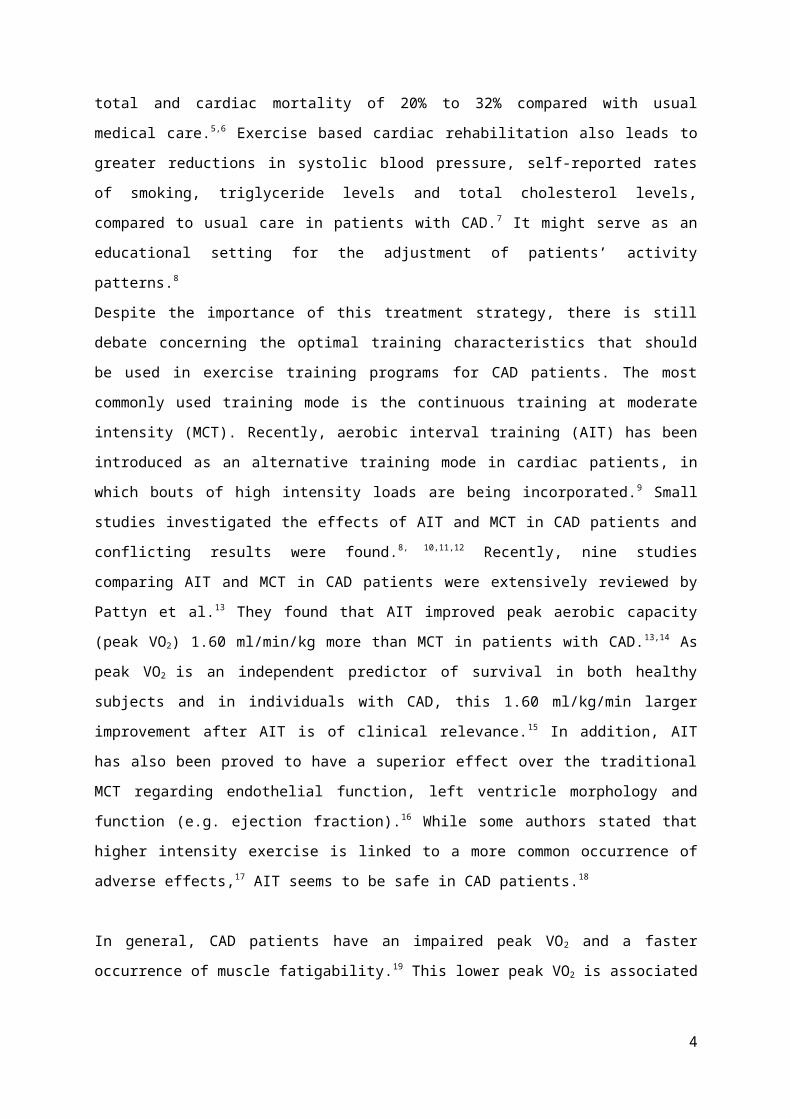
patients, with a reduction in total and cardiac mortality of 20% to 32% compared with usual medical
care.5,6 Exercise based cardiac rehabilitation also leads to greater reductions in systolic blood
pressure, self-reported rates of smoking, triglyceride levels and total cholesterol levels, compared to
usual care in patients with CAD.7 It might serve as an educational setting for the adjustment of
patients’ activity patterns.8
Despite the importance of this treatment strategy, there is still debate concerning the optimal
training characteristics that should be used in exercise training programs for CAD patients. The most
commonly used training mode is the continuous training at moderate intensity (MCT). Recently,
aerobic interval training (AIT) has been introduced as an alternative training mode in cardiac
patients, in which bouts of high intensity loads are being incorporated.9 Small studies investigated
the effects of AIT and MCT in CAD patients and conflicting results were found.8, 10,11,12 Recently, nine
studies comparing AIT and MCT in CAD patients were extensively reviewed by Pattyn et al. 13 They
found that AIT improved peak aerobic capacity (peak VO2) 1.60 ml/min/kg more than MCT in patients
with CAD.13,14 As peak VO2 is an independent predictor of survival in both healthy subjects and in
individuals with CAD, this 1.60 ml/kg/min larger improvement after AIT is of clinical relevance.15 In
addition, AIT has also been proved to have a superior effect over the traditional MCT regarding
endothelial function, left ventricle morphology and function (e.g. ejection fraction).16 While some
authors stated that higher intensity exercise is linked to a more common occurrence of adverse
effects,17 AIT seems to be safe in CAD patients.18
In general, CAD patients have an impaired peak VO2 and a faster occurrence of muscle fatigability.19
This lower peak VO2 is associated with a decreased muscle strength, which is caused by a lower
activity of local muscular enzymes and by a smaller cross-section of the muscle.19 After 3 months of
exercise-based cardiac rehabilitation, including a combination of endurance training and moderate
resistance training, peak VO2, isometric and isokinetic leg strength and endurance were significantly
improved compared to baseline values.20
3

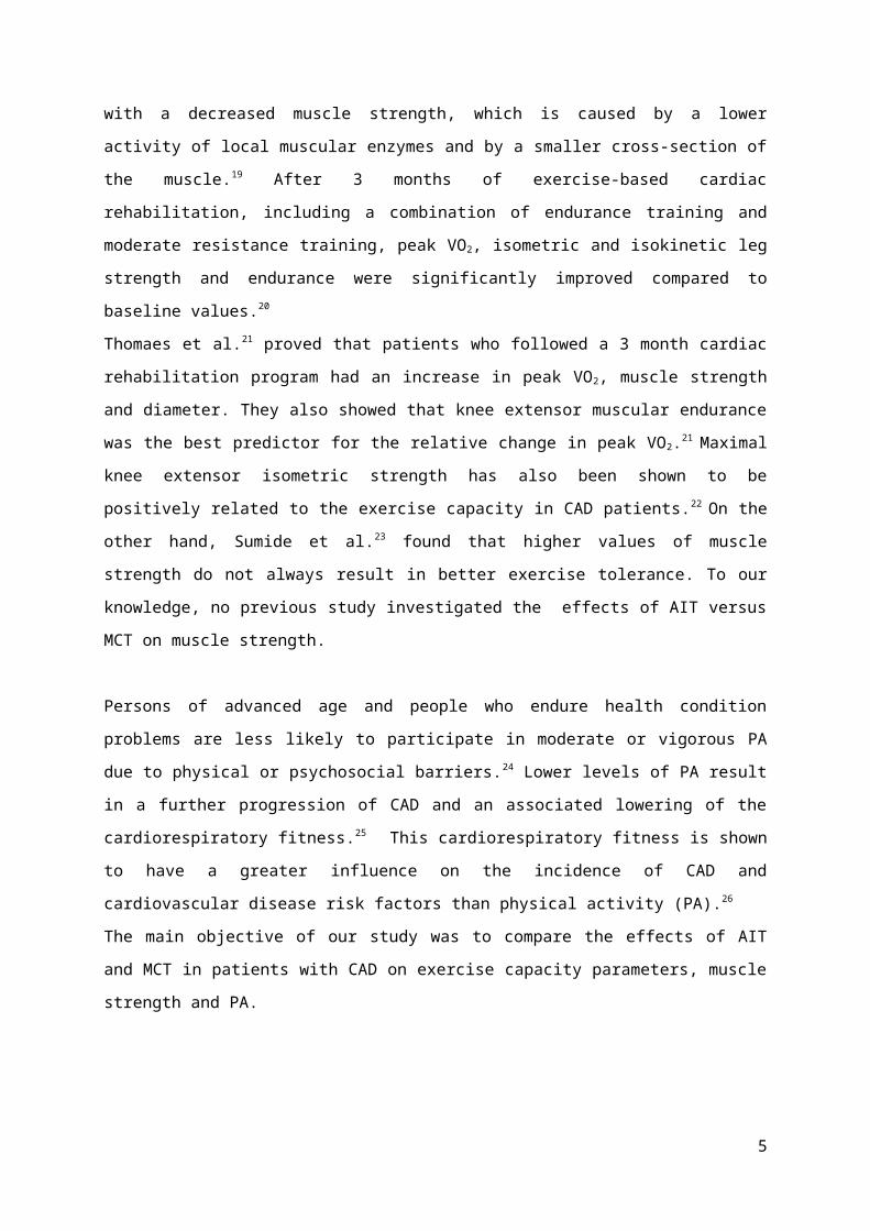
Thomaes et al.21 proved that patients who followed a 3 month cardiac rehabilitation program had an
increase in peak VO2, muscle strength and diameter. They also showed that knee extensor muscular
endurance was the best predictor for the relative change in peak VO2.21 Maximal knee extensor
isometric strength has also been shown to be positively related to the exercise capacity in CAD
patients.22 On the other hand, Sumide et al.23 found that higher values of muscle strength do not
always result in better exercise tolerance. To our knowledge, no previous study investigated the
effects of AIT versus MCT on muscle strength.
Persons of advanced age and people who endure health condition problems are less likely to
participate in moderate or vigorous PA due to physical or psychosocial barriers.24 Lower levels of PA
result in a further progression of CAD and an associated lowering of the cardiorespiratory fitness.25
This cardiorespiratory fitness is shown to have a greater influence on the incidence of CAD and
cardiovascular disease risk factors than physical activity (PA).26
The main objective of our study was to compare the effects of AIT and MCT in patients with CAD on
exercise capacity parameters, muscle strength and PA.
4
Methods
Participants
One-hundred CAD patients, aged between 40 and 75 years, were included in the Saintex study
between February 2011 and December 2012.27 All patients were investigated in the cardiac
rehabilitation center of the UZ Leuven Gasthuisberg. Patients with CAD were defined as having a
history of myocardial infarction (MI), coronary artery bypass grafting (CABG) or percutaneous
coronary intervention (PCI) and patients had a left ventricular ejection fraction (LVEF) of at least 40%.
Further inclusion conditions were an optimal medical treatment of the patients, stable
pharmacotherapy and normal symptoms. Exclusion criteria were significant illness during the last 6
weeks, recent CABG (<30 days), severe ventricular arrhythmia with functional or prognostic
significance, myocardial ischemia, hemodynamic deterioration, exercise-induced arrhythmia or
another heart disease that limits exercise tolerance, co-morbidities that influence a one year
prognosis, functional and mental disability, a habit of regular vigorous exercise, acute chronic
inflammatory disease, glomerular filtration rate (GFR) <25ml/min/1.73m², hemoglobin<10g/dl,
severe chronic obstructive pulmonary disease (COPD) (FEV1<50%) and participation in another
clinical trial. After screening, eligible and interested subjects were randomized in 2 groups (AIT and
MCT) on a 1:1 basis, using an online randomization procedure. Subjects signed a written consent
form prior to participation in accordance with the Declaration of Helsinki. The study was approved by
the local ethics committee.
Measurements
Cardiopulmonary exercise test. A maximal graded exercise test until exhaustion on a cycle-ergometer
was performed before the exercise intervention and after 12 weeks. The test protocol started with a
load of 20 Watts, and workload increased by 20 Watts every minute. Heart rate (HR) and a twelve
lead-ECG were continuously recorded during the test, and blood pressure was measured every 2
minutes and at the end of the test. Breath-by-breath gas exchange was measured continuously,
providing ventilation (Ve), oxygen uptake (VO2) and carbon dioxide production (VCO2). Peak VO2 was
defined as the mean VO2-value reached during the last full bout of 30 seconds of the exercise test.
The criteria of Binder et al.28 for a maximal exercise test were used. The patient’s weight was
measured with a Tefal progress sensitive computer.
5

Muscle strength. The Jamar hand-held dynamometer (Saehan corp., Korea) was used to measure
maximal hand grip strength. Patients were seated with the upper arm parallel to the body, the elbow
flexed at 90° and the wrist positioned neutral. Patients were instructed to develop a maximal
contraction during 3 seconds. Instructors encouraged the subjects to perform the strength test with
maximal effort. The test was repeated 3 times for each hand alternatively, with a break of 10 seconds
between the measurements. The average of the 3 attempts was used for analysis. The Jamar
dynamometer has shown to be valid for measuring hand grip strength.29
The Biodex system 3 PRO (Biodex medical systems, USA) was used to measure the patient’s isokinetic
extension and flexion strength, and the maximal isometric extension strength. The patient had to sit
upright on the Biodex chair with back support, with the hip joint in a 90° flexion-angle. The patients’
trunk and legs were firmly stabilized using straps around the chest, hips and the legs. To avoid any
compensation, the subjects were instructed to hold their hands on their thighs during their maximal
efforts. The instructor vocally encouraged each subject in a standardized way to complete every part
of the test to the maximum. The subject started with isometric knee extension at 60° during 5
seconds, repeated 4 times and interrupted by 30 seconds of rest. The highest value, corrected for
weight, was used in the analyses. After 1 minute of rest, an isokinetic test with an angular velocity of
60°/sec was performed, consisting of 4 knee extension and flexion movements as fast as possible.
After a rest period of 30 seconds, the same procedure was repeated at an angular velocity of
180°/sec. The average of the 4 repetitions at each angular speed was used in the analyses.
Muscle strength was measured before and after the 12 weeks intervention period.
Sensewear. The sensewear bodymonitor (Bodymedia) is an armband enabling a continuous collection
of data about energy expenditure.30 In particular, the amount of steps, the duration and intensity of
activity (sedentary, moderate, vigorous), total energy expenditure, active energy expenditure and
average level of PA expressed as average metabolic equivalent, were determined.
The Sensewear was given to the patients at the beginning and at the end of the exercise training
intervention. The device was worn during the day at the upper dominant arm during seven
consecutive days. In a population of COPD patients, Van Remoortel et al.31 concluded that the
sensewear armband is a valid monitor to measure standardized PA.
6
Exercise intervention
The exercise training took place 3 times a week for 12 weeks (see figure 1). Each training session was
performed on a cycle ergometer and was supervised by educated physiotherapist. The AIT group
started with a ten-minute warming up (60-70% peak HR), then cycled four times a 4-minute interval
at high intensity (90-95% peak HR) while each interval was separated by a 3 min active pause interval
at 50-70% of peak HR. The total exercise time in the AIT group was 38 minutes.
The MCT group started with a 5-min warming up, followed by 37 minutes of cycling at moderate
intensity (70-75% of peak HR), and ended with 5 min cool-down. Each session of the MCT group had
a duration of 47 minutes. Both programs were isocaloric9.
HR was continuously monitored during training to assure patients trained within their target HRs. The
Borg 6-20 scale was taken to evaluate the patients’ rate of perceived exertion. Apart from the
exercise training, patients had the possibility to attend six multi-disciplinary educational sessions.
An overview can be seen in figure 1, where the study design and intervention programs are outlined.
Figure 1. Flowchart of the study design and intervention program
AIT 12 weeks - 3x/week - 38min MCT 12 weeks - 3x/week - 47min
90-95% 90-95% 90-95% 90-95%Peak HR 60-70% 4' 50-70% 4' 50-70% 4' 50-70% 4' 50-70% Peak HR 60-70% 60-70%
3' 3' 3' 3' Time (min) 5' 5'
12 weeks after start:*CPET*Muscular strength: Biodex and Jamar*Sensewear
Time (min) 10'
Baseline measurements:*CPET*Muscular strength: Biodex and Jamar*Sensewear
Inclusion criteria:*men/women*40-75year*CAD=CABG/MI/PCI*LVEF>40%Randomisation (n=100)
37'
70-75%
AIT = 50 patients MCT = 50 patients
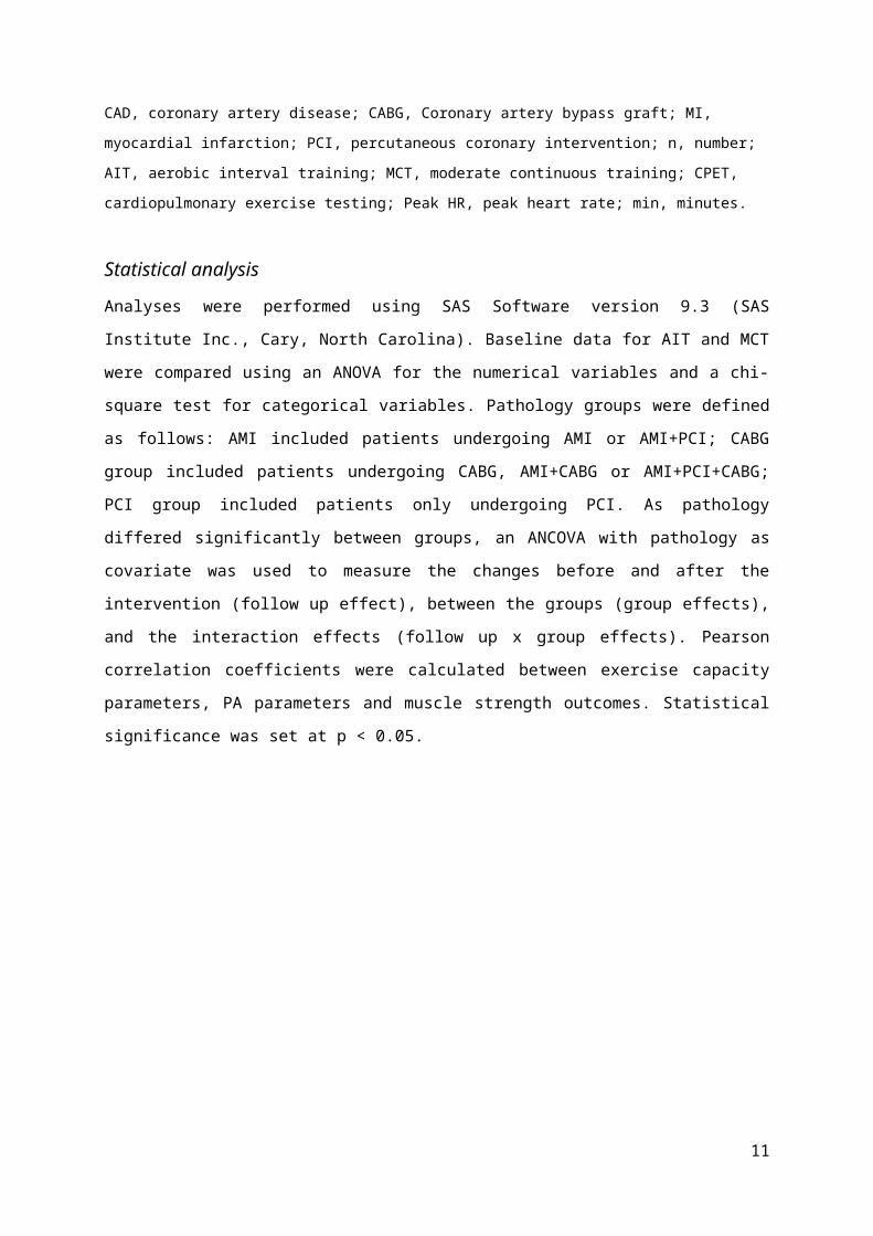
CAD, coronary artery disease; CABG, Coronary artery bypass graft; MI, myocardial infarction; PCI, percutaneous
coronary intervention; n, number; AIT, aerobic interval training; MCT, moderate continuous training; CPET,
cardiopulmonary exercise testing; Peak HR, peak heart rate; min, minutes.
7
Statistical analysis
Analyses were performed using SAS Software version 9.3 (SAS Institute Inc., Cary, North Carolina).
Baseline data for AIT and MCT were compared using an ANOVA for the numerical variables and a chi-
square test for categorical variables. Pathology groups were defined as follows: AMI included
patients undergoing AMI or AMI+PCI; CABG group included patients undergoing CABG, AMI+CABG or
AMI+PCI+CABG; PCI group included patients only undergoing PCI. As pathology differed significantly
between groups, an ANCOVA with pathology as covariate was used to measure the changes before
and after the intervention (follow up effect), between the groups (group effects), and the interaction
effects (follow up x group effects). Pearson correlation coefficients were calculated between exercise
capacity parameters, PA parameters and muscle strength outcomes. Statistical significance was set at
p < 0.05.
8

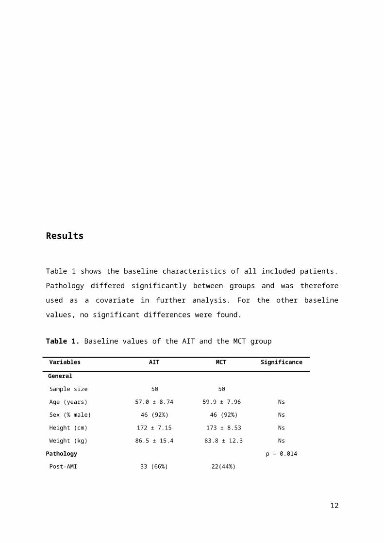
Results
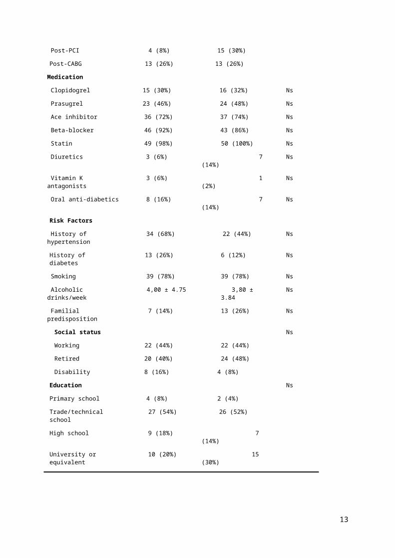
Table 1 shows the baseline characteristics of all included patients. Pathology differed significantly
between groups and was therefore used as a covariate in further analysis. For the other baseline
values, no significant differences were found.
Table 1. Baseline values of the AIT and the MCT group
Variables AIT MCT Significance
General
Sample size 50 50
Age (years) 57.0 ± 8.74 59.9 ± 7.96 Ns
Sex (% male) 46 (92%) 46 (92%) Ns
Height (cm) 172 ± 7.15 173 ± 8.53 Ns
Weight (kg) 86.5 ± 15.4 83.8 ± 12.3 Ns
Pathology p = 0.014
Post-AMI 33 (66%) 22(44%)
Post-PCI 4 (8%) 15 (30%)
Post-CABG 13 (26%) 13 (26%)
Medication
Clopidogrel 15 (30%) 16 (32%) Ns
Prasugrel 23 (46%) 24 (48%) Ns
Ace inhibitor 36 (72%) 37 (74%) Ns
Beta-blocker 46 (92%) 43 (86%) Ns
Statin 49 (98%) 50 (100%) Ns
Diuretics 3 (6%) 7 (14%) Ns
Vitamin K antagonists 3 (6%) 1 (2%) Ns
Oral anti-diabetics 8 (16%) 7 (14%) Ns
Risk Factors
History of hypertension 34 (68%) 22 (44%) Ns
History of diabetes 13 (26%) 6 (12%) Ns
Smoking 39 (78%) 39 (78%) Ns
Alcoholic drinks/week 4,00 ± 4.75 3,80 ± 3.84 Ns
Familial predisposition 7 (14%) 13 (26%) Ns
9

Social status Ns
Working 22 (44%) 22 (44%)
Retired 20 (40%) 24 (48%)
Disability 8 (16%) 4 (8%)
Education Ns
Primary school 4 (8%) 2 (4%)
Trade/technical school 27 (54%) 26 (52%)
High school 9 (18%) 7 (14%)
University or equivalent 10 (20%) 15 (30%)
Data presented as mean ± SD or numbers and percentages. AIT, aerobic interval training; MCT, moderate
continuous training; Ns, not significant; AMI, acute myocardial infarction; PCI, percutaneous coronary
intervention; CABG, coronary artery bypass graft; ACE, angiotensin-converting-enzyme. Statistical significance
was set at p < 0.05.
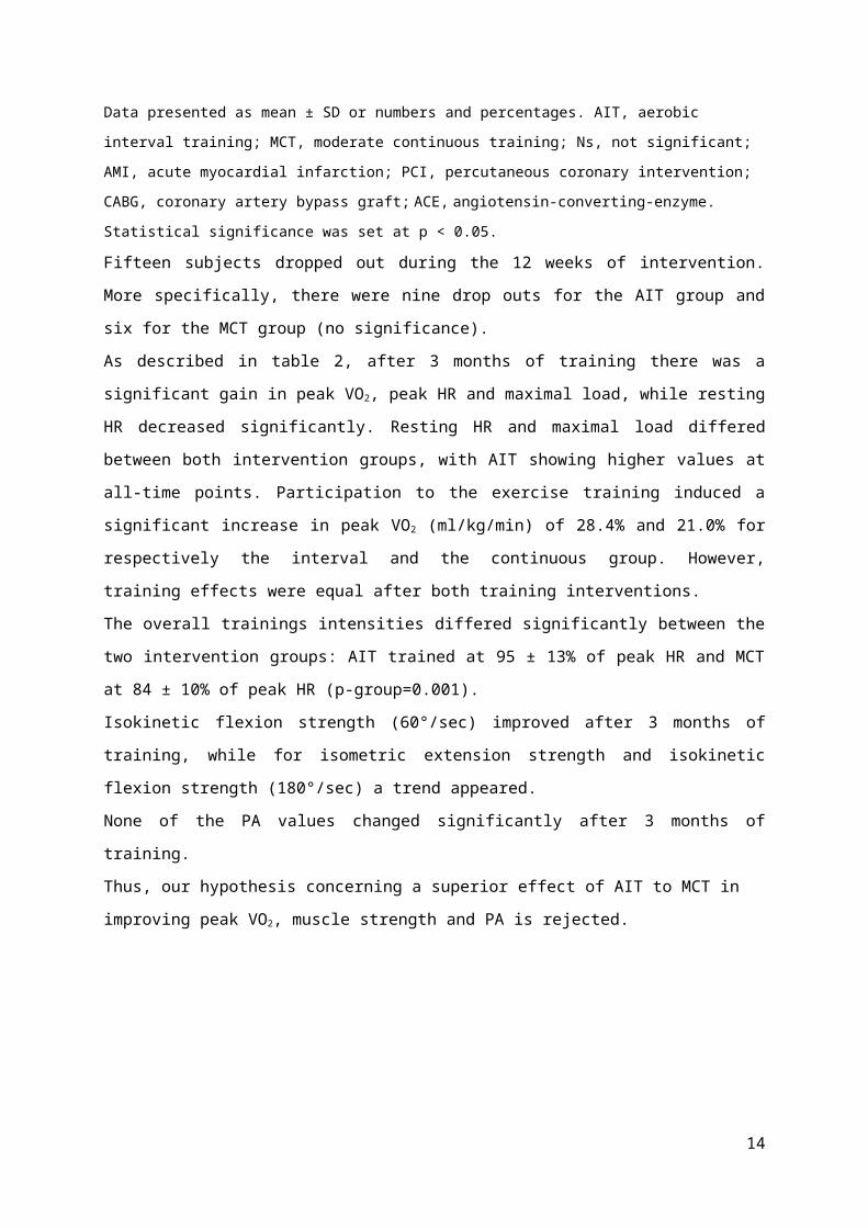
Fifteen subjects dropped out during the 12 weeks of intervention. More specifically, there were nine
drop outs for the AIT group and six for the MCT group (no significance).
As described in table 2, after 3 months of training there was a significant gain in peak VO 2, peak HR
and maximal load, while resting HR decreased significantly. Resting HR and maximal load differed
between both intervention groups, with AIT showing higher values at all-time points. Participation to
the exercise training induced a significant increase in peak VO2 (ml/kg/min) of 28.4% and 21.0% for
respectively the interval and the continuous group. However, training effects were equal after both
training interventions.
The overall trainings intensities differed significantly between the two intervention groups: AIT
trained at 95 ± 13% of peak HR and MCT at 84 ± 10% of peak HR (p-group=0.001).
Isokinetic flexion strength (60°/sec) improved after 3 months of training, while for isometric
extension strength and isokinetic flexion strength (180°/sec) a trend appeared.
None of the PA values changed significantly after 3 months of training.
Thus, our hypothesis concerning a superior effect of AIT to MCT in improving peak VO2, muscle
strength and PA is rejected.
10

Table 2. Exercise capacity, muscle strength and PA at baseline and after 3 months of AIT or MCT
VARIABLES AIT MCT F GROUP F FOLLOW-UP F INTERACTION
Pre Post Pre PostEXERCISE CAPACITYPEAK VO2 (ML/MIN) 1865 ± 501 2366 ± 536 1894 ± 455 2263 ± 503 1.44ns 35.84***
35.8435.84**
0.89ns
PEAK VO2 (ML/KG/MIN) 21.8 ± 5.49 28.0 ± 6.22 22.9 ± 5.76 27.7 ± 6.64 0.00ns 38.91*** 0.58ns
MAXIMAL LOAD (WATT) 162 ± 39.2 207 ± 46.9 163 ± 42.1 202 ± 45.8 4.48* 20.11*** 0.01ns
PEAK HR (B.P.M.) 139 ± 21.2 147 ± 19.7 136 ± 21.2 141 ± 21.2 3.66α 4.45* 0.31ns
RER peak 1.21 ± 0.10 1.21 ± 0.09 1.20 ± 0.11 1.22 ± 0.06 0.00ns 1.02ns 0.46ns
Resting HR (b.p.m) 68.7 ± 11.5 65.4 ± 10.4 64.1 ± 10.5 60.9 ± 10.1 8.23** 4.28* 0.00ns
Body weight (kg) 86.6 ± 15.4 85.9 ± 15.5 83.9 ± 12.3 82.9 ± 12.0 0.74ns 0.34ns 0.01ns
MUSCLE STRENGTHHand grip right (kg) 41.7 ± 8.89 43.9 ± 10.1 40.9 ± 8.95 40.1 ± 8.19 2.72ns 0.26ns 1.09ns
Hand grip left (kg) 39.3 ± 9.46 41.8 ± 10.1 38.9 ± 8.80 38.8 ± 7.80 0.98ns 0.68ns 0.85ns
Isometric ext (Nm) 177 ± 42.8 195 ± 34.4 177 ± 46.1 185 ± 43.1 0.65ns 3.65α 0.54ns
Isokinetic ext at 60°/s (Nm) 162 ± 44.2 170 ± 35.1 162 ± 45.7 167 ± 43.5 0.06ns 1.07ns 0.03ns
Isokinetic flex at 60°/s (Nm) 102 ± 29.3 113 ± 25.5 102 ± 29.7 114 ± 28.8 0.00ns 6.37* 0.05ns
Isokinetic ext at 180°/s (Nm) 104 ± 26.0 108 ± 26.5 104 ± 28.8 113 ± 31.4 0.36ns 2.17ns 0.34ns
Isokinetic flex at 180°/s (Nm) 88.7 ± 23.1 96.5 ± 21.3 89.3 ± 28.7 96.5 ± 25.0 0.02ns 3.75α 0.01ns
PASteps (per week) 54850 ±17286 48982 ± 15871 53849 ± 32429 54224 ± 25262 0.01ns 0.41ns 0.60ns
Moderate PA (hours/7days) 10.8 ± 6.30 9.72 ± 5.86 11.1 ± 8.18 10.9 ± 6.38 0.08ns 0.31ns 0.16ns
Vigorous PA (hours/7days) 0.22 ± 0.45 0.20 ± 0.26 0.21 ± 0.28 0.32 ± 0.49 0.38ns 0.56ns 0.96ns
TOTAL ENERGY EXPENDITURE
(KCAL/7DAYS)
15481 ± 3537 14314 ± 3425 15505 ± 3801 14954 ± 3677 0.00ns 1.92ns 0.26ns
ACTIVE ENERGY EXPENDITURE
(KCAL/7DAYS)
3682 ± 2461 3205 ± 1776 3654 ± 2644 3522 ± 1840 0.00ns 0.61ns 0.21ns
Average MET (MET) 1.61 ± 0.30 1.55 ± 0.25 1.58 ± 0.29 1.61 ± 0.28 0.00ns 0.14ns 0.73ns
PA DURATION (HOURS) 11.1 ± 6.50 9.92 ± 5.91 11.3 ± 8.33 11.2 ± 6.71 0.09ns 0.27ns 0.19ns
Values are mean ± SD or F-values. Pre, before intervention; post, after intervention; AIT, aerobic interval training; MCT, moderate continuous training; peak VO2: peak
oxygen uptake; HR, heart rate; b.p.m., beats per minute; RER peak, peak respiratory exchange ratio; ext, extension strength; flex, flexion strength, Nm, Newton meters; PA,
physical activity; MET, average metabolic equivalent; ***P<0.001; **p<0.01; *p<0.05; αp=0.06; ns: not significant. Statistical analyses are performed by ANCOVA (adjusted
for pathology).
11

The correlations between exercise capacity and PA (table 3A and B), exercise capacity and muscle
strength (table 3C and D) and muscle strength and PA (table 3E and F) are shown in table 3. Figure 2
(A and B) shows the relation between peak VO2, isokinetic extension strength at 180°/s and active
energy expenditure. These parameters are outlined as specific values of respectively; exercise
capacity, muscle strength and physical activity. All three parameters correlated significantly, except
for the correlation between changes in active energy expenditure and changes in isokinetic extension
strength at 180°/s which was weak and not significant (r=0.195; p=0.129). Moderate correlations
were found for peak VO2 and isokinetic extension strength at 180°/s and its changes.
Relative peak VO2 correlated significantly with all the PA parameters except for the correlation with
total energy expenditure (r=0.107; p=0.128). However, changes in total energy expenditure had a
moderate and significant correlation with changes in relative peak VO2 (r=0.322; p=0.008). All
correlations between relative peak VO2 and muscle strength parameters (see table 3C) were
significant and moderate/strong, except for the correlation with hand grip strength which was weak
(but still significant). Only changes in isokinetic extension strength at 180°/s had a moderate and
significant correlation with changes in relative peak VO2. Other correlations in table 3D were shown
to be weak and not significant. Correlations between parameters of PA and muscle strength and its
changes are variable and outlined in table 3E-F.
Figure 2A-B. Correlations between exercise capacity, muscle strength and PA
Values are Pearson correlation coefficients. peak VO2: peak oxygen uptake; Δ, changes; ext, extension strength;
ns, not significant; ***p<0.001; *p<0.05.
12
peak VO2
Isokinetic extActive energy expenditure
Δpeak VO2
ΔIsokinetic extΔActive energy
expenditure
r=0.574***r=0.333***
r=0.180*
r=0.412***r=0.253*
r=0.195ns

Table 3A-B-C-D-E-F. Correlations after 3 months of AIT or MCT
VARIABLES RELATIVE PEAK VO2
(ML/KG/MIN)
Steps (per day) 0,231**
MODERATE PA(HOURS/7DAYS) 0,411***
Vigorous PA (hours/7days) 0,334***
Total energy expenditure (kcal) 0,107ns
Average MET (MET) 0,372***
Active energy expenditure (kcal) 0,333***
PA duration (hours) 0,418***
C) Correlations between exercise capacity and muscle strength D) Correlations between
changes in exercise capacity and muscle strength
13
A) Correlations between exercise capacity and PA B) Correlations between changes in exercise capacity and PA
VARIABLES Δ RELATIVE PEAK
VO2
(ML/KG/MIN)
Δ Steps (per day) 0.149ns
Δ MODERATE PA(HOURS/7DAYS) 0.269*
Δ VIGOROUS PA (HOURS/7DAYS) 0.303*
Δ TOTAL ENERGY EXPENDITURE
(KCAL)
0.322**
Δ Average MET (MET) 0.144ns
Δ ACTIVE ENERGY EXPENDITURE
(KCAL)
0.253*
Δ PA DURATION (HOURS) 0.283*
VARIABLES RELATIVE PEAK
VO2
(ML/KG/MIN)
Hand grip right (kg) 0,228**
Hand grip left (kg) 0,344***
Isometric Ext (Nm) 0,507***
Isokinetic Ext at 60°/s (Nm) 0,552***
Isokinetic Flex at 60°/s (Nm) 0,563***
Isokinetic Ext at 180°/s (Nm) 0,574***
Isokinetic Flex at 180°/s (Nm) 0,582***
VARIABLES Δ RELATIVE PEAK
VO2
(ML/KG/MIN)
Δ HAND GRIP RIGHT (KG) 0,082ns
Δ HAND GRIP LEFT (KG) 0,095ns
Δ Isometric Ext (Nm) 0,016ns
Δ ISOKINETIC EXT AT 60°/S (NM) 0,030ns
Δ ISOKINETIC FLEX AT 60°/S (NM) 0,126ns
Δ ISOKINETIC EXT AT 180°/S (NM) 0,412***
Δ ISOKINETIC FLEX AT 180°/S (NM) 0,141ns
E) Correlations between muscle strength and PA
VARIABLES Hand grip right
(kg)
Hand grip left
(kg)
Isometric Ext (Nm) Isokinetic Ext at 60°/s
(Nm)
Isokinetic Flex at 60°/s
(Nm)
Isokinetic Ext at
180°/s (Nm)
Isokinetic Flex at
180°/s (Nm)
Steps (per day) -0,048ns 0,025ns 0,108ns 0,119ns 0,187* 0,093ns 0,229**
Moderate PA (hours/7days) -0,017ns 0,103ns 0,259** 0,303*** 0,284*** 0,212* 0,289***
Vigorous PA (hours/7days) 0,099ns 0,083ns 0,187* 0,240** 0,192* 0,269** 0,243**
Total energy expenditure (kcal) 0,279*** 0,335*** 0,115ns 0,215* 0,200*** 0,204* 0,173*
Average MET (MET) 0,067ns 0,105ns 0,289*** 0,329*** 0,308*** 0,271** 0,283***
Active energy expenditure (kcal) 0,065ns 0,179* 0,227** 0,276*** 0,270** 0,180* 0,281***
PA duration (hours) -0,010ns 0,106ns 0,262** 0,307*** 0,287*** 0,220** 0,294***
F) Correlations between changes in muscle strength and PA
VARIABLES Δ HAND GRIP
RIGHT (KG)
Δ HAND GRIP
LEFT (KG)
Δ Isometric Ext
(Nm)
Δ ISOKINETIC EXT AT
60°/S (NM)
Δ ISOKINETIC FLEX AT
60°/S (NM)
Δ ISOKINETIC EXT AT
180°/S (NM)
Δ ISOKINETIC FLEX AT
180°/S (NM)
Δ Steps (per day) -0,038ns -0,032ns 0,039ns 0,176ns 0,150ns 0,090ns 0,150ns
Δ MODERATE PA (HOURS/7DAYS) 0,160ns 0,110ns 0,198ns 0,190ns 0,082ns 0,219ns 0,166ns
Δ VIGOROUS PA (HOURS/7DAYS) 0,247α 0,233β 0,141ns 0,209ns -0,080ns 0,310* 0,064ns
Δ TOTAL ENERGY EXPENDITURE
(KCAL)
0,116ns 0,082ns 0,039ns 0,126ns 0,083ns 0,168ns 0,154ns
Δ Average MET (MET) 0,206ns 0,053ns 0,220ns 0,258* 0,039ns 0,216ns 0,121ns
14
Δ ACTIVE ENERGY EXPENDITURE
(KCAL)
0,160ns 0,133ns 0,194ns 0,231β 0,082ns 0,195ns 0,154ns
Δ PA DURATION (HOURS) 0,175ns 0,125ns 0,198ns 0,194ns 0,072ns 0,231β 0,164ns
Values are Pearson correlation coefficients; peak VO2: peak oxygen uptake; kcal, kilocalories; MET, metabolic equivalent; average MET, average metabolic equivalent; PA,
physical activity; Δ, change; Ext, extension strength; Flex, flexion strength; Nm, Newton meters; ns, not significant; ***p<0.001; **p<0.01; *p<0.05; αp=0.06; βp=0.07.
15
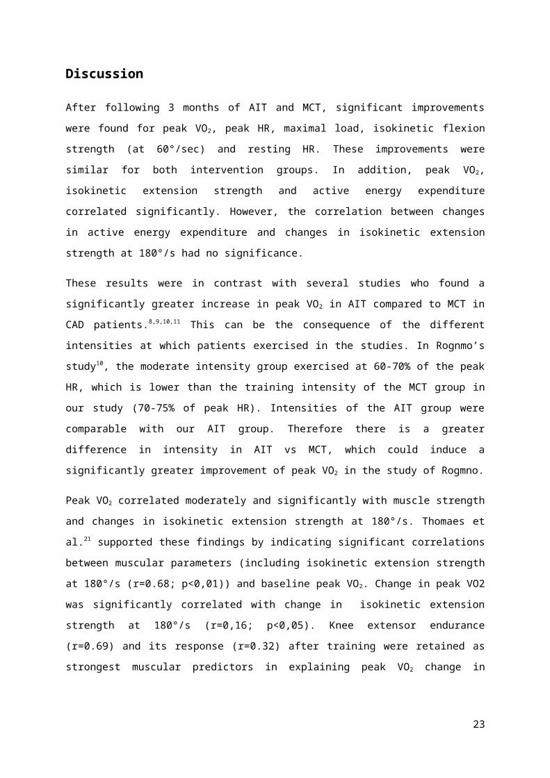
Discussion
After following 3 months of AIT and MCT, significant improvements were found for peak VO2, peak
HR, maximal load, isokinetic flexion strength (at 60°/sec) and resting HR. These improvements were
similar for both intervention groups. In addition, peak VO2, isokinetic extension strength and active
energy expenditure correlated significantly. However, the correlation between changes in active
energy expenditure and changes in isokinetic extension strength at 180°/s had no significance.
These results were in contrast with several studies who found a significantly greater increase in peak
VO2 in AIT compared to MCT in CAD patients.8,9,10,11 This can be the consequence of the different
intensities at which patients exercised in the studies. In Rognmo’s study10, the moderate intensity
group exercised at 60-70% of the peak HR, which is lower than the training intensity of the MCT
group in our study (70-75% of peak HR). Intensities of the AIT group were comparable with our AIT
group. Therefore there is a greater difference in intensity in AIT vs MCT, which could induce a
significantly greater improvement of peak VO2 in the study of Rogmno.
Peak VO2 correlated moderately and significantly with muscle strength and changes in isokinetic
extension strength at 180°/s. Thomaes et al.21 supported these findings by indicating significant
correlations between muscular parameters (including isokinetic extension strength at 180°/s (r=0.68;
p<0,01)) and baseline peak VO2. Change in peak VO2 was significantly correlated with change in
isokinetic extension strength at 180°/s (r=0,16; p<0,05). Knee extensor endurance (r=0.69) and its
response (r=0.32) after training were retained as strongest muscular predictors in explaining peak
VO2 change in patients with CAD. Although, in our study no measures of muscle endurance were
obtained, it’s likely that an increase in endurance strength took place regarding the improving results
on the successive maximal exercise tests.
Remarkably, there was a significant training effect for isokinetic flexion at 60°/s and a trend for
isokinetic flexion at 180°/s, while no effect for the isokinetic knee extensors was found. Cycling
includes a repeated extension and flexion movement, while more power output is produced during
the extension phase. Seat height, pedaling rate, fatigue, power output, physical condition and shoe-
pedal interface are factors altering the muscular activation pattern during cycling. 32 Cycling with a
higher seat will induce a diminished knee angle, which will lead to a higher relative hamstrings (knee
flexors) activation compared to the quadriceps (knee extensor) muscles. During the exercise sessions,
patients were possibly seated higher than their optimal position, so a significant isokinetic knee
flexors strengthening was induced while isokinetic knee extensor strength remained unchanged.
16
Both exercise capacity and muscle strength are inversely associated with mortality in both healthy
and diseased populations.33 In CAD patients, being less mobile, having fear of movement, being ill,
lying in bed and sarcopenia are some of the underlying causes of a decline in muscle strength.19 It is
important to intervene in this vicious cycle where post-operative CAD patients are less active and
become less mobile. Including patients in gradually exercise trainings programs with appropriate
guiding and multidisciplinary support to overcome these problems, is essential.
No significant effects on PA for both intervention groups were found, which means that there was no
transfer effect from the higher amount of exercise training during the sessions to an increase in
leisure-time PA. These findings are in discrepancy with those of Weinstein et al. 34 who found a
significant increase in PA after a 10-week intervention program in patients with pulmonary arterial
hypertension.
A significant relationship was found between an increase in vigorous PA and isokinetic extension
strength at 180°/s. Goodpaster et al.35 constructed a randomized control trial to investigate the
influence of regular PA on muscle strength and muscle fat infiltration. They proved that PA prevents
the loss of knee extension strength, which is known to be a common risk in an advancing aged
population.35
Strength and limitations
This study is strengthened by the combination of measurements of exercise capacity, PA and muscle
strength and correlations between these parameters after an AIT or MCT intervention, in a large
study group of CAD patients.
On the other hand, this study has some limitations. The adaptable height of the bicycle seat and
position of the handle-bars could have been potential confounders for changes in muscle strength
during exercise training. In addition, the irregularity in pedaling rate from 60 to 90 rounds per minute
could also have altered the muscular activation pattern, the change in power output and the amount
of work that was produced.
Future research
More intensive research in a large CAD population group is needed for a better understanding of the
optimal guidelines concerning programs for CAD patients and for further clarification of the
relationship between exercise capacity, muscle strength and PA.
17

Conclusions
Exercise based cardiac rehabilitation is a keystone in the multidisciplinary treatment of patients with
CAD. After an AIT or MCT intervention of 12 weeks, patients with CAD significantly increased exercise
capacity and isokinetic flexion strength, but not PA. Changes were similar after both interventions.
Peak VO2, isokinetic extension strength at 180°/s and active energy expenditure correlated
significantly, except for relation between changes in active energy expenditure and changes in
isokinetic extension strength at 180°/s which was weak and not significant.
18
Rerefence list
1. Pubmed Health. Coronary artery disease,
http://www.ncbi.nlm.nih.gov/pubmedhealth/PMH0004449/ [Accessed 25 february 2013].
2. The National Heart, Lung, and Blood Institute (NHLBI). What is Artherosclerosis?
https://www.nhlbi.nih.gov/health/health-topics/topics/atherosclerosis/ [Accessed 28 february 2013].
3. World health organisation (WHO). The top 10 causes of death,
http://www.who.int/mediacentre/factsheets/fs310/en/ [Accessed 3 march 2013].
4. Vlayen J, De Backer G, Peers J et al. Artherosclerotic cardiovascular diseases in Belgium: a vost-of-
illness analysis. Cardiovasc Drugs Ther 2008; 22(6): 487-494.
5. O'Connor GT, Buring JE, Yusuf S. Goldhaber SZ et al. An overview of randomised trials of
rehabilitation with exercise after myocardial infarction. Circulation 1989; 80(2): 234-244.
6. Oldridge NB, Guyatt GH, Fischer ME et al. Cardiac rehabilitation after myocardial infarction.
Combined experience of randomized clinical trials. JAMA 1988; 260(7): 945-950.
7. Taylor RS, Brown A, Ebrahim S et al. Exercise-based rehabilitation for patients with coronary heart
disease: systematic review and meta-analysis of randomized controlled trials. Am J Med 2004;
116(10): 682-692.
8. Moholdt TT, Amundsen BH, Rustad LA et al. Aerobic interval training versus continuous moderate
exercise after coronary artery bypass surgery: a randomized study of cardiovascular effects and
quality of life. Am Heart J 2009; 158(6): 1031-1037.
9. Wisløff U, Støylen A, Loennechen JP et al. Superior cardiovascular effect of aerobic interval training
versus moderate continuous training in heart failure patients: a randomized study. Circulation 2007;
115(24): 3086-3094.
10. Rognmo Ø, Hetland E, Helgerud J et al. High intensity aerobic interval exercise is superior to
moderate intensity exercise for increasing aerobic capacity in patients with coronary artery disease.
Eur J Cardiovasc Prev Rehabil 2004; 11(3): 216-222.
11. Warburton DE, McKenzie DC, Haykowsky MJ et al. Effectiveness of high-intensity interval training
for the rehabilitation of patients with coronary artery disease. Am J Cardiol 2005; 95(9): 1080-1084.
19

12. Myers J, Prakash M, Froelicher V et al. Exercise capacity and mortality among men referred for
exercise testing. N Engl J Med 2002; 346(11): 793-801.
13. Pattyn N, Coeckelberghs E, Buys R et al. Aerobic Interval Training vs. Moderate Continuous
Training in Coronary Artery Disease Patients: A Systematic Review and Meta-Analysis. Sports Med
2014 Feb 19.
14. Hottenrott K, Ludyga S, Schulze S. Effects of High Intensity Training and Continuous Endurance
Training on Aerobic Capacity and Body Composition in Recreationally Active Runners. J Sports Sci
Med 2012; 11(3): 483–488.
15. Kavanagh T, Mertens DJ, Hamm LF et al. Prediction of Long-Term Prognosis in 12 169 Men
Referred for Cardiac Rehabilitation. Circulation 2002; 106(6): 666-671.
16. Cornish AK, Broadbent S, Cheema BS. Interval training for patients with coronary artery disease: a
systematic review. Eur J Appl Physiol 2011; 111(4): 579-589.
17. Thompson PD, Franklin BA, Balady GJ et al. Exercise and acute cardiovascular events placing the
risks into perspective: a scientific statement from the American Heart Association Council on
Nutrition, PA, and Metabolism and the Council on Clinical Cardiology. Circulation 2007; 115(17):
2358-2368.
18. Rognmo Ø, Moholdt T, Bakken H et al. Cardiovascular risk of high- versus moderate-intensity
aerobic exercise in coronary heart disease patients. Circulation 2012; 126(12): 1436-1440.
19. Ghroubi S, Chaari M, Elleuch H et al. The isokinetic assessment of peripheral muscle function in
patients with coronary artery disease: correlations with cardiorespiratory capacity. Ann Readapt Med
Phys 2007; 50(5): 295-301.
20. Cornelissen VA, Defoor JG, Stevens A et al. Effect of creatine supplementation as a potential
adjuvant therapy to exercise training in cardiac patients: a randomized controlled trial. Clin Rehabil
2010; 24(11): 988-999.
21. Thomaes T, Thomis M, Onkelinx S et al. Muscular strength and diameter as determinants of
aerobic power and aerobic power response to exercise training in CAD patients. Acta Cardiol 2012;
67(4): 399-406.
22. Kamiya K, Mezzani A, Hotta K et al. Quadriceps isometric strength as a predictor of exercise
capacity in coronary artery disease patients. Eur J Prev Card 2013.
20

23. Sumide T, Shimada K, Ohmura H et al. Relationship between exercise tolerance and muscle
strength following cardiac rehabilitation: comparison of patients after cardiac surgery and patients
with myocardial infarction. J Cardiol 2009; 54(2): 273-281.
24. Casa DJ. Preventing Sudden Death in Sport and physical activity. 2012. p33-41.
25. Hambrecht R, Niebauer J, Marburger C et al. Various intensities of leisure time PA in patients with
coronary artery disease: effects on cardiorespiratory fitness and progression of coronary
atherosclerotic lesions. J Am Coll Cardiol 1993; 22(2): 468-477.
26. Sassen B, Cornelissen VA, Kiers H et al. Physical fitness matters more than PA in controlling
cardiovascular disease risk factors. Eur J Cardiovasc Prev Rehabil 2009; 16(6): 677-683.
27. Conraads VM, Van Craenenbroeck EM, Pattyn N et al. Rationale and design of a randomized trial
on the effectiveness of aerobic interval training in patients with coronary artery disease: the
SAINTEX-CAD study. Int J Cardiol 2013; 168(4): 3532-3536.
28. Binder RK, Wonisch M, Corra U et al. Methodological approach to the first and second lactate
threshold in incremental cardiopulmonary exercise testing. Eur J Cardiovasc Prev Rehabil 2008; 15(6):
726-734.
29. Mathiowetz V. Comparison of Rolyan and Jamar dynamometers for measuring grip strength.
Occup Ther Int 2002; 9(3): 201-209.
30. Almeida GJ, Wasko MC, Jeong K et al. PA measured by the SenseWear Armband in women with
rheumatoid arthritis. Phys Ther 2011; 91(9): 1367-1376.
31. Van Remoortel H, Raste Y, Louvaris Z et al. Validity of six activity monitors in chronic obstructive
pulmonary disease: a comparison with indirect calorimetry. PLoS One 2012; 7(6):e39198.
32. Hug F, Dorel S. Electromyographic analysis of pedaling: a review. J Electromyogr Kinesiol 2009
Apr;19(2): 182-98.
33. Rantanen T, Volpato S, Ferrucci L, Heikkinen E, Fried LP, Guralnik JM. Handgrip strength and
cause-specific and total mortality in older disabled women: exploring the mechanism. J Am Geriatr
Soc 2003 May;51(5): 636-41.
34. Weinstein AA, Chin LM, Keyser RE et al. Effect of aerobic exercise training on fatigue and PA in
patients with pulmonary arterial hypertension. Respir Med 2013; 107(5): 778-784.
21

35. Goodpaster BH, Chomentowski P, Ward BK et al. Effects of PA on strength and skeletal muscle fat
infiltration in older adults: a randomized controlled trial. J Appl Physiol 2008; 105(5): 1498-1503.
22

Manuscript Submission Guidelines
European Journal of Preventive Cardiology
1. Peer review policy 2. Article types 3. Authorship 4. How to submit your manuscript 5. Journal contributor's publishing agreement
5.1 SAGE Choice and Open Access6. Statements and conventions
6.1 Acknowledgments6.2 Declaration of conflicting interests6.3 Funding acknowledgement6.4 Other statements and conventions
7. Permissions 8. Manuscript style
8.1 File types8.2 Journal style8.3 Reference style8.4 Manuscript preparation
9. After acceptance 9.1 Proofs9.2 E-Prints9.3 SAGE production9.4 OnlineFirst publication
10. Further information
1. Peer review policy
Submitted papers will undergo full peer review, and written comments, when available, will be returned with all refereed manuscripts. Reports for provisionally accepted papers will include a review by a statistician which authors will be required to follow when revising their manuscript. The final decision on the acceptance or rejection of a manuscript will be made by the Editors.
Back to top
2. Article types
European Journal of Preventive Cardiology publishes papers on all aspects of clinical and public health disciplines that address the causes and prevention of cardiovascular disease, as well as cardiovascular rehabilitation and exercise physiology.
The following are the various article types which the journal publishes. Authors must adhere to the page/word counts given here. The word counts cover all sections of a manuscript, including title, author names/affiliation, abstract, keywords, figure/table legends and references:
23

Type of paperMaximum number of pages Maximum number of words
Full original research paper 6 journal pages5,000 words (with each table or figure reducing that word count by 250)
Review paper 10 journal pages8,700 words (with each table or figure reducing that word count by 250)
Short report 2 journal pages1,300 words (with each table or figure reducing that word count by 250)
Commentary 2 journal pages1,300 words (with each table or figure reducing that word count by 250)
Editorial 2 journal pages1,300 words (with each table or figure reducing that word count by 250)
Letter to the Editor 1 journal page700 words (with each table or figure reducing that word count by 250)
A manuscript submitted to the Journal will be considered for publication on the understanding that it has not been published before (in print or electronically, including on a web site), that it has not been accepted for publication elsewhere, and that it is not under consideration by another publication. Authors who submit papers to the Journal must document that all persons acknowledged have seen and approved the mention of their name in the paper.
Back to top
3. Authorship
Papers should only be submitted for consideration once the authorization of all contributing authors has been gathered. Those submitting papers should carefully check that all those whose work contributed to the paper are acknowledged as contributing authors.
The list of authors should include all those who can legitimately claim authorship. This is all those who:
1. have made a substantial contribution to the concept and design, acquisition of data or analysis and interpretation of data
2. drafted the article or revised it critically for important intellectual content3. approved the version to be published.
Authors should meet the conditions of all of the points above. Each author should have participated sufficiently in the work to take public responsibility for appropriate portions of the content.
When a large, multicentre group has conducted the work, the group should identify the individuals who accept direct responsibility for the manuscript. These individuals should fully meet the criteria for authorship.
Acquisition of funding, collection of data, or general supervision of the research group alone does not
24

constitute authorship, although all contributors who do not meet the criteria for authorship should be listed in the Acknowledgments section.Please refer to the ICMJE Authorship guidelines at http://www.icmje.org/ethical_1author.html.
Back to top
4. How to submit your manuscript
Before submitting your manuscript, please ensure you carefully read and adhere to all the guidelines and instructions to authors provided below. Manuscripts not conforming to these guidelines may be returned.
Manuscripts should be submitted online, through Editorial Manager at http://www.edmgr.com/ejpc, where further instructions are available.
In the instance that you cannot submit online, please contact the journal’s Editorial Office at [email protected].
Back to top
5. Journal contributor’s publishing agreement
Before publication SAGE requires the author as the rights holder to sign a Journal Contributor’s Publishing Agreement. SAGE’s Journal Contributor’s Publishing Agreement is an exclusive licence agreement which means that the author retains copyright in the work but grants SAGE the sole and exclusive right and licence to publish for the full legal term of copyright. Exceptions may exist where an assignment of copyright is required or preferred by a proprietor other than SAGE. In this case copyright in the work will be assigned from the author to the society. For more information please visit our Frequently Asked Questions on the SAGE Journal Author Gateway.
The Journal and SAGE take issues of copyright infringement, plagiarism or other breaches of best practice in publication very seriously. We seek to protect the rights of our authors and we always investigate claims of plagiarism or misuse of articles published in the Journal. Equally, we seek to protect the reputation of the Journal against malpractice. Submitted articles may be checked with duplication-checking software. Where an article is found to have plagiarised other work or included third-party copyright material without permission or with insufficient acknowledgement, or where the authorship of the article is contested, we reserve the right to take action including, but not limited to: publishing an erratum or corrigendum (correction); retracting the article (removing it from the journal); taking up the matter with the head of department or dean of the author's institution and/or relevant academic bodies or societies; banning the author from publication in the journal or all SAGE journals, or appropriate legal action.
5.1 SAGE Choice and Open Access
If you or your funder wish your article to be freely available online to non subscribers immediately upon publication (gold open access), you can opt for it to be included in SAGE Choice, subject to payment of a publication fee. The manuscript submission and peer review procedure is unchanged. On acceptance of your article, you will be asked to let SAGE know directly if you are choosing SAGE Choice. To check journal eligibility and the publication fee, please visit SAGE Choice. For more information on open access options and compliance at SAGE, including self author archiving deposits (green open access) visit SAGE Publishing Policies on our Journal Author Gateway.
Back to top
6. Statements and conventions
25
6.1. Acknowledgements
Any acknowledgements should appear first at the end of your article prior to your Declaration of Conflicting Interests (if applicable), any notes and your References.
All contributors who do not meet the criteria for authorship should be listed in an ‘Acknowledgements’ section. Examples of those who might be acknowledged include a person who provided purely technical help, writing assistance, or a department chair who provided only general support. Authors should disclose whether they had any writing assistance and identify the entity that paid for this assistance.
6.2 Declaration of conflicting interests
Within your Journal Contributor’s Publishing Agreement you will be required to make a certification with respect to a declaration of conflicting interests. It is the policy of the EJPC to require a declaration of ccccconflicting interests from all authors enabling a statement to be carried within the paginated pages of all published articles.
Please include any declaration at the end of your manuscript after any acknowledgements and prior to the references, under a heading ‘Declaration of Conflicting Interests’. If no declaration is made the following will be printed under this heading in your article: ‘None Declared’. Alternatively, you may wish to state that ‘The Author(s) declare(s) that there is no conflict of interest’.
When making a declaration the disclosure information must be specific and include any financial relationship that all authors of the article has with any sponsoring organization and the for-profit interests the organization represents, and with any for-profit product discussed or implied in the text of the article.
Any commercial or financial involvements that might represent an appearance of a conflict of interest need to be additionally disclosed in the covering letter accompanying your article to assist the Editor in evaluating whether sufficient disclosure has been made within the Declaration of Conflicting Interests provided in the article.
For more information please visit the SAGE Journal Author Gateway.
6.3 Funding Acknowledgement
To comply with the guidance for research funders, authors and publishers issued by the Research Information Network (RIN), the EJPCadditionally requires all authors to acknowledge their funding in a consistent fashion under a separate heading. Please visit Funding Acknowledgement s on the SAGE Journal Author Gateway to confirm the format of the acknowledgment text in the event of funding or state in your acknowledgments that: This research received no specific grant from any funding agency in the public, commercial, or not-for-profit sectors.
6.4 Other statements and conventions
6.4.1 Research ethicsAll papers reporting animal and human studies must include whether written consent was obtained from the local Ethics Committee or Institutional Review Board. Please ensure that you have provided the full name and institution of the review committee and an Ethics Committee reference number.
We accept manuscripts that report human and/or animal studies for publication only if it is made clear that investigations were carried out to a high ethical standard. Studies in humans which might be interpreted as experimental (e.g. controlled trials) should conform to the Declaration of Helsinkihttp://www.wma.net/en/30publications/10policies/b3/index.html and typescripts must include a statement that the research protocol was approved by the appropriate ethical committee. In line with the Declaration of Helsinki 1975, revised Hong Kong 1989, we encourage authors to register their clinical trials (athttp://clinicaltrials.gov or other suitable databases identified by the ICMJE,http://www.icmje.org/publishing_10register.html). If your trial has been registered, please state this on the Title Page. When reporting experiments on animals, indicate on the Title Page which guideline/law on the care and use of laboratory animals was followed.
6.4.2 Patient consent
26

Authors are required to ensure the following guidelines are followed, as recommended by the International Committee of Medical Journal Editors, Uniform Requirements for Manuscripts Submitted to Biomedical Journals. Patients have a right to privacy that should not be infringed without informed consent. Identifying information, including patients' names, initials, or hospital numbers, should not be published in written descriptions, photographs, and pedigrees unless the information is essential for scientific purposes and the patient (or parent or guardian) gives written informed consent for publication. Informed consent for this purpose requires that a patient who is identifiable be shown the manuscript to be published.
Identifying details should be omitted if they are not essential. Complete anonymity is difficult to achieve, however, and informed consent should be obtained if there is any doubt. For example, masking the eye region in photographs of patients is inadequate protection of anonymity. If identifying characteristics are altered to protect anonymity, such as in genetic pedigrees, authors should provide assurance that alterations do not distort scientific meaning and editors should so note. When informed consent has been obtained it should be indicated in the submitted article.
6.4.3 Units: The Systéme International (SI)The EJPC employs SI Units (see Quantities, Units, and Symbols, 2nd edn. London: The Royal Society of Medicine; 1975). All submitted papers should use this system, which should only be departed from where long-established clinical usage demands it (e.g. the measurement of blood pressure in mmHg). Where helpful, other units of measurement may be included in parentheses. Whenever possible, renin should be expressed in terms of the International Standard Renin Unit [Bangham et al.: Clin Sci 1975, 48(suppl):135s-159s]. Derived SI units may also be used, and for basic and derived units prefixes to denote multiples and submultiples may be used.
Back to top
7. Permissions
Authors are responsible for obtaining permission from copyright holders for reproducing any illustrations, tables, figures or lengthy quotations previously published elsewhere. For further information including guidance on fair dealing for criticism and review, please visit our Frequently Asked Questions on the SAGE Journal Author Gateway.
Back to top
8. Manuscript style
8.1 File types
Only electronic files conforming to the journal's guidelines will be accepted. Preferred formats for the text and tables of your manuscript are Word DOC, RTF, XLS. LaTeX files are also accepted. Please also refer to additional guideline on submitting artwork below.
8.2 Journal Style
The EJPC conforms to the SAGE house style. Click here to review guidelines on SAGE UK House Style.
8.3 Reference Style
The EJPC adheres to the SAGE Vancouver reference style. Click here to review the guidelines on SAGE Vancouver to ensure your manuscript conforms to this reference style.
If you use EndNote to manage references, download the SAGE Vancouver output file by following this
27

link and save to the appropriate folder (normally for Windows C:\Program Files\EndNote\Styles and for Mac OS X Harddrive:Applications:EndNote:Styles). Once you’ve done this, open EndNote and choose “Select Another Style...” from the dropdown menu in the menu bar; locate and choose this new style from the following screen.
8.4 Manuscript Preparation
The text should be double-spaced throughout and with a minimum of 3cm for left and right hand margins and 5cm at head and foot. Text should be standard 10 or 12 point. Number pages consecutively, beginning with the title page. All original papers must be arranged in sections under the headings and in the order indicated below (begin each on a separate page):
Title pageo The title page should carry the full title of the paper, consisting of no more than 20 words
(only common abbreviations should be used if absolutely necessary); titles should be clear and brief, conveying the message of the paper
o Manuscripts should have no more than 10 authors. In cases where there are more than 10 authors, the corresponding author will be contacted and asked to amend.
o All authors’ names: the full first name, middle name/initial (optional) and last name of each author should appear; if the work is to be attributed to a department or institution, its full name and location should be included. Persons listed as authors should be those who substantially contributed to the study's conception, design, and performance.
o The affiliations of all the authors; when authors are affiliated to more than one institution, their names should be connected using a,b,c. These letters should follow the surname but precede the address; they should be used only for the second and subsequent addresses.
o Information about previous presentations of the whole or part of the work presented in the article.
o The sources of any support, for all authors, for the work in the form of grants, equipment, drugs, or any combination of these.
o Disclaimers, if any.o The name and address of the author responsible for correspondence concerning the
manuscript, and the name and address of the author to whom requests for reprints should be made. If reprints are not to be made available, a statement to this effect should be included.
o The peer-review process as well as publication will be delayed if you do not provide up to date e-mail address, telephone and fax numbers.
o Word count: please list full word count (including references). Structured abstract
The second page should carry an abstract not exceeding 250 words and should include sections on Background, Design, Methods, Results and Conclusions. Please list abstract wordcount at the end of the abstract.
KeywordsThe abstract should be followed by a list of 3-10 keywords which will assist the cross-indexing of the article and will be published. The terms used should be from the Medical Subject Headings list of the Index Medicus.
Suggest reviewersIt is required to indicate at least two potential reviewers who have a sufficient area of expertise to review your submission. This will help Editors and shorten the period of your manuscript review.
TextFull papers of an experimental or observational nature should be divided into sections headed Introduction, Methods, Results and Discussion.Use of abbreviations should be kept to an absolute minimum; abbreviations and abbreviated phrases should be written out at first mention followed by the abbreviation in parentheses. Avoid those not accepted by international bodies. Systéme International (SI) units should be used where appropriate.
8.4.1 Your Title, Keywords and Abstracts: Helping readers find your article online
The title, keywords and abstract are key to ensuring readers find your article online through online search engines such as Google. Please refer to the information and guidance on how best to title your article, write your abstract and select your keywords by visiting SAGE’s Journal Author Gateway Guidelines on How
28

to Help Readers Find Your Article Online.
8.4.2 Corresponding Author Contact details
Provide full contact details for the corresponding author including email, mailing address and telephone numbers. Academic affiliations are required for all co-authors. These details should be presented separately to the main text of the article to facilitate anonymous peer review.
8.4.3 Guidelines for submitting artwork, figures and other graphics
For guidance on the preparation of illustrations, pictures and graphs in electronic format, please visit SAGE’sManuscript Submission Guidelines.
Figures supplied in colour will appear in colour online regardless of whether or not these illustrations are reproduced in colour in the printed version. For specifically requested colour reproduction in print, you will receive information regarding the costs from SAGE after receipt of your accepted article.
TablesEach table should be typed on a separate sheet in double spacing. Tables should not be submitted as photographs. Each table MUST have a title and should be assigned an arabic numeral, e.g. (Table 3). Vertical rules should not be used. Tables should not duplicate the content of the text. Each table should consist of at least two columns.
Table headingsIf applicable, table headings should indicate whether the figures used represent percentages, by (%) after the figure, or units. Columns should always have headings.
Table footnotesInformation should be listed in the following order:(i) Abbreviations and symbols should be defined in the order in which they appear in the table (reading across each line rather than down columns); spell out ALL abbreviations and symbols used in the table, even if they have already been listed in previous tables or the text itself when giving a key, use a comma rather than =, e.g. H, hypertensive NOT H=hypertensive.(ii) Any additional comments should follow the explanation of abbreviations and symbols.(iii) Keys to the P values should be listed in the following order (note the use of asterisks for probability): *P<0.05, **P<0.01, ***P<0.001; asterisks are the only symbols that should be used with P values; DO NOT use @ or #.
Checklist for data in tables
the data are consistent with those cited in the relevant parts in the text, totals add up correctly, percentages have been calculated correctly.
Illustration Size and presentationWhen preparing illustrations, it should be kept in mind that they will be printed in the Journal either at column width (about 84mm wide) or at page width (about 170 mm wide). Figures should be professionally drawn and photographed; freehand or typewritten lettering is unacceptable. Photomicrographs must have internal scale markers. If photographs of people are used, their identities must be obscured or the picture must be accompanied by written permission to use the photograph. Photographs may be cropped or deleted at the discretion of the Editor.
Legends for illustrationsAll illustrations must have legends. These should be typed using double spacing, beginning on a separate page, each with an Arabic numeral corresponding to the illustration to which it refers. All abbreviations used in the illustration must be defined in the legend. Internal scales should be explained, and staining methods for photomicrographs identified.
Figures sent by hardcopyAll illustrations should have a label pasted on the back bearing the figure number, the title of the paper, the author's name and an arrow indicating the top of the figure. Avoid writing directly on the back of prints. Do not mount illustrations.
29

PhotographsSupply halftone illustrations (photographs) as sharp, glossy, black-and-white prints, preferably to a width of 84mm or, when the illustration demands it, to a width of 170mm.
Line drawingsArtwork should be submitted either as glossy prints or as high-quality laser prints; dot-matrix printers do not produce artwork suitable for publication.
Colour reproductionThe cost of colour reproduction will be borne by the author. An estimate will be provided before the illustration is processed.
8.4.4 Guidelines for submitting supplemental files
This journal is able to host approved supplemental materials online, alongside the full-text of articles. Supplemental files will be subjected to peer-review alongside the article. For more information please refer to SAGE’s Guidelines for Authors on Supplemental Files.
8.4.5 English Language Editing services
Non-English speaking authors who would like to refine their use of language in their manuscripts might consider using a professional editing service. Visit English Language Editing Services on our Journal Author Gateway for further information.
Back to top
9. After acceptance
9.1 Proofs
We will email a PDF of the proofs to the corresponding author.
9.2 E-Prints
SAGE provides authors with access to a PDF of their final article. For further information please visit Author e-prints on our Journal Author Gateway.
9.3 SAGE Production
At SAGE we place an extremely strong emphasis on the highest production standards possible. We attach high importance to our quality service levels in copy-editing, typesetting, printing, and online publication (http://online.sagepub.com/). We also seek to uphold excellent author relations throughout the publication process.
We value your feedback to ensure we continue to improve our author service levels. On publication all corresponding authors will receive a brief survey questionnaire on your experience of publishing in the EJPC with SAGE.
9.4 OnlineFirst Publication
EJPC benefits from Online First, a feature offered through SAGE’s electronic journal platform, SAGE Journals. It allows final revision articles (completed articles in queue for assignment to an upcoming issue) to be hosted online prior to their inclusion in a final print and online journal issue which significantly reduces the lead time between submission and publication. For more information please visit our Online First Fact Sheet.
30

Back to top
10. Further information
Any correspondence, queries or additional requests for information on the manuscript submission process should be sent to the Editorial Office as follows:
Esther Molenaar (Editorial Assistant):Email: [email protected]
Rosemary Allpress (Editorial Assistant):Email: [email protected]
For Professor Diederick Grobbee please contact Giene de Vries:E-mail: [email protected]
Populaire samenvatting
Het progressief vernauwen van kransslagaders noemen we coronair vaatlijden. Vandaag is dit met
12,8% de meest voorkomende doodsoorzaak volgens de Wereldgezondheidsorganisatie.
31
Onderzoek heeft aangetoond dat deelname aan cardiale revalidatie de kans op overlijden vermindert
met 20-32% bij hartpatiënten. Onderbouwde trainingsprogramma’s zijn een fundamenteel
onderdeel binnen dit revalidatieproces. Echter, welke trainingskarakteristieken het meest effect
hebben in deze patiëntenpopulatie is nog onduidelijk.
Daarom onderzochten we of Aerobe Interval Training (AIT, 90-95% van maximale hartslag) efficiënter
is dan Matig intense Continue Training (MCT, 70-75% van maximale hartslag). Dit zou kunnen leiden
tot een kortere revalidatieduur met bijhorend minder kosten en een snellere re-integratie in de
maatschappij.
Honderd patiënten met coronair vaatlijden werden gelijk verdeeld over een AIT en MCT groep.
Gedurende 3 maanden trainden ze 3x/week op een fietsergometer. De maximale
inspanningscapaciteit, spierkracht (handknijpkracht en kracht van bovenbenen) en fysieke activiteit
werden gemeten voor en na de 3 maanden interventie.
Uit de resultaten bleek dat beide groepen significant verbeterden in maximale inspanningscapaciteit
en in één parameter van de spierkracht, maar dat fysieke activiteit onveranderd bleef. AIT en MCT
hadden dezelfde effecten. Maximale inspanningscapaciteit, spierkracht en fysieke activiteit bleken
onderling gerelateerd te zijn en hebben elk een effect binnen het revalidatieproces in deze
patiëntenpopulatie.
32