-cresyl sulfate aggravates cardiac dysfunction...
TRANSCRIPT

p-Cresyl Sulfate Aggravates Cardiac Dysfunction Associated WithChronic Kidney Disease by Enhancing Apoptosis of CardiomyocytesHui Han, MD, PhD;* Jinzhou Zhu, MD, PhD;* Zhengbin Zhu, MD; Jingwei Ni, MD; Run Du, MD, PhD; Yang Dai, PhD; Yanjia Chen, MD;Zhijun Wu, MD; Lin Lu, MD, PhD; Ruiyan Zhang, MD, PhD
Background-—Cardiovascular disease is the leading cause of death in patients with chronic kidney disease. A body of evidencesuggests that p-cresyl sulfate (PCS), a uremic toxin, is associated with the cardiovascular mortality rate of patients with chronickidney disease; however, the molecular mechanisms underlying this feature have not yet been fully elucidated.
Methods and Results-—We aimed to determine whether PCS accumulation could adversely affect cardiac dysfunction via directcytotoxicity to cardiomyocytes. In mice that underwent 5/6 nephrectomy, PCS promoted cardiac apoptosis and affected theratio of left ventricular transmitral early peak flow velocity to left ventricular transmitral late peak flow velocity (the E/A ratio)observed by echocardiography (n=8 in each group). Apocynin, an inhibitor of NADPH oxidase activity, attenuates this alteration ofthe E/A ratio (n=6 in each group). PCS also exhibited proapoptotic properties in H9c2 cells by upregulating the expression ofp22phox and p47phox, NADPH oxidase subunits, and the production of reactive oxygen species. Apocynin and N-acetylcysteinewere both able to suppress the effect of PCS, underscoring the importance of NADPH oxidase activation for the mechanism ofaction.
Conclusions-—This study demonstrated that the cardiac toxicity of PCS is at least partially attributed to induced NADPH oxidaseactivity and reactive oxygen species production facilitating cardiac apoptosis and resulting in diastolic dysfunction. ( J Am HeartAssoc. 2015; 4:e001852 doi: 10.1161/JAHA.115.001852)
Key Words: apoptosis • cardiac dysfunction • chronic kidney disease • nicotinamide adenine dinucleotide phosphateoxidase • p-cresyl sulfate
C ardiorenal syndrome refers to a broad spectrum ofdiseases involving both the heart and the kidneys.1
These 2 organs are closely involved in maintaining hemody-namic stability through an intricate network. Clinical studieshave proven that chronic kidney disease (CKD) is a major riskfactor for the development of cardiovascular complications,including atherosclerosis, vascular calcification, heart failure,and sudden cardiac death.2,3 Earlier stages of CKD areassociated with an increased cardiovascular risk.4 Despite theclear clinical evidence addressing this correlation, the path-
ophysiology underlying the development and progression ofcardiovascular impairment following renal dysfunctionremains to be elucidated.
CKD is characterized by progressive loss of glomerularfiltration rate over a period of months or years. Impairedurinary excretion of metabolites leads to accumulation ofvarious uremic toxins, including middle-molecular-weightcompounds (0.5 to 60 kD), free water-soluble low-molecu-lar-weight compounds (<0.5 kD), and protein-bound sol-utes.5,6 Although chronic hemodialysis and peritonealdialysis treatments are potent and efficient approaches toremoving most free water-soluble compounds, the prognosisis still poor for CKD patients, and cardiovascular disease is aleading cause of death.7–9 A possible culprit involved in CKDis protein-bound solutes, a large family of compounds thatcause various diseases.
p-Cresyl sulfate (PCS) is a protein-bound uremic toxinmolecule. Plasma PCS concentrations are determined mainlyby intestinal uptake of p-cresol, metabolism of p-cresol toPCS, and renal excretion of PCS.10,11 For CKD patients,albumin is the main binding protein of PCS in circulation.12
Previous studies have demonstrated that the accumulatedPCS plays a role in activating leukocyte radical production,
From the Department of Cardiology, Rui Jin Hospital (H.H., J.Z., Z.Z., J.N., R.D.,Z.W., L.L., R.Z.) and Institute of Cardiovascular Diseases (H.H., Y.D., Y.C., L.L.,R.Z.), Shanghai Jiao Tong University School of Medicine, Shanghai, China.
*Dr Han and Dr Zhu contributed equally to this article.
Correspondence to: Ruiyan Zhang, MD, PhD, Department of Cardiology, RuiJin Hospital, 197 Rui Jin 2nd road, Shanghai 200025, China. E-mail:[email protected]
Received January 24, 2015; accepted May 22, 2015.
ª 2015 The Authors. Published on behalf of the American Heart Association,Inc., by Wiley Blackwell. This is an open access article under the terms of theCreative Commons Attribution-NonCommercial License, which permits use,distribution and reproduction in any medium, provided the original work isproperly cited and is not used for commercial purposes.
DOI: 10.1161/JAHA.115.001852 Journal of the American Heart Association 1
ORIGINAL RESEARCH
by guest on June 7, 2018http://jaha.ahajournals.org/
Dow
nloaded from

disturbing the renin–angiotensin–aldosterone system, pro-moting renal tubular cell damage, and interfering with insulinsignaling pathways.13–17 The results of our previous studyrevealed that p-cresol disrupted proliferation, migration, tubeformation, and cell-cycle kinetics in endothelial progenitorcells, but its metabolite PCS did not.18 An increasing body ofclinical evidence suggests that serum PCS is a predictorof cardiovascular disease and mortality associated withCKD.19–21
Despite previous clinical implications, no study hasfocused on the role of PCS in cardiac injury; therefore, weinvestigated the effect of PCS in cardiotoxicity and cardiacdysfunction. We hope that our findings provide new insightsinto cardiorenal syndrome and facilitate the development ofpotential treatments.
Methods
Animals and Induction of Renal InsufficiencyMale C57BL/6J mice aged 5 weeks were obtained from theModel Animal Research Center of Nanjing University (Nanj-ing, People’s Republic of China) and housed in a specificpathogen–free facility under a 12-hour light/dark cycle withfree access to food and water. After 1 week of accommo-dation, the mice underwent 5/6 nephrectomy (Nx) with a 2-step surgical procedure or a sham operation. Surgicalprocedures were performed under pentobarbital anesthesia,as described previously.22 Briefly, anesthetized mice under-went removal of approximately two-thirds of the left kidneyand right subcapsular Nx after 1 week. Sham-operated miceunderwent laparotomy in parallel by decapsulating the kidneybefore wound closure. The animal experimental protocolcomplied with the Animal Management Rules of the ChineseMinistry of Health (document no. 55, 2001) and wasapproved by the animal care committee of Shanghai JiaoTong University.
Experimental ProtocolFor animal study 1, experimental mice were randomlydivided into 4 groups: sham control (n=8), sham treatedwith PCS (PCS; n=8), untreated 5/6 nephrectomized (Nx;n=8), and PCS-treated 5/6 nephrectomized mice (Nx/PCS;n=8). For animal study 2, Nx mice were randomly dividedinto 2 groups: 5/6 nephrectomized treated with PCS (Nx/PCS; n=6) and treated with both PCS and apocynin (Nx/PCS/apocynin; n=6). PCS and apocynin were administeredby oral gavage for 8 weeks. The concentrations of PCS andapocynin were adjusted for a daily intake of 100 and200 mg/kg, respectively. PCS was purchased from ShanghaiChempartner Co, Ltd. The product was >99% pure, and its
structure was confirmed by nuclear magnetic resonancespectroscopy.
Echocardiography and Doppler AnalysisTransthoracic echocardiography was performed noninvasivelywith a high-resolution ultrasound imaging system (Vevo 2100;VisualSonics, Inc) equipped with a 30-MHz mechanicaltransducer. The mice were lightly anesthetized with 2%isoflurane/100% oxygen and placed on a warming platform(set to 37°C) for the duration of the recordings. The heart ratewas monitored simultaneously by electrocardiography.
Two-dimensional guided M-mode echocardiography wasperformed in the parasternal long-axis view. Left ventricular(LV) end-diastolic diameter and end-systolic diameter weremeasured. The LV end-diastolic volume and end-systolicvolume were calculated, as described previously.23 Fractionalshortening and ejection fraction were calculated using thefollowing formulas: FS (%)=[(LVEDD�LVESD)/LVEDD]9100%and EF (%)=[(LVEDV�LVESV)/LVEDV]9100%. The formulasused the following abbreviations: FS, fractional shortening;LVEDD, LV end-diastolic diameter; LVESD, LV end-systolicdiameter; EF, ejection fraction; LVEDV, LV end-diastolicvolume; LVESV, LV end-systolic volume. Mitral valve flowDoppler was acquired in an apical 4-chamber view. LVdiastolic function was assessed by measuring the wave ratioof the LV transmitral early peak flow velocity to LV transmitrallate peak flow velocity (the E/A ratio). M-mode and Dopplermeasurement data represent 3 to 6 averaged cardiac cyclesfrom at least 2 scans per mouse.
Blood Pressure MeasurementsFor animal study 1, the tail systolic blood pressure wasmeasured in conscious mice using a Softron tail-cuff BP-98Anoninvasive sphygmomanometer (Softron Inc).
Histological AnalysisFormalin-fixed myocardial sections were deparaffinized andrehydrated, as described in detail previously.24 Picrosirius redstaining was used to assess the collagen content. Thesections were photographed using a microscope (Olympus),and the percentage of picrosirius red stain in the sections wascalculated.
Next, 5-lm sections of the left ventricle were prepared todetect apoptosis by in situ terminal deoxynucleotidyl-trans-ferase-mediated 20-deoxyuridine-50-triphosphate nick-endlabeling, or TUNEL. The procedures were performed accordingto the manufacturer’s instructions (TUNEL apoptosis assaykit; F. Hoffmann-La Roche Ltd). The percentage of apoptoticcells was determined as an apoptotic index, which was
DOI: 10.1161/JAHA.115.001852 Journal of the American Heart Association 2
p-Cresyl Sulfate Aggravates Cardiac Dysfunction Han et alORIG
INALRESEARCH
by guest on June 7, 2018http://jaha.ahajournals.org/
Dow
nloaded from

calculated by dividing the number of positively stainedcardiomyocyte nuclei by the total number of cardiomyocytenuclei and multiplying that value by 100.
Cell Culture and TreatmentH9c2 cardiac myoblasts were maintained in DMEM/high-glucose culture medium containing 10% FBS in a water-saturated atmosphere of 5% CO2 and 95% air at 37°C.Thecells were starved in DMEM/high-glucose culture mediumcontaining 0.5% FBS for 6 hours before exposure to PCS (0,250, 500, and 1000 lmol/L) for the designated times. Thecells were treated with 100 lmol/L apocynin or 10 mmol/LN-acetylcysteine (NAC) for 4 hours before they were incu-bated with 500 lmol/L PCS. H9c2 cells were obtained fromShanghai Institutes for Biological Sciences, Chinese Academyof Sciences (Shanghai, People’s Republic of China). DMEM/high-glucose culture medium and FBS were purchased fromGibco (Thermo Fisher Scientific). Apocynin and NAC werepurchased from Sigma-Aldrich.
Annexin V–FITC and Propidium Iodide Staining forDetecting Phosphatidylserine TranslocationCell apoptosis was measured with fluorescein isothiocyanate–labeled human recombinant annexin V (annexin V–FITC) andwith propidium iodide (PI) using a detection kit (BD Pharm-ingen), according to the manufacturer’s instructions. Briefly,the cells were harvested, washed once with cold PBS, andresuspended in 19 binding buffer at a concentration of19106 cells/mL. Next, 100 lL of the solution (19105 cells)was incubated with 5 lL annexin V–FITC and 5 lL PI solutionfor 15 minutes in the dark at room temperature, and 400 lLof 19 binding buffer was added. Samples were analyzed usingflow cytometry (BD FACSCalibur; BD Biosciences) within1 hour. Apoptotic cells, which stained positive for annexin V–FITC and negative or positive for PI, were counted andrepresented as a percentage of the total cell count.
Western Blot AnalysisH9c2 cells were cultured and treated in 6-well plates, washedtwice with PBS, and lysed with the ProteoJET Mammalian CellLysis Reagent (Fermentas; Thermo Fisher Scientific) to extractcytoplasmic proteins. After the protein concentration wasdetermined, the supernatants were stored at �80°C. Afterdenaturation, equal amounts of protein extracts were subjectedto SDS-PAGE and blotted onto polyvinylidene fluoride mem-branes. The membranes were blocked with Tris-buffered salinewith Tween 20 containing 5% nonfat milk powder and subse-quently probed overnight at 4°C with antibodies against B-celllymphoma 2 (Bcl-2, 1:1000), Bcl-2 Associated X Protein (Bax,
1:1000),b-actin (1:2000), p22phox (1:500), and p47phox (1:500),followed by incubationwith respective horseradish peroxidase–conjugated secondary antibodies (1:5000) for 1 hour at roomtemperature. Bands were detected using an enhanced chem-iluminescense system (Millipore) and quantified by scanningdensitometry. Each image was captured, and the intensity ofeach band was analyzed with Quantity One software (Bio-RadLaboratories Inc).
Caspase-3 Activity AssayThis assay is based on the ability of caspase-3 to changeacetyl-Asp-Glu-Val-Asp p-nitroanilide into p-nitroaniline, ayellow formazan product. Caspase-3 activity was determinedusing a caspase-3 activity kit (Beyotime), according to themanufacturer’s protocol. Briefly, treated cells were lysedwith lysis buffer on ice after washing with cold Hanks’balanced salt solution with calcium and magnesium. Afterthe reaction buffer and caspase-3 substrate were incubatedin 96-well plates at 37°C for 4 hours, the caspase-3 activitywas quantified with a microplate spectrophotometer at405 nm. Caspase-3 activity was expressed as the fold ofenzyme activity compared with that of synchronized cells.
Reactive Oxygen Species AssayIntracellular reactive oxygen species (ROS) levels wereassayed on the basis of 5-(and-6)-carboxy-20, 70- dichlorodi-hydrofluorescein diacetate, or carboxy-H2DCFDA, a reliablefluorogenic marker for ROS in live cells.25 The Image-iT LIVEGreen Reactive Oxygen Species Detection Kit (MolecularProbes; Thermo Fisher Scientific) was used, according to themanufacturer’s instructions. Briefly, cells were washed oncewith warm Hanks’ balanced salt solution with calcium andmagnesium and incubated in a sufficient amount of the25 lmol/L carboxy-H2DCFDA working solution for 30 min-utes in the dark at 37°C. During the last 5 minutes of theincubation, 1.0 lmol/L Hoechst 33342 was added to thecarboxy-H2DCFDA staining solution. The samples werewashed 3 times with warm Hanks’ balanced salt solutionwith calcium and magnesium. Images were captured imme-diately with a microscope (Olympus).
Statistical AnalysisData are presented as mean�SEM analyzed by paired orunpaired Student t test, unless otherwise stated. Statisticalsignificance was determined with 1-way ANOVA followed byStudent-Newman-Keuls post hoc analysis. A P value of <0.05was considered statistically significant. Data were analyzedusing GraphPad Prism 5 software (GraphPad Software Inc)and SPSS software (version 13.0; IBM Corp).
DOI: 10.1161/JAHA.115.001852 Journal of the American Heart Association 3
p-Cresyl Sulfate Aggravates Cardiac Dysfunction Han et alORIG
INALRESEARCH
by guest on June 7, 2018http://jaha.ahajournals.org/
Dow
nloaded from

Results
LV Dysfunction Assessed by Echocardiographyand DopplerAs shown in Figure 1A through 1C, although ejection fractionand fractional shortening were decreased in 5/6 nephrec-tomized mice compared with untreated mice, the differenceswere not statistically significant. Echocardiography revealedno change in ejection fraction or fractional shorteningregardless of whether or not PCS treatment was given. Otherrelevant data are summarized in Table 1.
Analysis of diastolic function revealed no change in the E/A ratio between the control and PCS-treated mice (control:2.01�0.07; PCS: 2.15�0.11; P>0.05). Compared with thecontrols, 5/6 nephrectomized mice showed a significantreduction in the E/A ratio (control: 2.01�0.07; Nx:1.64�0.08; P<0.05). The E/A ratio was further reduced in5/6 nephrectomized mice treated with PCS (Nx: 1.64�0.08;Nx/PCS: 1.32�0.08; P<0.05) (Figure 1D and 1E).
Effect of PCS on Histological ChangesSpecific staining for apoptosis using the TUNEL assay in theLV myocardium demonstrated significantly increased apopto-sis following PCS administration (control: 3.75�0.95%; PCS:7.75�0.85%; P<0.05). A similar phenomenon was observed inthe PCS-treated 5/6 nephrectomized mice; the effect of thesubtotal Nx was amplified (Nx: 12.5�1.94%; Nx/PCS:19.0�1.22%; P<0.05) (Figure 2A).
In addition, we observed a significant increase in theinterstitial and perivascular collagen in 5/6 nephrectomizedmice compared with controls (control: 0.63�0.10%; Nx:2.24�0.23%; P<0.05). Treatment with PCS further augmentedthe positive area of picrosirius red stain in the LV of 5/6nephrectomized mice (Nx/PCS: 4.40�0.32%). Similarly,administration of PCS in sham-operated mice resulted inincreased cardiac collagen content (PCS: 1.30�0.16%) (Fig-ure 2B).
Effect of PCS on Apoptosis in H9c2 CellsWe then studied the effect of PCS on phosphatidylserineexposure in H9c2 cells using flow cytometry. Annexin V–FITCand PI were used to determine whether the cells were viable(negative for both annexin V–FITC and PI), damaged (positivefor annexin V–FITC only), or dead (positive for both annexin V–FITC and PI or for PI only). Quantitative analysis showed thatPCS treatment for 24 hours enhanced apoptosis of H9c2cells in a dose-dependent manner. Specifically, comparedwith the control, PCS significantly increased the percentage ofapoptosis at the concentrations of 500 and 1000 lmol/L(control: 15.37�1.43%; 250 lmol/L PCS: 20.90�2.08%;
500 lmol/L PCS: 28.03�1.21%; 1000 lmol/L PCS:30.43�1.59%) (Figure 3A).
During the apoptotic process, the Bcl-2 protein family hasemerged as a key regulator, consisting of death agonists (Baxand Bak) and antagonists (Bcl-2 and Bcl-xL). As shown inFigure 3B, Western blots from the present study revealedsignificant upregulation of Bax and downregulation of Bcl-2expression following exposure to PCS.
Caspase-3 executes apoptosis and is involved in manyimportant events leading to the completion of apoptosis.Activation of caspase-3 serves as an early marker ofapoptosis in H9c2 cells. Our data showed that treatmentwith 500 and 1000 lmol/L PCS induced upregulation ofcaspase-3 activity compared with controls (250 lmol/L PCS:1.15-fold; 500 lmol/L PCS: 1.72-fold; 1000 lmol/L PCS:1.73-fold) (Figure 3C).
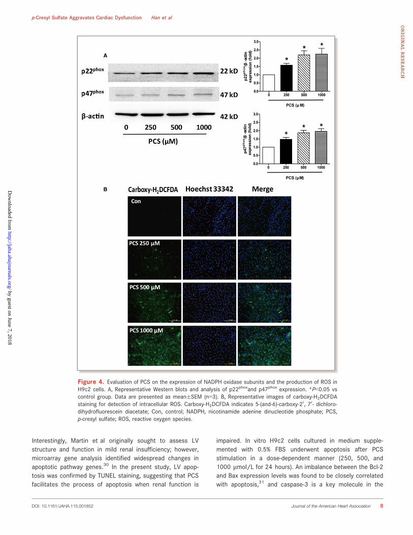
Effect of PCS on the NADPH Oxidase SubunitExpression and Intracellular ROS Production inH9c2 CellsWe examined whether nicotinamide adenine dinucleotidephosphate (NADPH) oxidase plays an important role in PCS-induced ROS production in H9c2 cells. The protein expressionlevels of p22phox and p47phox were evaluated by Westernblotting. The results of the Western blot analysis revealed thatPCS significantly increased the expression of p22phox in H9c2cells at the concentrations of 250, 500, and 1000 lmol/L(1.59-, 2.21-, and 2.25-fold, respectively, over controls).Similarly, p47phox expression was significantly upregulated byPCS treatment, although to a lesser extent than p22phox (1.48-,1.88-, and 1.97-fold, respectively, over controls) (Figure 4A).The fluorescent dye carboxy-H2DCFDA was used to monitorintracellular ROS production. The results revealed significantincrease in fluorescence intensity in the PCS-treated cells (500and 1000 lmol/L) relative to the controls, indicating that PCScan potentially increase the production of ROS (Figure 4B).
Effect of Apocynin and NAC on PCS-InducedApoptosis in H9c2 CellsBecause oxidative stress is a key factor contributing toinitiation of apoptosis in cardiomyocytes, we examinedwhether apocynin or NAC reduced PCS-induced cell damage.H9c2 cells were pretreated with 100 lmol/L apocynin or10 mmol/L NAC for 3 hours and then exposed to 500 lmol/L PCS for 21 hours. Consistent with previous results,500 lmol/L PCS elevated the expression of Bax anddecreased that of Bcl-2, indicating increased apoptosis byPCS. The blots showed that pretreatment with apocynin orNAC partially restored the imbalance of Bax and Bcl-2(Figure 5A).
DOI: 10.1161/JAHA.115.001852 Journal of the American Heart Association 4
p-Cresyl Sulfate Aggravates Cardiac Dysfunction Han et alORIG
INALRESEARCH
by guest on June 7, 2018http://jaha.ahajournals.org/
Dow
nloaded from

The assay of caspase-3 activity proved again thatapocynin or NAC could protect H9c2 cells from thedetrimental effect of PCS. Compared with the controls,cells treated with 500 lmol/L PCS significantly elevatedcaspase-3 activity, whereas pretreatment with apocynin orNAC could restrict this activation (500 lmol/L PCS: 1.70-fold; PCS and apocynin: 1.32-fold; PCS and NAC: 1.32-fold)(Figure 5B).
Effect of Apocynin on PCS-Induced LVDysfunction
Finally, we analyzed whether apocynin treatment couldameliorate PCS-induced alteration of diastolic function in5/6 nephrectomized mice. As shown in Figure 6, PCStreatment resulted in an E/A ratio of 1.30, whereas PCSand apocynin together elevated the ratio to 1.55 (Nx/PCS:
A B
D
E
C
Figure 1. Functional analysis of the left ventricle with echocardiography. A, Representative M-modeechocardiograms from control, PCS-treated, Nx, and Nx/PCS-treated mice. B, The mean left ventricular FSin control, PCS, Nx, and Nx/PCS mice. Data are presented as mean�SEM (n=6 to 8). C, The mean leftventricular EF in control, PCS, Nx, and Nx/PCS mice. Data are presented as mean�SEM (n=6 to 8). D,Representative Doppler echocardiograms from control, PCS, Nx, and Nx/PCS mice. E, The mean E/A ratioin control, PCS, Nx, and Nx/PCS mice. Data are presented as mean�SEM (n=6 to 8). *P<0.05 vs controlgroup. #P<0.05 vs Nx group. A indicates left ventricular transmitral late peak flow velocity; Con, control; E,left ventricular transmitral early peak flow velocity; EF, ejection fraction; FS, fractional shortening; Nx, 5/6nephrectomy; PCS, p-cresyl sulfate.
DOI: 10.1161/JAHA.115.001852 Journal of the American Heart Association 5
p-Cresyl Sulfate Aggravates Cardiac Dysfunction Han et alORIG
INALRESEARCH
by guest on June 7, 2018http://jaha.ahajournals.org/
Dow
nloaded from

Table 1. Characteristics of Animal Models in Animal Study 1
Parameters Con Mice(n=8) PCS Mice(n=8) Nx Mice(n=8) Nx/PCS Mice(n=8)
IVSd, mm 0.856�0.032 0.864�0.050 0.979�0.046* 0.983�0.050
LVEDD, mm 3.26�0.26 3.45�0.19 3.61�0.19 3.41�0.24
LVPWd, mm 0.847�0.043 0.853�0.042 0.897�0.031 0.925�0.048
IVSs, mm 1.481�0.086 1.497�0.069 1.624�0.080 1.691�0.070
LVESD, mm 1.99�0.19 2.07�0.14 2.37�0.16 2.22�0.18
LVPWs, mm 1.255�0.069 1.301�0.065 1.531�0.084* 1.592�0.078*
LVW/BW, mg/g 3.32�0.04 3.31�0.05 3.82�0.17* 3.69�0.13*
SBP, mm Hg 94.8�2.3 93.1�2.3 132.5�4.5* 133.9�4.8*
Values are mean�SEM. BW indicates body weight; Con, control; IVSd, interventricular septum diastole; IVSs, interventricular septum systole; LVEDD, left ventricular end-diastolic diameter;LVESD, left ventricular end-systolic diameter; LVPWd, left ventricular posterior wall diastole; LVPWs, left ventricular posterior wall systole; LVW, left ventricle weight; Nx, 5/6 nephrectomy;PCS, p-cresyl sulfate; SBP, systolic blood pressure.*P<0.05 vs Con mice.
A
B
Figure 2. Histological analysis of cardiac sections from control, PCS-treated, Nx, and Nx/PCS mice. A,Representative images (9200) and analysis of TUNEL staining in all 4 groups. *P<0.05 vs control group.#P<0.05 vs Nx group. Data are presented as mean�SEM (n=4). B, Representative images (9200) andanalysis of picrosirius red staining in the 4 groups. *P<0.05 vs control group. #P<0.05 vs Nx group. Data arepresented as mean�SEM (n=4). Con indicates control; Nx, 5/6 nephrectomy; PCS, p-cresyl sulfate; TUNEL,terminal deoxynucleotidyl-transferase-mediated 20-deoxyuridine-50-triphosphate nick-end labeling.
DOI: 10.1161/JAHA.115.001852 Journal of the American Heart Association 6
p-Cresyl Sulfate Aggravates Cardiac Dysfunction Han et alORIG
INALRESEARCH
by guest on June 7, 2018http://jaha.ahajournals.org/
Dow
nloaded from

1.30�0.07; Nx/PCS/apocynin: 1.55�0.07). Statistical analy-sis revealed significant differences in the E/A ratios betweenthe 2 groups. Other relevant data are summarized in Table 2.
DiscussionIn the present study, PCS was found to promote NADPHoxidase–derived oxidative stress, resulting in an elevatedpercentage of apoptosis in H9c2 cells. In vivo, PCS was foundto be involved in impaired LV diastolic function and increasedcardiac apoptosis. We also observed an increase in cardiacsynthesis of collagen, indicating possible compensation in theheart. Apocynin, an inhibitor of NADPH oxidase activity,26,27
was found to partially restore the PCS-induced decreasedcardiac function.
PCS is of particular interest among the various uremictoxins because of its highly protein-bound property. PCS is
about 94% bound to plasma protein, causing a predictabledecrease in the measured dialytic clearance.28 An increasingbody of evidence suggests that PCS is a predictor ofcardiovascular and/or all-cause mortality in CKD patients;however, whether this biomarker plays a key role in thepathological process of cardiorenal syndrome is still underinvestigation.
In our study, Doppler-derived mitral flow velocities revealedan altered E/A ratio in 5/6 nephrectomized mice treated withPCS, and such alteration is always accompanied by diastolicrelaxation abnormalities.29 Despite this finding, we did notobserve any significant changes in systolic function butcannot exclude the possibility that systolic changes mighthave been detected had the animals been studied at latertime points.
Previous experimental investigations have shown thatrenal impairment would mediate early cardiac apoptosis.
A
B C
Figure 3. Apoptotic analysis in H9c2 cells after incubation with various concentrations of PCS (0, 250,500, and 1000 lmol/L). A, Evaluation of apoptotic incidence by flow cytometry analysis via double stainingwith annexin V and fluorescein isothiocyanate and with propidium iodide. *P<0.05 vs control group. Dataare presented as mean�SEM (n=3). B, Representative Western blots showing the expression of Bax andBcl-2. C, Changes in caspase-3 activity. *P<0.05 vs control group. Data are presented as mean�SEM (n=3).Con indicates control; PCS, p-cresyl sulfate.
DOI: 10.1161/JAHA.115.001852 Journal of the American Heart Association 7
p-Cresyl Sulfate Aggravates Cardiac Dysfunction Han et alORIG
INALRESEARCH
by guest on June 7, 2018http://jaha.ahajournals.org/
Dow
nloaded from
Interestingly, Martin et al originally sought to assess LVstructure and function in mild renal insufficiency; however,microarray gene analysis identified widespread changes inapoptotic pathway genes.30 In the present study, LV apop-tosis was confirmed by TUNEL staining, suggesting that PCSfacilitates the process of apoptosis when renal function is
impaired. In vitro H9c2 cells cultured in medium supple-mented with 0.5% FBS underwent apoptosis after PCSstimulation in a dose-dependent manner (250, 500, and1000 lmol/L for 24 hours). An imbalance between the Bcl-2and Bax expression levels was found to be closely correlatedwith apoptosis,31 and caspase-3 is a key molecule in the
A
B
Figure 4. Evaluation of PCS on the expression of NADPH oxidase subunits and the production of ROS inH9c2 cells. A, Representative Western blots and analysis of p22phoxand p47phox expression. *P<0.05 vscontrol group. Data are presented as mean�SEM (n=3). B, Representative images of carboxy-H2DCFDAstaining for detection of intracellular ROS. Carboxy-H2DCFDA indicates 5-(and-6)-carboxy-20, 70- dichloro-dihydrofluorescein diacetate; Con, control; NADPH, nicotinamide adenine dinucleotide phosphate; PCS,p-cresyl sulfate; ROS, reactive oxygen species.
DOI: 10.1161/JAHA.115.001852 Journal of the American Heart Association 8
p-Cresyl Sulfate Aggravates Cardiac Dysfunction Han et alORIG
INALRESEARCH
by guest on June 7, 2018http://jaha.ahajournals.org/
Dow
nloaded from

apoptosis pathway.32 The data from this study demonstratethat PCS significantly amplifies the effect of serum limitationon H9c2 cell apoptosis, as shown by the increased proportionof cells positively stained with annexin V–FITC, decreasedBcl-2 expression, and the increased Bax and caspase-3fragment expression levels.
ROS-generating NADPH oxidase is a family of enzymesconsisting mainly of 7 members: Nox1, Nox2, Nox3, Nox4,Nox5, Duox1, and Duox2.33,34 Of these, Nox2 and Nox4 arethe predominant isoforms expressed in cardiomyocytes.Activation of these enzymes depends on the assembly ofvarious subunits, including p22phox, p67phox, p40phox, p47phox,and Rac2.35 In our study, PCS treatment was found toupregulate the expression of the NADPH oxidase subunitsp22phox and p47phox. An elevated intracellular ROS level wasalso observed. ROS and the related redox signaling potentiallyaffect apoptosis at several levels, including at upstreamsignaling pathways that are proapoptotic and at the mito-chondrial level.36,37 Indeed, apoptosis is markedly curbed in
A
B
Figure 5. Effects of apocynin and NAC on PCS-induced apop-totic process in H9c2 cells. A, Representative Western blots ofshowing the expression of Bax and Bcl-2. B, Changes in caspase-3activity. *P<0.05 vs control group. #P<0.05 vs PCS group. Dataare presented as mean�SEM (n=3). NAC indicates N-acetylcys-teine; PCS, p-cresyl sulfate.
A
B
Figure 6. Left ventricular transmitral flow velocity analysis withechocardiography. A, Representative Doppler echocardiogramsfrom Nx/PCS and with both PCS and apocynin (Nx/PCS/apocynin) mice. B, The mean E/A ratio from Nx/PCS and Nx/PCS/apocynin mice. Data are presented as mean�SEM (n=6).*P<0.05 vs Nx/PCS group. A indicates left ventricular transmitrallate peak flow velocity; E, left ventricular transmitral early peakflow velocity; Nx indicates 5/6 nephrectomy; PCS, p-cresylsulfate.
Table 2. Characteristics of Animal Models in Animal Study 2
ParametersNx/PCS Mice(n=6)
Nx/PCS/Apocynin Mice(n=6)
EF, % 64.14�1.86 64.64�2.18
FS, % 34.22�1.36 34.89�1.68
IVSd, mm 0.985�0.055 0.981�0.046
LVEDD, mm 3.50�0.16 3.91�0.17
LVPWd, mm 0.928�0.048 0.911�0.038
IVSs, mm 1.708�0.068 1.650�0.090
LVESD, mm 2.26�0.10 2.54�0.11
LVPWs, mm 1.606�0.081 1.574�0.097
LVW/BW, mg/g 3.68�0.11 3.72�0.12
SBP, mm Hg 139.8�4.5 137.5�4.7
Values are means�SEM. BW indicates body weight; EF, ejection fraction; FS, fractionalshortening; IVSd, interventricular septum diastole; IVSs, interventricular septum systole;LVEDD, left ventricular end-diastolic diameter; LVESD, left ventricular end-systolicdiameter; LVPWd, left ventricular posterior wall diastole; LVPWs, left ventricular posteriorwall systole; LVW, left ventricle weight; Nx, 5/6 nephrectomy; PCS, p-cresyl sulfate; SBP,systolic blood pressure.
DOI: 10.1161/JAHA.115.001852 Journal of the American Heart Association 9
p-Cresyl Sulfate Aggravates Cardiac Dysfunction Han et alORIG
INALRESEARCH
by guest on June 7, 2018http://jaha.ahajournals.org/
Dow
nloaded from
H9c2 cells pretreated with apocynin or NAC because eitheragent is capable of reversing the imbalance of Bcl-2 and Baxand restricting the activation of caspase-3. Based on thesefindings, we speculate that NADPH oxidase is at least partiallyinvolved in the observed PCS-related apoptosis and subse-quent diastolic dysfunction.
Apocynin is a naturally occurring methoxy-substitutedcatechol with activity that relies on its oxidation andsubsequent formation of the symmetrical dimer.27,38 Thisformatted diapocynin is metabolically active and inhibits theintracellular assembly of NADPH oxidases.27 Of note, apocy-nin also protects the heart from the E/A ratio alterations thatare exacerbated by PCS, indicating that cardiac diastolicfunction is reserved by an inhibition of NADPH oxidaseactivity.
The present study has several limitations. First, we did nottest our conclusion on gene knockout mouse models. If thetoxicity of PCS is reduced in Nox2 or Nox4 knockout mice, themechanisms should be more convincing. Second, our clinicaldata were limited. We are still working to establish a databaseof patients with cardiorenal syndrome. Such long-term anddetailed studies, however, are difficult to execute, given theneed for substantial follow-up durations.
In summary, the uremic toxin PCS was found to disturb thecellular redox balance and to contribute to detrimental cardiaceffects in cell culture. Animal experiments confirm theadverse effects of PCS in cardiomyocytes. Such factors areattributable to the presence of diastolic dysfunction observedby echocardiography. These findings might provide newknowledge about cardiorenal syndrome and thus representan important novel therapeutic target for the amelioration ofthe disease.
AcknowledgmentsWe thank Dr Yuehua Fang (Rui Jin Hospital, Shanghai, China) and DrJinquan Hu (Changzheng Hospital, Shanghai, China) fortheir excellent technical assistance and valuable comments on ourstudy.
Sources of FundingThis study was supported by grants from the National NaturalScience Foundation of China (81300178 and 81370401).
DisclosuresNone.
References1. Cruz DN. Cardiorenal syndrome in critical care: the acute cardiorenal and
renocardiac syndromes. Adv Chronic Kidney Dis. 2013;20:56–66.
2. Herzog CA, Asinger RW, Berger AK, Charytan DM, Diez J, Hart RG, Eckardt KU,Kasiske BL, McCullough PA, Passman RS, DeLoach SS, Pun PH, Ritz E.Cardiovascular disease in chronic kidney disease. A clinical update fromkidney disease: improving global outcomes (KDIGO). Kidney Int. 2011;80:572–586.
3. Matsushita K, Sang Y, Ballew SH, Shlipak M, Katz R, Rosas SE, Peralta CA,Woodward M, Kramer HJ, Jacobs DR, Sarnak MJ, Coresh J. Subclinicalatherosclerosis measures for cardiovascular prediction in CKD. J Am SocNephrol. 2015;26:439–447.
4. Gottlieb SS, Abraham W, Butler J, Forman DE, Loh E, Massie BM, O’Connor CM,Rich MW, Stevenson LW, Young J, Krumholz HM. The prognostic importance ofdifferent definitions of worsening renal function in congestive heart failure. JCardiac Fail. 2002;8:136–141.
5. Kurella M, Lo JC, Chertow GM. Metabolic syndrome and the risk for chronickidney disease among nondiabetic adults. J Am Soc Nephrol. 2005;16:2134–2140.
6. Duranton F, Cohen G, De Smet R, Rodriguez M, Jankowski J, Vanholder R,Argiles A. Normal and pathologic concentrations of uremic toxins. J Am SocNephrol. 2012;23:1258–1270.
7. Foley RN, Parfrey PS, Sarnak MJ. Clinical epidemiology of cardiovasculardisease in chronic renal disease. Am J Kidney Dis. 1998;32:S112–S119.
8. Anavekar NS, Pfeffer MA. Cardiovascular risk in chronic kidney disease. KidneyInt Suppl. 2004;66:S11–S15.
9. Go AS, Mozaffarian D, Roger VL, Benjamin EJ, Berry JD, Blaha MJ, Dai S, FordES, Fox CS, Franco S, Fullerton HJ, Gillespie C, Hailpern SM, Heit JA, HowardVJ, Huffman MD, Judd SE, Kissela BM, Kittner SJ, Lackland DT, Lichtman JH,Lisabeth LD, Mackey RH, Magid DJ, Marcus GM, Marelli A, Matchar DB,McGuire DK, Mohler ER III, Moy CS, Mussolino ME, Neumar RW, Nichol G,Pandey DK, Paynter NP, Reeves MJ, Sorlie PD, Stein J, Towfighi A, Turan TN,Virani SS, Wong ND, Woo D, Turner MB. Heart disease and stroke statistics–2014 update: a report from the American Heart Association. Circulation.2014;129:e28–e292.
10. Meyer TW, Hostetter TH. Uremic solutes from colon microbes. Kidney Int.2012;81:949–954.
11. Poesen R, Viaene L, Verbeke K, Augustijns P, Bammens B, Claes K, Kuypers D,Evenepoel P, Meijers B. Cardiovascular disease relates to intestinal uptakeof p-cresol in patients with chronic kidney disease. BMC Nephrol. 2014;15:87.
12. Viaene L, Annaert P, de Loor H, Poesen R, Evenepoel P, Meijers B. Albumin isthe main plasma binding protein for indoxyl sulfate and p-cresyl sulfate.Biopharm Drug Dispos. 2013;34:165–175.
13. Schepers E, Meert N, Glorieux G, Goeman J, Van der Eycken J, Vanholder R. p-Cresylsulphate, the main in vivo metabolite of p-cresol, activates leucocytefree radical production. Nephrol Dial Transplant. 2007;22:592–596.
14. Sun CY, Chang SC, Wu MS. Uremic toxins induce kidney fibrosis by activatingintrarenal renin-angiotensin-aldosterone system associated epithelial-to-mes-enchymal transition. PLoS One. 2012;7:e34026.
15. Watanabe H, Miyamoto Y, Honda D, Tanaka H, Wu Q, Endo M, Noguchi T,Kadowaki D, Ishima Y, Kotani S, Nakajima M, Kataoka K, Kim-Mitsuyama S,Tanaka M, Fukagawa M, Otagiri M, Maruyama T. p-Cresyl sulfate causes renaltubular cell damage by inducing oxidative stress by activation of NADPHoxidase. Kidney Int. 2013;83:582–592.
16. Sun CY, Hsu HH, Wu MS. p-Cresol sulfate and indoxyl sulfate induce similarcellular inflammatory gene expressions in cultured proximal renal tubular cells.Nephrol Dial Transplant. 2013;28:70–78.
17. Koppe L, Pillon NJ, Vella RE, Croze ML, Pelletier CC, Chambert S, Massy Z,Glorieux G, Vanholder R, Dugenet Y, Soula HA, Fouque D, Soulage CO. p-Cresylsulfate promotes insulin resistance associated with ckd. J Am Soc Nephrol.2013;24:88–99.
18. Zhu JZ, Zhang J, Yang K, Du R, Jing YJ, Lu L, Zhang RY. p-Cresol, but not p-cresylsulphate, disrupts endothelial progenitor cell function in vitro. NephrolDial Transplant. 2012;27:4323–4330.
19. Meijers BK, Bammens B, De Moor B, Verbeke K, Vanrenterghem Y, EvenepoelP. Free p-cresol is associated with cardiovascular disease in hemodialysispatients. Kidney Int. 2008;73:1174–1180.
20. Liabeuf S, Barreto DV, Barreto FC, Meert N, Glorieux G, Schepers E, TemmarM, Choukroun G, Vanholder R, Massy ZA. Free p-cresylsulphate is a predictorof mortality in patients at different stages of chronic kidney disease. NephrolDial Transplant. 2010;25:1183–1191.
21. Lin CJ, Pan CF, Chuang CK, Sun FJ, Wang DJ, Chen HH, Liu HL, Wu CJ. p-Cresylsulfate is a valuable predictor of clinical outcomes in pre-ESRD patients.Biomed Res Int. 2014;2014:526932.
22. Ni J, Zhang W, Zhu Z, Zhu J, Du R, Jing Y, Lu L, Zhang R. In vivo kinetics of theuremic toxin p-cresyl sulfate in mice with variable renal function. Ther ApherDial. 2014;18:637–642.
DOI: 10.1161/JAHA.115.001852 Journal of the American Heart Association 10
p-Cresyl Sulfate Aggravates Cardiac Dysfunction Han et alORIG
INALRESEARCH
by guest on June 7, 2018http://jaha.ahajournals.org/
Dow
nloaded from

23. Wilson KD, Li Z, Wagner R, Yue P, Tsao P, Nestorova G, Huang M, HirschbergDL, Yock PG, Quertermous T, Wu JC. Transcriptome alteration in the diabeticheart by rosiglitazone: implications for cardiovascular mortality. PLoS One.2008;3:e2609.
24. Finckenberg P, Inkinen K, Ahonen J, Merasto S, Louhelainen M, Vapaatalo H,Muller D, Ganten D, Luft F, Mervaala E. Angiotensin II induces connectivetissue growth factor gene expression via calcineurin-dependent pathways. AmJ Pathol. 2003;163:355–366.
25. Konorev EA, Zhang H, Joseph J, Kennedy MC, Kalyanaraman B. Bicarbonateexacerbates oxidative injury induced by antitumor antibiotic doxorubicin incardiomyocytes. Am J Physiol Heart Circ Physiol. 2000;279:H2424–H2430.
26. Zhang L, Li F, Zhi G, Zhang B, Chen YD. NADPH oxidase contributes to the leftventricular dysfunction induced by sinoaortic denervation in rats. Free RadicalRes. 2015;49:57–66.
27. Johnson DK, Schillinger KJ, Kwait DM, Hughes CV, McNamara EJ, Ishmael F,O’Donnell RW, Chang MM, Hogg MG, Dordick JS, Santhanam L, Ziegler LM,Holland JA. Inhibition of NADPH oxidase activation in endothelial cells byortho-methoxy-substituted catechols. Endothelium. 2002;9:191–203.
28. Martinez AW, Recht NS, Hostetter TH, Meyer TW. Removal of p-cresol sulfateby hemodialysis. J Am Soc Nephrol. 2005;16:3430–3436.
29. Oh JK, Appleton CP, Hatle LK, Nishimura RA, Seward JB, Tajik AJ. The noninvasiveassessment of left ventricular diastolic function with two-dimensional andDoppler echocardiography. J Am Soc Echocardiogr. 1997;10:246–270.
30. Martin FL, McKie PM, Cataliotti A, Sangaralingham SJ, Korinek J, Huntley BK,Oehler EA, Harders GE, Ichiki T, Mangiafico S, Nath KA, Redfield MM, Chen
HH, Burnett JC Jr. Experimental mild renal insufficiency mediates earlycardiac apoptosis, fibrosis, and diastolic dysfunction: a kidney-heart connection. Am J Physiol Regul Integr Comp Physiol. 2012;302:R292–R299.
31. Zhou C, Li X, Du W, Feng Y, Kong X, Li Y, Xiao L, Zhang P. Antitumoreffects of ginkgolic acid in human cancer cell occur via cell cycle arrestand decrease the Bcl-2/Bax ratio to induce apoptosis. Chemotherapy.2010;56:393–402.
32. Yang C, Wang Y, Liu H, Li N, Sun Y, Liu Z, Yang P. Ghrelin protects H9c2cardiomyocytes from angiotensin II-induced apoptosis through theendoplasmic reticulum stress pathway. J Cardiovasc Pharmacol.2012;59:465–471.
33. Geiszt M. NADPH oxidases: new kids on the block. Cardiovasc Res.2006;71:289–299.
34. Lambeth JD. NOX enzymes and the biology of reactive oxygen. Nat RevImmunol. 2004;4:181–189.
35. Brandes RP, Weissmann N, Schroder K. NADPH oxidases in cardiovasculardisease. Free Radic Biol Med. 2010;49:687–706.
36. Filomeni G, Ciriolo MR. Redox control of apoptosis: an update. Antioxid RedoxSignal. 2006;8:2187–2192.
37. Matsuzawa A, Ichijo H. Stress-responsive protein kinases in redox-regulatedapoptosis signaling. Antioxid Redox Signal. 2005;7:472–481.
38. Stolk J, Hiltermann TJ, Dijkman JH, Verhoeven AJ. Characteristics of theinhibition of NADPH oxidase activation in neutrophils by apocynin, a methoxy-substituted catechol. Am J Respir Cell Mol Biol. 1994;11:95–102.
DOI: 10.1161/JAHA.115.001852 Journal of the American Heart Association 11
p-Cresyl Sulfate Aggravates Cardiac Dysfunction Han et alORIG
INALRESEARCH
by guest on June 7, 2018http://jaha.ahajournals.org/
Dow
nloaded from

Lu and Ruiyan ZhangHui Han, Jinzhou Zhu, Zhengbin Zhu, Jingwei Ni, Run Du, Yang Dai, Yanjia Chen, Zhijun Wu, Lin
Enhancing Apoptosis of CardiomyocytesCresyl Sulfate Aggravates Cardiac Dysfunction Associated With Chronic Kidney Disease by−p
Online ISSN: 2047-9980 Dallas, TX 75231
is published by the American Heart Association, 7272 Greenville Avenue,Journal of the American Heart AssociationThe doi: 10.1161/JAHA.115.001852
2015;4:e001852; originally published June 11, 2015;J Am Heart Assoc.
http://jaha.ahajournals.org/content/4/6/e001852World Wide Web at:
The online version of this article, along with updated information and services, is located on the
for more information. http://jaha.ahajournals.orgAccess publication. Visit the Journal at
is an online only OpenJournal of the American Heart AssociationSubscriptions, Permissions, and Reprints: The
by guest on June 7, 2018http://jaha.ahajournals.org/
Dow
nloaded from