© copyright annals of internal medicine, 2011 ann int med. 154 (1): itc1-1. terms of use the in...
TRANSCRIPT

© Copyright Annals of Internal Medicine, 2011Ann Int Med. 154 (1): ITC1-1.
Terms of Use
The In the Clinic® slide sets are owned and copyrighted by the American College of Physicians (ACP). All text, graphics, trademarks, and other intellectual property incorporated into the slide sets remain the sole and exclusive property of ACP. The slide sets may be used only by the person who downloads or purchases them and only for the purpose of presenting them during not-for-profit educational activities. Users may incorporate the entire slide set or selected individual slides into their own teaching presentations but may not alter the content of the slides in any way or remove the ACP copyright notice. Users may make print copies for use as hand-outs for the audience the user is personally addressing but may not otherwise reproduce or distribute the slides by any means or media, including but not limited to sending them as e-mail attachments, posting them on Internet or Intranet sites, publishing them in meeting proceedings, or making them available for sale or distribution in any unauthorized form, without the express written permission of the ACP. Unauthorized use of the In the Clinic slide sets constitutes copyright infringement.

© Copyright Annals of Internal Medicine, 2011Ann Int Med. 154 (1): ITC1-1.
* For Best Viewing:
Open in Slide Show Mode Click on icon or
From the View menu, select the Slide Show option
* To help you as you prepare a talk, we have included the relevant text from ITC in the notes pages of each slide

© Copyright Annals of Internal Medicine, 2011Ann Int Med. 154 (1): ITC1-1.
in the clinic
Transient Ischemic Attack

© Copyright Annals of Internal Medicine, 2011Ann Int Med. 154 (1): ITC1-1.
What is a transient ischemic attack?
Classical definition: Episode of focal cerebral, retinal, or spinal cord ischemia causing transient neurologic dysfunction lasting <24 h (most TIAs resolve in <1 h)
But : despite complete symptom resolution… 20%-50% w/TIA have evidence of acute tissue infarction on MRI
AHA revised definition: TIA should only refer to transient episode of neurologic dysfunction due to ischemia w/o evidence acute infarction, regardless symptom duration
Intent: Better align terminology w/actual disease process
Encourage neurodiagnostic testing to ID brain injury
© Copyright Annals of Internal Medicine, 2011Ann Int Med. 154 (1): ITC1-1.
What is a transient ischemic attack?
TIA and ischemic stroke fundamentally identical disease processes
Differ in:
Duration & degree of cerebral ischemia
Whether permanent detectable brain injury ensues (radiographic/clinical)
Shared pathophysiology supports data extrapolation from selected clinical trials of stroke Rxs to pts w/TIA
Most TIA studies to date use “classical definition”
this review uses “classical definition”

© Copyright Annals of Internal Medicine, 2011Ann Int Med. 154 (1): ITC1-1.
What causes TIA and what are the risk factors?
Mechanism of TIA is…
Large artery atherosclerosis (~20%-25%)
Cardioembolism (10%-15%)
Small vessel disease (10%-15%)
Unusual diverse rare causes (~5%)
Undetermined cause (~50%)

© Copyright Annals of Internal Medicine, 2011Ann Int Med. 154 (1): ITC1-1.
Common Risk Factors for TIA and Stroke
Age >50 y
Hypertension
Diabetes mellitus
Elevated cholesterol levels
Smoking
Carotid stenosis
Hx TIA or stroke
Hx paroxysmal or persistent AF
Hx CAD or PAD
Family Hx CAD, CVD, or PAD before age 60 y

© Copyright Annals of Internal Medicine, 2011Ann Int Med. 154 (1): ITC1-1.
What is the spectrum of presentations for patients with TIA? Symptoms of TIA
Impaired speech and/or language
Visual loss in one or both eyes
Double vision
Facial drooping
Swallowing dysfunction
Unilateral weakness
Unilateral sensory loss
Impaired limb coordination
Vertigo
Gait dysfunction
© Copyright Annals of Internal Medicine, 2011Ann Int Med. 154 (1): ITC1-1.
What is the spectrum of presentations for patients with TIA?
Sxs depend on area of ischemia (brain, retina, or spinal cord)
Lethargy or loss of consciousness atypical ? alternative Dx
Vertigo possible, particularly w/other typical symptoms
isolated vertigo only very rarely due to TIA
Most TIAs quite brief:
60% <1 h, 71% <2 h
Only 14% >6 h
Deficits often resolved by time of medical attention

© Copyright Annals of Internal Medicine, 2011Ann Int Med. 154 (1): ITC1-1.
How should clinicians use history and physical examination to evaluate patients with suspected TIA? Neurologic exam
May be normal (most resolve by time seen); if persistent deficits at evaluation more likely stroke
“Localize the lesion” often challenging (patient recall & description of symptoms may not reliably reflect deficit)
Unilateral weakness often perceived as heaviness; unilateral sensory loss often perceived as tingling, pain
Nonspecific gait dysfunction common; lateropulsion suggests focal deficit
Abnormal speech common challenge to differentiate if aphasia or dysarthria
Blurred vision may be cortical visual field deficit, monocular visual loss, or diplopia
© Copyright Annals of Internal Medicine, 2011Ann Int Med. 154 (1): ITC1-1.
How should clinicians use history and physical examination to evaluate patients with suspected TIA?
Check for any of the following:
Carotid artery bruit
Cardiac arrhythmia or murmur
Impaired distal pulses
Fundi: chronic hypertensive vascular changes
May aid assessment of vascular risk factors
Identify cause of current event

© Copyright Annals of Internal Medicine, 2011Ann Int Med. 154 (1): ITC1-1.
What other disorders should clinicians consider in patients with suspected TIA?
TIA/stroke Abrupt onset; fixed focal findings refer to arterial distribution DW-MRI abnormal in 20%-50% w/transient symptoms
Seizure Abrupt onset and termination; often includes responsiveness, involuntary movements, and/or incontinence during; usually lethargy or confusion after Focal findings occur + resolve over hrs to days May accompany stroke (~2%-3%)
Hypo-/hyperglycemia Usually occurs in pts w/diabetes (diagnose by measuring blood glucose); may be accompanied by seizure
© Copyright Annals of Internal Medicine, 2011Ann Int Med. 154 (1): ITC1-1.
Complicated migraine Severe headache usually precedes/follows; sensory & visual disturbances often prominent; sensory symptoms spread over affected area slowly Younger pts, those w/history severe headache; MRI usually normal; stroke may accompany
Mass lesion e.g., tumor, abscess, herpes encephalitis, subdural hematoma, demyelination Focal symptoms over h to d; may not follow cerebrovascular territory; 1° cancer, fever, trauma, or immunosuppr’n often present Brain imaging distinguishes from stroke
Cervical or lumbar spine disease Focal symptoms isolated to peripheral nerve root distribution in single limb, associated w/depressed reflexes in that region Symptom recurrence may be demonstrated w/maneuvers (e.g., cervical stretch test)

© Copyright Annals of Internal Medicine, 2011Ann Int Med. 154 (1): ITC1-1.
Benign paroxysmal positional vertigo Very brief (seconds) spells of vertigo precipitated by head movement Reproducible w/Dix-Hallpike maneuver Associated w/specific characteristics (torsional nystagmus w/latency, fatigability)
Psychogenic (functional) May look like stroke Often not in vascular territory; findings often nonanatomical/ inconsistent MRI usually normal More likely conversion disorder than malingering

© Copyright Annals of Internal Medicine, 2011Ann Int Med. 154 (1): ITC1-1.
What diagnostic tests are helpful in diagnosis? Brain CT: To exclude brain hemorrhage, large mass lesions, and
identify old areas of infarction
Brain MRI: Evidence acute infarction in 20%-50% with TIA
Carotid US: Sensitivity & specificity 80%-90% for carotid bifurcation stenosis >70%; not helpful for TIA in vertebrobasilar system
Transcranial doppler US: Sensitivity ~70%; specificity 30%-50% for intracranial stenosis >50%
MRA, CTA: Sensitivity & specificity 80%-90% for stenosis >50%; Gd-enhanced cervical MRA ups accuracy; CTA requires IV contrast
Catheter angiography: Reference standard; invasive procedure; use when revascularization considered; requires contrast
ECHO: To detect intracardiac thrombus, tumors, valve disorders
EKG: To identify AF
Serum glucose & hemoglobin A1C: To detect latent diabetes mellitus

© Copyright Annals of Internal Medicine, 2011Ann Int Med. 154 (1): ITC1-1.
Serum lipids: To detect hyperlipidemia
Clotting studies: Obtain baseline prothrombin/partial thromboplastin time to prep for possible anticoagulation
Toxicology screen: To detect sympathomimetic drug use
Blood cultures: If patient febrile, especially if endocarditis, embolic stroke, or TIA suspected
ESR: Patient >50 y: unexplained TIA or headache + possible giant cell arteritis
Rheumatologic serologies: If systemic vasculitis or autoimmune-mediated hypercoagulable state or valvular disease suspected
VDRL/RPR testing: Unexplained TIA + possible meningovascular syphilis
Hb electrophoresis: For anemia and atypical cerebral vasculopathy
Genetic testing: For rare inherited diseases associated with stroke

© Copyright Annals of Internal Medicine, 2011Ann Int Med. 154 (1): ITC1-1.
When should clinicians consult a neurologist for the diagnosis of TIA?
When patient has suspected TIA
Patients evaluated urgently by stroke specialists have lower rates of subsequent stroke
Compared with patients in nonspecialty settings
Early evaluation & therapy associated w/80% reduction in risk early recurrent stroke

© Copyright Annals of Internal Medicine, 2011Ann Int Med. 154 (1): ITC1-1.
CLINICAL BOTTOM LINE: Diagnosis… Transient neurologic symptoms characterize TIA
Evaluate: Are the symptoms due to cerebral ischemia?
If so, what is the mechanism by which it occurred?
Perform tests to identify and treat the causes of TIA
Vascular imaging, electrocardiography, echocardiography
Lab testing, basic blood work
Brain MRI-DWI: Sensitive to acute infarction (seen in ~33% of TIA patients despite symptom resolution)

© Copyright Annals of Internal Medicine, 2011Ann Int Med. 154 (1): ITC1-1.
What is the role of risk assessment in the acute care of patients with TIA? Essential component of management
Overall: 5.2% stroke risk w/in 7 days most w/in first 48 h
Up to 20% stroke risk w/in 90 days in patients with high-risk features of TIA
Risk of early stroke varies by health care setting:
Lowest risk in patients receiving emergency Rx from specialist stroke services (0.9% at 7 days)
Highest risk in general population-based studies w/o urgent Rx (11.0% at day 7)
Observed risk also depends on study methodology higher risks in studies w/active outcome ascertainment

© Copyright Annals of Internal Medicine, 2011Ann Int Med. 154 (1): ITC1-1.
How should clinicians assess the risk for subsequent stroke in patients after TIA?
Score Risk Stroke Risk
2 Days 7 Days 90 Days
0-3 Low 1.0 1.2 3.1
4-5 Moderate 4.1 5.9 9.8
6-7 High 8.1 11.7 17.8
Clinical risk scores: ABCD2 Score Age: ≥60 y = 1 point
Systolic BP ≥140 mm Hg and/or diastolic BP ≥90 mm Hg = 1 point
Unilateral weakness = 2 points
Diabetes: yes = 1 point
Symptom duration: ≥60 min = 2 points; 10-59 min = 1 point

© Copyright Annals of Internal Medicine, 2011Ann Int Med. 154 (1): ITC1-1.
Brain imaging studies: CT or MRI
Cerebral infarction = significantly increased short-term stroke risk
On CT: indicates background presence of CVD
On MRI-DWI: provides information about acute clinical event
Vascular imaging studies
Estimate risk
Identifies candidates for revascularization
If large-vessel stenosis causes TIA: high short-term stroke risk
© Copyright Annals of Internal Medicine, 2011Ann Int Med. 154 (1): ITC1-1.
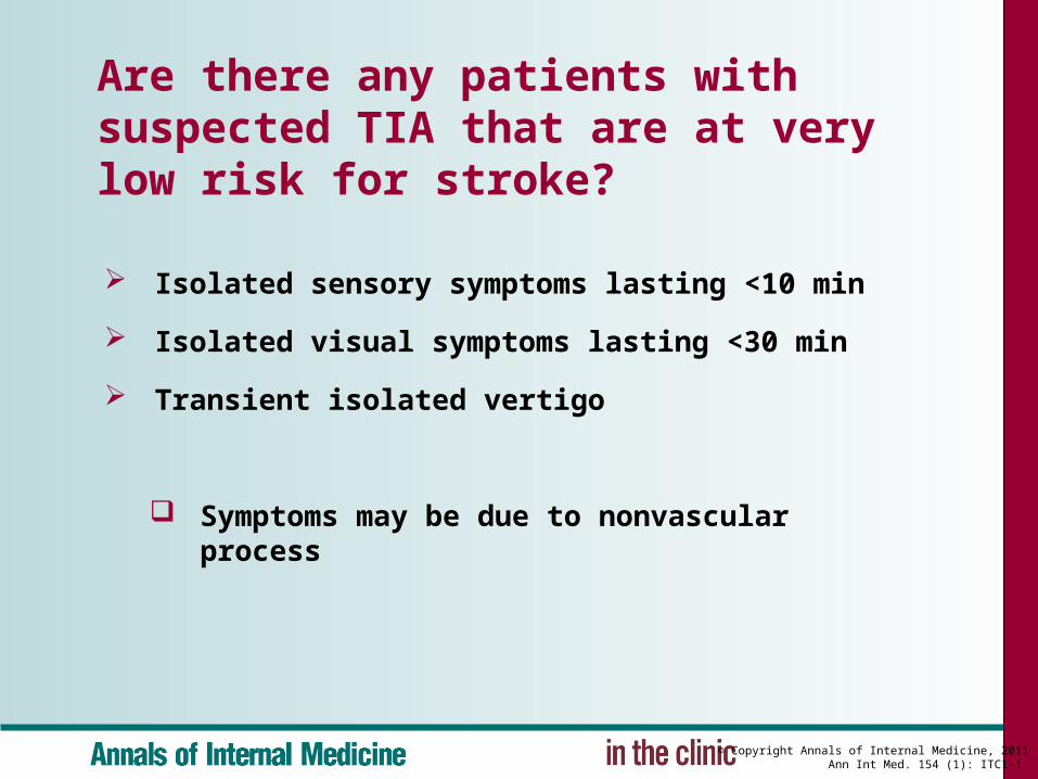
Are there any patients with suspected TIA that are at very low risk for stroke?
Isolated sensory symptoms lasting <10 min
Isolated visual symptoms lasting <30 min
Transient isolated vertigo
Symptoms may be due to nonvascular process
© Copyright Annals of Internal Medicine, 2011Ann Int Med. 154 (1): ITC1-1.
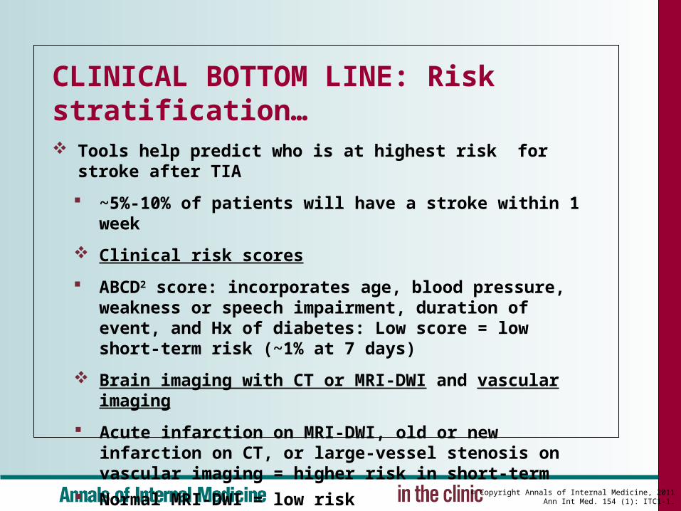
CLINICAL BOTTOM LINE: Risk stratification… Tools help predict who is at highest risk for stroke after TIA
~5%-10% of patients will have a stroke within 1 week
Clinical risk scores
ABCD2 score: incorporates age, blood pressure, weakness or speech impairment, duration of event, and Hx of diabetes: Low score = low short-term risk (~1% at 7 days)
Brain imaging with CT or MRI-DWI and vascular imaging
Acute infarction on MRI-DWI, old or new infarction on CT, or large-vessel stenosis on vascular imaging = higher risk in short-term
Normal MRI-DWI = low risk
© Copyright Annals of Internal Medicine, 2011Ann Int Med. 154 (1): ITC1-1.
When should clinicians hospitalize patients with suspected TIA?
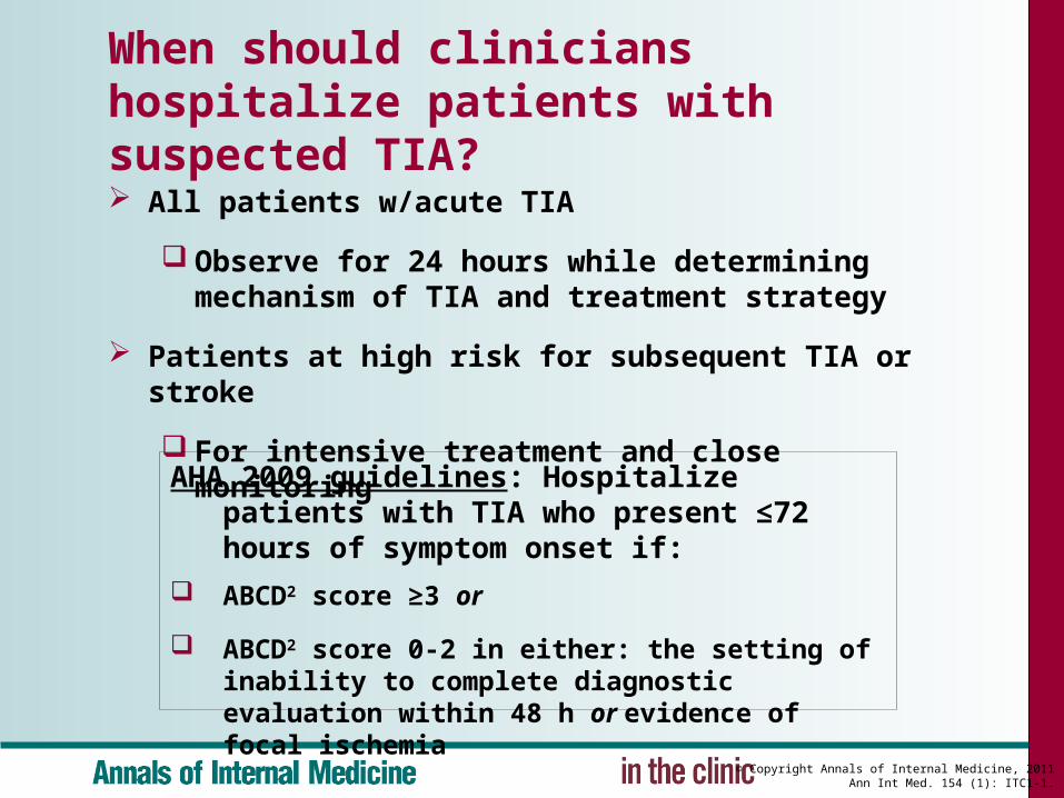
All patients w/acute TIA
Observe for 24 hours while determining mechanism of TIA and treatment strategy
Patients at high risk for subsequent TIA or stroke
For intensive treatment and close monitoring
AHA 2009 guidelines: Hospitalize patients with TIA who present ≤72 hours of symptom onset if:
ABCD2 score ≥3 or
ABCD2 score 0-2 in either: the setting of inability to complete diagnostic evaluation within 48 h or evidence of focal ischemia
© Copyright Annals of Internal Medicine, 2011Ann Int Med. 154 (1): ITC1-1.
What drug therapy should clinicians consider for patients having acute TIA?
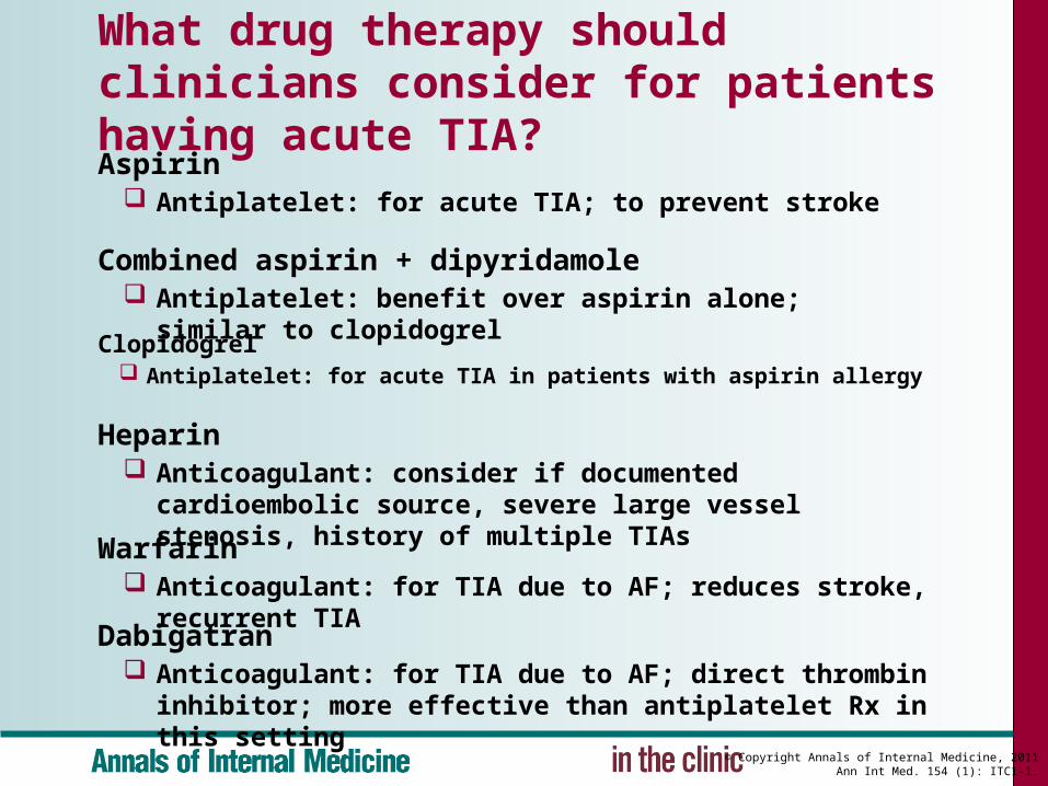
Clopidogrel Antiplatelet: for acute TIA in patients with aspirin allergy
Heparin Anticoagulant: consider if documented cardioembolic
source, severe large vessel stenosis, history of multiple TIAs
Warfarin Anticoagulant: for TIA due to AF; reduces stroke, recurrent TIA
Dabigatran Anticoagulant: for TIA due to AF; direct thrombin inhibitor;
more effective than antiplatelet Rx in this setting
Aspirin Antiplatelet: for acute TIA; to prevent stroke
Combined aspirin + dipyridamole Antiplatelet: benefit over aspirin alone; similar to clopidogrel

© Copyright Annals of Internal Medicine, 2011Ann Int Med. 154 (1): ITC1-1.
How should clinicians manage antihypertensive medications in the setting of acute TIA?
In the first 24 h, optimize cerebral blood flow
Avoid BP-lowering agents (i.e., permissive HTN)
Use IV normal saline to ensure euvolemia
Position patient with head of bed flat
If patient stable for 24 h
Restart antihypertensive medications
Do so more cautiously in patients with TIA due to severe large-vessel stenosis

© Copyright Annals of Internal Medicine, 2011Ann Int Med. 154 (1): ITC1-1.
When should clinicians use anticoagulant and antiplatelet therapy to prevent recurrent TIA or stroke?
Warfarin anticoagulation
If TIA or stroke due to AF: Reduces risk of future stroke
Oral anticoagulation
If TIA due to other high-risk cardioembolic sources
Aspirin + clopidogrel combination
For patients unable to take warfarin for reasons other than bleeding risk
Antiplatelet therapy If no cardioembolic TIA source; clopidogrel or ASA-ER DP better than
aspirin to prevent recurrent vascular events after stroke

© Copyright Annals of Internal Medicine, 2011Ann Int Med. 154 (1): ITC1-1.
What other drug therapy has a role in preventing recurrent TIA or stroke?
Statins
Reduce risk for recurrent vascular events after stroke or TIA
Recommended target LDL <70 mg/dL with atherosclerotic stroke or TIA
Multiple mechanisms ameliorate stroke risk
Antihypertensive Agents
Use outside acute period
Long-term aggressive BP lowering significantly reduces future vascular event risk
Beneficial even in normotensive pts w/stroke or TIA

© Copyright Annals of Internal Medicine, 2011Ann Int Med. 154 (1): ITC1-1.
What surgical or other nondrug therapies can be used to prevent recurrent TIA or stroke? CEA
If carotid stenosis ≥50%: reduces future stroke risk (especially when stenosis >70%)
Best if performed within 2 weeks of TIA, especially when carotid stenosis 50%-69%
Carotid angioplasty and stenting
Alternative to CEA
Use if surgery contraindicated (high carotid bifurcation, previous neck radiation, extremely high cardiac risk)
© Copyright Annals of Internal Medicine, 2011Ann Int Med. 154 (1): ITC1-1.
How should clinicians follow patients with TIA?
Focus on controlling vascular risk factors
Emphasize
Healthy diet and regular exercise
Medication compliance
Smoking cessation
Pay close attention to new neurologic symptoms that suggest recurrent TIA or stroke
In absence of new symptoms, no routine follow-up studies needed

© Copyright Annals of Internal Medicine, 2011Ann Int Med. 154 (1): ITC1-1.
CLINICAL BOTTOM LINE: Treatment… Immediate evaluation important to determine cause of TIA and
strategies to reduce the risk for recurrent TIA or stroke
Hospitalization appropriate for most patients with acute TIA
Administer antiplatelet therapy promptly
Administer long-term anticoagulation to patients with a cardioembolic source, such as AF
Consider carotid revascularization for patients with carotid stenosis >50%, preferably by CEA and within 2 weeks of TIA
Provide long-term antithrombotic therapy and control of vascular risk factors to all patients