chapter 10: blood. blood the only fluid tissue in the body classified as a connective tissue ...
TRANSCRIPT
Chapter 10: Blood
Blood
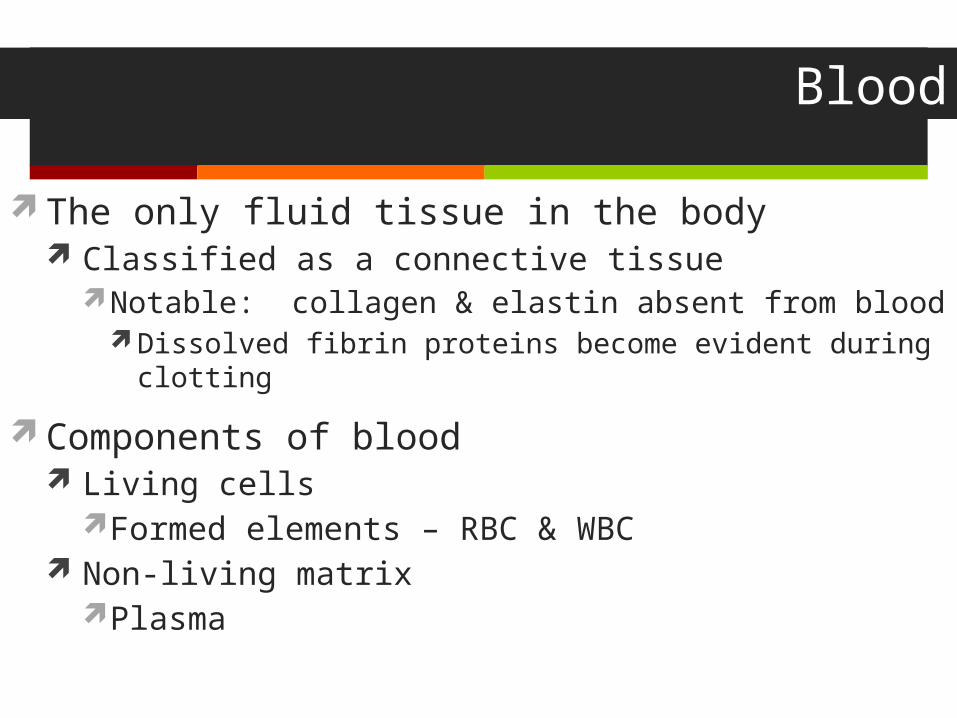
The only fluid tissue in the body Classified as a connective tissue
Notable: collagen & elastin absent from blood Dissolved fibrin proteins become evident during clotting
Components of blood Living cells
Formed elements – RBC & WBC Non-living matrix
Plasma
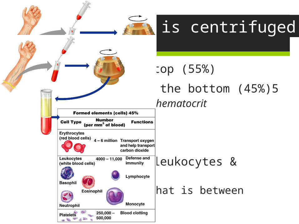
When Blood is centrifuged
Plasma rises to the top (55%) Erythrocytes sink to the bottom (45%)5
This is known as the hematocritNormal hematocrit:
47% +/- 5%: males42% +/-5%: females
Buffy coat contains leukocytes & platelets (<1%) Thin, whitish layer that is between erythrocytes &
plasma
Physical Characteristics of Blood
Sticky, opaque fluid – metallic taste
More dense than water, 5x more viscous
Colors: Oxygen rich blood – scarlet red (bright) Oxygen poor blood – dull red
pH is between 7.35 – 7.45
Blood temperature is always slightly higher than body temperature – around 100.4F
In a healthy man, blood volume is about 5-6 liters or about 6 quarts Women have less blood, about 3.5 – 5 liters
Blood makes up about 8% of body weight
Functions of Blood
All concerned with: substance distribution regulating blood levels of particularly substances bodily protection
Distribution: Oxygen to lungs Nutrients to digestive system Transporting metabolic wastes from elimination sites
(lungs, kidneys, liver) Hormones from glands to target organs
Functions of Blood
Regulation: Maintain appropriate body temperature by absorbing and
distributing heat through body, encouraging skin for heat loss
Maintain normal pH, blood holds an “alkaline” reserve to raise pH when necessary
Maintain adequate fluid volume in circulation Salts & blood protein – prevent excess fluid loss
Protection Prevents blood loss Prevents infection

Blood Plasma
Straw colored, sticky liquid
Composed of approximately 90% water
Includes over 100 dissolved substances: Nutrients Salts (electrolytes) Respiratory gases (CO2 & O2) Hormones Plasma proteins Waste products
Uric acid, creatinine, lactic acid, ammonium salts
Blood Plasma: Plasma Proteins
Most abundant solutes in plasma
Made mostly by the liver
Proteins include: Albumin: regulates osmotic pressure
The pressure that keeps water in the blood Essentially carries all proteins around the blood High albumin almost always caused by dehydration Low albumin can come from liver disease, kidney disease, & malnutrition
Globulins: Alpha, beta: transport proteins that bind to lipids, metal ions, fat-soluble vitamins Gamma: antibodies released during an immune response
Clotting proteins: help to stop blood loss when a blood vessel is injured Antibodies: help protect the body from pathogens (disease)
Blood Plasma
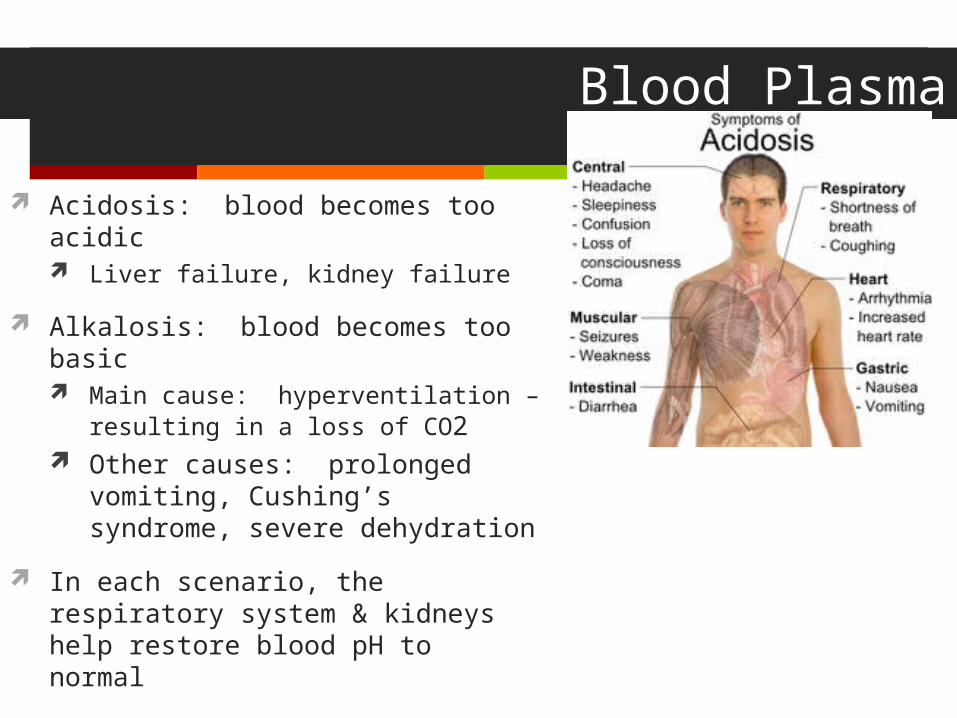
Acidosis: blood becomes too acidic Liver failure, kidney failure
Alkalosis: blood becomes too basic Main cause: hyperventilation –
resulting in a loss of CO2 Other causes: prolonged
vomiting, Cushing’s syndrome, severe dehydration
In each scenario, the respiratory system & kidneys help restore blood pH to normal
Formed Elements
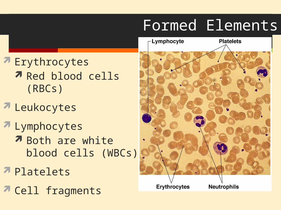
Erythrocytes Red blood cells (RBCs)
Leukocytes
Lymphocytes Both are white
blood cells (WBCs)
Platelets
Cell fragments
Formed Elements Erythrocytes
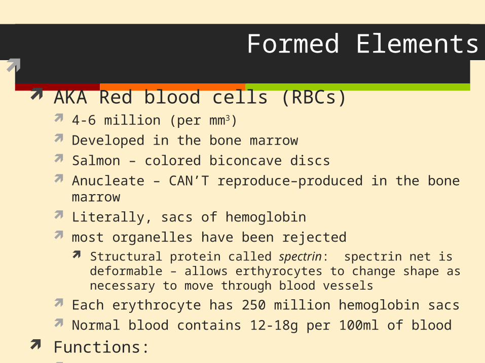
AKA Red blood cells (RBCs) 4-6 million (per mm3) Developed in the bone marrow Salmon – colored biconcave discs Anucleate – CAN’T reproduce–produced in the bone marrow Literally, sacs of hemoglobin most organelles have been rejected
Structural protein called spectrin: spectrin net is deformable – allows erthyrocytes to change shape as necessary to move through blood vessels
Each erythrocyte has 250 million hemoglobin sacs Normal blood contains 12-18g per 100ml of blood
Functions: Transport oxygen to lung capillary beds Also transports small amounts of CO2 (about 20%)
Formed Elements Erythrocytes
Women have lower RBC count (4.32 -5, versus 5.1 – 5.8 mm3) As RBCs increase, blood viscosity increases, blood flow slows
Function: Respiratory gas transport Hemoglobin in RBC binds to oxygen
14-20 (g/100ml) – infants 13-18 – adult males 12 – 16 – adult females
Hemoglobin is inside RBCs to prevents the protein molecule from breaking apart and leaking through the bloodstream
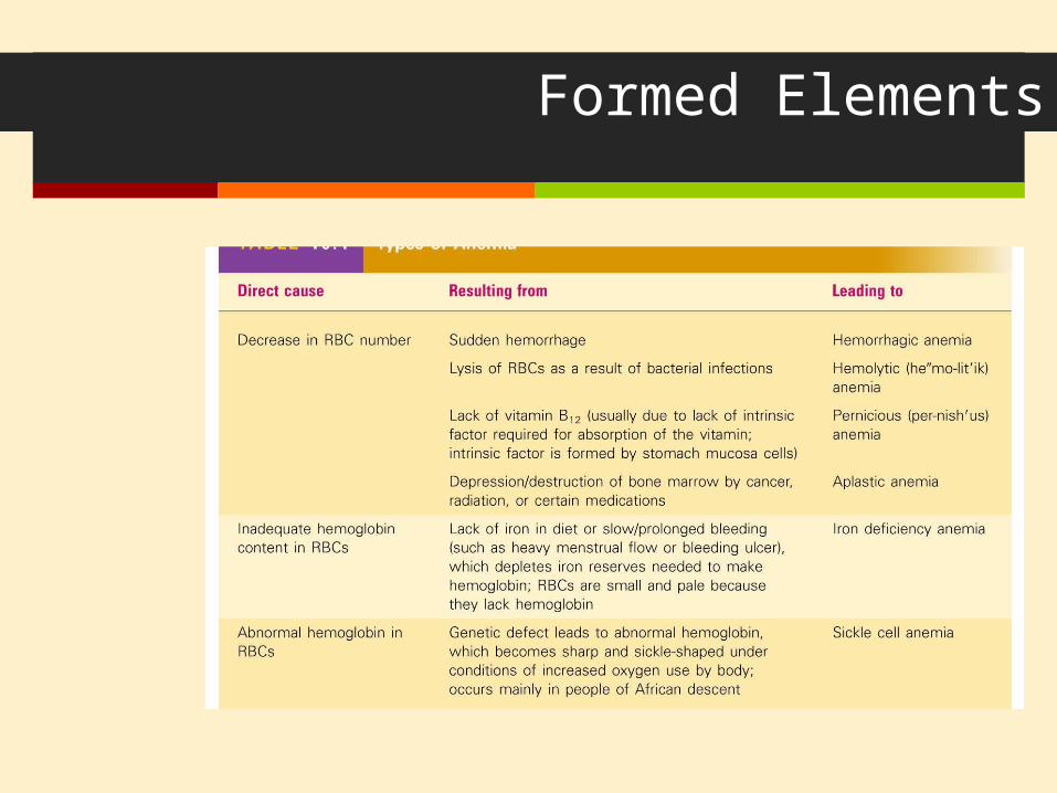
Formed Elements Diseases of RBCs
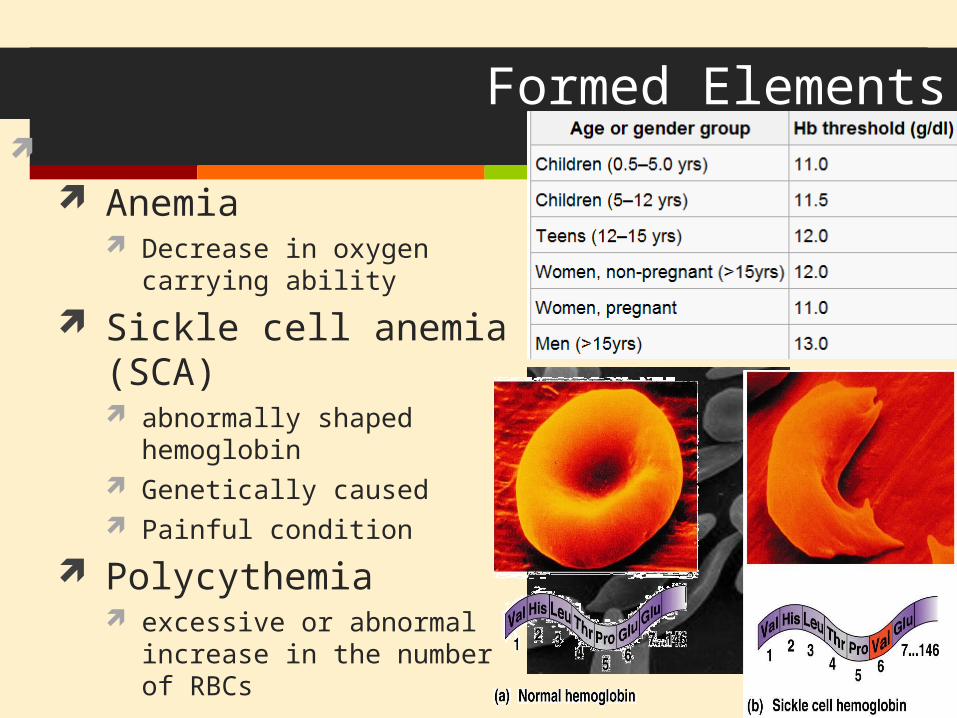
Anemia Decrease in oxygen carrying ability
Sickle cell anemia (SCA) abnormally shaped hemoglobin Genetically caused Painful condition
Polycythemia excessive or abnormal increase in
the number of RBCs
Formed Elements
Formed Elements: Leukocytes Essential for body’s defense against pathogens
Complete cells with a nucleus & organelles
Can move in/out of blood vessels on their own (diapedesis)
Move with ameboid motion
Respond to chemicals released by damaged tissues
4,000 – 11,000 per mm3 of blood
Formed Elements: Types of Leukocytes Granulocytes
Possess lobed nuclei Will show granules in cytoplasm when stained
Include: neutrophils, eosinophils, & basophils
Agranulocytes Lack of visible granules Nuclei are spherical, oval or kidney – shaped Include: lymphocytes & monocytes
List of the WBCs from most to least abundant Neutrophils Never Lymphocytes Let Monocytes Monkeys Eosinophils Eat Basophils Bananas
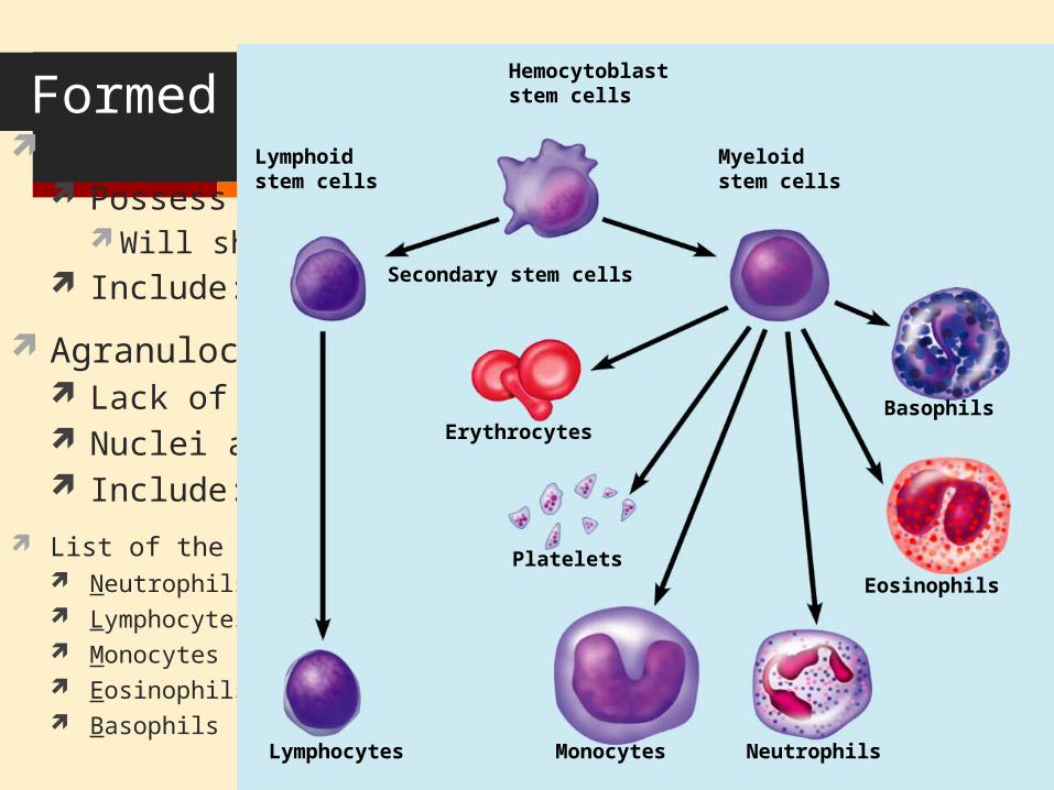
Hemocytoblaststem cells
Secondary stem cells
Basophils
Eosinophils
NeutrophilsMonocytesLymphocytes
Erythrocytes
Platelets
Lymphoidstem cells
Myeloidstem cells
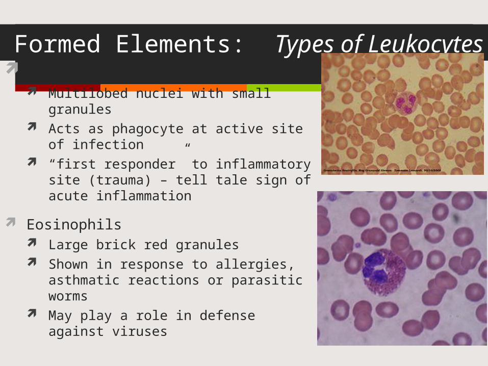
Formed Elements: Types of Leukocytes Neutrophils
Multilobed nuclei with small granules Acts as phagocyte at active site of
infection “first responder” to inflammatory site
(trauma) – tell tale sign of acute inflammation
Eosinophils Large brick red granules Shown in response to allergies, asthmatic
reactions or parasitic worms May play a role in defense against viruses
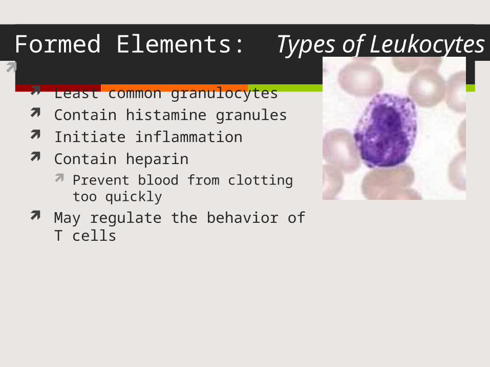
Formed Elements: Types of Leukocytes Basophils
Least common granulocytes Contain histamine granules Initiate inflammation Contain heparin
Prevent blood from clotting too quickly May regulate the behavior of T cells
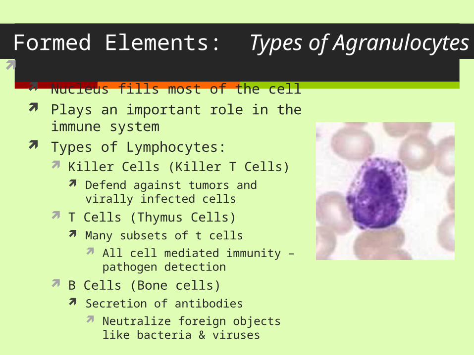
Formed Elements: Types of Agranulocytes Lymphocytes
Nucleus fills most of the cell Plays an important role in the immune
system Types of Lymphocytes:
Killer Cells (Killer T Cells) Defend against tumors and virally infected
cells T Cells (Thymus Cells)
Many subsets of t cells All cell mediated immunity – pathogen
detection B Cells (Bone cells)
Secretion of antibodies Neutralize foreign objects like bacteria &
viruses
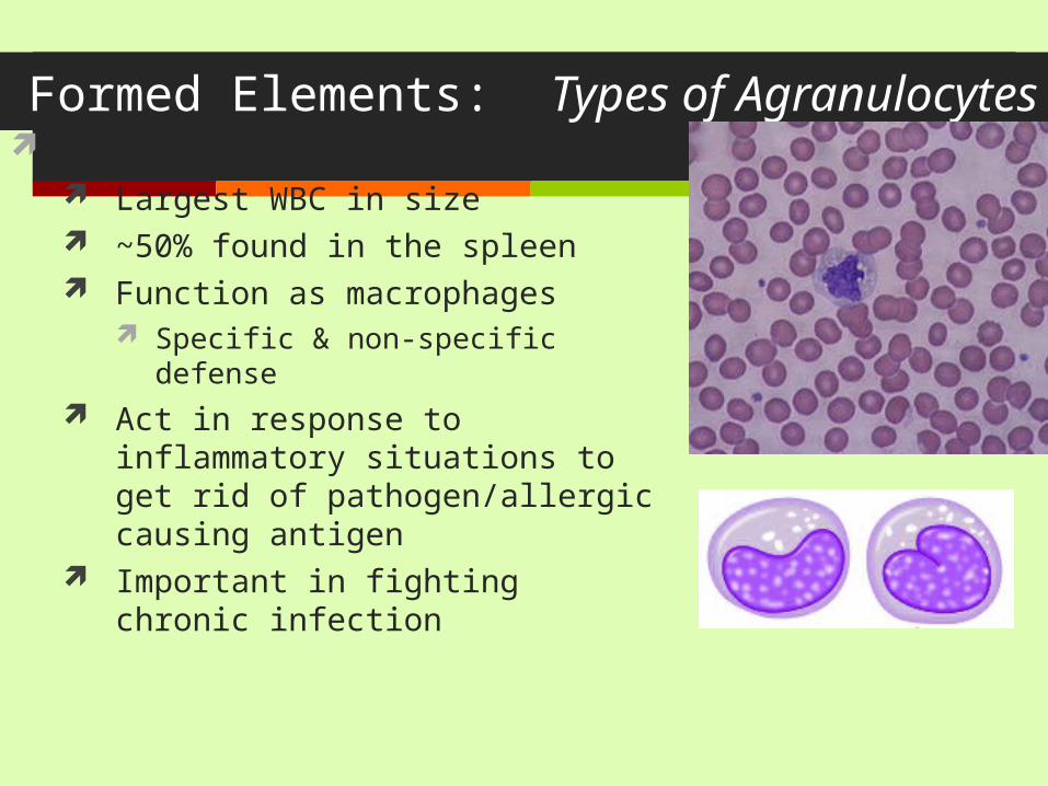
Formed Elements: Types of Agranulocytes Monocytes
Largest WBC in size ~50% found in the spleen Function as macrophages
Specific & non-specific defense Act in response to inflammatory situations
to get rid of pathogen/allergic causing antigen
Important in fighting chronic infection
WBC Abnormalities Leukocytosis
WBC count above 11,000 leukocytes/mm3
Generally indicates an infection But not indicative of any specific infection
It’s diagnostically similar to a fever
Leukopenia Abnormally low leukocyte level
Commonly caused by certain drugs such as corticosteroids and anticancer agents An important indicator of infection risk
Leukemia Bone marrow becomes cancerous, turns out excess WBC 4 types: ALL (acute lymphoblastic), CLL, AML (acute myelogenous), & CML
Can be acute or chronic Acute is more common in children, chronic in the elderly
Can be lymphoid or myeloid Lymphoid is more common in children, myeloid is rare in children
http://www.youtube.com/watch?v=tDTLC2swhlQ
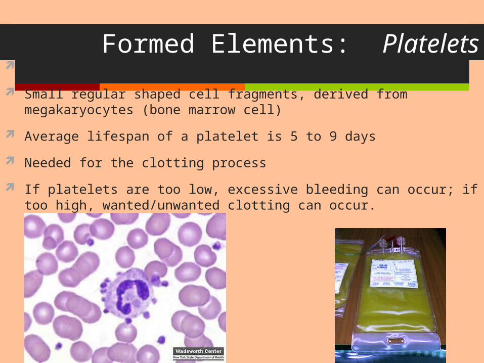
Formed Elements: Platelets Aka thrombocytes
Small regular shaped cell fragments, derived from megakaryocytes (bone marrow cell)
Average lifespan of a platelet is 5 to 9 days
Needed for the clotting process
If platelets are too low, excessive bleeding can occur; if too high, wanted/unwanted clotting can occur.
Hematopoiesis Blood cell formation in red bone marrow
All blood cells (red & white) derive from a common stem cell (hemocytoblast)
Differentation: Lymphoid stem cells produce lymphocytes Myeloid stem cell produces all other formed elements
These stem cells are self-renewing I.e. – some never develop into RBCs or WBCs – they stay
stem cells so they can mitotically divide into more stem cells.
Erthyrocyte formation Erthyrocytes are unable to divide, grow & synthesize proteins
Wear out in 100 – 120 days
When they are no longer usable, they will be removed by phagocytosis by the liver or spleen
Lost cells are replaced by hemocytoblasts
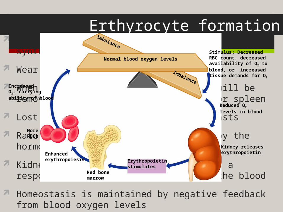
Rate of RBC production is controlled by the hormone erthyropoietin
Kidneys produce most erythropoietin as a response to reduced oxygen levels in the blood
Homeostasis is maintained by negative feedback from blood oxygen levels
Reduced O2
levels in blood
Stimulus: DecreasedRBC count, decreasedavailability of O2 toblood, or increasedtissue demands for O2
IncreasedO2- carryingability of blood
Erythropoietinstimulates
Kidney releaseserythropoietinEnhanced
erythropoiesis
Red bonemarrow
MoreRBCs
Normal blood oxygen levels
Imbalance
Imbalance

Formation of WBCs and platelets
Controlled by hormones Colony stimulating factors (CSFs) and interleukins prompt bone
marrow to generate leukocytes Thrombopoietin stimulates production of platelets
Hemostasis
Stoppage of bleeding resulting from a break in a blood vessel
Hemostasis involves three phases Vascular spasms
Vasoconstriction causes the blood vessel to spasmSpasms narrow the blood vessel and decreases
overall blood loss
Hemostasis Platelet plug formation
Collagen fibers that make up the blood vessel become exposed by the break
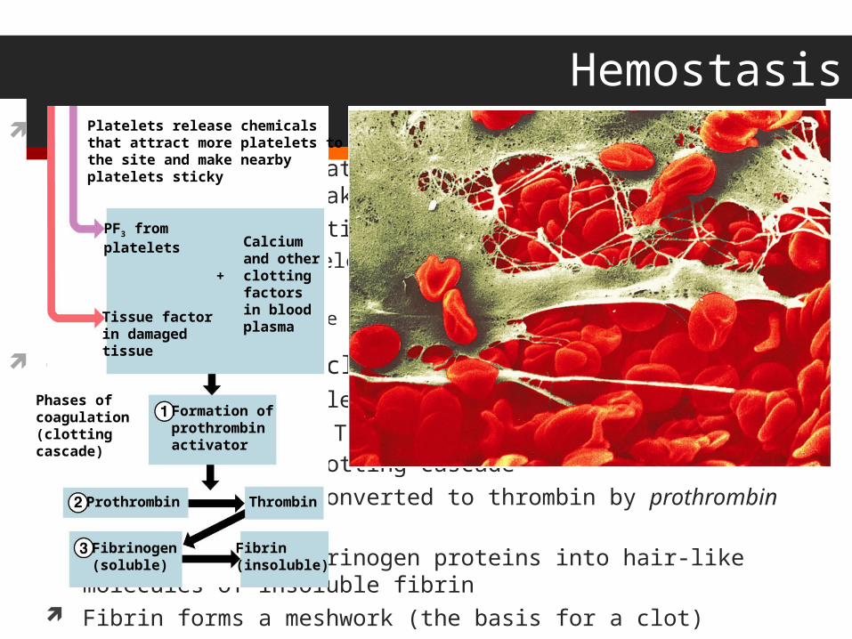
Platelets become sticky & cling to the collagen fibers Those platelets release chemicals (PF3) to attract more platelets
The platelets pile up to form a platelet plug
Coagulation (blood clotting) Injured tissues release TF (tissue factor) PF3 interacts with TF, clotting factors & calcium ions to initiate the
clotting cascade Prothrombin gets converted to thrombin by prothrombin activator. Thrombin joins fibrinogen proteins into hair-like molecules of insoluble
fibrin Fibrin forms a meshwork (the basis for a clot)
Platelets release chemicalsthat attract more platelets tothe site and make nearbyplatelets sticky
PF3 fromplatelets Calcium
and otherclottingfactorsin bloodplasma
Formation ofprothrombinactivator
Prothrombin
Fibrinogen(soluble)
Fibrin(insoluble)
Thrombin
Tissue factorin damagedtissue
Phases ofcoagulation(clottingcascade)
+
Hemostasis
Blood usually clots within 3 to 6 minutes
The clot remains as endothelium regenerates
The clot is broken down after tissue repair
Undesirable Clotting Thrombus
A clot in an unbroken blood vessel – normal during injury but should be disposed of once the risk of excessive bleeding has past.
Can be deadly in areas like the heart More likely to occur when there are problems in the heart (arrhythmia,
heart valve replacement and/or recent heart attack
Embolus A thrombus that breaks away and floats freely in the bloodstream
In general, an embolus is ANY detached, itinerant intravascular mass – they can be solid, liquid or gas
Can later clog vessels in critical areas such as the brain
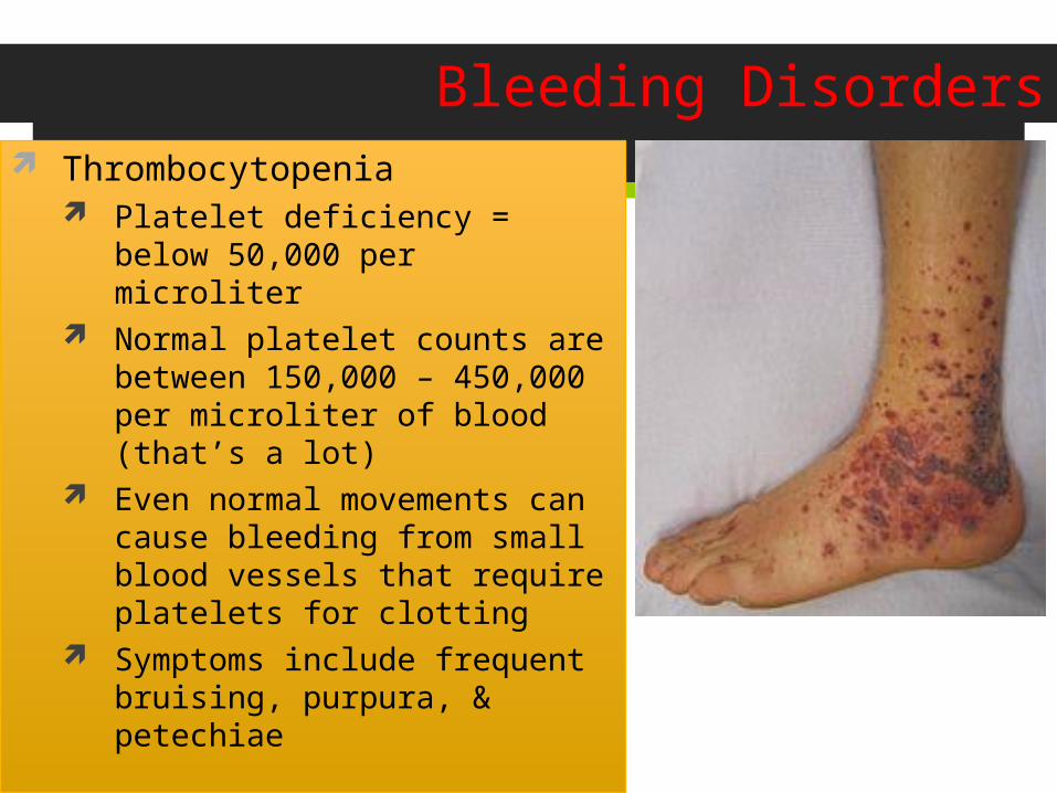
Bleeding Disorders Thrombocytopenia
Platelet deficiency = below 50,000 per microliter
Normal platelet counts are between 150,000 – 450,000 per microliter of blood (that’s a lot)
Even normal movements can cause bleeding from small blood vessels that require platelets for clotting
Symptoms include frequent bruising, purpura, & petechiae
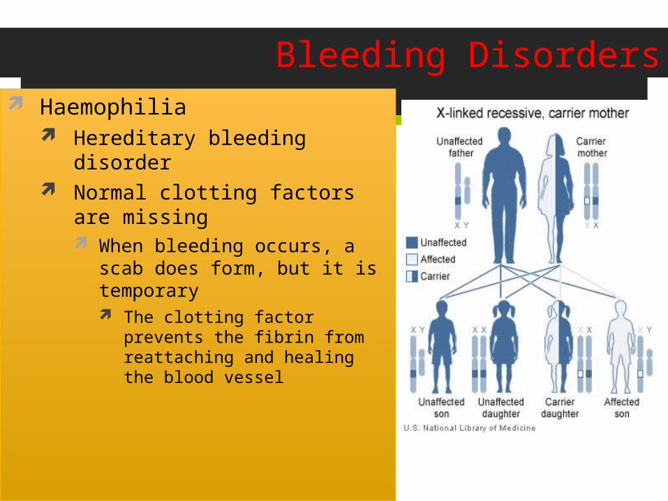
Bleeding Disorders Haemophilia
Hereditary bleeding disorder Normal clotting factors are missing
When bleeding occurs, a scab does form, but it is temporary The clotting factor prevents the fibrin
from reattaching and healing the blood vessel
Blood Typing & Transfusions
Large losses of blood have serious consequences Loss of 15–30% causes weakness Loss of over 30% causes shock, which can be fatal
Transfusions are the only way to replace blood quickly
Transfused blood must be of the same blood group
Blood is tested for a large number of diseases before giving to the patient who needs blood Tested for:
All forms of HIV, All forms of hepatitis, syphillis, CMV, and West Nile virus – just to name a few Complications:
Hemolytic reactions: you have antibodies again the donor’s RBCs – symptoms include fever, chills, increased heart rate, shortness of breath, rapid drop in blood pressure. Transfusion must be stopped immediately before kidney damage occurs
Allergic reactions: while blood banks look at the complete chemical make up of donor blood, if a patient is unaware of allergies and these chemicals are in blood – an allergic reaction can occur If severe, Usually easily fixed by dosing epinephrine to stop anaphylaxis
Human Blood Groups
Blood contains genetically determined proteins
Antigens (a substance the body recognizes as foreign) may be attacked by the immune system
Antibodies are the “recognizers”
Blood is “typed” by using antibodies that will cause blood with certain proteins to clump (agglutination)
There are over 30 common red blood cell antigens
The most vigorous transfusion reactions are caused by ABO and Rh blood group antigens
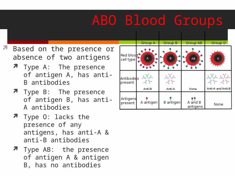
ABO Blood Groups
Based on the presence or absence of two antigens Type A: The presence of
antigen A, has anti-B antibodies
Type B: The presence of antigen B, has anti-A antibodies
Type O: lacks the presence of any antigens, has anti-A & anti-B antibodies
Type AB: the presence of antigen A & antigen B, has no antibodies

ABO Blood Groups
Blood type AB can receive A, B, AB, and O blood Universal recipient
Blood type B can receive B and O blood
Blood type A can receive A and O blood
Blood type O can receive O blood Universal donor
Rh Blood Groups
Named because of the presence or absence of one of eight Rh antigens (agglutinogen D) that was originally defined in Rhesus monkeys
Most Americans are Rh+ (Rh positive)
Problems can occur in mixing Rh+ blood into a body with Rh– (Rh negative) blood
Dangers of an Rh mismatch
Danger occurs only when the mother is Rh– and the father is Rh+, and the child inherits the Rh+ factor
RhoGAM shot can prevent buildup of anti-Rh+ antibodies in mother’s blood
The mismatch of an Rh– mother carrying an Rh+ baby can cause problems for the unborn child The first pregnancy usually proceeds without problems The immune system is sensitized after the first pregnancy In a second pregnancy, the mother’s immune system produces
antibodies to attack the Rh+ blood (hemolytic disease of the newborn)
Blood typing
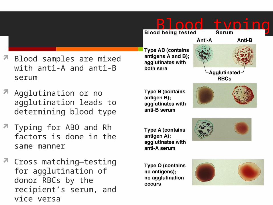
Blood samples are mixed with anti-A and anti-B serum
Agglutination or no agglutination leads to determining blood type
Typing for ABO and Rh factors is done in the same manner
Cross matching—testing for agglutination of donor RBCs by the recipient’s serum, and vice versa
Developmental Aspects of Blood
Sites of blood cell formation The fetal liver and spleen are early sites of blood cell formation Bone marrow takes over hematopoiesis by the seventh month
Fetal hemoglobin differs from hemoglobin produced after birth It can carry much more oxygen than adult hemoglobin By 12 weeks of life, fetal hemoglobin is replaced by adult hemoglobin
Physiologic jaundice results in infants in which the liver cannot rid the body of hemoglobin breakdown products fast enough