body temperature is the balance between heat produced in the body and heat loss from the body. heat...
TRANSCRIPT


Body temperature is the balance between heat produced in the body and heat loss from the body.

Core Temperature – temperature of the deep tissues of the body such as abdominal or pelvic cavities. It is relatively constant
Surface Temperature – temperature of the skin and subcutaneous tissue. It fluctuates depending on the blood supply to the skin and the amount of heat loss to the external environment.


Physiologically ◦ Receptors in the skin, abdomen, and spinal cord send messages to
the autonomic nervous system that sends the message to the hypothalamus.
◦ The hypothalamus acts as a central thermostat, receiving input from sensors that detect hot or cold temperatures and initiates body responses mainly in the cardiovascular system via vasoconstriction or vasodilation that decrease heat production and increase heat loss.
Behavioral◦ When an individual perceives he is hot or
cold, he changes his behavior such as: moves to the shade or sun, regulates the thermostat, removes extra clothes or puts on sweater.

Radiation
◦Conduction
Evaporation
Convection

◦ Transfer of heat from the surface of one object to the surface of another without contact
◦ Blood flows from the core internal organs carrying heat to skin and surface blood vessels.
◦ Amount of heat carried to the surface depends on the extent of vasoconstriction and vasodilation regulated by hypothalamus.
◦ 85% of body heat is loss via radiation to the environment
◦ Usually from the body to a cooler surface

Transfer of heat from one object to another with direct contact.
When the warm skin touches a cooler object, the heat is loss
Increase amount loss by applying a ice pack, bathing with cool water
Decrease amount of heat loss by applying blankets
Gains heat via conduction when contact is made with a warmer object

Transfer of heat when a liquid is changed to a gas.
When body temperature rises, hypothalamus signals the sweat glands to release sweat. Sweat evaporates from the skin, resulting in heat loss.

Transfer of heat away by air movements.
Air currents carry away the heat
An electric fan promotes heat loss through convection
Vasoconstriction◦ Decreases the amount of blood that reaches the
surface and thus is not lost.

Increase Basal Metabolism Rate
◦ Movement / exercise
◦ Shivering

Age Activity / Exercise / Sleep Hormones Stress Environment Medication Illness

Children: 98.60 - 99.60 F, oral
Adults: 98.6 + /-1 degree F, oral
Older Adults: 97.6 + / -1 degree F, oral

Rectal temperature is usually 1 degree higher than oral
Axillary temperature is usually 1 degree lower than oral

Pyrexia, hyperthermia, fever – an elevation in body temperature
Hyperpyrexia – a very high fever such as: 1050 F
Febrile – having a fever; Afebrile – without a fever
Hypothermia – decreased body temperature below 970 F
The nurse needs to look at the relationship of the vital signs to each other, to previous findings, and to other assessment data.
If the temperature is abnormal, the nurse should notify the physician.
Reassess the temperature more often than ordered – nursing intervention / judgment


Oral
◦Rectal
Axillary
Tympanic
Temporal

Most common and convenient
Sublingual pocket very vascular and responds to changes in core temperature
Disadvantage – varies with what the patient has had in their mouth, mouth breathers
Not safe for infants, children, elderly, disoriented, epileptic, or unconscious
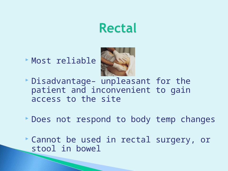
Most reliable
Disadvantage– unpleasant for the patient and inconvenient to gain access to the site
Does not respond to body temp changes
Cannot be used in rectal surgery, or stool in bowel

Safest, Noninvasive, Easily accessible
Does not always give a true reading of core temperature
Used for infants, children, patients with oral problems or mouth breathers, and irrational patients.

Utilizes the ear which is readily accessible, minimal discomfort, and reflects core temperature
Disadvantage – cost of the equipment and inaccurate reading because of incorrect positioning of the instrument.

One of the newer methods for evaluating a person’s temperature is the temporal area of the forehead.
The temporal artery is a branch of the internal carotid artery, but courses within a millimeter or two of the skin's surface over the lateral forehead and is readily accessible.


Battery operated unit with an attached heat sensitive probe and disposable probe cover.
More rapid reading
Can be used in all methods of assessing the temperature
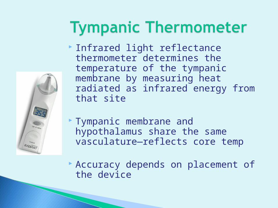
Infrared light reflectance thermometer determines the temperature of the tympanic membrane by measuring heat radiated as infrared energy from that site
Tympanic membrane and hypothalamus share the same vasculature—reflects core temp
Accuracy depends on placement of the device

Designed specifically to be completely non-invasive
Scanner captures naturally emitted infrared heat from the arterial blood supply, locking in the highest temperature it senses

Measures the temperature by movement of chemical through the calibrated glass.
Must be shaken down to 950 before taking a reading.
Disadvantage – must follow hazard precautions if it should break.

Shakes the thermometer to get below 960 or inserts into probe cover
Places in posterior sublingual pocket Remain in mouth for 3 - 5 minutes Remove and wipe from stem to bulb (clean to dirty) Reads at eye level, rotates slowly until mercury
visible and reads to nearest tenth of degree Shakes the thermometer down, cleans with
soapy cold water, or pushes ejection button to release probe cover

Preparation: same
Places the patient in Sim’s position and drape
Apply gloves
Prepares thermometer- lubricates tip

Dominant hand hold thermometer, other hand, separate buttocks to expose anus
Instruct patient to take a deep breath. Insert the thermometer gently into the anus: infant- ½ inch, adult 1 ½ inches.
Do not force insertion
Hold in place for 3-5 minutes
Wipe secretions, and discard tissue, read temp

Gain access to the axillary area
Make sure area is dry
Place thermometer in the center of axilla. Fold patients arm straight down and place arm across chest
Leave in place 6-10 minutes
Remove, read, Write down

Easy way to remember how long to take a temperature
5 min. = oral +3 min. = rectal 8 min. = axillary

Position patient upright or on their side
Attach probe cover to the unit
Turn head to one side and insert into ear canal ◦ Adult – pull pinna upward and back◦ Child – pull pinna downward and back
Remove after reading displayed
Remove probe cover and place in storage unit

With just a light stroke across the temporal artery area of the forehead, an accurate reproducible temperature is measured in about 3 seconds - eliminating any discomfort caused by thermometer inserted into the ear, mouth, or rectum.


A wave of blood created by contraction of the left ventricle of the heart
Heart is a large pump that forces blood to enter the arteries with each heartbeat, causing pressure pulses or pulse waves

The heart pumps 4-6 liters each minute.
This volume is the cardiac output

Stroke volume- amount of blood that enters the arteries with each ventricle contraction◦ Normally heart empties about 70% of its volume
with each contraction. That is about 70 ml. of blood in an adult
Heart rate- beats per minute
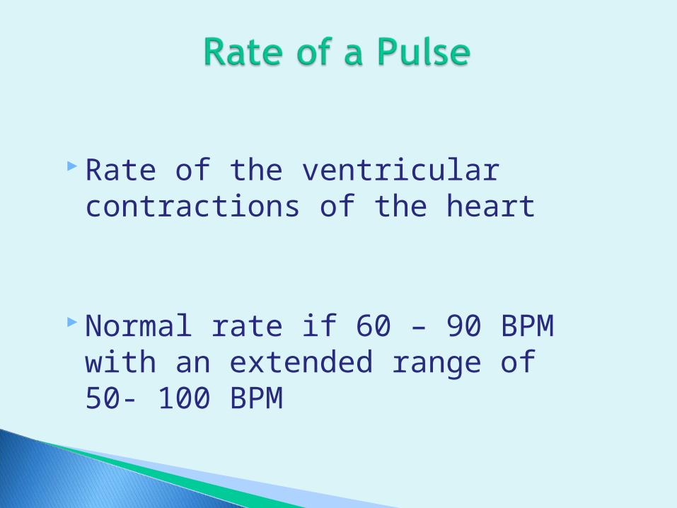
Rate of the ventricular contractions of the heart
Normal rate if 60 – 90 BPM with an extended range of 50- 100 BPM
Tachycardia◦ Increased heart rate – over 100 BPM◦ Usually occur when sympathetic nervous
system is stimulated
Bradycardia◦ Decreased heart rate – below 60 BPM◦ Usually occur with parasympathetic nervous
system is stimulated

Age Sex Exercise Temperature Medications Emotions / Stress Hemorrhage Illness Position

Pattern of the beats and the intervals between the beats.
Equal time should lapse between beats of a normal pulse making a normal rhythm
An irregular rhythm is dysrhythmia

May consist of an early beat, late beat, or missed beat
Irregular rhythm may be random, irregular rhythm or a predictable pattern of irregular beats.
Threatens the heart’s ability to provide adequate cardiac output.

Etiology = A contraction of the heart that fails to transmit a pulse wave to the peripheral pulse sites
Assess by apical – radial pulse simultaneously for one minute, compare
Difference between the apical and radial pulse rates is the pulse deficit.

Nurse A takes the apical pulse and gets a rate of 88.
Nurse B takes the radial pulse and gets a rate of 76.
The pulse deficit is 12.
88 – 76 = 12

Strength of the pulse reflects the volume of blood ejected against the arterial wall with each heartbeat.
Usually the volume / strength is the same with each heartbeat.

Absent ◦ Not palpable
Weak, thready◦ Difficult to feel, readily obliterated with pressure
from the fingers
Normal◦ Easily detectable with moderate pressure
Bounding, full◦ Obliterated, only with difficulty

A healthy normal artery should feel:◦ Straight◦ Smooth◦ Soft◦ Pliable
That makes it easy to assess the pulse

If pulses on both sides of the body are equal, they are known as bilaterally equal
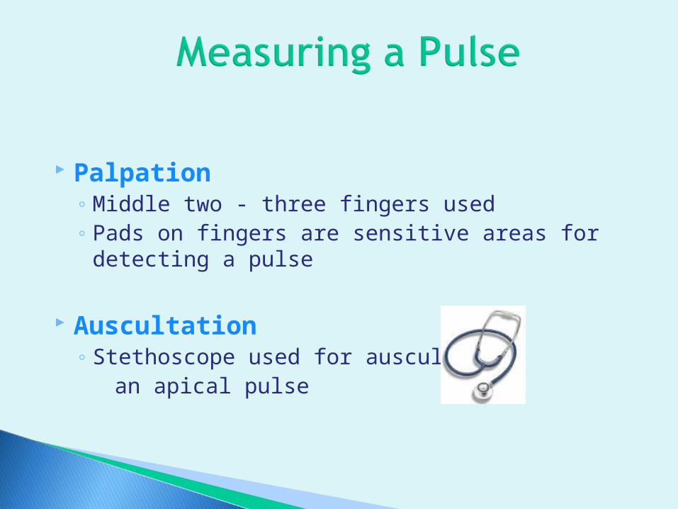
Palpation◦ Middle two - three fingers used◦ Pads on fingers are sensitive areas for detecting a
pulse
Auscultation◦ Stethoscope used for auscultating an apical pulse

Where arteries lie over bony surfaces:◦ Temporal
◦ Carotid artery - in the neck
◦ Brachial – inner aspect of the elbow
◦ Radial – inner aspect of wrist on thumb side
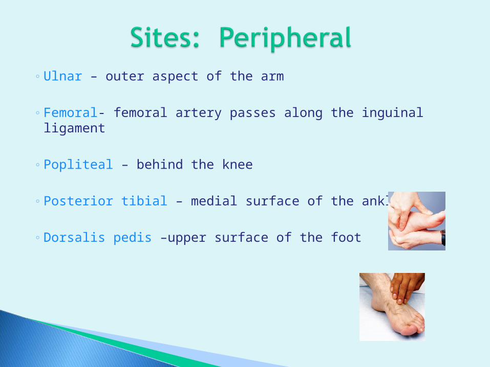
◦ Ulnar – outer aspect of the arm
◦ Femoral- femoral artery passes along the inguinal ligament
◦ Popliteal – behind the knee
◦ Posterior tibial – medial surface of the ankle
◦ Dorsalis pedis –upper surface of the foot

Taken with a stethoscope at apex of the heart S1- low pitched and dull, lub at apex of
heart, fifth intercostal space, just left of the midclavicular line
S2 – Higher and shorter, dub at the base of heart, second intercostal space, right or left of sternal border
Each lub-dub is one heart beat Counted for one full minute
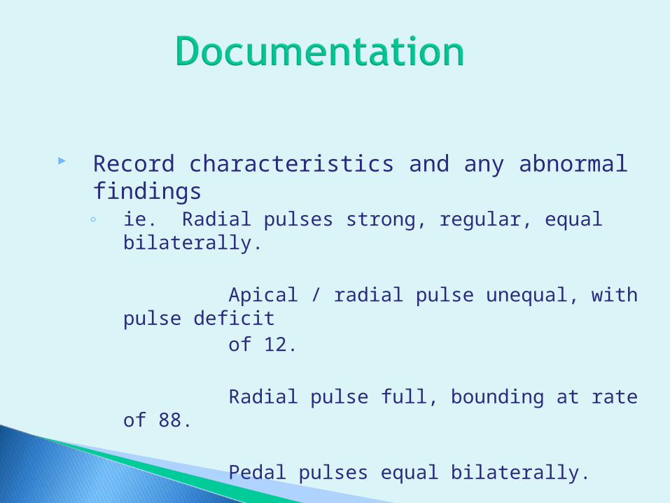
Record characteristics and any abnormal findings◦ ie. Radial pulses strong, regular, equal
bilaterally.
Apical / radial pulse unequal, with pulse deficit
of 12.
Radial pulse full, bounding at rate of 88.
Pedal pulses equal bilaterally.

The act of breathing
Supplying oxygen to the body cells and removing carbon dioxide from the body cells

Inhalation of air into the lungs and exhalation of gases from the lungs to the atmosphere
Three components:1. Ventilation – mechanical movement of air in and out
of the lungs2. Diffusion – movement of oxygen and carbon dioxide
between alveoli and RBC’s3. Perfusion – distribution of blood to and from the
pulmonary capillaries in the lungs

Cellular level
Oxygen diffuses from hemoglobin in the RBC to the cells of the body for use in production of heat and energy
Carbon dioxide if given off from the body cell as waste by-product of metabolism
Respiratory center in the brain stem and chemoreceptors in the carotid artery and aorta.◦ Regulated by levels of carbon dioxide, oxygen, and hydrogen
ion concentration (pH) in the arterial blood ◦ Most important is level of CO2. Elevation of carbon dioxide
causes the respiratory control system in the brain to increase the rate and depth of breathing to move out the carbon dioxide
If arterial oxygen levels drop, chemoreceptors signal the brain to increase the rate and depth of ventilation

Accurate measurements requires observation of the patient’s chest wall movement
Best to assess when resting
Prior data collection:◦ Normal breathing pattern◦ Health problems◦ Medications or therapies◦ Relationship of respirations to
cardiovascular function

Normally 12 – 20 breaths per minute
Terminology:◦Eupnea – normal rate and depth
◦Bradypnea – abnormally slow respirations
◦Tachypnea – abnormally fast respirations
◦Apnea – cessation or absence of breathing

Exercise – increases the rate
Stress – increases the rate
Increased altitude – increases the rate
Medications – narcotics and analgesics lower the rates

Regularity of expirations and inspirations
Normally, they are evenly spaced in an adult.
Normally, irregular in an infant
Irregular respirations in an adult should be reported.
Degree of movement in the chest wall
Described as normal, deep, or shallow
Normal inspiration and expiration or volume of air exchanges with each breath in an adult is about 500 ml. of air. This in know as tidal volume

Assess the chest◦ Does it expand symmetrically with inspiration◦ Is there retraction of intercostal spaces between the
ribs on inspiration◦ Are the respirations diaphragmatic or costal◦ Are accessory muscles used to augment
Difficulty with breathing is known as dyspnea

Respirations should be quiet
Abnormal breathing sounds are:◦ Stridor – shrill, harsh, inspiratory crowing sounds that occurs
with upper airway obstruction
◦ Wheeze – high-pitched, musical, whistling sound that occurs with partial obstruction in the smaller bronchi
◦ Stertor- snoring respiration produced by secretions in the trachea and large bronchi

Hypoventilation – rate and depth decreased
Hyperventilation – rate and depth increased
Cheyne-Stokes – irregular, alternating periods of apnea and hyperventilation
Kussmaul –abnormally deep, regular and increased in rate.

Unobtrusively observes the patient’s respirations while seeming to be involved in another activity
Counts the rate for 30 seconds unless otherwise indicated
Washes hands if contact made with the patient or furnishings in the room
Documents
Reports abnormalities


A measure of the pressure exerted by the blood on the walls of an artery as it flows through the arteries.
Systolic Pressure◦ The pressure of the blood as a result of
contraction of the ventricles, that is the pressure of the height of the blood wave
Diastolic Pressure◦ The pressure when the ventricles are at rest.
Diastolic pressure in the minimal pressure exerted against the arterial walls at all times

The difference between the systolic and diastolic pressure

Systolic 100 – 120 mm. Hg.
______________Diastolic 60-80 mm. Hg.

The volume of blood pumped by the heart (stroke volume) during one minute (heart rate).
The blood pressure depends on the cardiac output and peripheral resistance
B/P = CO X R

Resistance of blood flow determined by the tone of vascular musculature and diameter of blood vessels

Volume of blood circulating with the vascular system
Normal is about 5000 ml.
If volume increases, the B/P goes up; if volume decreases, the B/P drops.

Thickness of blood Hematocrit– percentage of RBC’s in blood
compared to plasma.
When hematocrit rises, the blood is thicker and the heart must work harder to push it around. This causes the B/P to go up.

Vessels walls normally have elasticity and can adjust (dilate and constrict) to accommodate the needs of the body.
When vessels lose their elasticity and is replaced by fibrous tissue that does not stretch, there is greater resistance to blood flow, and the B/P rises.

1. Age
2. Stress and emotions
3. Gender
4. Medications
5. Disease / Illness
6. Body position and exercise
Hypertension is an elevated B/P◦ Occurs on two or more occasions◦ Diastolic pressure if 80 mm.Hg. or above◦ Systolic pressure is 120 mm Hg or above
Hypotension is a lowered B/P◦ Systolic pressure is 90 mm.Hg or below
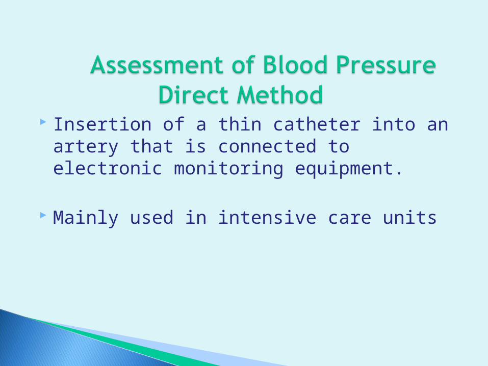
Insertion of a thin catheter into an artery that is connected to electronic monitoring equipment.
Mainly used in intensive care units
Measured by auscultation or palpation

Sphygmomanometer
Stethoscope
Cuff

Aneroid—glass enclosed circular gauge containing a needle that registers millimeters calibrations
Mercury – Upright tube containing mercury. Pressure created by inflation of cuff moves column of mercury upward against gravity.
Cuff – come in various sizes and must have the correct size

Used to hear sounds on which you base the B/P
Be sure the earpieces point toward the nose and not the back of the head

Brachial is the most common
Alternative sites:◦Lower extremities—popliteal artery behind the knee.

Electronic blood pressure machines

Cuff too wide Cuff too narrow Cuff wrapped too
loosely Deflating cuff too
slowly Deflating cuff too
quickly Stethoscope that
fits poorly Inaccurate
inflation level
False low reading False high reading False high reading
False high diastolic reading
False low systolic and false high diastolic
False low systolic and false high diastolic
False low systolic reading

Assessment◦ Reviews chart for data RT blood pressure◦ Evaluates size of arm and accessibility◦ Explains to patient
Washes hands Positions patient and removes any
clothing Extends the arm with palm facing up Applies the cuff 1-2 inches above
inner aspect of elbow with bladder over brachial artery

Palpates the brachial pulse, places the stethoscope over the brachial pulse area
Tightens the valve on the cuff and inflates about 30-40 mm. over normal or inflate to about 180 –200 mm.
Releases valve slowly controlling rate of descent which will deflate the cuff.
Watch the calibrations on the gauge as the air is released and note changes in the sounds.

Removes the cuffAssists patient to comfortable position
Documents Reports unusual findings