© 2015 pearson education, inc. figure 5.1 flat bones consist of a layer of spongy bone sandwiched...
TRANSCRIPT

© 2015 Pearson Education, Inc.
Figure 5.1 Flat bones consist of a layer of spongy bone sandwiched between two thin layers of compact bone.

© 2015 Pearson Education, Inc.
Figure 5.2 Classification of bones on the basis of shape.

© 2015 Pearson Education, Inc.
Figure 5.2a Classification of bones on the basis of shape.
© 2015 Pearson Education, Inc.
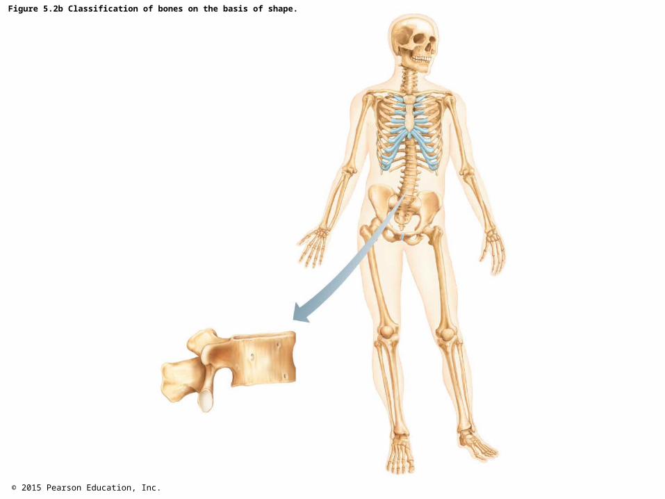
Figure 5.2b Classification of bones on the basis of shape.

© 2015 Pearson Education, Inc.
Figure 5.2c Classification of bones on the basis of shape.

© 2015 Pearson Education, Inc.
Figure 5.2d Classification of bones on the basis of shape.

© 2015 Pearson Education, Inc.
Figure 5.3 The structure of a long bone (humerus of arm).

© 2015 Pearson Education, Inc.
Figure 5.3a The structure of a long bone (humerus of arm).

© 2015 Pearson Education, Inc.
Figure 5.3b The structure of a long bone (humerus of arm).

© 2015 Pearson Education, Inc.
Figure 5.3c The structure of a long bone (humerus of arm).

© 2015 Pearson Education, Inc.
Figure 5.4 Microscopic structure of compact bone.

© 2015 Pearson Education, Inc.
Figure 5.4a Microscopic structure of compact bone.

© 2015 Pearson Education, Inc.
Figure 5.4b Microscopic structure of compact bone.

© 2015 Pearson Education, Inc.
Figure 5.4c Microscopic structure of compact bone.

© 2015 Pearson Education, Inc.
Table 5.1 Bone Markings (1 of 3).

© 2015 Pearson Education, Inc.
Table 5.1 Bone Markings (2 of 3).

© 2015 Pearson Education, Inc.
Table 5.1 Bone Markings (3 of 3).

© 2015 Pearson Education, Inc.
Figure 5.5 Stages of long-bone formation in an embryo, fetus, and young child.

© 2015 Pearson Education, Inc.
Figure 5.6 Growth and remodeling of long bones.

© 2015 Pearson Education, Inc.
Focus on Careers, Radiologic Technologist.

© 2015 Pearson Education, Inc.
Table 5.2 Common Types of Fractures.

© 2015 Pearson Education, Inc.
Homeostatic Imbalance 5.1 This child suffering from rickets is a member of the el-Molo tribe in Kenya, whose diet consists primarily of fish.
© 2015 Pearson Education, Inc.
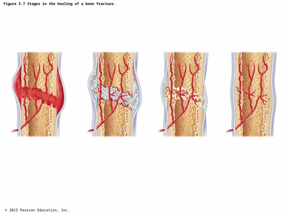
Figure 5.7 Stages in the healing of a bone fracture.

© 2015 Pearson Education, Inc.
Figure 5.8 The human skeleton.

© 2015 Pearson Education, Inc.
Figure 5.8a The human skeleton.

© 2015 Pearson Education, Inc.
Figure 5.8b The human skeleton.

© 2015 Pearson Education, Inc.
Figure 5.9 Human skull, lateral view.

© 2015 Pearson Education, Inc.
Figure 5.10 Human skull, superior view (top of cranium removed).

© 2015 Pearson Education, Inc.
Figure 5.11 Human skull, inferior view (mandible removed).

© 2015 Pearson Education, Inc.
Figure 5.12 Human skull, anterior view.

© 2015 Pearson Education, Inc.
Figure 5.13 Paranasal sinuses.

© 2015 Pearson Education, Inc.
Figure 5.13a Paranasal sinuses.

© 2015 Pearson Education, Inc.
Figure 5.13b Paranasal sinuses.

© 2015 Pearson Education, Inc.
Figure 5.14 Anatomical location and structure of the hyoid bone.

© 2015 Pearson Education, Inc.
Figure 5.15 The fetal skull.

© 2015 Pearson Education, Inc.
Figure 5.15a The fetal skull.

© 2015 Pearson Education, Inc.
Figure 5.15b The fetal skull.

© 2015 Pearson Education, Inc.
Figure 5.16 The vertebral column.

© 2015 Pearson Education, Inc.
Figure 5.17 The C-shaped spine typical of a newborn.

© 2015 Pearson Education, Inc.
Figure 5.18 Abnormal spinal curvatures.
© 2015 Pearson Education, Inc.
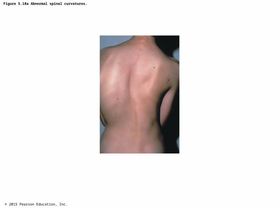
Figure 5.18a Abnormal spinal curvatures.

© 2015 Pearson Education, Inc.
Figure 5.18b Abnormal spinal curvatures.

© 2015 Pearson Education, Inc.
Figure 5.18c Abnormal spinal curvatures.

© 2015 Pearson Education, Inc.
Figure 5.19 A typical vertebra, superior view.

© 2015 Pearson Education, Inc.
Figure 5.20 Regional characteristics of vertebrae.

© 2015 Pearson Education, Inc.
Figure 5.20a Regional characteristics of vertebrae.

© 2015 Pearson Education, Inc.
Figure 5.20b Regional characteristics of vertebrae.

© 2015 Pearson Education, Inc.
Figure 5.20c Regional characteristics of vertebrae.

© 2015 Pearson Education, Inc.
Figure 5.20d Regional characteristics of vertebrae.

© 2015 Pearson Education, Inc.
Figure 5.21 Sacrum and coccyx, posterior view.
© 2015 Pearson Education, Inc.
Figure 5.22 The bony thorax (thoracic cage).

© 2015 Pearson Education, Inc.
Figure 5.22a The bony thorax (thoracic cage).

© 2015 Pearson Education, Inc.
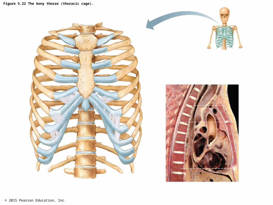
Figure 5.22b The bony thorax (thoracic cage).

© 2015 Pearson Education, Inc.
Figure 5.23 Bones of the shoulder girdle.

© 2015 Pearson Education, Inc.
Figure 5.23a Bones of the shoulder girdle.

© 2015 Pearson Education, Inc.
Figure 5.23b Bones of the shoulder girdle.

© 2015 Pearson Education, Inc.
Figure 5.23c Bones of the shoulder girdle.

© 2015 Pearson Education, Inc.
Figure 5.23d Bones of the shoulder girdle.

© 2015 Pearson Education, Inc.
Figure 5.24 Bones of the right arm and forearm.

© 2015 Pearson Education, Inc.
Figure 5.24a Bones of the right arm and forearm.

© 2015 Pearson Education, Inc.
Figure 5.24b Bones of the right arm and forearm.

© 2015 Pearson Education, Inc.
Figure 5.24c Bones of the right arm and forearm.

© 2015 Pearson Education, Inc.
Figure 5.25 Bones of the right hand, anterior view.

© 2015 Pearson Education, Inc.
Figure 5.26 The bony pelvis.

© 2015 Pearson Education, Inc.
Figure 5.26a The bony pelvis.

© 2015 Pearson Education, Inc.
Figure 5.26b The bony pelvis.

© 2015 Pearson Education, Inc.
Figure 5.26c The bony pelvis.

© 2015 Pearson Education, Inc.
Figure 5.27 Bones of the right thigh and leg.

© 2015 Pearson Education, Inc.
Figure 5.27a Bones of the right thigh and leg.

© 2015 Pearson Education, Inc.
Figure 5.27b Bones of the right thigh and leg.

© 2015 Pearson Education, Inc.
Figure 5.27c Bones of the right thigh and leg.

© 2015 Pearson Education, Inc.
Figure 5.28 Bones of the right foot, superior view.

© 2015 Pearson Education, Inc.
Figure 5.29 Arches of the foot.

© 2015 Pearson Education, Inc.
A Closer Look 5.1 Joint Ventures.

© 2015 Pearson Education, Inc.
A Closer Look 5.1a Joint Ventures.
© 2015 Pearson Education, Inc.
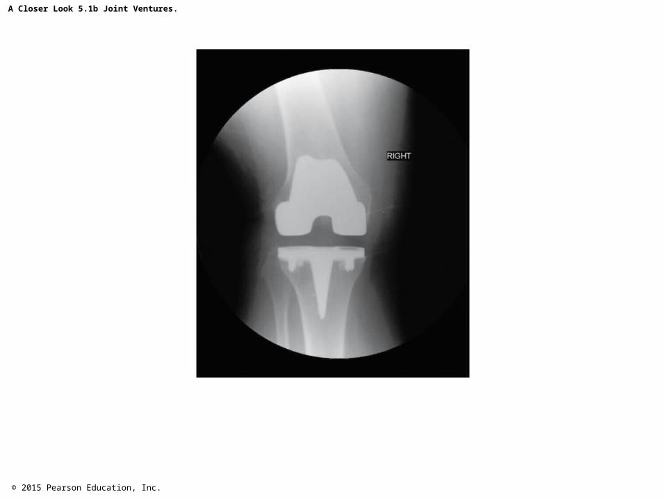
A Closer Look 5.1b Joint Ventures.

© 2015 Pearson Education, Inc.
A Closer Look 5.1c Joint Ventures.

© 2015 Pearson Education, Inc.
Figure 5.30 Types of joints.

© 2015 Pearson Education, Inc.
Figure 5.30a Types of joints.

© 2015 Pearson Education, Inc.
Figure 5.30b Types of joints.

© 2015 Pearson Education, Inc.
Figure 5.30c Types of joints.
© 2015 Pearson Education, Inc.
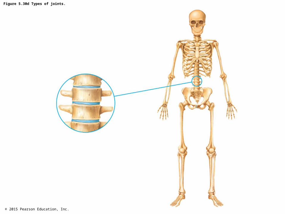
Figure 5.30d Types of joints.

© 2015 Pearson Education, Inc.
Figure 5.30e Types of joints.

© 2015 Pearson Education, Inc.
Figure 5.30f Types of joints.

© 2015 Pearson Education, Inc.
Figure 5.30g Types of joints.

© 2015 Pearson Education, Inc.
Figure 5.30h Types of joints.

© 2015 Pearson Education, Inc.
Table 5.3 Summary of Joint Classes.

© 2015 Pearson Education, Inc.
Figure 5.31 General structure of a synovial joint.

© 2015 Pearson Education, Inc.
Figure 5.32 Types of synovial joints.

© 2015 Pearson Education, Inc.
Figure 5.32a Types of synovial joints.

© 2015 Pearson Education, Inc.
Figure 5.32b Types of synovial joints.

© 2015 Pearson Education, Inc.
Figure 5.32c Types of synovial joints.

© 2015 Pearson Education, Inc.
Figure 5.32d Types of synovial joints.
© 2015 Pearson Education, Inc.
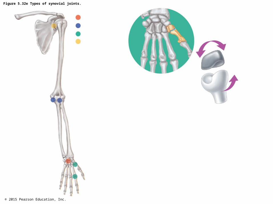
Figure 5.32e Types of synovial joints.

© 2015 Pearson Education, Inc.
Figure 5.32f Types of synovial joints.

© 2015 Pearson Education, Inc.
Figure 5.33 X-ray image of a hand deformed by rheumatoid arthritis.

© 2015 Pearson Education, Inc.
Figure 5.34 Ossification centers in the skeleton of a 12-week-old fetus are indicated by the darker areas. Lighter regions are still fibrous or cartilaginous.
© 2015 Pearson Education, Inc.
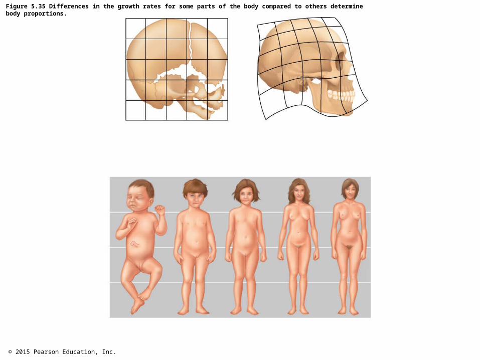
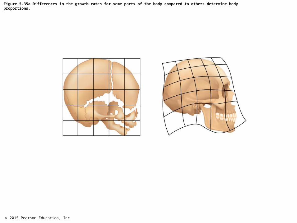
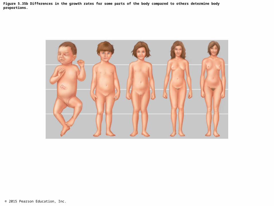
Figure 5.35 Differences in the growth rates for some parts of the body compared to others determine body proportions.
© 2015 Pearson Education, Inc.
Figure 5.35a Differences in the growth rates for some parts of the body compared to others determine body proportions.
© 2015 Pearson Education, Inc.
Figure 5.35b Differences in the growth rates for some parts of the body compared to others determine body proportions.

© 2015 Pearson Education, Inc.
Figure 5.36 Osteoporosis.

© 2015 Pearson Education, Inc.
Figure 5.37 Vertebral collapse due to osteoporosis.

© 2015 Pearson Education, Inc.
Systems in Sync 5 Homeostatic Relationships between the Skeletal System and Other Body Systems.