© 2010 mcgraw-hill higher education. all rights reserved. ankle and lower leg rehabilitation
TRANSCRIPT

© 2010 McGraw-Hill Higher Education. All rights reserved.
Ankle and Lower Leg Rehabilitation

© 2010 McGraw-Hill Higher Education. All rights reserved.Figure 15-1
© 2010 McGraw-Hill Higher Education. All rights reserved.
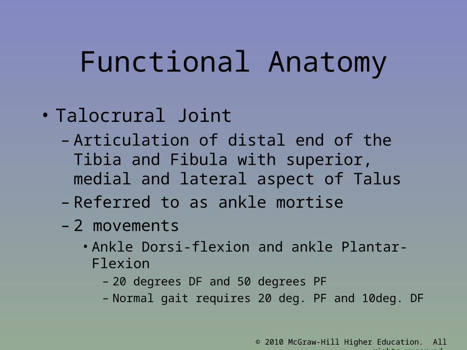
Functional Anatomy
• Talocrural Joint– Articulation of distal end of the Tibia and
Fibula with superior, medial and lateral aspect of Talus
– Referred to as ankle mortise– 2 movements
• Ankle Dorsi-flexion and ankle Plantar-Flexion– 20 degrees DF and 50 degrees PF– Normal gait requires 20 deg. PF and 10deg. DF

© 2010 McGraw-Hill Higher Education. All rights reserved.
Functional Anatomy
• Talocrural joint ligaments– Lateral: anterior talofibular ligament
(ATFL), Calcaneofibular Ligament (CFL), Posterior talofibular ligament (PTFL)
– Medial: Deltoid Ligament; anterior, middle and posterior bands
– Anterior & Posterior Tibiofibular ligament• Distal portion of interosseous membrane

© 2010 McGraw-Hill Higher Education. All rights reserved.
• Talocrural muscles– Posterior to lateral malleolous create plantar flexion and toe
flexion• Superficial: gastrocnemius• Middle: soleus & plantaris• Deep: posterior tibialis, flexor digitorum longus, flexor
hallucis longus
– Anterior muscles will dorsiflex the ankle and extend the toes• Ext. halicus longus, tibialis anterior, extensor digitorum,
peroneal tertius

© 2010 McGraw-Hill Higher Education. All rights reserved.
• Subtalar joint– Articulation of calcaneus and talus
• Pronation and supination– Occur in 3 planes simultaneously– Supination: Foot moves into plantar flexion,
adduction, and inversion– Pronation: Foot moves into abduction, dorsiflexion
and eversion
© 2010 McGraw-Hill Higher Education. All rights reserved.
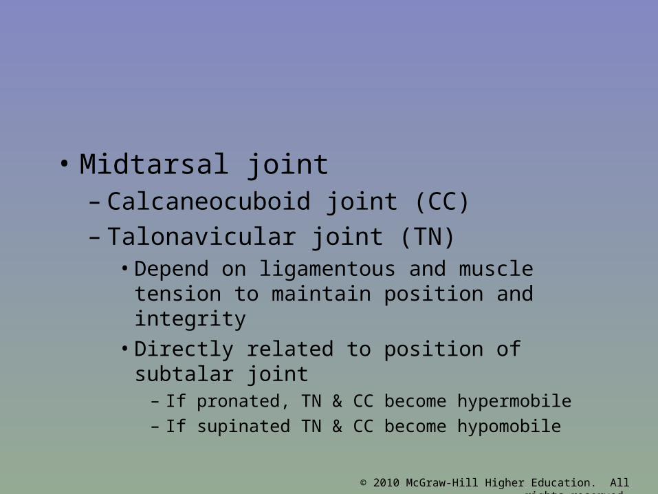
• Midtarsal joint– Calcaneocuboid joint (CC)– Talonavicular joint (TN)
• Depend on ligamentous and muscle tension to maintain position and integrity
• Directly related to position of subtalar joint– If pronated, TN & CC become hypermobile– If supinated TN & CC become hypomobile

© 2010 McGraw-Hill Higher Education. All rights reserved.
• MT joint during pronation– Hypermobile 1st ray and increase pressure
on other metatarsals• Peroneal tendon unable to stabilize 1st ray
because mechanical advantage lost at cuboid pulley
• Also hypermobility at articulation between 1st metatarsal and 1st cuneiform

© 2010 McGraw-Hill Higher Education. All rights reserved.

© 2010 McGraw-Hill Higher Education. All rights reserved.
Functional Anatomy
• MT joint during supination– Less surface area between tarsal
articulation=less movement=hypomobility– Foot rigid and tight– More weight and stress placed on 1st and
5th metatarsal because of less mobility at 1st ray

© 2010 McGraw-Hill Higher Education. All rights reserved.

© 2010 McGraw-Hill Higher Education. All rights reserved.

© 2010 McGraw-Hill Higher Education. All rights reserved.

© 2010 McGraw-Hill Higher Education. All rights reserved.
Functional Anatomy
• Ankle more unstable in plantar flexion– Shape of talus: Wider anteriorly and more
narrow posteriorly• In Dorsi flexion talus gripped tightly in talocrural
joint• In plantar flexion less stable because narrow
aspect of talus exposed– Also less stable with inversion
» Distal end of tibia doesn’t extend as far as distal end of fibula

© 2010 McGraw-Hill Higher Education. All rights reserved.

© 2010 McGraw-Hill Higher Education. All rights reserved.

© 2010 McGraw-Hill Higher Education. All rights reserved.
Biomechanics of Normal Gait
• 2 phases: stance or support phase & swing or recovery phase– Stance: initial contact at heel strike and
ends at toe off– Swing: time immediately after toe off, leg
moved from behind body to a position in front of body in preparation of heel strike

© 2010 McGraw-Hill Higher Education. All rights reserved.
• Foot at stance phase– Shock absorber to impact forces at heel
strike and adapt to uneven surface– At push off functions as rigid lever to
transmit explosive force– Lateral aspect of calcaneus with subtalar
joint in supination to forefoot contact on medial surface of foot and subtalar joint pronation
• Pronation distributes forces to many structures

© 2010 McGraw-Hill Higher Education. All rights reserved.
• Foot begins to re-supinate and returns subtalar joint to neutral ay 70 to 90 % of support phase
• Foot becomes rigid and stable to allow greater amount of force at push off

© 2010 McGraw-Hill Higher Education. All rights reserved.
Assessing the Lower Leg and Ankle
• History– Past history– Mechanism of injury– When does it hurt?– Type of, quality of, duration of pain?– Sounds or feelings?– How long were you disabled?– Swelling?– Previous treatments?

© 2010 McGraw-Hill Higher Education. All rights reserved.
• Observations– Postural deviations?– Genu valgum or varum?– Is there difficulty with walking?– Deformities, asymmetries or swelling?– Color and texture of skin, heat, redness?– Patient in obvious pain?– Is range of motion normal?
• Palpation– Begin with bony landmarks and progress
to soft tissue– Attempt to locate areas of deformity,
swelling and localized tenderness

© 2010 McGraw-Hill Higher Education. All rights reserved.
• Ankle Stability Tests– Anterior drawer test
• Used to determine damage to anterior talofibular ligament primarily and other lateral ligament secondarily
• A positive test occurs when foot slides forward and/or makes a clunking sound as it reaches the end point
– Talar tilt test• Performed to determine extent of inversion or eversion
injuries• With foot at 90 degrees calcaneus is inverted and excessive
motion indicates injury to calcaneofibular ligament and possibly the anterior and posterior talofibular ligaments
• If the calcaneus is everted, the deltoid ligament is tested

© 2010 McGraw-Hill Higher Education. All rights reserved.
Anterior Drawer Test
Talar Tilt TestBump Test

© 2010 McGraw-Hill Higher Education. All rights reserved.
• Functional Tests
– While weight bearing the following should be performed
• Walk on toes (plantar flexion)• Walk on heels (dorsiflexion)• Hops on injured ankle• Start and stop running• Change direction rapidly• Run figure eights

© 2010 McGraw-Hill Higher Education. All rights reserved.
• Footwear– Can be an important factor in reducing injury– Shoes should not be used in activities they were not
made for
• Preventive Taping and Orthoses– Tape can provide some prophylactic protection– However, improperly applied tape can disrupt normal
biomechanical function and cause injury– Lace-up braces have even been found to be effective
in controlling ankle motion

© 2010 McGraw-Hill Higher Education. All rights reserved.

© 2010 McGraw-Hill Higher Education. All rights reserved.
Figure 15-4

© 2010 McGraw-Hill Higher Education. All rights reserved.
• Neuromuscular Control Training– Can be enhanced by training in controlled
activities on uneven surfaces or a balance board
Figure 15-5 & 6

© 2010 McGraw-Hill Higher Education. All rights reserved.

© 2010 McGraw-Hill Higher Education. All rights reserved.

© 2010 McGraw-Hill Higher Education. All rights reserved.

© 2010 McGraw-Hill Higher Education. All rights reserved.
PHASE I
• Decrease pain and swelling– PRICE– Modalities: pulsed ultrasound, electrical
stimulation (Interferential, High Volt)– Massage– Pain-free AROM exercises

© 2010 McGraw-Hill Higher Education. All rights reserved.
Phase II-ROM• Increase ROM:
– AROM, PROM exercises– Progress to weight bearing ROM ex.
• Maintain CV fitness
• Maintain Core Stability
• Restore Balance and proprioception– Double leg and single leg balance
progression
• Continue to assist healing process and pain management

© 2010 McGraw-Hill Higher Education. All rights reserved.
Phase III-Strengthening
• Continue ROM exercises• Continue to assist healing process and pain
management• Continue and progress CV fitness• Continue and progress Core stability • Evaluate and treat other biomechanical deficiencies• Begin strengthening programs for foot and ankle as
well as entire lower kinetic chain – Progress to functional activities and plyometrics

© 2010 McGraw-Hill Higher Education. All rights reserved.
Phase IV
• Continue all of Phase III
• Add sport specific movement exercises– Rehab should be equally, if not more
difficult than their practice for their sport– Running progression– Speed and agility– Sport specific movement
• Goal of Phase IV is return to their sport

© 2010 McGraw-Hill Higher Education. All rights reserved.
Phase V-Maintenance
• Continue to monitor and rehabilitate athlete through their return to activity– Observe for setbacks or decrease in
performance– Ensure activity and movement is
coordinated and unconscious• Athlete should not be limited at all by their
injury

© 2010 McGraw-Hill Higher Education. All rights reserved.
Recognition and Management of Injuries to the Ankle
• Ankle Injuries: Sprains– Single most common injury in athletics caused by sudden
inversion or eversion moments
• Inversion Sprains– Most common and result in injury to the lateral ligaments– Anterior talofibular ligament is injured with inversion,
plantar flexion and internal rotation– Occasionally the force is great enough for an avulsion
fracture to occur w/ the lateral malleolus

© 2010 McGraw-Hill Higher Education. All rights reserved.
• Severity of sprains is graded (1-3)
• With inversion sprains the foot is forcefully inverted or occurs when the foot comes into contact w/ uneven surfaces

© 2010 McGraw-Hill Higher Education. All rights reserved.

© 2010 McGraw-Hill Higher Education. All rights reserved.
•Eversion Ankle Sprains-(Represent 5-10% of all ankle sprains)
• Etiology – Bony protection and
ligament strength decreases likelihood of injury
– Eversion force resulting in damage to deltoid and possibly fx of the fibula
– Deltoid can also be impinged and contused with inversion sprains

© 2010 McGraw-Hill Higher Education. All rights reserved.
• Syndesmotic Sprain– Etiology
• Injury to the distal tibiofemoral joint (anterior/posterior tibiofibular ligament)
• Torn w/ increased external rotation or dorsiflexion
• Injured in conjunction w/ medial and lateral ligaments
• May require extensive period of time in order to return to play
Figure 15-13

© 2010 McGraw-Hill Higher Education. All rights reserved.
• Graded Ankle Sprains – Signs of Injury
• Grade 1– Mild pain and disability; weight bearing is minimally impaired; point tenderness
over ligaments and no laxity
• Grade 2– Feel or hear pop or snap; moderate pain w/ difficulty bearing weight; tenderness
and edema– Positive talar tilt and anterior drawer tests– Possible tearing of the anterior talofibular and calcaneofibular ligaments
• Grade 3– Severe pain, swelling, hemarthrosis, discoloration– Unable to bear weight– Positive talar tilt and anterior drawer– Instability due to complete ligamentous rupture

© 2010 McGraw-Hill Higher Education. All rights reserved.
– Care• Must manage pain and swelling• Apply horseshoe-shaped foam pad for focal compression • Apply wet compression wrap to facilitate passage of cold from
ice packs surrounding ankle• Apply ice for 20 minutes and repeat every hour for 24 hours• Continue to apply ice over the course of the next 3 days• Keep foot elevated as much as possible• Avoid weight bearing for at least 24 hours • Begin weight bearing as soon as tolerated• Return to participation should be gradual and dictated by healing
process
© 2010 McGraw-Hill Higher Education. All rights reserved.
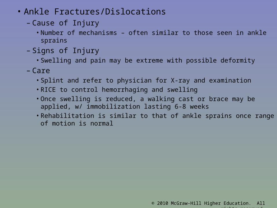
• Ankle Fractures/Dislocations– Cause of Injury
• Number of mechanisms – often similar to those seen in ankle sprains
– Signs of Injury• Swelling and pain may be extreme with possible deformity
– Care• Splint and refer to physician for X-ray and examination• RICE to control hemorrhaging and swelling• Once swelling is reduced, a walking cast or brace may be applied, w/
immobilization lasting 6-8 weeks• Rehabilitation is similar to that of ankle sprains once range of motion is
normal

© 2010 McGraw-Hill Higher Education. All rights reserved.

© 2010 McGraw-Hill Higher Education. All rights reserved.
• Tendinitis– Cause of Injury
• Singular cause or collection of mechanisms
– Footwear, mechanics, trauma, overuse, limited flexibility
– Signs of Injury• Pain & inflammation• Crepitus• Pain with AROM & PROM
– Care• Rest, NSAIDs, modalities• Orthotics for foot
mechanic

© 2010 McGraw-Hill Higher Education. All rights reserved.
• Tibial and Fibular Fractures– Cause of Injury
• Result of direct blow or indirect trauma• Fibular fractures seen with tibial fractures or as the result of direct
trauma
– Signs of Injury• Pain, swelling, soft tissue insult• Leg will appear hard and swollen (Volkman’s contracture)• Deformity – may be open or closed
– Care• Immediate treatment should include splinting to immobilize and ice,
followed by medical referral• Restricted weight bearing for weeks/months depending on severity

© 2010 McGraw-Hill Higher Education. All rights reserved.

© 2010 McGraw-Hill Higher Education. All rights reserved.
• Stress Fracture of Tibia or Fibula– Cause of Injury
• Common overuse condition, particularly in those with structural and biomechanical insufficiencies
• Result of repetitive loading during training and conditioning
– Signs of Injury• Pain with activity
• Pain more intense after exercise than before
• Point tenderness; difficult to discern bone and soft tissue pain
• Bone scan results (stress fracture vs. periostitis)

© 2010 McGraw-Hill Higher Education. All rights reserved.
• Care– Eliminate offending activity– Discontinue stress inducing activity 14 days– Use crutch for walking– Weight bearing may return when pain subsides– After pain free for 2 weeks athlete can gradually
return to activity– Biomechanics must be addressed

© 2010 McGraw-Hill Higher Education. All rights reserved.
• Medial Tibial Stress Syndrome (Shin Splints)– Cause of Injury
• Pain in anterior portion of shin• Stress fractures, muscle strains, chronic anterior
compartment syndrome, periosteum irritation• Caused by repetitive microtrauma• Weak muscles, improper footwear, training errors, varus
foot, tight heel cord, hypermobile or pronated feet and even forefoot supination can contribute to MTSS
• May also involve, stress fractures or exertional compartment syndrome

© 2010 McGraw-Hill Higher Education. All rights reserved.
• Shin Splints (continued)– Signs of Injury
• Diffuse pain about distomedial aspect of lower leg• As condition worsens ambulation may be painful, morning pain and
stiffness may also increase
• Can progress to stress fracture if not treated
– Care• Physician referral for X-rays and bone scan• Activity modification
• Correction of abnormal biomechanics• Ice massage to reduce pain and inflammation
• Flexibility program for gastroc-soleus complex
• Arch taping and orthotics

© 2010 McGraw-Hill Higher Education. All rights reserved.
• Shin Contusion– Cause of Injury
• Direct blow to lower leg (impacting periosteum anteriorly)
– Signs of Injury• Intense pain, rapidly forming hematoma w/ jelly like consistency
• Increased warmth
– Care• RICE, NSAID’s and analgesics as needed
• Maintaining compression for hematoma (which may need to aspirated)
• Fit with doughnut pad and orthoplast shell for protection

© 2010 McGraw-Hill Higher Education. All rights reserved.
• Compartment Syndrome– Cause of Injury
• Rare acute traumatic syndrome due to direct blow or excessive exercise
• May be classified as acute, acute exertional or chronic
– Signs of Injury • Excessive swelling compresses muscles, blood supply
and nerves• Deep aching pain and tightness is experienced• Weakness with foot and toe extension and occasionally
numbness in dorsal region of foot

© 2010 McGraw-Hill Higher Education. All rights reserved.
Figure 15-20

© 2010 McGraw-Hill Higher Education. All rights reserved.
– Care• If severe acute or chronic case, may present as medical
emergency that requires surgery to reduce pressure or release fascia
• RICE, NSAID’s and analgesics as needed – Avoid use of compression wrap = increased pressure
• Surgical release is generally used in recurrent conditions
– May require 2-4 month recovery (post surgery)
• Conservative management requires activity modification, icing and stretching
– Surgery is required if conservative management fails
© 2010 McGraw-Hill Higher Education. All rights reserved.
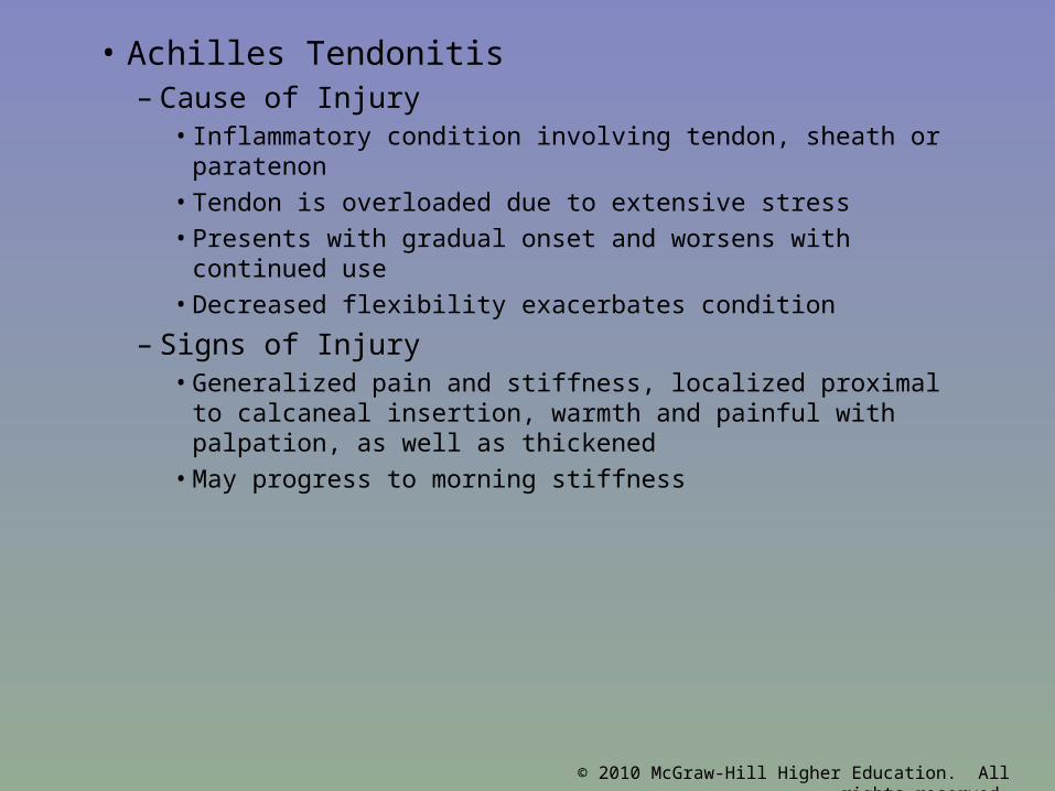
• Achilles Tendonitis– Cause of Injury
• Inflammatory condition involving tendon, sheath or paratenon
• Tendon is overloaded due to extensive stress
• Presents with gradual onset and worsens with continued use
• Decreased flexibility exacerbates condition
– Signs of Injury• Generalized pain and stiffness, localized proximal to
calcaneal insertion, warmth and painful with palpation, as well as thickened
• May progress to morning stiffness

© 2010 McGraw-Hill Higher Education. All rights reserved.
– Care• Resistant to quick resolution due to slow healing
nature of tendon• Must reduce stress on tendon, address structural
faults (orthotics, mechanics, flexibility)• Aggressive stretching and use of heel lift may be
beneficial• Use of anti-inflammatory medications is suggested

© 2010 McGraw-Hill Higher Education. All rights reserved.
• Achilles Tendon Rupture– Cause
• Occurs w/ sudden stop and go; forceful plantar flexion w/ knee moving into full extension
• Commonly seen in athletes > 30 years old• Generally has history of chronic inflammation
– Signs of Injury• Sudden snap (kick in the leg) w/ immediate pain which
rapidly subsides• Point tenderness, swelling, discoloration; decreased ROM• Obvious indentation and positive Thompson test

© 2010 McGraw-Hill Higher Education. All rights reserved.
Figure 15-20

© 2010 McGraw-Hill Higher Education. All rights reserved.
– Care• Usual management involves surgical repair for
serious injuries • Non-operative treatment consists of RICE,
NSAID’s, analgesics, and a non-weight bearing cast for 6 weeks to allow for proper tendon healing
• Must work to regain normal range of motion followed by gradual and progressive strengthening program