zoonotic cestode infections:emphasis on neurocysticercosis and echinococcosis
TRANSCRIPT

Zoonotic cestode infections: Emphasis on Neurocysticercosis
and Hydatidosis
Zoonotic cestode infections: Emphasis on Neurocysticercosis
and Hydatidosis Aquil Mohmad
M-5527 M.V.Sc Scholar

Diseases and infections that are naturally transmitted between vertebrate animals and human.
(WHO)
Cestode zoonoses are emerging and spreading worldwide and are classified as neglected zoonotic diseases .
Asia currently has the greatest burden of cystic echinococcosis (CE) and cysticercosis .
( Xiaoning et al 2013)

Sparganosis Hymenolepises Coenuriasis Diphyllobothriasis Cysticercosis
Echinococcosis Bertieliasis Dipylidiasis Inermicapsifer infection

First described by Patrick Manson in 1882 from china
Caused by plerocercoid larvae of Pseudophyllidea tape worms of the genus Spirometra

.
Painful subconjunctival horseshoe shaped swelling around the limbus in the superior part of the bulbar conjunctiva with surrounding chemosis and congestion
( R. Nath 2015)
larvae can lodge in the brain parenchyma or spinal cord producing seizures, headache, hemiparesis, paralysis even death .

Spargana worms

.
Theodor Bilharz (1851) first discovered H. nana in small intestine of an Egyptian male


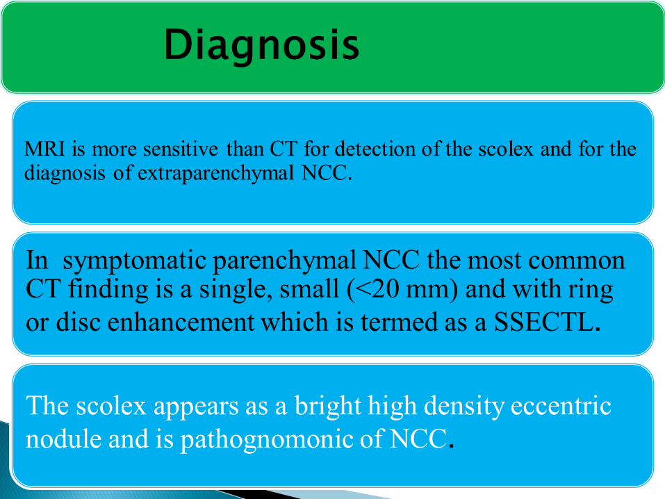
In India it is an important emerging disease of the
central nervous system second to tuberculosis ( B.B. Singh 2010 )
It is the single most common cause of epilepsy in the developing countries
It is common in communities where pigs are allowed to roam freely and the basic sanitary facilities are lacking .
( K N Prasad et al 2008)


Areas suggested to be highly endemicAreas suggested to be moderatelyAreas with no or very few casesAreas with no available report

Scolex
Adult Taenia solium
Three most common sites ofinfection in human (primary host)1. Brain (Neurocysticercosis)2. Muscles (Cysticercosis)3. Intestine (Taeniasis
Eggs
Onchosphere
hatches,
penetrate
intestinal wall
and ciruculate to
musculature of
pig (intermediate
host
Ingestion of raw vegetables,fruits, salad contaimnated with eggs
Consumption ofinfected pork
Cysticerci1
2
3


(Pratibhi, 2011)


(Monika Sharma et al , 2013 )

(Monika Sharma ;2013)

Sheep strain (G1) is the most cosmopolitan form and is that most commonly associated with human infections.
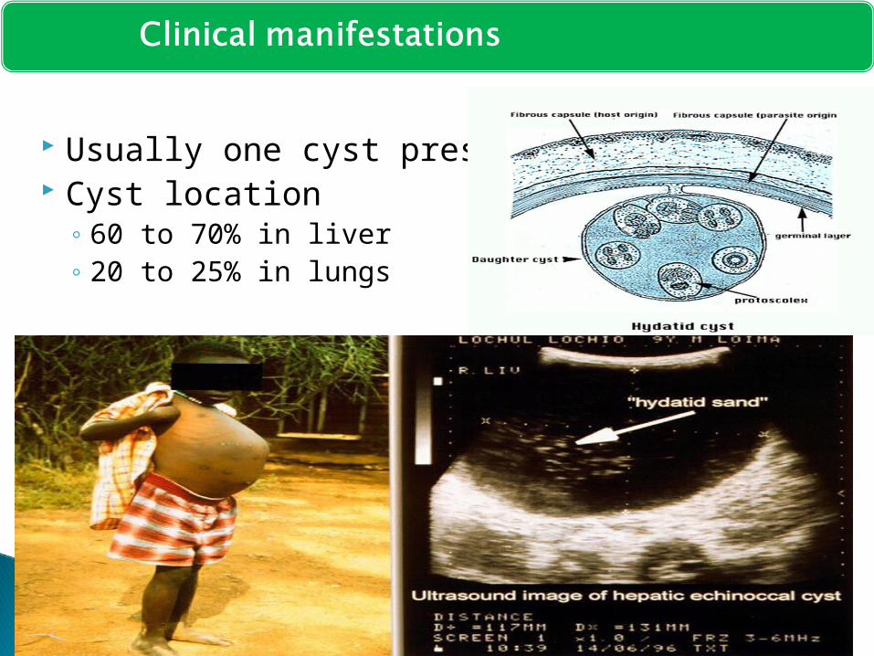
Usually one cyst present Cyst location
◦ 60 to 70% in liver◦ 20 to 25% in lungs


WHO Type Cyst Morphology
Unilocular anechoic lesion with double line containing hydatid sand.
Multiseptated rosette like honeycomb cyst
Cyst with detached membrane (waterlily sign)
Cyst with daughter cysts in solid matrix
Cyst with heterogenous hypoechoic hyperechoic contents.
no daughter cysts
Solid and calcified wall
CE 1
CE 2
CE 3A
CE 3B
CE 4
CE 5


Thank you
Thank you