ypfΦ: a filamentous phage acquired by yersinia pestis...derbise and carniel the yersinia pestis...
TRANSCRIPT

MINI REVIEW ARTICLEpublished: 15 December 2014
doi: 10.3389/fmicb.2014.00701
Ypf�: a filamentous phage acquired by Yersinia pestisAnne Derbise* and Elisabeth Carniel
Yersinia Research Unit, Department of Microbiology, Institut Pasteur, Paris, France
Edited by:
Bhabatosh Das, Translational HealthScience and Technology Institute,India
Reviewed by:
Jasna Rakonjac, Massey University,New ZealandFrançois-Xavier Barre, CNRS, France
*Correspondence:
Anne Derbise, Yersinia Research Unit,Department of Microbiology, InstitutPasteur, 28 rue du Dr. Roux,75724 Paris Cedex 15, Francee-mail: [email protected]
Yersinia pestis, the plague bacillus, has an exceptional pathogenicity for humans. Theplague bacillus emerged very recently (≈3,000 years ago) from the enteropathogenY. pseudotuberculosis. Early after its emergence,Y. pestis became infected by a filamentousphage named Ypf�. During the microevolution of the plague bacillus, the phage remainedin the various lineages as an unstable extrachromosomal element. However, in the subbranch that caused the third plague pandemic, Ypf� integrated itself into the bacterialchromosome to become a stable prophage.The genome of this phage has the same geneticorganization as that of other filamentous phages such as the Vibrio cholerae CTX� phage,and shares high sequence identity with the CUS-1 filamentous phage of a high-virulenceEscherichia coli K1 clone. In addition to genes involved in phage physiology,Ypf� carries ateach extremity of its genome two open reading frames with no predicted functions. Thisfilamentous phage confers some selective properties to Y. pestis during the infectiousprocess, which may explain why it was conserved during Y. pestis microevolution, despiteits instability as an extrachromosomal element in most branches.
Keywords: filamentous bacteriophage, plague,Yersinia pestis
INTRODUCTIONYersinia pestis, one of the most dangerous bacterial pathogens ofhumans, is the causative agent of plague, a zoonotic disease trans-mitted from animals to humans by fleabites. After injection intothe dermis, the bacteria migrate to the draining lymph node, wherethey cause the pathognomonic bubo. Bubonic plague, the mostcommon clinical form, is fatal in less than a week in 40–70%of the patients if left untreated. Pneumonic plague, the secondmost common form of the disease, results from human-to-humantransmission of the bacillus by aerosols, and is systematically andrapidly lethal if effective antibiotic therapy is not delivered beforeor at the onset of symptoms. Despite considerable progress inplague prevention and cure, this infection has not been eradicated.Natural plague foci still persist in numerous countries in Africa,Asia, and the Americas. Several genetic elements conferring viru-lence properties have been horizontally acquired by Y. pestis. Thisincludes three plasmids: (i) pYV, which encodes a type III secre-tion system and toxins that subvert the defenses of the mammalianhosts, (ii) pFra, a large replicon which codes for a capsule that con-fers some resistance to the antibacterial activity of macrophages,and (iii) pPla, whose main product (Pla) is an important viru-lence factor that has protease and plasminogen activator activities.Another horizontally acquired element is the High PathogenicityIsland which allows Y. pestis to utilize the host iron and to causesepticemia. The most recently described mobile element is a fila-mentous phage, named Ypf� that plays a role in the capacity ofthe plague bacillus to multiply and disseminate in mice (Derbiseet al., 2007). The purpose of this review is to present the currentknowledge about Ypf� and to discuss its impact on the plaguebacillus physiology.
CHARACTERISTICS OF Ypf�Ypf� forms filamentous particles that are secreted by the Y.pestis cells into the culture supernatant without affecting bacterial
growth or lyzing the cells. Low titers of phages are producedin standard in vitro growth conditions. Ypf� has a filamentousvirion 1,200 nm in length and 8 nm in diameter, which containsa circular positive single strand DNA molecule (Figure 1A). Thephage has the capacity under laboratory conditions to infect thethree pathogenic Yersinia species (Y. pestis and the enteropathogensY. pseudotuberculosis and Y. enterocolitica). The phage infectivityrates, determined with an antibiotic tagged version of Ypf� rangefrom <2 × 10⁻⁹ to 10−1 depending on the isolates. Y. pestis strainsare the most susceptible to a Ypf� infection (99% of the strains),followed by Y. enterocolitica (50% of the strains), while only 30%of the Y. pseudotuberculosis strains are infected. The infectivityspectrum of Ypf� is not restricted to Yersinia since Escherichia colistrains (TOP10, ECOR31) were also successfully transduced withYpf�.
No Ypf� receptor on the bacterial surface has been identifiedyet. Deletion of genes encoding pili-like structures (pilA,psaA) thatwere potential candidate receptors did not affect the susceptibilityof Y. pestis to Ypf� (Chouikha et al., 2010). It could be possiblethat Ypf� uses several receptor molecules at the bacterial surface,a hypothesis that is supported by the wide host range of this phage.Many filamentous phages use the TolQAR complex as a membranereceptor for entry into the recipient cell. Since TolQAR is highlyconserved in Gram negative bacteria (Click and Webster, 1997;Heilpern and Waldor, 2000), it could serve as a secondary receptorfor Ypf� entry (Russel et al., 1988).
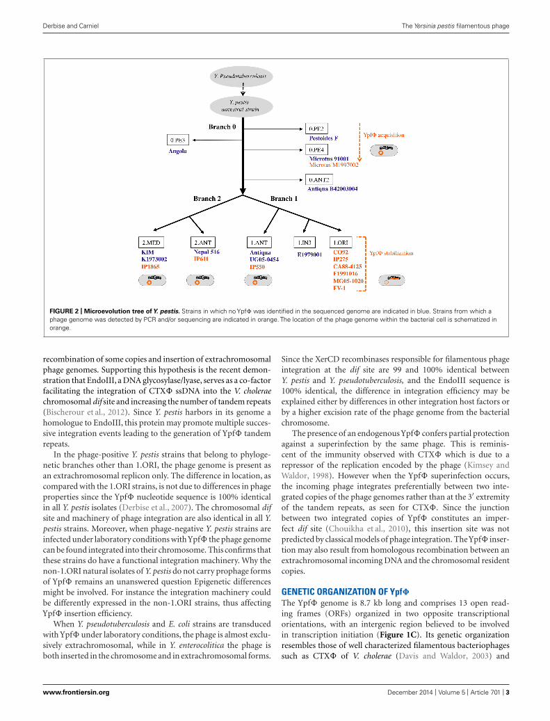
ORIGIN AND DISTRIBUTION OF Ypf�Yersinia pestis is a clonal species that emerged recently fromY. pseudotuberculosis, a much less virulent bacterium (Achtmanet al., 1999). Molecular phylogenetic studies combined with wholegenome sequencing showed that after its divergence from Y. pseu-dotuberculosis, Y. pestis evolved along one branch (branch 0),before the split into two main branches (1 and 2) and several sub
www.frontiersin.org December 2014 | Volume 5 | Article 701 | 1

Derbise and Carniel The Yersinia pestis filamentous phage
FIGURE 1 | (A) Electronic micrograph of Ypf�. (B) Schematic representation of the proposed XerC-dependent phage integration into the chromosomal dif siteof Yersinia pestis. (C) Ypf�, CUS-1 and CTX-1 genomic organization.
branches (Figure 2; Achtman et al., 2004). The Ypf� prophagegenome was initially identified in a comparative study of thegenomes of one strain each of Y. pestis and Y. pseudotuberculo-sis. An analysis of an extensive set of isolates showed that thephage is systematically absent from Y. pseudotuberculosis. In Y.pestis, the phage is present in isolates from the three main phy-logenetic branches, suggesting its acquisition early after Y. pestisemergence (Derbise et al., 2007). However, Ypf� was not detectedin all Y. pestis isolates. The phage is systematically present instrains from sub branch 1.ORI (Figure 2). This branch cor-responds to the Y. pestis isolates that caused the third plaguepandemic and that are found in most plague foci worldwidetoday. In the other sub branches Ypf� is detected in some iso-lates only. Furthermore, only a portion of the bacterial cellsthat were found to be phage-positive harbors the phage genome(Derbise et al., 2007; Li et al., 2008). This indicates that Ypf�is not stably maintained in the non-1.ORI branches and thatthe phage is easily lost upon Y. pestis subcultures in vitro. Thecapacity to stabilize the phage genome in the bacterial chro-mosome as a prophage is thus a property that was acquiredlate during Y. pestis evolution, and only in the third pandemiclineage.
LOCATION OF Ypf� GENOME IN THE BACTERIAL CELLIn sub branch 1.ORI, Ypf� is present mostly as an integratedprophage although extrachromosomal forms are also detected.As observed for several other filamentous prophages (Gonzalezet al., 2002; Huber and Waldor, 2002) Ypf� integrates its genomeinto the chromosomal dif site of the host bacterium. dif is arecombinational locus used by the XerCD recombinases of thebacterial host to resolve chromosome dimers (Das et al., 2013).Ypf� insertion reconstitutes an intact dif site at the 3′ extremityof the prophage sequence (Figure 1B). Sequence analysis of theYpf� encapsidated genome indicates the presence of a potentialpair of binding sites for XerC and XerD in inverted orientations(Figure 1B). Similarly to the Vibrio cholerae filamentous phageCTX� such structure may constitute a XerCD substrate for recom-bination with the bacterial dif site, leading to the integration ofthe phage genome into the host chromosome (Figure 1B; Valet al., 2005). In the Y. pestis chromosome, the Ypf� genome formstandem repeats of two to four copies. Although two copies is thepredominant form, more copies can be detected in the same bacte-rial population, suggesting constant and dynamic rearrangementsbetween tandem repeats. Variations in the number of tandemrepeats may also result from continuous excision by homologous
Frontiers in Microbiology | Virology December 2014 | Volume 5 | Article 701 | 2
Derbise and Carniel The Yersinia pestis filamentous phage
FIGURE 2 | Microevolution tree ofY. pestis. Strains in which no Ypf� was identified in the sequenced genome are indicated in blue. Strains from which aphage genome was detected by PCR and/or sequencing are indicated in orange. The location of the phage genome within the bacterial cell is schematized inorange.
recombination of some copies and insertion of extrachromosomalphage genomes. Supporting this hypothesis is the recent demon-stration that EndoIII, a DNA glycosylase/lyase, serves as a co-factorfacilitating the integration of CTX� ssDNA into the V. choleraechromosomal dif site and increasing the number of tandem repeats(Bischerour et al., 2012). Since Y. pestis harbors in its genome ahomologue to EndoIII, this protein may promote multiple succes-sive integration events leading to the generation of Ypf� tandemrepeats.
In the phage-positive Y. pestis strains that belong to phyloge-netic branches other than 1.ORI, the phage genome is present asan extrachromosomal replicon only. The difference in location, ascompared with the 1.ORI strains, is not due to differences in phageproperties since the Ypf� nucleotide sequence is 100% identicalin all Y. pestis isolates (Derbise et al., 2007). The chromosomal difsite and machinery of phage integration are also identical in all Y.pestis strains. Moreover, when phage-negative Y. pestis strains areinfected under laboratory conditions with Ypf� the phage genomecan be found integrated into their chromosome. This confirms thatthese strains do have a functional integration machinery. Why thenon-1.ORI natural isolates of Y. pestis do not carry prophage formsof Ypf� remains an unanswered question Epigenetic differencesmight be involved. For instance the integration machinery couldbe differently expressed in the non-1.ORI strains, thus affectingYpf� insertion efficiency.
When Y. pseudotuberculosis and E. coli strains are transducedwith Ypf� under laboratory conditions, the phage is almost exclu-sively extrachromosomal, while in Y. enterocolitica the phage isboth inserted in the chromosome and in extrachromosomal forms.
Since the XerCD recombinases responsible for filamentous phageintegration at the dif site are 99 and 100% identical betweenY. pestis and Y. pseudotuberculosis, and the EndoIII sequence is100% identical, the difference in integration efficiency may beexplained either by differences in other integration host factors orby a higher excision rate of the phage genome from the bacterialchromosome.
The presence of an endogenous Ypf� confers partial protectionagainst a superinfection by the same phage. This is reminis-cent of the immunity observed with CTX� which is due to arepressor of the replication encoded by the phage (Kimsey andWaldor, 1998). However when the Ypf� superinfection occurs,the incoming phage integrates preferentially between two inte-grated copies of the phage genomes rather than at the 3′ extremityof the tandem repeats, as seen for CTX�. Since the junctionbetween two integrated copies of Ypf� constitutes an imper-fect dif site (Chouikha et al., 2010), this insertion site was notpredicted by classical models of phage integration. TheYpf� inser-tion may also result from homologous recombination between anextrachromosomal incoming DNA and the chromosomal residentcopies.
GENETIC ORGANIZATION OF Ypf�The Ypf� genome is 8.7 kb long and comprises 13 open read-ing frames (ORFs) organized in two opposite transcriptionalorientations, with an intergenic region believed to be involvedin transcription initiation (Figure 1C). Its genetic organizationresembles those of well characterized filamentous bacteriophagessuch as CTX� of V. cholerae (Davis and Waldor, 2003) and
www.frontiersin.org December 2014 | Volume 5 | Article 701 | 3

Derbise and Carniel The Yersinia pestis filamentous phage
Ff (f1, fd, and M13) of E. coli (Model and Russel, 1988).Eight ORFs are organized in three modules involved in phagereplication (YPO2274 and YPO2275), morphogenesis (YPO2276-YPO2278), and secretion (YPO2279 and YPO2280). The roleof these functional modules was confirmed after disruption ofgenes predicted to be involved in morphogenesis (YPO2277),secretion (YPO2279), or replication (YPO2274), as each muta-tion abolished the production of phage particles (Chouikha et al.,2010).
Two additional ORFs (YPO2280a and YPO2281) of the Ypf�prophage, located immediately adjacent to the attR, have no pre-dictable functions. CUS-1, a very similar filamentous prophageof an E. coli K1 high-virulence strain is almost identical (99%nucleotide identity) over its 7.1 kb segment, covering, besidesthe YPO2280a and YPO2281 ORF homologs, all phage genesrequired for regulation, replication and assembly (Figure 1C;Gonzalez et al., 2002). The YPO2280a and YPO2281 homologsare absent from CTX�, which carries at this position two otherORFs, encoding the cholera toxin, the major virulence factor of V.cholerae.
The attL-adjacent segment of Ypf� is composed of two ORFs(YPO2271 and YPO2272) that are absent from CTX� and that arereplaced by two other unrelated ORFs (orf1 and orf1a) in CUS-1(Figure 1C). These two ORFs have no homologs in the databasesand are therefore of unknown functions.
REGULATION OF Ypf� PRODUCTIONYpf� carries YPO2273, a gene homologous to RstR, which is atranscriptional repressor of CTX�. YPO2273 is located at the sameposition as rstR in the phage genome, thus suggesting that it mightalso regulate the bacteriophage replication. However its regulatoryrole awaits experimental demonstration. In addition, Ypf� is reg-ulated by the Yersinia global regulator RovA, which binds to theputative promoter regions of YPO2274. In the absence of RovA,transcription of the phage genes YPO2274 to YPO2279 is highlyincreased (Cathelyn et al., 2006). Whether RovA interferes withYPO2273 expression is not known.
ROLE OF Ypf� IN Y. pestis PATHOGENESISIn Y. pestis, a slight increase in the LD50 of the Ypf� deleted strain(≈sevenfold) was observed in the mouse experimental model ofbubonic plague. Furthermore, in vivo competitive assays showedthat the presence of the phage conferred some advantages to thehost bacteria, allowing a better colonization of their mammalianhost. Deletion of the prophage genomes from the bacterial chro-mosome had no impact on Y. pestis capacity to grow in vitro, tobe taken up by fleas and to multiply in their gut. Therefore, Ypf�is not a major virulence factor of Y. pestis, but seems to confer ahigher fitness to its bacterial host during the infection of mam-mals. This is similar to the effect of closely related phage CUS-1in E. coli O18:K1:H7 invasive extra intestinal clones. Decreasedin vivo fitness of an E. coli K1 mutant in the CUS-1 prophagecontaining an interrupted puvA gene (encoding a virion proteinrequired for binding to the host receptors; Figure 1C) suggeststhat CUS-1 plays a role in E. coli virulence (Gonzalez et al., 2001).Overall, in contrast to the filamentous phage CTX�, that is cru-cial for the pathogenicity of V. cholerae, Ypf� and CUS-1 have
moderate impact on bacterial fitness and pathogenicity. Nev-ertheless, the fact that episomal Ypf� has been maintained inthe different Y. pestis branches despite the observed high rate ofthe phage loss in vitro, suggests that the presence of this mobileelement provides an overall selective advantage to the plaguebacillus.
CONCLUSIONThe Ypf� filamentous phage has been acquired by Y. pestis afterits divergence from Y. pseudotuberculosis, first as an extrachromo-somal replicon, and subsequently as a stable, integrated prophagein the 1-ORI branch, the contemporary pathogen and the cause ofthe last plague pandemic. Whether the stabilization of the phagegenome in this branch participated in its current pandemic spreadis not known. Another yet unanswered question is why Ypf� iscapable of integrating itself into the bacterial chromosome in thislineage and not in the other ones, despite identical site and machin-ery of integration. Finally, identifying the functions of the ORFslocated at each of the termini of the Ypf� prophage genome couldbring important insights into the function of the phage.
REFERENCESAchtman, M., Morelli, G., Zhu, P., Wirth, T., Diehl, I., Kusecek, B., et al. (2004).
Microevolution and history of the plague bacillus, Yersinia pestis. Proc. Natl.Acad. Sci. U.S.A. 101, 17837–17842. doi: 10.1073/pnas.0408026101
Achtman, M., Zurth, K., Morelli, G., Torrea, G., Guiyoule, A., and Carniel,E. (1999). Yersinia pestis, the cause of plague, is a recently emerged clone ofYersinia pseudotuberculosis. Proc. Natl. Acad. Sci. U.S.A. 96, 14043–14048. doi:10.1073/pnas.96.24.14043
Bischerour, J., Spangenberg, C., and Barre, F. X. (2012). Holliday junction affinityof the base excision repair factor Endo III contributes to cholera toxin phageintegration. EMBO J. 31, 3757–3767. doi: 10.1038/emboj.2012.219
Cathelyn, J. S., Crosby, S. D., Lathem, W. W., Goldman, W. E., and Miller,V. L. (2006).RovA, a global regulator of Yersinia pestis, specifically required for bubonic plague.Proc. Natl. Acad. Sci. U.S.A. 103, 13514–13519. doi: 10.1073/pnas.0603456103
Chouikha, I., Charrier, L., Filali, S., Derbise, A., and Carniel, E. (2010). Insights intothe infective properties of YpfPhi, the Yersinia pestis filamentous phage. Virology407, 43–52. doi: 10.1016/j.virol.2010.07.048
Click, E. M., and Webster, R. E. (1997). Filamentous phage infection: requiredinteractions with the TolA protein. J. Bacteriol. 179, 6464–6471.
Das, B., Martinez, E., Midonet, C., and Barre, F. X. (2013). Integrative mobileelements exploiting Xer recombination. Trends Microbiol. 21, 23–30. doi:10.1016/j.tim.2012.10.003
Davis, B. M., and Waldor, M. K. (2003). Filamentous phages linked to virulenceof Vibrio cholerae. Curr. Opin. Microbiol. 6, 35–42. doi: 10.1016/S1369-5274(02)00005-X
Derbise, A., Chenal-Francisque, V., Pouillot, F., Fayolle, C., Prevost, M. C., Medigue,C., et al. (2007). A horizontally acquired filamentous phage contributes tothe pathogenicity of the plague bacillus. Mol. Microbiol. 63, 1145–1157. doi:10.1111/j.1365-2958.2006.05570.x
Gonzalez, M. D., Lichtensteiger, C. A., Caughlan, R., and Vimr, E. R. (2002). Con-served filamentous prophage in Escherichia coli O18:K1:H7 and Yersinia pestisbiovar orientalis. J. Bacteriol. 184, 6050–6055. doi: 10.1128/JB.184.21.6050-6055.2002
Gonzalez, M. D., Lichtensteiger, C. A., and Vimr, E. R. (2001). Adaptation ofsignature-tagged mutagenesis to Escherichia coli K1 and the infant-rat modelof invasive disease. FEMS Microbiol. Lett. 198, 125–128. doi: 10.1111/j.1574-6968.2001.tb10630.x
Heilpern, A. J., and Waldor, M. K. (2000). CTXphi infection of Vibriocholerae requires the tolQRA gene products. J. Bacteriol. 182, 1739–1747. doi:10.1128/JB.182.6.1739-1747.2000
Huber, K. E., and Waldor, M. K. (2002). Filamentous phage integrationrequires the host recombinases XerC and XerD. Nature 417, 656–659. doi:10.1038/nature00782
Frontiers in Microbiology | Virology December 2014 | Volume 5 | Article 701 | 4
Derbise and Carniel The Yersinia pestis filamentous phage
Kimsey, H. H., and Waldor, M. K. (1998). CTXphi immunity: application in thedevelopment of cholera vaccines. Proc. Natl. Acad. Sci. U.S.A. 95, 7035–7039. doi:10.1073/pnas.95.12.7035
Li, Y., Dai, E., Cui, Y., Li, M., Zhang, Y., Wu, M., et al. (2008). Different regionanalysis for genotyping Yersinia pestis isolates from China. PLoS ONE 3:e2166.doi: 10.1371/journal.pone.0002166
Model, P., and Russel, M. (1988). “Filamentous bacteriophage,” in The Bacterio-phages, ed. R. Calendar (New York and London: Plenum press).
Russel, M., Whirlow, H., Sun, T. P., and Webster, R. E. (1988). Low-frequencyinfection of F- bacteria by transducing particles of filamentous bacteriophages.J. Bacteriol. 170, 5312–5316.
Val, M. E., Bouvier, M., Campos, J., Sherratt, D., Cornet, F., Mazel, D.,et al. (2005). The single-stranded genome of phage CTX is the form used forintegration into the genome of Vibrio cholerae. Mol. Cell 19, 559–566. doi:10.1016/j.molcel.2005.07.002
Conflict of Interest Statement: The authors declare that the research was conductedin the absence of any commercial or financial relationships that could be construedas a potential conflict of interest.
Received: 24 September 2014; accepted: 26 November 2014; published online: 15December 2014.Citation: Derbise A and Carniel E (2014) Ypf�: a filamentous phage acquired byYersinia pestis. Front. Microbiol. 5:701. doi: 10.3389/fmicb.2014.00701This article was submitted to Virology, a section of the journal Frontiers in Microbiology.Copyright © 2014 Derbise and Carniel. This is an open-access article distributed underthe terms of the Creative Commons Attribution License (CC BY). The use, distributionor reproduction in other forums is permitted, provided the original author(s) or licensorare credited and that the original publication in this journal is cited, in accordance withaccepted academic practice. No use, distribution or reproduction is permitted whichdoes not comply with these terms.
www.frontiersin.org December 2014 | Volume 5 | Article 701 | 5