yijing yu, ioannis prassas and eleftherios p. …sites.utoronto.ca/acdclab/pubs/pm/24854536.pdf ·...
TRANSCRIPT

Biol. Chem. 2014; 395(9): 931–943
Review
Yijing Yu, Ioannis Prassas and Eleftherios P. Diamandis*
Putative kallikrein substrates and their (patho)biological functions
Abstract: Human tissue kallikreins (KLKs) represent the largest contiguous group of protease genes within our genome. All 15 KLK genes co-localize within approxi-mately 260 kb in human chromosome 19q13.3–13.4 (14 640 kb→274 990 kb). They are widely expressed in several tis-sues and mediate a wide range of critical physiological and pathological processes. Despite the recent developments in KLK research, elucidation of their physiological sub-strate repertoires remains a largely unfulfilled goal. Phage display, positional scanning and combinatorial peptide library screens have provided some valuable insights into the preferred specificities of these powerful enzymes. More recently, advances in proteomic technologies have enabled more systemic approaches towards identification of KLK substrates in a physiological setting. The advent of degradomic technologies has brought to light sev-eral putative physiological substrates and has allowed a deeper appreciation of the in vivo functional roles of KLKs. The aim of this review is to provide an overview of the different techniques that have been utilized towards the elucidation of the substrate specificities of these enzymes and elaborate on their emerging in vivo substrates.
Keywords: degradomics; kallikreins; physiological sub-strate; proteases; proteomics; substrates profiling.
DOI 10.1515/hsz-2014-0129Received February 13, 2014; accepted April 8, 2014; previously published online April 11, 2014
IntroductionSince the first report by Eugen Werle in 1934 about an ‘unknown substance present in large amounts in the pancreas of humans’ (he named it kallikrein by deri-vation from the Greek word for pancreas) research has grown exponentially on the kallikrein enzymes. At present, the PubMed query ‘Kallikreins’ retrieves more than 30 000 research papers and 3000 reviews related to these enzymes. The human tissue kallikrein family of peptidases (KLKs) belongs to the protease family S1 within the PA clan. The 15 KLK genes are located on chromosome 19q13.3–q13.4, distant from the plasma kallikrein (KLK1B), which is located in chromosome 4q35 (Yousef and Diamandis, 2001). All KLK genes have five coding exons of similar size, and a conserved intron phase pattern (Yousef and Diamandis, 2001). These enzymes are initially produced as inactive pre-pro-enzymes, which, subsequently, are (irreversibly) activated through proteolytic removal of their amino-terminal signal peptide sequences. Upon activation, the activity of mature kallikreins is tightly regulated by various endogenous inhibitors, including metal ions, lympho-epithelial kazal-type inhibitor, serpins and mac-roglobulins (Goettig et al., 2010).
A sequence comparison of the 15 KLKs reveals a 45–65% identity among the classic kallikreins (KLKs 1–3) and a lower 30–40% identity among the remaining members of the family (KLK4–15). Expectedly, the most conserved areas are those adjacent to the catalytic domains of these enzymes, which are mainly formed by three amino acids: histidine (His), aspartic acid (Asp) and serine (Ser) (Borgono and Diamandis, 2004). Recent efforts have managed to solve the crystal structures of several human kallikreins, such as KLKs 1, 3, 4, 5, 6 and 7. These studies have offered essential information regarding the distinct regulatory, activation and inhibitory mechanisms of each
*Corresponding author: Eleftherios P. Diamandis, MD, PhD, Pathology and Laboratory Medicine Department, Mount Sinai Hospital Suite 6-201, Box 32, 60 Murray Street Toronto, ON, M5T 3L9, Canada, Phone: +14165868443, Fax: +14166195521, e-mail: [email protected] Yu: Department of Laboratory Medicine and Pathobiology, University of Toronto, Toronto, ON, Canada, M5S 1A8; and Department of Pathology and Laboratory Medicine, Mount Sinai Hospital, Toronto, ON, Canada, M5T 3L9Ioannis Prassas: Department of Pathology and Laboratory Medicine, Mount Sinai Hospital, Toronto, ON, Canada, M5T 3L9Eleftherios P. Diamandis: Department of Laboratory Medicine and Pathobiology, University of Toronto, Toronto, ON, Canada, M5S 1A8; Department of Pathology and Laboratory Medicine, Mount Sinai Hospital, Toronto, ON, Canada, M5T 3L9; and Department of Clinical Biochemistry, University Health Network, Toronto, ON, Canada, M5G 2C4
Brought to you by | Carnegie Mellon UniversityAuthenticated
Download Date | 1/23/15 10:44 PM
932 Y. Yu et al.: Putative substrates of kallikreins
KLK (Katz et al., 1998; Bernett et al., 2002; Carvalho et al., 2002; Debela et al., 2006a, 2007a,b).
From a clinical perspective, PSA (KLK3) is without doubt the most studied kallikrein (an established marker for the clinical management of prostate cancer). Other than PSA, it has been demonstrated that many KLKs are also aberrantly expressed in a plethora of cancer types (e.g., ovarian, breast, colon, lung), highlighting a poten-tial application for these enzymes as single or combined cancer markers (Bhoola et al., 2007). Beyond their applica-bility as biomarkers, it is now established that kallikreins participate in a wide range of other physiological and pathobiological processes, such as skin desquamation, neuron degeneration, tissue remodeling, wound healing and enamel tooth maturation (Ogawa et al., 2000; Caubet et al., 2004; Ghosh et al., 2004; Oikonomopoulou et al., 2006; Yamakoshi et al., 2013). These findings were largely based on the results of recently developed KLK transgenic and knockout models (Hansson et al., 2002; Simmer et al., 2009; Yoshida, 2010).
In light of their overlapping in vivo activities, it has become evident that elucidation of the exact substrate repertoire of each KLK will significantly enhance our understanding of their physiological functions. In this direction, several steps have been undertaken, which are discussed in the first part of this review. The second part provides an update of all known in vivo substrates for each kallikrein.
Profiling the substrate specificity of KLKsTraditionally, the classic method of determining whether a protein is a substrate for a given protease, is the direct incubation of the candidate substrate with the protease of interest followed by subsequent examination for can-didate proteolytic products (Schilling and Overall, 2007). This hypothesis-driven approach has been widely used for the identification of putative kallikrein substrates. For instance, in the 1990s it was observed that the stratum corneum tryptic enzyme (KLK5) and chymotryptic enzyme (KLK7) were abundantly expressed in granular keratino-cytes and stratum corneum of the skin (Hansson et al., 1994; Brattsand and Egelrud, 1999), but their exact physi-ological substrates were only revealed in 2004, when Caubet et al. demonstrated that co-incubation of KLK5 and KLK7 with corneodesmosin (CDSN), desmoglein 1 (DSG1) and desmocollin 1 (DSC1) led to degradation of all three proteins (Caubet et al., 2004). Since then, the
same approach has been used for the identification of a several putative KLK substrates, including extracellular matrix collagens by KLK6 (Ghosh et al., 2004), fibronec-tin by KLK7 (Ramani and Haun, 2008) and laminin by KLK5 (Michael et al., 2005). The obvious disadvantage of this method is the lack of robustness and the bias that emanates from the arbitrary pre-selection of the candi-date substrates. Furthermore, the fact that a protease can cleave a protein in vitro does not necessarily mean that this cleavage is also happening in vivo, where proteases and endogenous substrates are under completely differ-ent micro-environmental dynamics.
Combinatorial scanning of peptide libraries, phage display and matrix substrate library screening are addi-tional methods that have been used for the profiling of KLK specificities (Matsumura et al., 2005; Debela et al., 2006b; Li et al., 2008; de Veer et al., 2012). With the exception of KLKs 9 and 15, these methods have been widely used for the characterization of the active-site profiling of the prime and non-prime preferences of most KLKs (Coombs et al., 1998; Cloutier et al., 2002; Sotiropoulou et al., 2003; Matsumura et al., 2005; Debela et al., 2006b; Borgono et al., 2007a; Li et al., 2008; Sharma et al., 2008). Despite some minor discrepancies, all three methods clearly dis-tinguish KLKs in two groups based on their P1-preference: a) the tryptic-like KLKs (4, 5, 6, 11 and 14), which display a strong preference for basic P1 residues (arginine/lysine) and b) the chymotryptic-like KLKs (3, 7 and 9), which prefer aromatic or more bulky P1 amino acid residues (phenylalanine/tryptophan). A common limitation of these methods is their inability to provide direct insights into how KLKs interact with their substrates in an ex vivo or in vivo setting (Table 1 summarizes the limitations of each of these methods). This problem has been recently tackled by the emergence of powerful mass spectrometry-based technologies, as discussed in detail below.
Proteomic identification of KLK protease cleavage sites
Proteomic identification of protease cleavage sites (PICS) is a powerful new method, which allows the determina-tion of both prime and non-prime site preference of an enzyme after incubation with a proteome-derived peptide library (Schilling and Overall, 2007). The ingenuity of the method lies in the chemical protection of the primary amines of all peptides, which allows enrichment of the neo-N-termini of the prime-side protease products after incubation with the protease of interest. The newly gen-erated termini are biotinylated and detected with liquid
Brought to you by | Carnegie Mellon UniversityAuthenticated
Download Date | 1/23/15 10:44 PM

Y. Yu et al.: Putative substrates of kallikreins 933
chromatography coupled to mass spectrometry [LC-mul-tiple sclerosis (MS)], while the non-prime sites are pre-dicted by blasting the prime sequence against the human proteome database. This technique has been successfully used to delineate the physiological substrates of many proteases, including thrombin, matrix metalloprotease 2, cathepsin G and caspase-3 (Schilling and Overall, 2008) and is currently optimized against certain KLKs (unpub-lished data).
Protein topography and migration analysis platform
Protein topography and migration analysis platform (PRO-TOMAP) is another method that was recently invented by Ben Cravatt et al. at the Scripps Research Institute (Dix et al., 2012). This method allows characterization of global proteolytic events in biological systems, by combining 1D SDS-PAGE with liquid chromatography (LC)-tandem mass spectrometry (LC-MS/MS). In a classical PROTOMAP anal-ysis, complex biological samples are first separated in a 1D
gel, based on their size (MW). Gel bands are then excised, trypsin-digested and analyzed with LC-MS/MS. Data from each gel band is incorporated into multiple ‘peptographs’, which facilitate identification of modifications in migra-tion and topography between control (low or no enzyme activity samples) and experimental (protease treated) samples.
Based on this data, candidate substrate proteins are identified and a quantitative assessment of the efficiency of cleavage by the enzyme is provided. This approach has been recently applied to analyze several proteolytic net-works in humans (Niessen et al., 2011; Dix et al., 2012; Shen et al., 2012). Among others, PROTOMAP has also been used to study KLK4 substrates in a prostate cancer cell model, which identified KLK4 as a major regulator of several of TGFβ1-related proteins (Clements, 2013).
Cell surface protease degradomics
Secreted proteases are in a constant interplay with extra-cellular molecules (cytokines, chemokines, and growth
Table 1 Overview of the techniques commonly used for protease substrate identification.
Methods Primary specificity information
Natural substrate information
Limitations/disadvantages Reference
In vitro degradation assay
Yes Yes Pre-selection of putative substrates allows biases. Lack of throughput.
Caubet et al., 2004; Ghosh et al., 2004; Michael et al., 2005; Ramani and Haun, 2008
Phage display Yes No Possible bias via under-representation of particular sequences; false-positive interaction of peptide sequence with the solid support, system is lacking protein-protein, or protein-cell interactions common in physiological conditions.
Coombs et al., 1998; Cloutier et al., 2002; Li et al., 2008; Sharma et al., 2008
PSL Yes No Induced-fit neighboring bias (binding of an amino acid influences can depend on the type of amino acids that is beside it), lack of information about non-prime sites; system is lacking protein-protein, or protein-cell interactions common in physiological conditions.
Sotiropoulou et al., 2003; Matsumura et al., 2005; Debela et al., 2006b; Borgono et al., 2007a
PICS Yes No Overlapping cleavage sites between the enzymes used to create the library and the enzyme of interest, inefficient re-constructed non-prime sites.
Schilling and Overall, 2007
2D-DIGE No Yes Low throughput, time-consuming (2–3 days per run), not suitable for membrane bound or small ( < 10 kDa) proteins.
Bredemeyer et al., 2004
PROTOMAP No Yes Non-optimal sensitivity, non-complete coverage of explicit proteolysis sites.
Niessen et al., 2011; Dix et al., 2012; Shen et al., 2012; Clements, 2013
Cell surface degradomics
Possible Yes Absolute need for quantitation (e.g., ITRAQ, ICAT, SILAC) to reduce false positives.
Schilling and Overall, 2007; Butler et al., 2009; Guillon-Munos et al., 2011
COFRADIC Yes Yes Time and labor intensive. Staes et al., 2011
Brought to you by | Carnegie Mellon UniversityAuthenticated
Download Date | 1/23/15 10:44 PM

934 Y. Yu et al.: Putative substrates of kallikreins
factors), adhesion proteins and cell surface receptors. The objective of degradomic studies is the characterization of all proteolytic fragments that follow treatment of cells (or tissues) with an active enzyme. In cell culture models, cleaved peptides can be detected in the culture super-natant, with the use of tandem mass spectrometry and database mining in a standard fashion. Quantification of these methods can be achieved with differential tagging of proteins prior to enzymatic treatment, such as in the case of isobaric tags for relative and absolute quantification (iTRAQ) and isotope-coded affinity tags (ICAT), or with metabolic labeling of cells such as in the case of stable isotope labeling by amino acids (in cell culture models)(Schilling and Overall, 2007; Butler et al., 2009). For instance, the degradome of KLK12 has been recently inves-tigated using a similar approach. By comparing the spec-tral count of peptides among different biological samples (enzyme treated vs non-treated controls), a novel extracel-lular matrix protein, cysteine rich angiogenic inducer 61 (CYR61), has been identified as a putative KLK12 substrate in breast cancer cells (Guillon-Munos et al., 2011).
Terminal amino isotope labeling of substrates
Terminal amino isotope labeling of substrates (TAILS) is another quantitative MS-based method that has been recently implemented towards the identification of the substrate repertoire of selected KLKs. This method is based on quantitation of N-terminal fragments of each protein (N-terminal peptides) through identification of alterations in relative protein abundance among treated and non-treated samples (Kleifeld et al., 2010). Enrich-ment occurs through modification of N-termini and lysine residues before trypsin digestion. Blocked peptides cannot bind to hyperbranched polyglycerols (HPGs) polymer, which is specifically used in TAILS. The trypsin-digested
non-N-terminal peptides binding to the column are removed from the samples (Kleifeld et al., 2010). Most importantly, the amines blocked by reductive dimeth-ylation can use both heavy [i.e., d(2)C13-formaldehyde] or light [i.e., d(0)C12-formaldehyde] labeling for different biological samples, which result in quantitative com-parison of multiple samples (Kleifeld et al., 2010). This method has been recently applied in the kallikrein field. The degradomic profiling of KLKs 4, 5, 6 and 7 have been investigated in an ovarian cancer cells model (Shahinian et al., 2013). Among the lists of putative kallikrein sub-strates the most significant findings included the growth differentiation factor 15 (GDF 15) and the macrophage migration inhibitory factor (MIF) (Shahinian et al., 2013).
All the above techniques have been used for the iden-tification of KLKs physiological substrates (as discussed in the next chapter). Of note, some MS-based methods (e.g., COFRADIC method) that have been successfully used for the characterization of other enzymatic families, has not been utilized in the KLK scene yet (Bredemeyer et al., 2004; Staes et al., 2011). A summary of the MS-based approaches that has been used for the characterization of KLK substrates is depicted in Figure 1.
Putative KLK substrates in several physiological and pathological processes
Regulation of kinin signaling pathway by KLKs
Since its initial discovery, the functional roles of KLK1 have been thoroughly studied (Chao et al., 2006). Similar to plasma kallikrein (KLKB1), KLK1 targets the kinin-medi-ated signaling pathway and is an important regulator of
Figure 1 Summary of current MS-based methods towards identification of the substrate repertoire of a protease.
Brought to you by | Carnegie Mellon UniversityAuthenticated
Download Date | 1/23/15 10:44 PM

Y. Yu et al.: Putative substrates of kallikreins 935
blood pressure, cell proliferation, neovascularization promotion and platelet aggregation through cleavage of the low molecular weight kininogen (LK) into bradykinin (Borgono and Diamandis, 2004). The exact cleavage sites of LK by KLK1 have been identified such as GFSPFR↓SSRIG and MISLM↓KRPPG (Lima et al., 2008). Using kinin releasing assays, it has been shown that KLK2 and KLK13 may also display some activity against LK, however at sig-nificantly lower levels compared to KLK1 (Deperthes et al., 1997; Andrade et al., 2011). Whether other KLKs (other than KLK1) can release vasoactive kinin from kininogens is a subject of an active debate in the literature (Bourgeois et al., 1997).
Semen function
Among 11 KLKs detected in seminal plasma, KLK3 (also known as prostate specific antigen, PSA) is the major component of seminal fluid, with a concentration of 0.5–3 mg/mL (Veveris-Lowe et al., 2007). The majority of KLK3 is found in seminal fluid in its active state, free from any inhibitors (Veveris-Lowe et al., 2007). In 1980s, the first identified natural substrates for seminal KLK3 were semenogelin I, II and fibronectin (Lilja et al., 1987; Webber et al., 1995). Since then, fibronectin has been proposed as a putative substrate for more KLKs, including KLK2, 4, 5, 6, 7, 8 and 14 (Deperthes et al., 1996; Ghosh et al., 2004; Michael et al., 2005; Obiezu et al., 2006; Sher et al., 2006; Borgono et al., 2007c; Ramani and Haun, 2008). The spermatozoa mixture of semenogelins and fibronectin creates a gel like coagulum, which prevents sperm from acidic pH, but limits the mobility of sperm. KLK3-medi-ated degradation of semenogelins and fibronectin results in semen liquefaction, which allows sperm to be mobile. Both semenogelins I and II can be digested by KLK3 into multiple peptides in vitro, at KLK3-preferred cleavage sites (P1: leucine or tyrosine) (Robert and Gagnon, 1996; Malm et al., 2000). KLKs 2, 5 and 14 have also been shown to be able to hydrolyze semenogelins in vitro (Lovgren et al., 1999; Michael et al., 2005; Borgono et al., 2007c), but whether these enzymes possess a functional in vivo role in semen remains questionable (Lovgren et al., 1999). For instance, the activity of KLK2 in seminal fluid is dras-tically compromised by immediate binding to endog-enous inhibitors (e.g., inhibition of KLK2 from protein C inhibitor) (Deperthes et al., 1996). Interestingly, KLK14 was found to be significantly lower in asthenospermic cases and in patients with delayed liquefaction. It is now established that KLK14 can activate pro-KLK3, which suggests a possible role for KLK14 as a main regulator of
semen’s coagulation and liquefaction cascade (Emami et al., 2008).
Skin-related KLK substrates
In human epidermis, 9 KLKs (KLK1, 5–8, 10–11, 13–14) have been found in stratum corneum (SC), stratum granulosum (SG) and epidermis appendages (such as eccrine sweat glands, hair follicles and nerves). Active forms of KLK5, KLK7 and KLK14 have been detected in SC extracts, while active KLK8 has been recently demonstrated in human sweat (Brattsand et al., 2005; Eissa et al., 2011). Desquama-tion is a physiological biweekly process, in which termi-nally differentiated keratinocytes (known as corneocytes) are shed off from skin surface in a continuous renewal process. Regulated degradation of corneodesmosomes is crucial for balanced barrier breakdown and maintenance of skin barrier integrity. Corneodesomosomes are junc-tional proteins, consisting of corneodesmosin (CDSN), desmogleins (DSG 1, 4) and desmocollins (DSC 1), which collectively mediate keratinocytes adhesion (Caubet et al., 2004). KLK 5 and 7 were initially detected as the major desquamatory enzymes, since they are highly expressed in granular keratinocytes and intercellular spaces of SC. Using in vitro digestion systems, both KLK5 and KLK7 could potently cleave CDSN, DSC1, while DSG1 was mainly degraded by KLK5 (Caubet et al., 2004). Interestingly, DSG1 has also been shown to be cleaved by other skin KLKs, such as KLK6, 13 and 14 (Borgono et al., 2007b). The KLK-mediated degradation of DSG1 can be inhibited by epidermal KLK inhibitor lymphoepithelial-kazal-type 5 inhibitor (LEKTI) (Fortugno et al., 2011). Loss of function of LEKTI can cause Netherton syndrome (NS), a severe disease with aberrant skin barrier and deregulated prote-olysis of SC. Excessive degradation of DSG1 and DSC1, has been observed in NS and Spink 5-/- mice (Spink: the gene encoding serine protease inhibitor kazal-type 5), mainly because of hyperactivity of KLK5 and KLK7 (Descargues et al., 2006; Hachem et al., 2006). Finally, KLK5 has also been recently shown to proteolytically activate pro-filla-grin to natural moisturizing factor highlighting a criti-cal role for KLK5 in maintenance of skin barrier function (Sakabe et al., 2013).
KLK substrates involved in innate immunity
Recent research has suggested that KLKs contribute to human innate immunity through activation or degra-dation of antimicrobial peptides. Cathelicidin-related
Brought to you by | Carnegie Mellon UniversityAuthenticated
Download Date | 1/23/15 10:44 PM

936 Y. Yu et al.: Putative substrates of kallikreins
antimicrobial peptides are a family of polypeptides found in macrophages and leukocytes that play critical roles for microbial defense. Cathelicidins vary in size from 12 to 80 amino acid residues and their activity is controlled by proteolytic processing of the pro-form (hCAP18 in humans) to a mature peptide (LL-37). In skin, activation of human cathelicidin peptides has been attributed to KLK5 and KLK7. According to Yamasaki et al., KLK5 and KLK7 can activate precursor hCAP18 into mature form LL-37, and further generate higher activity antimicro-bial peptides (middle size), while excessive digestion of cathelicidin by the two enzymes results in significant loss of antimicrobial activities (Yamasaki et al., 2006). More recently, our group has shown that KLK8 and KLK14 can also cleave synthetic LL-37 into shorter active anti-microbial peptides, suggesting additional antimicrobial roles for these enzymes in the skin (Eissa et al., 2011). Similar regulation of microbial defense by KLKs has been also demonstrated in studies with cervico-vaginal fluids (CVF). Defensing-1α, which is abundantly expressed in CVF have been proposed as putative substrates for KLK5. Interestingly, similar fragmentation pattern was found in ex vivo cleavage of defensing-1α mix with CVF, however the biological relevance of KLKs in this process is under investigation (Shaw and Diamandis, 2008).
KLK substrates in the central nervous system
KLK6 is the one of the most abundant serine protease in human brain, the dysregulation of which has been asso-ciated with many neurological diseases, including Par-kinsons disease (PD), Alzheimer’s disease (AD), MS and spinal cord injury (PCI) (Scarisbrick et al., 1997; Ogawa et al., 2000; Mitsui et al., 2002; Scarisbrick et al., 2006). Most of the insights into the functional roles of KLKs in the neural system come from studies in relevant animal models. For instance, in 2002 Bernett et al. reported that KLK6 was able to degrade human myelin basic protein (MBP), a finding that implicates KLK6 in the pathogenesis of several demyelinating diseases (e.g., MS) (Bernett et al., 2002). Interestingly, the use of KLK6-neutralizing antibod-ies significantly relieved the pathological symptoms of in a rodent model of MS (Blaber et al., 2004). Moreover, α-synuclein, which is involved in the aggregation of Lewy bodies in PD, can also be cleaved by KLK6. In fact, using an α-synuclein transgenic mouse model, Spencer et al showed that lentiviral delivery of KLK6 resulted in clear-ance of α-synuclein (Spencer et al., 2013). Furthermore, proteolysis of amyloid precursor protein by KLK6 has also been demonstrated, indicating a potential contribution of
KLK6 in the development of AD (Magklara et al., 2003). Lastly, in spinal cord injury studies, it has been shown that KLK6 can decrease neurite outgrowth through pro-teolytic digestion of certain laminins (Scarisbrick et al., 2006). Other than KLK6, KLK8 is also abundant in brain tissues. KLK8 has been shown to also mediate cleavage of MBP (He et al., 2001). More recently, KLK8 has been involved in memory and anxiety regulation through cleav-age of ephrin type-B receptor 2(EphB2) in the amygdala (Attwood et al., 2011)
Extracellular matrix-related KLK substrates
The extracellular matrix (ECM) is the non-cellular con-stituent found in all tissues that provides vital physical support for the cells and regulates central biochemical functions, such as cell differentiation, cell-cell interac-tion and tissue homeostasis. KLKs have long been known to interact with many proteins of the ECM. For example, KLK 3, 5, 6, 13 and 14 are all able to cleave recombi-nant laminins in vitro (Webber et al., 1995; Borgono and Diamandis, 2004; Kapadia et al., 2004; Felber et al., 2005; Michael et al., 2005). Similarly, collagens can also be cleaved by KLK2 (Cloutier et al., 2002), KLK5 (Michael et al., 2005), KLK6 (Magklara et al., 2003; Ghosh et al., 2004) and KLK13 (Kapadia et al., 2004). As mentioned above, fibronectin (another major ECM glycoprotein) is also a common substrate for several KLKs (Webber et al., 1995; Ghosh et al., 2004; Michael et al., 2005; Obiezu et al., 2006; Ramani and Haun, 2008). Recent studies with KLK12 have revealed that this enzyme mediates signaling properties through cleavage of the mitogen CTGF, Cyr61 and NOV proteins (CCN family), which all belong to an ECM-associated signaling protein family (Guillon-Munos et al., 2011). Kallikreins can also regulate urokinase-type plasmainogen activator receptor (uPAR) and its ligand serine protease urokinase-type plasminogen activator (uPA), which is essential for cell signaling, cell-ECM inter-action and ECM proteolysis (Smith and Marshall, 2010). It has been reported that both KLK2 and KLK4 can convert zymogen pro-uPA into active uPA, which may contribute to increased invasion potential of prostate (Takayama et al., 2001) and ovarian cancers (Beaufort et al., 2006). Lastly, KLKs can also interact with cell adhesion ECM proteins, by transiently expressing human KLK6 in the HEK293 cells, Klucky et al. observed a significantly enhanced cleavage of E-cadherin (Klucky et al., 2007). Along this line, KLK6 induced keratinocyte proliferation and migration in a KLK6 transgenic mice, was also associated with decreased level of E-cadherin in epidermal keratinocytes. Similarly,
Brought to you by | Carnegie Mellon UniversityAuthenticated
Download Date | 1/23/15 10:44 PM
Y. Yu et al.: Putative substrates of kallikreins 937
KLK7 was able to generate soluble E-cadherin fragments via in vitro proteolysis (Johnson et al., 2007).
Secreted substrates of KLKs
Kallikreins have been implicated in the regulation of several hormones, growth factors and cytokines. For instance, KLK1 has been reported to activate proinsu-lin into mature form insulin (Yoi et al., 1979). Moreover, there are several studies suggesting that KLKs regu-late several growth factors. KLKs 2, 3, 4, 5 and 14 have all been shown to cleave insulin-like growth factors binding proteins (IGFBPs) (Rehault et al., 2001, Mat-sumura et al., 2005; Michael et al., 2006; Borgono et al., 2007c). Cleavage of IGFBPs by KLKs and concomitant loss of IGF-binding capacity has been associated with increases in IGF bioavailability, which may partially account for the involvement of KLKs in tumor growth
and metastasis (Rehault et al., 2001). Furthermore, kallikreins have been implicated in the pathogen-esis of many inflammatory diseases via regulation of cytokines, such as IL-1β. IL-1β belongs to the interleu-kin 1 family, which have an essential role in inflamma-tion, tumorigenesis and autoimmune diseases (Kasza, 2013). IL-1β is synthesized as precursor proteins, which is activated by caspase-1 [also known as IL-1β-converting enzyme (ICE)]. In skin, keratinocytes express IL-1β but not ICE (Nylander-Lundqvist and Egelrud, 1997). On the contrary, KLK7 rather than ICE converts pro-IL-1β into mature form IL-1β (Nylander- Lundqvist and Egelrud, 1997). Finally, it has been known for more than 20 years KLK3 can cleave latent hTGF-β into mature form in the conditioned medium of prostate cancer cells (Killian et al., 1993). More recently it was shown that more KLKs (4, 5, 6 and 7) can cleave TGFβ-1, indicating KLKs as important mediators of tumor progression through regu-lation of TGFβ-1 signaling (Shahinian et al., 2013).
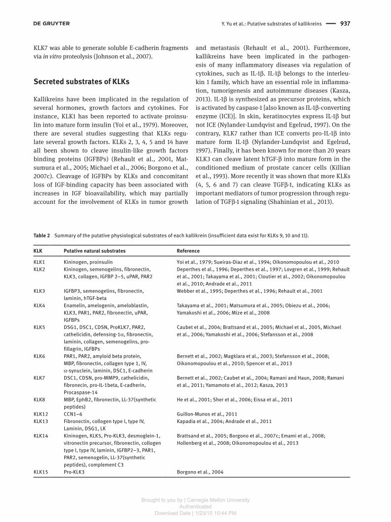
Table 2 Summary of the putative physiological substrates of each kallikrein (insufficient data exist for KLKs 9, 10 and 11).
KLK Putative natural substrates Reference
KLK1 Kininogen, proinsulin Yoi et al., 1979; Sueiras-Diaz et al., 1994; Oikonomopoulou et al., 2010KLK2 Kininogen, semenogelins, fibronectin,
KLK3, collagen, IGFBP 2–5, uPAR, PAR2 Deperthes et al., 1996; Deperthes et al., 1997; Lovgren et al., 1999; Rehault
et al., 2001; Takayama et al., 2001; Cloutier et al., 2002; Oikonomopoulou et al., 2010; Andrade et al., 2011
KLK3 IGFBP3, semenogelins, fibronectin, laminin, hTGF-beta
Webber et al., 1995; Deperthes et al., 1996; Rehault et al., 2001
KLK4 Enamelin, amelogenin, ameloblastin, KLK3, PAR1, PAR2, fibronectin, uPAR, IGFBPs
Takayama et al., 2001; Matsumura et al., 2005; Obiezu et al., 2006; Yamakoshi et al., 2006; Mize et al., 2008
KLK5 DSG1, DSC1, CDSN, ProKLK7, PAR2, cathelicidin, defensing-1α, fibronectin, laminin, collagen, semenogelins, pro-fillagrin, IGFBPs
Caubet et al., 2004; Brattsand et al., 2005; Michael et al., 2005, Michael et al., 2006; Yamakoshi et al., 2006; Stefansson et al., 2008
KLK6 PAR1, PAR2, amyloid beta protein, MBP, fibronectin, collagen type 1, IV, α-synuclein, laminin, DSC1, E-cadherin
Bernett et al., 2002; Magklara et al., 2003; Stefansson et al., 2008; Oikonomopoulou et al., 2010; Spencer et al., 2013
KLK7 DSC1, CDSN, pro-MMP9, cathelicidin, fibronecin, pro-IL-1beta, E-cadherin, Procaspase-14
Bernett et al., 2002; Caubet et al., 2004; Ramani and Haun, 2008; Ramani et al., 2011; Yamamoto et al., 2012; Kasza, 2013
KLK8 MBP, EphB2, fibronectin, LL-37(synthetic peptides)
He et al., 2001; Sher et al., 2006; Eissa et al., 2011
KLK12 CCN1–6 Guillon-Munos et al., 2011KLK13 Fibronectin, collogen type I, type IV,
Laminin, DSG1, LK Kapadia et al., 2004; Andrade et al., 2011
KLK14 Kininogen, KLK5, Pro-KLK3, desmoglein-1, vitronectin precursor, fibronectin, collogen type I, type IV, laminin, IGFBP2–3, PAR1, PAR2, semenogelin, LL-37(synthetic peptides), complement C3
Brattsand et al., 2005; Borgono et al., 2007c; Emami et al., 2008; Hollenberg et al., 2008; Oikonomopoulou et al., 2013
KLK15 Pro-KLK3 Borgono et al., 2004
Brought to you by | Carnegie Mellon UniversityAuthenticated
Download Date | 1/23/15 10:44 PM

938 Y. Yu et al.: Putative substrates of kallikreins
KLK-regulated surface receptors
Proteinase-activated receptors (PARs) constitute a four-member family of G protein-coupled receptors with ubiquitous expression and crucial signaling roles in the pathobiology of several pathologies, including cancer, central nervous pathologies, gastrointestinal diseases and cardiovascular abnormalities (Caliendo et al., 2012; Ramachandran et al., 2013). These seven-transmembrane receptors are known targets of extracellular proteases, which activate PARs through proteolytic cleavage and unveiling of a tethered ligand that stimulates their activ-ity. Emerging literature has established a tight connection between PARs and KLK activity (Hollenberg et al., 2008). We have recently shown that KLKs 1, 2, 4, 5, 6 and 14 can cleave and regulate PAR activity (Oikonomopoulou et al., 2010). Along this line, KLKs 5, 6 and 14 (and not KLK7 and 8) were shown to activate PAR2 (Stefansson et al., 2008),
while KLK14 was also shown to be able to inactivate PAR1 (Hollenberg et al., 2008). It is becoming evident that the interaction of KLKs with PARs is tissue and disease-dependent. For instance, in the setting of prostate cancer, KLK4 has been shown to co-localize with PAR2 and to trigger calcium signaling through PAR1 and PAR2 activa-tion (Mize et al., 2008). In melanoma, KLK6 activation has been linked to increased metastasis and prolifera-tion through enhancing of PAR2 activation (Krenzer et al., 2011). Similarly, a critical association between PARs and several KLKs has been suggested in many types of neu-rodegenerative diseases (e.g., MS) (Noorbakhsh et al., 2006). Lastly, in skin pathologies, hyperactivity of KLK5 (a known state in Netherton syndrome) can promote the expression of proinflammatroy cytokines and chemokine via activation of PAR2 (Briot et al., 2009). The interplay between KLKs and PARs in the skin is definitely an excit-ing subject of ongoing research.
Figure 2 Physiological kallikrein substrates in various processes.
Brought to you by | Carnegie Mellon UniversityAuthenticated
Download Date | 1/23/15 10:44 PM

Y. Yu et al.: Putative substrates of kallikreins 939
Other KLK substrates
A unique role for KLK4 has been established in the tooth, during enamel maturation (Simmer et al., 2009). Elegant work in the field has revealed that amelobast-produced MMP20 activate KLK4, which, in turn, cleaves amelogenin, enamelin and ameloblastin leading to enamel maturation (Yamakoshi et al., 2006). Recently, in vitro degradation assays have suggested that KLK14 can cleave complement C3 to C3a fragments, implicating a potential role for KLK14 in inflammatory response regulation (Oikonomopoulou et al., 2013). Lastly, KLK7 has been shown to cleave pro-caspase-14 into intermediate forms during terminal differ-entiation (Yamamoto et al., 2012).
An updated overview of all physiological KLK sub-strates is presented in Table 2.
ConclusionDespite the exciting advances in kallikreins research, the exact physiological and pathological roles of KLKs remain obscure. Primarily known as cancer biomarkers (e.g., PSA), kallikreins have recently grown into key molecules in a plethora of (patho) physiological conditions. Skin, central nervous system, reproductive system and tooth are a few characteristic examples where KLKs play criti-cal roles in maintenance of normal physiology (Figure 2). Despite the great advances in our understanding of the KLK (patho)biology, the identification of the physiologi-cal substrates of these enzymes remains largely an open question. The advent of high-throughput proteomic tech-niques, together with the development of KLK-specific animal models, have recently offered ample new insights regarding the putative KLK substrates and their implica-tions in health and disease. The big picture depicts that kallikreins in vivo do not act in isolation, but as part of an interconnected web of proteases (Ohler et al., 2010) with overlapping roles and activities. Undoubtedly, elucidation of the exact KLK substrate repertoires will benefit greatly from the ongoing development of KLK-specific activity-based probes and highly selective KLK inhibitors.
ReferencesAndrade, D., Assis, D.M., Santos, J.A., Alves, F.M., Hirata, I.Y.,
Araujo, M.S., Blaber, S.I., Blaber, M., Juliano, M.A., and Juliano, L. (2011). Substrate specificity of kallikrein-related pepti-dase 13 activated by salts or glycosaminoglycans and a search for natural substrate candidates. Biochimie 93, 1701–1709.
Attwood, B.K., Bourgognon, J.M., Patel, S., Mucha, M., Schiavon, E., Skrzypiec, A.E., Young, K.W., Shiosaka, S., Korostynski, M., Piechota, M., et al. (2011). Neuropsin cleaves EphB2 in the amygdala to control anxiety. Nature 473, 372–375.
Beaufort, N., Debela, M., Creutzburg, S., Kellermann, J., Bode, W., Schmitt, M., Pidard, D., and Magdolen, V. (2006). Interplay of human tissue kallikrein 4 (hK4) with the plasminogen activa-tion system: hK4 regulates the structure and functions of the urokinase-type plasminogen activator receptor (uPAR). Biol. Chem. 387, 217–222.
Bernett, M.J., Blaber, S.I., Scarisbrick, I.A., Dhanarajan, P., Thompson, S.M., and Blaber, M. (2002). Crystal structure and biochemical characterization of human kallikrein 6 reveals that a trypsin-like kallikrein is expressed in the central nervous system. J. Biol. Chem. 277, 24562–24570.
Bhoola, K.D., Misso, N.L., Naran, A., and Thompson, P.J. (2007). Cur-rent status of tissue kallikrein inhibitors: importance in cancer. Curr. Opin. Investig. Drugs 8, 462–468.
Blaber, S.I., Ciric, B., Christophi, G.P., Bernett, M.J., Blaber, M., Rodriguez, M., and Scarisbrick, I.A. (2004). Targeting kallikrein 6 proteolysis attenuates CNS inflammatory disease. FASEB J. 18, 920–922.
Borgono, C.A., and Diamandis, E.P. (2004). The emerging roles of human tissue kallikreins in cancer. Nat. Rev. Cancer 4, 876–890.
Borgono, C.A., Michael, I.P., and Diamandis, E.P. (2004). Human tissue kallikreins: physiologic roles and applications in cancer. Mol. Cancer Res. 2, 257–280.
Borgono, C.A., Gavigan, J.A., Alves, J., Bowles, B., Harris, J.L., Sotiro-poulou, G., and Diamandis, E.P. (2007a). Defining the extended substrate specificity of kallikrein 1-related peptidases. Biol. Chem. 388, 1215–1225.
Borgono, C.A., Michael, I.P., Komatsu, N., Jayakumar, A., Kapa-dia, R., Clayman, G.L., Sotiropoulou, G., and Diamandis, E.P. (2007b). A potential role for multiple tissue kallikrein serine proteases in epidermal desquamation. J. Biol. Chem. 282, 3640–3652.
Borgono, C.A., Michael, I.P., Shaw, J.L., Luo, L.Y., Ghosh, M.C., Soosaipillai, A., Grass, L., Katsaros, D., and Diamandis, E.P. (2007c). Expression and functional characterization of the cancer-related serine protease, human tissue kallikrein 14. J. Biol. Chem. 282, 2405–2422.
Bourgeois, L., Brillard-Bourdet, M., Deperthes, D., Juliano, M.A., Juliano, L., Tremblay, R.R., Dube, J.Y., and Gauthier, F. (1997). Serpin-derived peptide substrates for investigating the sub-strate specificity of human tissue kallikreins hK1 and hK2. J. Biol. Chem. 272, 29590–29595.
Brattsand, M. and Egelrud, T. (1999). Purification, molecular clon-ing, and expression of a human stratum corneum trypsin-like serine protease with possible function in desquamation. J. Biol. Chem. 274, 30033–30040.
Brattsand, M., Stefansson, K., Lundh, C., Haasum, Y., and Egelrud, T. (2005). A proteolytic cascade of kallikreins in the stratum corneum. J. Invest. Dermatol. 124, 198–203.
Bredemeyer, A.J., Lewis, R.M., Malone, J.P., Davis, A.E., Gross, J., Townsend, R.R., and Ley, T.J. (2004). A proteomic approach for the discovery of protease substrates. Proc. Natl. Acad. Sci. USA 101, 11785–11790.
Briot, A., Deraison, C., Lacroix, M., Bonnart, C., Robin, A., Besson, C., Dubus, P., and Hovnanian, A. (2009). Kallikrein 5
Brought to you by | Carnegie Mellon UniversityAuthenticated
Download Date | 1/23/15 10:44 PM
940 Y. Yu et al.: Putative substrates of kallikreins
induces atopic dermatitis-like lesions through PAR2-mediated thymic stromal lymphopoietin expression in Netherton syndrome. J. Exp. Med. 206, 1135–1147.
Butler, G.S., Dean, R.A., Smith, D., and Overall, C.M. (2009). Mem-brane protease degradomics: proteomic identification and quantification of cell surface protease substrates. Methods Mol. Biol. 528, 159–176.
Caliendo, G., Santagada, V., Perissutti, E., Severino, B., Fiorino, F., Frecentese, F., and Juliano, L. (2012). Kallikrein protease acti-vated receptor (PAR) axis: an attractive target for drug develop-ment. J. Med. Chem. 55, 6669–6686.
Carvalho, A.L., Sanz, L., Barettino, D., Romero, A., Calvete, J.J., and Romao, M.J. (2002). Crystal structure of a prostate kallikrein isolated from stallion seminal plasma: a homologue of human PSA. J. Mol. Biol. 322, 325–337.
Caubet, C., Jonca, N., Brattsand, M., Guerrin, M., Bernard, D., Schmidt, R., Egelrud, T., Simon, M., and Serre, G. (2004). Degradation of corneodesmosome proteins by two serine proteases of the kallikrein family, SCTE/KLK5/hK5 and SCCE/KLK7/hK7. J. Invest. Dermatol. 122, 1235–1244.
Chao, J., Bledsoe, G., Yin, H., and Chao, L. (2006). The tissue kallikrein-kinin system protects against cardiovascular and renal diseases and ischemic stroke independently of blood pressure reduction. Biol. Chem. 387, 665–675.
Clements, J. (2013). KLK4 is a key regulator of the tumour microen-vironment in prostate cancer. 5th International Symposium on Kallikreins and Kallikrein-Related Peptides, 28 September – 1 October 2013, Toronto, Canada.
Cloutier, S.M., Chagas, J.R., Mach, J.P., Gygi, C.M., Leisinger, H.J., and Deperthes, D. (2002). Substrate specificity of human kal-likrein 2 (hK2) as determined by phage display technology. Eur. J. Biochem 269, 2747–2754.
Coombs, G.S., Bergstrom, R.C., Pellequer, J.L., Baker, S.I., Navre, M., Smith, M.M., Tainer, J.A., Madison, E.L., and Corey, D.R. (1998). Substrate specificity of prostate-specific antigen (PSA). Chem. Biol. 5, 475–488.
de Veer, S.J., Swedberg, J.E., Parker, E.A., and Harris, J.M. (2012). Non-combinatorial library screening reveals subsite coop-erativity and identifies new high-efficiency substrates for kallikrein-related peptidase 14. Biol. Chem. 393, 331–341.
Debela, M., Magdolen, V., Grimminger, V., Sommerhoff, C., Mess-erschmidt, A., Huber, R., Friedrich, R., Bode, W., and Goettig, P. (2006a). Crystal structures of human tissue kallikrein 4: activ-ity modulation by a specific zinc binding site. J. Mol. Biol. 362, 1094–1107.
Debela, M., Magdolen, V., Schechter, N., Valachova, M., Lottspeich, F., Craik, C.S., Choe, Y., Bode, W., and Goettig, P. (2006b). Specificity profiling of seven human tissue kallikreins reveals individual subsite preferences. J. Biol. Chem. 281, 25678–25688.
Debela, M., Goettig, P., Magdolen, V., Huber, R., Schechter, N.M., and Bode, W. (2007a). Structural basis of the zinc inhibition of human tissue kallikrein 5. J. Mol. Biol. 373, 1017–1031.
Debela, M., Hess, P., Magdolen, V., Schechter, N.M., Steiner, T., Huber, R., Bode, W., and Goettig, P. (2007b). Chymotryptic specificity determinants in the 1.0 A structure of the zinc-inhibited human tissue kallikrein 7. Proc. Natl. Acad. Sci. USA 104, 16086–16091.
Deperthes, D., Frenette, G., Brillard-Bourdet, M., Bourgeois, L., Gauthier, F., Tremblay, R.R., and Dube, J.Y. (1996). Potential
involvement of kallikrein hK2 in the hydrolysis of the human seminal vesicle proteins after ejaculation. J. Androl. 17, 659–665.
Deperthes, D., Marceau, F., Frenette, G., Lazure, C., Tremblay, R.R., and Dube, J.Y. (1997). Human kallikrein hK2 has low kininoge-nase activity while prostate-specific antigen (hK3) has none. Biochim. Biophys. Acta 1343, 102–106.
Descargues, P., Deraison, C., Prost, C., Fraitag, S., Mazereeuw-Hautier, J., D’Alessio, M., Ishida-Yamamoto, A., Bodemer, C., Zambruno, G., and Hovnanian, A. (2006). Corneodesmosomal cadherins are preferential targets of stratum corneum trypsin- and chymotrypsin-like hyperactivity in Netherton syndrome. J. Invest. Dermatol. 126, 1622–1632.
Dix, M.M., Simon, G.M., Wang, C., Okerberg, E., Patricelli, M.P., and Cravatt, B.F. (2012). Functional interplay between caspase cleavage and phosphorylation sculpts the apoptotic proteome. Cell 150, 426–440.
Eissa, A., Amodeo, V., Smith, C.R., and Diamandis, E.P. (2011). Kal-likrein-related peptidase-8 (KLK8) is an active serine protease in human epidermis and sweat and is involved in a skin barrier proteolytic cascade. J. Biol. Chem. 286, 687–706.
Emami, N., Deperthes, D., Malm, J., and Diamandis, E.P. (2008). Major role of human KLK14 in seminal clot liquefaction. J. Biol. Chem. 283, 19561–19569.
Felber, L.M., Borgono, C.A., Cloutier, S.M., Kundig, C., Kishi, T., Ribeiro Chagas, J., Jichlinski, P., Gygi, C.M., Leisinger, H.J., Diamandis, E.P., et al. (2005). Enzymatic profiling of human kallikrein 14 using phage-display substrate technology. Biol. Chem. 386, 291–298.
Fortugno, P., Bresciani, A., Paolini, C., Pazzagli, C., El Hachem, M., D’Alessio, M., and Zambruno, G. (2011). Proteolytic activation cascade of the Netherton syndrome-defective protein, LEKTI, in the epidermis: implications for skin homeostasis. J. Invest. Dermatol. 131, 2223–2232.
Ghosh, M.C., Grass, L., Soosaipillai, A., Sotiropoulou, G., and Dia-mandis, E.P. (2004). Human kallikrein 6 degrades extracellular matrix proteins and may enhance the metastatic potential of tumour cells. Tumour Biol. 25, 193–199.
Goettig, P., Magdolen, V., and Brandstetter, H. (2010). Natural and synthetic inhibitors of kallikrein-related peptidases (KLKs). Biochimie 92, 1546–1567.
Guillon-Munos, A., Oikonomopoulou, K., Michel, N., Smith, C.R., Petit-Courty, A., Canepa, S., Reverdiau, P., Heuze-Vourc’h, N., Diamandis, E.P., and Courty, Y. (2011). Kallikrein-related pepti-dase 12 hydrolyzes matricellular proteins of the CCN family and modifies interactions of CCN1 and CCN5 with growth factors. J. Biol. Chem. 286, 25505–25518.
Hachem, J.P., Wagberg, F., Schmuth, M., Crumrine, D., Lissens, W., Jayakumar, A., Houben, E., Mauro, T.M., Leonardsson, G., Bratt-sand, M., et al. (2006). Serine protease activity and residual LEKTI expression determine phenotype in Netherton syndrome. J. Invest. Dermatol. 126, 1609–1621.
Hansson, L., Stromqvist, M., Backman, A., Wallbrandt, P., Carl-stein, A., and Egelrud, T. (1994). Cloning, expression, and characterization of stratum corneum chymotryptic enzyme. A skin-specific human serine proteinase. J. Biol. Chem. 269, 19420–19426.
Hansson, L., Backman, A., Ny, A., Edlund, M., Ekholm, E., Ekstrand Hammarstrom, B., Tornell, J., Wallbrandt, P., Wennbo, H., and Egelrud, T. (2002). Epidermal overexpression of stratum cor-
Brought to you by | Carnegie Mellon UniversityAuthenticated
Download Date | 1/23/15 10:44 PM

Y. Yu et al.: Putative substrates of kallikreins 941
neum chymotryptic enzyme in mice: a model for chronic itchy dermatitis. J. Invest. Dermatol. 118, 444–449.
He, X.P., Shiosaka, S., and Yoshida, S. (2001). Expression of neurop-sin in oligodendrocytes after injury to the CNS. Neurosci. Res. 39, 455–462.
Hollenberg, M.D., Oikonomopoulou, K., Hansen, K.K., Saifeddine, M., Ramachandran, R., and Diamandis, E.P. (2008). Kallikreins and proteinase-mediated signaling: proteinase-activated receptors (PARs) and the pathophysiology of inflammatory diseases and cancer. Biol. Chem. 389, 643–651.
Johnson, S.K., Ramani, V.C., Hennings, L., and Haun, R.S. (2007). Kallikrein 7 enhances pancreatic cancer cell invasion by shed-ding E-cadherin. Cancer 109, 1811–1820.
Kapadia, C., Ghosh, M.C., Grass, L., and Diamandis, E.P. (2004). Human kallikrein 13 involvement in extracellular matrix degra-dation. Biochem. Biophys. Res. Commun. 323, 1084–1090.
Kasza, A. (2013). IL-1 and EGF regulate expression of genes impor-tant in inflammation and cancer. Cytokine 62, 22–33.
Katz, B.A., Liu, B., Barnes, M., and Springman, E.B. (1998). Crystal structure of recombinant human tissue kallikrein at 2.0 A reso-lution. Protein Sci. 7, 875–885.
Killian, C.S., Corral, D.A., Kawinski, E., and Constantine, R.I. (1993). Mitogenic response of osteoblast cells to prostate-specific antigen suggests an activation of latent TGF-beta and a proteo-lytic modulation of cell adhesion receptors. Biochem. Biophys. Res. Commun. 192, 940–947.
Kleifeld, O., Doucet, A., auf dem Keller, U., Prudova, A., Schilling, O., Kainthan, R.K., Starr, A.E., Foster, L.J., Kizhakkedathu, J.N., and Overall, C.M. (2010). Isotopic labeling of terminal amines in complex samples identifies protein N-termini and protease cleavage products. Nat. Biotechnol. 28, 281–288.
Klucky, B., Mueller, R., Vogt, I., Teurich, S., Hartenstein, B., Breu-hahn, K., Flechtenmacher, C., Angel, P., and Hess, J. (2007). Kallikrein 6 induces E-cadherin shedding and promotes cell pro-liferation, migration, and invasion. Cancer Res. 67, 8198–8206.
Krenzer, S., Peterziel, H., Mauch, C., Blaber, S.I., Blaber, M., Angel, P., and Hess, J. (2011). Expression and function of the kallikrein-related peptidase 6 in the human melanoma micro-environment. J. Invest. Dermatol. 131, 2281–2288.
Li, H.X., Hwang, B.Y., Laxmikanthan, G., Blaber, S.I., Blaber, M., Golubkov, P.A., Ren, P., Iverson, B.L., and Georgiou, G. (2008). Substrate specificity of human kallikreins 1 and 6 determined by phage display. Protein Sci. 17, 664–672.
Lilja, H., Oldbring, J., Rannevik, G., and Laurell, C.B. (1987). Seminal vesicle-secreted proteins and their reactions during gelation and liquefaction of human semen. J. Clin. Invest. 80, 281–285.
Lima, A.R., Alves, F.M., Angelo, P.F., Andrade, D., Blaber, S.I., Blaber, M., Juliano, L., and Juliano, M.A. (2008). S(1)’ and S(2)’ subsite specificities of human plasma kallikrein and tissue kal-likrein 1 for the hydrolysis of peptides derived from the brady-kinin domain of human kininogen. Biol. Chem. 389, 1487–1494.
Lovgren, J., Airas, K., and Lilja, H. (1999). Enzymatic action of human glandular kallikrein 2 (hK2). Substrate specificity and regulation by Zn2+ and extracellular protease inhibitors. Eur. J. Biochem. 262, 781–789.
Magklara, A., Mellati, A.A., Wasney, G.A., Little, S.P., Sotiropoulou, G., Becker, G.W., and Diamandis, E.P. (2003). Characterization of the enzymatic activity of human kallikrein 6: autoactivation, substrate specificity, and regulation by inhibitors. Biochem. Biophys. Res. Commun. 307, 948–955.
Malm, J., Hellman, J., Hogg, P., and Lilja, H. (2000). Enzymatic action of prostate-specific antigen (PSA or hK3): substrate specificity and regulation by Zn(2+), a tight-binding inhibitor. Prostate 45, 132–139.
Matsumura, M., Bhatt, A.S., Andress, D., Clegg, N., Takayama, T.K., Craik, C.S., and Nelson, P.S. (2005). Substrates of the prostate-specific serine protease prostase/KLK4 defined by positional-scanning peptide libraries. Prostate 62, 1–13.
Michael, I.P., Sotiropoulou, G., Pampalakis, G., Magklara, A., Ghosh, M., Wasney, G., and Diamandis, E.P. (2005). Biochemi-cal and enzymatic characterization of human kallikrein 5 (hK5), a novel serine protease potentially involved in cancer progres-sion. J Biol. Chem. 280, 14628–14635.
Michael, I.P., Pampalakis, G., Mikolajczyk, S.D., Malm, J., Sotiropou-lou, G., and Diamandis, E.P. (2006). Human tissue kallikrein 5 is a member of a proteolytic cascade pathway involved in seminal clot liquefaction and potentially in prostate cancer progression. J. Biol. Chem. 281, 12743–12750.
Mitsui, S., Okui, A., Uemura, H., Mizuno, T., Yamada, T., Yamamura, Y., and Yamaguchi, N. (2002). Decreased cer-ebrospinal fluid levels of neurosin (KLK6), an aging-related protease, as a possible new risk factor for Alzheimer’s disease. Ann. NY Acad. Sci. 977, 216–223.
Mize, G.J., Wang, W., and Takayama, T.K. (2008). Prostate-specific kallikreins-2 and -4 enhance the proliferation of DU-145 pros-tate cancer cells through protease-activated receptors-1 and -2. Mol. Cancer Res. 6, 1043–1051.
Niessen, S., Hoover, H., and Gale, A.J. (2011). Proteomic analysis of the coagulation reaction in plasma and whole blood using PROTOMAP. Proteomics 11, 2377–2388.
Noorbakhsh, F., Tsutsui, S., Vergnolle, N., Boven, L.A., Shariat, N., Vodjgani, M., Warren, K.G., Andrade-Gordon, P., Hollenberg, M.D., and Power, C. (2006). Proteinase-activated receptor 2 modulates neuroinflammation in experimental autoimmune encephalomyelitis and multiple sclerosis. J. Exp. Med. 203, 425–435.
Nylander-Lundqvist, E. and Egelrud, T. (1997). Formation of active IL-1 beta from pro-IL-1 beta catalyzed by stratum corneum chymotryptic enzyme in vitro. Acta Derm. Venereol. 77, 203–206.
Obiezu, C.V., Michael, I.P., Levesque, M.A., and Diamandis, E.P. (2006). Human kallikrein 4: enzymatic activity, inhibition, and degradation of extracellular matrix proteins. Biol. Chem. 387, 749–759.
Ogawa, K., Yamada, T., Tsujioka, Y., Taguchi, J., Takahashi, M., Tsuboi, Y., Fujino, Y., Nakajima, M., Yamamoto, T., Akatsu, H., et al. (2000). Localization of a novel type trypsin-like serine protease, neurosin, in brain tissues of Alzheimer’s disease and Parkinson’s disease. Psychiatry Clin. Neurosci. 54, 419–426.
Ohler, A., Debela, M., Wagner, S., Magdolen, V., and Becker-Pauly, C. (2010). Analyzing the protease web in skin: meprin metallo-proteases are activated specifically by KLK4, 5 and 8 vice versa leading to processing of proKLK7 thereby triggering its activa-tion. Biol. Chem. 391, 455–460.
Oikonomopoulou, K., Hansen, K.K., Saifeddine, M., Tea, I., Blaber, M., Blaber, S.I., Scarisbrick, I., Andrade-Gordon, P., Cottrell, G.S., Bunnett, N.W., et al. (2006). Proteinase-activated receptors, targets for kallikrein signaling. J. Biol. Chem. 281, 32095–32112.
Brought to you by | Carnegie Mellon UniversityAuthenticated
Download Date | 1/23/15 10:44 PM

942 Y. Yu et al.: Putative substrates of kallikreins
Oikonomopoulou, K., Diamandis, E.P., and Hollenberg, M.D. (2010). Kallikrein-related peptidases: proteolysis and signaling in cancer, the new frontier. Biol. Chem. 391, 299–310.
Oikonomopoulou, K., DeAngelis, R.A., Chen, H., Diamandis, E.P., Hollenberg, M.D., Ricklin, D., and Lambris, J.D. (2013). Induc-tion of complement C3a receptor responses by kallikrein-related peptidase 14. J. Immunol. 191, 3858–3866.
Ramachandran, R., Noorbakhsh, F., Defea, K., and Hollenberg, M.D. (2013). Targeting proteinase-activated receptors: therapeutic potential and challenges. Nat. Rev. Drug Discov. 11, 69–86.
Ramani, V.C. and Haun, R.S. (2008). The extracellular matrix protein fibronectin is a substrate for kallikrein 7. Biochem. Biophys. Res. Commun. 369, 1169–1173.
Ramani, V.C., Kaushal, G.P., and Haun, R.S. (2011). Proteolytic action of kallikrein-related peptidase 7 produces unique active matrix metalloproteinase-9 lacking the C-terminal hemopexin domains. Biochim. Biophys. Acta 1813, 1525–1531.
Rehault, S., Monget, P., Mazerbourg, S., Tremblay, R., Gutman, N., Gauthier, F., and Moreau, T. (2001). Insulin-like growth factor binding proteins (IGFBPs) as potential physiological substrates for human kallikreins hK2 and hK3. Eur. J. Biochem. 268, 2960–2968.
Robert, M. and Gagnon, C. (1996). Purification and characterization of the active precursor of a human sperm motility inhibitor secreted by the seminal vesicles: identity with semenogelin. Biol. Reprod. 55, 813–821.
Sakabe, J., Yamamoto, M., Hirakawa, S., Motoyama, A., Ohta, I., Tatsuno, K., Ito, T., Kabashima, K., Hibino, T., and Tokura, Y. (2013). Kallikrein-related peptidase 5 functions in proteolytic processing of profilaggrin in cultured human keratinocytes. J. Biol. Chem. 288, 17179–17189.
Scarisbrick, I.A., Towner, M.D., and Isackson, P.J. (1997). Nervous system-specific expression of a novel serine protease: regula-tion in the adult rat spinal cord by excitotoxic injury. J. Neurosci. 17, 8156–8168.
Scarisbrick, I.A., Sabharwal, P., Cruz, H., Larsen, N., Vandell, A.G., Blaber, S.I., Ameenuddin, S., Papke, L.M., Fehlings, M.G., Reeves, R.K., et al. (2006). Dynamic role of kallikrein 6 in trau-matic spinal cord injury. Eur. J. Neurosci. 24, 1457–1469.
Schilling, O. and Overall, C.M. (2007). Proteomic discovery of pro-tease substrates. Curr. Opin. Chem. Biol. 11, 36–45.
Schilling, O. and Overall, C.M. (2008). Proteome-derived, database-searchable peptide libraries for identifying protease cleavage sites. Nat. Biotechnol. 26, 685–694.
Shahinian, H., Loessner, D., Biniossek, M.L., Kizhakkedathu, J.N., Clements, J.A., Magdolen, V., and Schilling, O. (2013). Secretome and degradome profiling shows that Kallikrein-related peptidases 4, 5, 6, and 7 induce TGFbeta-1 signaling in ovarian cancer cells. Mol. Oncol. 8, 68–82.
Sharma, N., Oikonomopoulou, K., Ito, K., Renaux, B., Diamandis, E.P., Hollenberg, M.D., and Rancourt, D.E. (2008). Substrate specificity determination of mouse implantation serine pro-teinase and human kallikrein-related peptidase 6 by phage display. Biol. Chem. 389, 1097–1105.
Shaw, J.L. and Diamandis, E.P. (2008). A potential role for tissue kallikrein-related peptidases in human cervico-vaginal physiol-ogy. Biol. Chem. 389, 681–688.
Shen, C., Yu, Y., Li, H., Yan, G., Liu, M., Shen, H., and Yang, P. (2012). Global profiling of proteolytically modified proteins in human metastatic hepatocellular carcinoma cell lines reveals CAPN2 centered network. Proteomics 12, 1917–1927.
Sher, Y.P., Chou, C.C., Chou, R.H., Wu, H.M., Wayne Chang, W.S., Chen, C.H., Yang, P.C., Wu, C.W., Yu, C.L., and Peck, K. (2006). Human kallikrein 8 protease confers a favorable clinical out-come in non-small cell lung cancer by suppressing tumor cell invasiveness. Cancer Res. 66, 11763–11770.
Simmer, J.P., Hu, Y., Lertlam, R., Yamakoshi, Y., and Hu, J.C. (2009). Hypomaturation enamel defects in Klk4 knockout/LacZ knockin mice. J. Biol. Chem. 284, 19110–19121.
Smith, H.W. and Marshall, C.J. (2010). Regulation of cell signalling by uPAR. Nat. Rev. Mol. Cell Biol 11, 23–36.
Sotiropoulou, G., Rogakos, V., Tsetsenis, T., Pampalakis, G., Zafiropoulos, N., Simillides, G., Yiotakis, A., and Diamandis, E.P. (2003). Emerging interest in the kallikrein gene family for understanding and diagnosing cancer. Oncol. Res. 13, 381–391.
Spencer, B., Michael, S., Shen, J., Kosberg, K., Rockenstein, E., Patrick, C., Adame, A., and Masliah, E. (2013). Lentivirus mediated delivery of neurosin promotes clearance of wild-type alpha-synuclein and reduces the pathology in an alpha-synu-clein model of LBD. Mol. Ther. 21, 31–41.
Staes, A., Impens, F., Van Damme, P., Ruttens, B., Goethals, M., Demol, H., Timmerman, E., Vandekerckhove, J., and Gevaert, K. (2011). Selecting protein N-terminal peptides by combined fractional diagonal chromatography. Nat. Protoc. 6, 1130–1141.
Stefansson, K., Brattsand, M., Roosterman, D., Kempkes, C., Bocheva, G., Steinhoff, M., and Egelrud, T. (2008). Activation of proteinase-activated receptor-2 by human kallikrein-related peptidases. J. Invest. Dermatol. 128, 18–25.
Sueiras-Diaz, J., Jones, D.M., Ashworth, D., Horton, J., Evans, D.M., and Szelke, M. (1994). Cleavage of human kininogen fragments at Met-Lys by human tissue kallikrein. Braz. J. Med. Biol. Res. 27, 1935–1942.
Takayama, T.K., McMullen, B.A., Nelson, P.S., Matsumura, M., and Fujikawa, K. (2001). Characterization of hK4 (prostase), a prostate-specific serine protease: activation of the precursor of prostate specific antigen (pro-PSA) and single-chain urokinase-type plasminogen activator and degradation of prostatic acid phosphatase. Biochemistry 40, 15341–15348.
Veveris-Lowe, T.L., Kruger, S.J., Walsh, T., Gardiner, R.A., and Clements, J.A. (2007). Seminal fluid characterization for male fertility and prostate cancer: kallikrein-related serine proteases and whole proteome approaches. Semin. Thromb. Hemost. 33, 87–99.
Webber, M.M., Waghray, A., and Bello, D. (1995). Prostate-specific antigen, a serine protease, facilitates human prostate cancer cell invasion. Clin. Cancer Res. 1, 1089–1094.
Yamakoshi, Y., Hu, J.C., Fukae, M., Yamakoshi, F., and Simmer, J.P. (2006). How do enamelysin and kallikrein 4 process the 32-kDa enamelin? Eur. J. Oral Sci. 114 Suppl 1, 45–51; discus-sion 93–45, 379–380.
Yamakoshi, Y., Simmer, J.P., Bartlett, J.D., Karakida, T., and Oida, S. (2013). MMP20 and KLK4 activation and inactivation interac-tions in vitro. Arch. Oral Biol. 58, 1569–1577.
Yamamoto, M., Miyai, M., Matsumoto, Y., Tsuboi, R., and Hibino, T. (2012). Kallikrein-related peptidase-7 regulates caspase-14 maturation during keratinocyte terminal differentiation by generating an intermediate form. J. Biol. Chem. 287, 32825–32834.
Yamasaki, K., Schauber, J., Coda, A., Lin, H., Dorschner, R.A., Schechter, N.M., Bonnart, C., Descargues, P., Hovnanian, A., and Gallo, R.L. (2006). Kallikrein-mediated proteolysis regu-
Brought to you by | Carnegie Mellon UniversityAuthenticated
Download Date | 1/23/15 10:44 PM

Y. Yu et al.: Putative substrates of kallikreins 943
lates the antimicrobial effects of cathelicidins in skin. FASEB J. 20, 2068–2080.
Yoi, O.O., Seldin, D.C., Spragg, J., Pinkus, G.S., and Austen, K.F. (1979). Sequential cleavage of proinsulin by human pancreatic kallikrein and a human pancreatic kininase. Proc. Natl. Acad. Sci. USA 76, 3612–3616.
Yoshida, S. (2010). Klk8, a multifunctional protease in the brain and skin: analysis of knockout mice. Biol. Chem. 391, 375–380.
Yousef, G.M. and Diamandis, E.P. (2001). The new human tissue kallikrein gene family: structure, function, and association to disease. Endocr. Rev. 22, 184–204.
Yijing Yu completed her Msc Degree in the Molecular Science program at Ryerson University, Ontario, Canada. Currently, she is a PhD candidate at the Department of Laboratory Medicine and Pathobiology at the University of Toronto, Ontario, Canada. Her main research interests include the identification of novel biologi-cal substrates of kallikreins using degradomic approaches and the development of new therapeutic targets for skin diseases.
Ioannis Prassas obtained his PhD from the Department of Labo-ratory Medicine and Pathobiology at the University of Toronto, Ontario, Canada. Currently, he is training as a post-doctoral fellow in the Lunenfeld Tanenbaum Research Institute, Mount Sinai Hos-pital, Toronto, Onrario, Canada. His main research activities evolve around the use of mass spectrometry for the identification of novel therapeutic targets and the development of new therapeutic agents.
Eleftherios Diamandis currently serves as the Division Head of Clini-cal Biochemistry at Mount Sinai Hospital and Biochemist-in-Chief at the University Health Network and is Professor & Head, Clinical Biochemisty, Department of Laboratory Medicine and Pathobiol-ogy, University of Toronto, Ontario, Canada. His research activities evolve around discovery and validation of cancer biomarkers, prot-eomics, mass spectrometry and translational research.
Brought to you by | Carnegie Mellon UniversityAuthenticated
Download Date | 1/23/15 10:44 PM