yectoi(u) inyeresk research international ltd ... · trimethyl-p-phenyl ammonium chloride....
TRANSCRIPT

YECTOI(U) INYERESK RESEARCH INTERNATIONAL LTDNUSSEL3URGH (SCOTLAND) W J HARRIS 15 FEE 63INtLMISIFIEODAIT-92-C-2144
F/G 6/2 tL
Eu.....I

kWWA uanuw Q"I4ITONt TEST CHAR7
""No "qw IW "pr AV

'AD
ADA17 9 "LNIOCOI OF ACETYLCHOLINESTERASE .;..'-
GENE IN A MICROBIAL VECTOR
Annual Report
-o. .°..-
1 May 1982-31 January 1983
WILLIAM J. HARRIS, Ph.D.
15 February 1983
Supported by
U.S. ARMY MEDICAL RESEARCH AND DEVELOPMENT COMMANDFort Detrick, Frederick, Maryland 21701-5012
Contract No. DAMD 17-82-C-2144
Inveresk Research International Limited •Inveresk Gate, Musselburgh, EH21 7UB Scotland
Approved for Public Releases Distribution Unlimited
The findings in this report are not to be construed as an officialDepartment of the Army position unless so designated by otherauthroi sed documents.
L. 1
M'ACETYLCHOLINESTERA'SEI*-~E. GNE NAMICROBIAL .VET~~ .
Annual Report . . .,.-.
1 May 1982-31 January 1983
WILLIAM J. HARRIS, Ph.D..
15 February 1983
Supported by
( U.S. ARMY MEDICAL RESEARCH AND DEVELOPMENT COMMANDFort Detrick, Frederick, Maryland 21701-5012
C Contract No. DAND 17-82-C-2144
Inveresk Research International LimitedInveresk Gate, Musselburgh, EH21 7UB Scotland
Approved for Public Releases Distribution Unlimited
The findings in this report are not to be construed as an officialDepartment of the Army position unless so designated by otherauthroised documents.
gel
SECURITY CLASSIFICATION OF THIS PAGE (Whefl Dta Enteed)
READ ISTRUCTIONSREPORT DOCUMENTATION PAGE BEFORE COMPLETING FORM
1. REPORT NUMBER 2. GOVT ACCESSION NO 3. RECIPIENT'S CATALOG NUMBER
44. TITLE (and Sublide) S. TYPE OF REPORT & PERIOD COVERED
CLONING OF ACETYLCHOLINESTERASE GENE Annual
IN A MICROBIAL VECTOR May 1982-31 Jan. 19836. PERFORMING ORG. REPORT NUMBER
7. AUTHOR(*) S. CONTRACT OR GRANT NUMBER(*)
WILLIAM J. HARRIS, Ph.D. DAMD-17-82-C-2144
9. PERFORMING ORGANIZATION NAME AND ADDRESS 10. PROGRAM ELEMENT. PROJECT. TASK
INVERESK RESEARCH INTERNATIONAL LIMITED AREA& WORK UNIT NUMBERS
INVERESK GATE, MUSSELBURGH, EH21 7UBSCOTLAND
11. CONTROLLING OFFICE NAME AND ADDRESS 12. REPORT DATE
12. NUMBER OF PAGES
14. MONITORING AGENCY NAME A ADDRESS(If dLfferen tram Controlling Office) IS. SECURITY CLASS. (of this report)
UnclassifiedISa. DECLASSI IlCATION/DOWNGRADING
SCHEDULE
I. DISTRIBUTION STATEMENT (of thl Report)
17. DISTRIBUTION STATEMENT (of the abstract entered In Block 20, if iffieraen broe Report)
II. SUPPLEMENTARY NOTES
IS. KEY WORDS (Comtinue an reverse side it necessary ani Idntltj. by block number)
21L A§SYR ACT C m an reers neearn d d ,-* i y block mtobev)
Techniques to distinguish between the activities of human acetylcholinester-ase and pseudocholinesterase have been developed. Human neuroblastoma celllines have been shown to synthesise human acetylcholinesterase and the levelof enzyme within these cells can be induced by treatment of cells with sodiumbutyrate or dibutyryl cAMP. These cell lines should be suitable as a sourceof mRNA for the cloning of the human gene into E. coli.
D0 S FOR W 3 cA r TOo oF T IS mAv ls is oEn
SECURITY CLASSIFICATIONI OF TIMIS PAGE (Whe nee EntaeE)0

2
INDEX
Page
SUMMARY 3
FOREWORD 4
INTRODUCTION 5
PROGRESS TO DATE 7
METHODOLOGY 8
RESULTS 12
DISCUSSION 16
FUTURE WORK 17
METHODOLOGY 19
TABLES1. Levels of AChE and ChE in Neuroblastoma Cell Lines 20
IMR-32 and CHP-1262. Levels of AChE and ChE in Neuroblastoma Cell Lines 21
Under Different Growth Conditions in 24-Well Plates3. The Effects of Chemicals on AChE Levels in 22
Neuroblastoma Cells
FIGURES1. Effects of Specific Inhibitors on ChEs 232. Lineweaver-Burke Plots of ChE (Human Serum) and AChE 25
(Bovine Erythrocyte) Using Acetylthiocholine asSubstrate
3. Liquid Assay of AChe in Cell Extracts and Commercial 27Serum Samples
4. Effects of Specific Inhibitors on ChE Activity in 29Polyacrylamide Gels
5. Foetal Calf Serun and Human Erythrocyte AChE in 31Polyacrylamide Gels
6. Effect of Inducers on ChEs in IMR-32 Neuroblastoma 33Cells After 4 Days of Exposure
7. Effect of Nerve Growth Factor and 12-0-Tetradecanoyl-phorbol 35-13-acetate (TPA) on Cholinesterases in IMR-32 Cells
8. AChE Levels in IMR-32 Cells After 10 Days in Culture 379. Time Course of the Action of Sodium Butyrate on ChE 39
Levels in Neuroblastoma Cells
REFERENCES 41
DISTRIBUTION LIST 44
1? U 5'

3
SUMMARY
Nature of Project
As an aid to the design of effective antidotes to chemical agents, theU.S. Army would like to study in detail the structure of the humanenzyme acetylcholinesterase (AChE). This requires the availability ofreasonable quantities of the pure enzyme. One way in which this maybe achieved is the introduction of the gene from human cells intobacteria by genetic engineering techniques. This is the aim of thecurrent project.
The approach proposed in this study was as follows:
1. Obtain human neuroblastoma cells expressing high levels of AChEactivity.
2. Manipulate growth conditions so that expression of enzyme levelsis maximised.
3. Isolate mRNA from such cells and transcribe into cDNA.
4. Insert into a plasmid vector which will give low expression.
5. Transfer the gene into a high expression system.
Progress to Date
Objectives 1 and 2 described above have been achieved and work uponObjective 3 is under way.
Cell line CHP-126 has been found to produce 10-fold higher basallevels of AChE activity compared with cell line IMR-32.
Moreover, the AChE activity can be induced 3-4-fold by treatment ofcells with either sodium butyrate or dibutyryl cAMP. Data relating tothe induction of mRNA synthesis by these agents and the detection ofin vitro translation of the mRNA into AC-related protein should beforthcoming in the next 3 months. 4
4
FOREWORD
In conducting the research described in this report, the investigatorsadhered to the Cruelty to Animals Act, 1876 and to the guidelinesissued by the Advisory Committee on Genetic Manipulation, formerly theGenetic Manipulation Advisory Group, as defined in the health andSafety (Genetic Manipulation) Regulations, 1978.
N.
*

5
INTRODUCTION
Acetylcholine is an example of a transmitter substance which isreleased from nerve endings in response to electrical impulses and, bydiffusing across gaps, causes excitation of post-synaptic cells andcontinuation of nerve impulses. Nerves which transmit impulses byrelease of acetylcholine (cholinergic) include motor fibres tostriated muscle, parasympathetic fibres to smooth muscle, and fibresconnecting the central nervous system to sympathetic ganglia. Whileacetylcholine acts as a transmitter substance in small concentration,it is a paralytic substance in high concentration. Careful control ofthe amount of acetylcholine is therefore essential. This is achievedby the action of the enzyme acetylcholinesterase (AChE) which hydro-lyses acetylcholine to acetate and choline before another impulse canbe transmitted. Agents which inhibit AChE, therefore, cause a varietyof toxic effects, often fatal. Such agents include various chemicalagents and organophosphorous insecticides.
The U.S. Army would like to produce effective antidotes to chemicalagents suitable for both treatment of personnel after exposure to suchagents and also possible treatment to protect, prior to such potentialexposure. Such agents act by binding to the active site of humanACHE. Consequently, from a thorough knowledge of the structure and 3-dimensional arrangement of active chemical groupings within the activesite of the human AChE, it would be possible to custom-build chemicalsto replace or prevent binding of chemical agents within the activesite.
Properties of AChE
The most detailed information regarding the structure and propertiesof AChE has been obtained from studies with electric eel(Electrophorus electricus) and the electric ray (Torpedo marmorata),though general properties have been studied with impure preparationsfrom rat, mouse and chicken skeletal muscle; bovine brain; and humanerythrocytes and plasma (1). In most vertebrate tissues, AChE isextracted along with a related enzyme which hydrolyses either butyryl-choline or propionylcholine in addition to acetylcholine. This enzymeis generally referred to as cholinesterase (EC 3.1.1.8.). Human AchEcan be distinguished from cholinesterase in blood samples; AChE isfound only in erythrocytes and is inhibited by high concentrations ofacetylcholine. Cholinesterase, on the other hand, will hydrolysebutyrylcholine, propionylcholine and benzoylcholine (2). A comparisonof AChE from fish and non-human vertebrate tissue has been describedby Massoulie (3), and while the enzymes are similar in overallstructure and organisation, it is clear that significant speciesvariations exist at the molecular level in terms of size and shape.Generally, the enzyme exists in three globular forms and threecollagen-tailed forms, the globular forms being monomers, dimers andtetramers of uniform 70-80,000 dalton peptides. Similar data fromhuman muscle or ganglia are not available but the human erythrocyteenzyme is generally membrane bound and is extracted as a glycoprotein
NON:

6
with CHO:protein ratio 0:16 (4). Purification is achieved using theproperty of the anionic site by affinity chromatography withtrimethyl-p-phenyl ammonium chloride. Extraction of the enzyme in thepresence of Triton X-100 results in a homogenous preparation of 6.5-7.OS consisting of a dimer of subunits of molecular weight 80,000. Onremoval of Triton X-100, the enzyme aggregates into at least 8multiple molecular weight species equivalent to 6-14 subunits, thoughaggregation can be prevented by chaotroplc ions (5, 6).
Unfortunately, it is not possible to prepare large quantities of purehuman AChE from human material or from human cells in cell culture.In this contract, therefore, it is proposed to transfer the gene forhuman AChE from human cells into the bacterium E. coli, therebypermitting the routine preparation of large quantities of the purehuman enzyme for structural studies.
Rationale for Project Design
The methodology of cloning directly from the genome of human neuro-blastoma cells was rejected. It was considered that the human genomeis too complex and too many colonies would need to be screened. Also,it is possible that the AChE gene within the genome is "split" andwould not be transcribed and translated in bacteria into a proteinrecognisable as ACHE. We proposed the following approach:
1. Obtain a neuroblastoma cell line which expresses a high level ofAChE activity.
2. Manipulate growth conditions so that expression of AChE activityis maximised.
3. Isolate mRNA from such cells and subfractionate polyA + -RNA toenrich for species containing AChE or mRNA.
4. Transcribe this into cDNA.
5. Insert into plasmid pAT153, which will give low expression.
6. Transfer into a high expression vector host system by a methodbased upon insertion of the AChE gene into bacteriophage M13 mp-7and induction of transcription through the lac operon by isopropylthiogalactoside (IPTG).
I-
S ~ '!
7
PROGRESS TO DATE
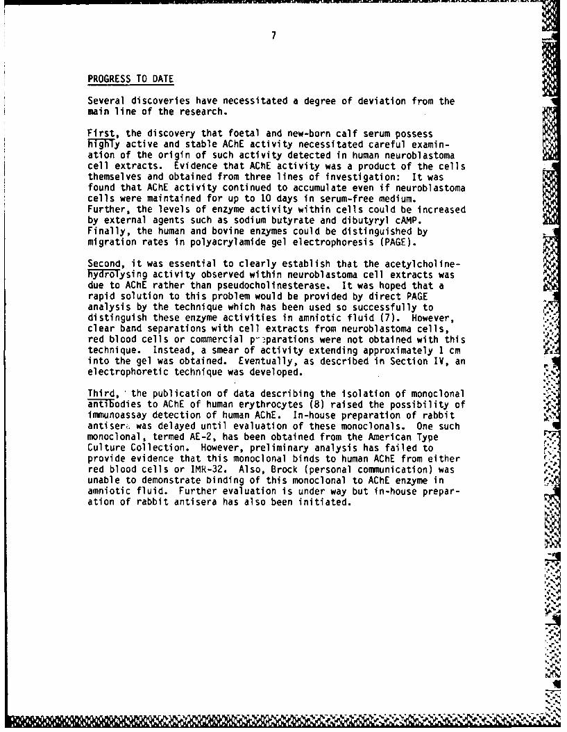
Several discoveries have necessitated a degree of deviation from themain line of the research.
First, the discovery that foetal and new-born calf serum possesshighy active and stable AChE activity necessitated careful examin-ation of the origin of such activity detected in human neuroblastomacell extracts. Evidence that AChE activity was a product of the cellsthemselves and obtained from three lines of investigation: It wasfound that AChE activity continued to accumulate even if neuroblastomacells were maintained for up to 10 days in serum-free medium.Further, the levels of enzyme activity within cells could be increasedby external agents such as sodium butyrate and dibutyryl cAMP.Finally, the human and bovine enzymes could be distinguished bymigration rates in polyacrylamide gel electrophoresis (PAGE).
Second, it was essential to clearly establish that the acetylcholine-hFydroysing activity observed within neuroblastoma cell extracts wasdue to AChE rather than pseudocholinesterase. It was hoped that arapid solution to this problem would be provided by direct PAGEanalysis by the technique which has been used so successfully todistinguish these enzyme activities in amniotic fluid (7). However,clear band separations with cell extracts from neuroblastoma cells,red blood cells or commercial p-parations were not obtained with thistechnique. Instead, a smear of activity extending approximately 1 cminto the gel was obtained. Eventually, as described in Section IV, anelectrophoretic technique was developed.
Third, the publication of data describing the isolation of monoclonalantibodies to AChE of human erythrocytes (8) raised the possibility ofimmunoassay detection of human ACHE. In-house preparation of rabbitantiserr was delayed until evaluation of these monoclonals. One suchmonoclonal, termed AE-2, has been obtained from the American TypeCulture Collection. However, preliminary analysis has failed toprovide evidence that this monoclonal binds to human AChE from eitherred blood cells or IMR-32. Also, Brock (personal communication) wasunable to demonstrate binding of this monoclonal to AChE enzyme inamniotic fluid. Further evaluation is under way but in-house prepar-ation of rabbit antisera has also been initiated.

8
METHODOLOGY
Materials
Human neuroblastoma cell line IMR-32, PG3, Lot 27961, was obtainedfrom Flow Laboratories, Irvine, Scotland, on 8 June 1982. Neuro-blastoma cell line CHP-126 was received from Professor M. Glick,University of Pennsylvania, on 20 May 1982. RPMI-1640, foetal calfserum and nonessential amino acids were purchased from Gibco EuropeLimited, Paisley, Scotland. Anti-erythrocyte membrane antibody andanti-human cholinesterase were obtained from Dakopathy, Denmark.Human serum cholinesterase and Proteinase K were products ofBoehringer. Lysivane was obtained from Professor D. Brock, Universityof Edinburgh. All other enzymes and associated reagents werepurchased from Sigma, Poole, England, and general laboratory chemicalswere either from BDH or Sigma. Phenol for RNA extractions was AnalaRgrade from BDH and was redistilled before use.
Growth and Maintenance of Neuroblastoma Cells
a) Cell Culture
Neuroblastoma cells were routinely grown as a monolayer at 37°C inan atmosphere of 5% CO (100% humidity). The cells were culturedin plastic Falcon flasis or 1 litre glass Roux bottles containingRPMI-1640 medium supplemented with foetal calf serum (10% v/v),non-essential amino acids (1% v/v), glutamine (297 ug.mll),pyruvate (200 pg.mll), penicillin (6.3 ug.ml) and streptomycin(10 vg.mll). In some experiments "serum-free" medium was usedwhich lacked foetal calf serum and contained insulin (20 ug.mll),transferrin (20 vg.mll), ethanolaine (20 PM), sodium selenite $
(2.5 nM) and mercaptoethanol (10- M). Medium was changed every2-3 days until the cells formed a confluent monolayer, spentmedium was decanted and a small volume of fresh medium added.Cells were deteched from the surface of the culture flask bygentle shaking with a few glass beads, diluted with fresh mediumand seeded into new flasks or tissue culture multi-well plates ata split ratio of 1:2.
b) Freezing Cells
Cells were cultured to confluence and suspended in a volume ofmedium equal to that in which the cells were grown. They werecollected by centrifugation at room temperature at 1,000 r.p.m.for 5 min and resuspended in fresh medium (with serum) at aconcentration of at least 106 cells.ml (1 ml of medium per 25 cm2
flask). The cell suspension was added to a plastic ampoule forfreezing and sterile dimethylsulphoxide added to a finalconcentration of 10% (v/v). Ampoules were cooled slowly byplacing them within a polystyrene box at -70C overnight.Ampoules were finally immersed in liquid nitrogen.
S'.'1, !l~ l ili. 1 , .r , " , .'N . .,.... :, ',/.,:'X'.', -,'- 5',,',,,",'-.,", 2 '., '.Z,2,5,' , . ' ,, .. ,"2.' 2. "2 , 2," gr . , ;7

9
c) Thawing Cells
Ampoules were removed from liquid nitrogen and the contents thawedquickly by placing in a water bath at 37°C. Cells were then -
centrifuged for 5 min at 1,000 r.p.m., and medium containingdimethylsulphoxide removed with a sterile pasteur pipette; cellswere resuspended in fresh medium in a flask of the same size asthat in which the cells were originally grown.
Induction of AChE Activity in Neuroblastoma Cells (
A range of chemicals known to cause induction of enzyme activitiesand/or differentiation of cell cultures was compared with IMR-32 andCHP-126. Cells were seeded at 1:2, split from confluent flasks into24-well plates and grown for 4-7 days in standard medium with 10%foetal calf serum and supplemented, where appropriate, with varyingconcentrations of the potential inducers dlmethylsulphoxide (DMSO),5-bromodeoxyuridine (5-BrdU), papaverine, sodium butyrate, dibutyrylcAMP (dB-CMP), nerve growth factor, prostaglandin El (PgE1) or 12-0-tetradecanoyl -phorbol-13-acetate (TPA).
Preparation of Cell Extracts
A number of variations of a standard method have been described fordisrupting mammalian cells and releasing AChE activity (e.g., 9, 10).The method involves non-ionic detergent treatment (Triton X-100, .2.Lubrol WX) together with mechanical disruption (sonication, homogeni-sation, osmotic shock) and removal of cell debris by centrifugation.The effects of sonication, homogenisation and osmotic shock on therelease, of AChE activity fro6 IMR-32 cells were compared. Confluentmonolayers from three 150 cm Falcon flasks were suspended in 2.25 mlphosphate buffer (0.1 M, pH 7.4) containing 1% (v/v) Triton X-100, andthe suspension was divided into three aliquots and (a) left at 4°C for1 h; (b) placed in a Kerry sonic water bath for 1 min, then held at4°C for 1 h; and (c) homogenised with 6 strokes of a ground-glasspestle and held at 4°C for 1 h.
All three samples were centrifuged at 100,000 g for 1 h in a BeckmanModel L5-50 ultracentrifuge at 50C. Liquid assays demonstrated littledifference in AChE activity among the preparations. Thus, for routinepreparation, gentle methods of cell disruption such as (a) were used.
Enzyme Assays
a) Acety1chollnesterase (AChE) Assay
A spectrophotometric assay to detect the liberation of thiocholineusing Ellman's reagent with increased absorbance at 412 nm (11). TIAchE activity was distinguished from pseudocholinesterase (ChE)activity with specific inhibitors, AChE being inhibited by 8 PM1,5-bi s-4-al lyl dimethyl ammoni um-phenyl pentone-1,3-di bromide
w
$1 .N.
10
(Figure 1) (BW 284C51), in agreement with data of Austin and Berry(12). Lysivane (ethopropazine hydrochloride) at 11.4 Pm speci-fically inhibited ChE activity (Figure 1) (13).
b) ChE Assay
Cholinesterase activity was monitored by the decrease in absor-bance at 240 nm with benzoyl choline as substrate (14). The assaywas performed in quartz glass semi-micro cuvettes with a reactionmix containing 120 mMv sodium phosphate, pH 7.2/0.16% (v/v) TritonX-100/0.25 mt benzoyl choline. AChE was unreactive in this assay(personal observation).
c) Assay of AChE in Microtitre Plates
The general assay described in a) above was modified to permitrapid assay of samples in 96-well microtitre plates. The finalconcentration of all reagents in the reaction mix was the sameexcept that the substrate concentration was reduced to 0.6 M andtotal assay volume was 0.25 ml. All reagents except substratewere placed in the wells and reaction was started by the additionof substrate and mixing. If bubbles formed during this stage,they were removed by gentle blowing on the plate or in severecases by placing the plate under vacuum for 2-3 min. The absor-bance of each well was measured as soon as possible at 405 nm in aTitertek multiscan and again after 10 min incubation of plates atroom temperature. The multiscan recorded the absorbance withinthe 96 wells in 10-20 sec. Normally lysivane was included at28 M to inhibit ChE activity.
d) Protein Estimation
Protein assays were carried out according to Lowry et al. (15)using bovine serum albumin as standard. When non-ionic detergentwas present in cell extracts, an equivalent amount was added tostandards and reagent blank. Prior to measuring absorbance,samples were centrifuged at 500 g for 10 min to remove theprecipitate.
Polyacrylamide Gel Electrophoresis
The method used for non-denaturing gels was the simplified proceduredescribed by Clark (16). A gel system described by Gratzl et al. (17)was also used. Denaturing gel electrophoresis was varried out asdetailed by Laemmli (18).
Activity Staining of Gels
After electrophoresis, AChE was detected in the gels using specificactivity stains. Three different methods were used:
11
a) The method routinely used was that of Koelle (19) as described byChubb and Smith (20). Gels were soaked for 1 h at room temper-ature in a pre-incubation mixture of 1.69 M sodium sulehate/ 70 mMsodium maleate, pH 6.5. Sometimes gentle warming (<37 C) wasrequired to keep the sodium sulphate in solution. The incubationmixture (100 ml/gel) consisted of 1.69 M Na2SO4/70 nmM sodiummaleate, pH 6.5/4 mM CuS04 /20 mM glycine/33 nml MgC12 /0.4 mM Tri-Cl, pH 6.5/2 mM acetylthiocholine iodide, and was freshly preparedeach time. The gels were incubated at 37°C between 3 and 16 hbefore washing twice in distilled water and soaking in saturated,aqueous dithiooximide for 30 min.
To specifically stain AChE 14.2 gM lysivane was included in theincubation mix. When only cholinesterase activity was required,10 PM BW284C41 was used.
b) A modification of the method of Gratzl et al. (17) was also tried.After electrophoresis the gel was washed twice for 30 min each in100 mM sodium phosphate, pH 7.4/0.1% Triton X-100 and then placedin fresh buffer containing, in addition, 0.5 mM DTNB and 0.5 nlacetylthiocholine iodide. The gel was incubated at 37°C untilyellow bands appeared, corresponding to the AChE activity.
c) The method of Galbraith and Watts (21) was used. The gel wassoaked in a solution of 6.4 nl N-methylindoxyl acetate/0.2 Msodium phosphate, pH 7.4, containing 50 mg.ml i Fast Blue RR at30°C for 1 h. Some difficulty was experienced in keeping thereagents in solution.
Protein Staining of Gels
Gels were soaked in 0.1% Coomassie Blue R250 in 25% (v/v) methanol/10%(v/v) acetic acid for 1 h and destained in several changes of 25%(v/v) methanol/5% (v/v) acetic acid until the background was clear.
Gels to be dried were soaked for 1-2 h in 2% DMSO and dried on a Bio-Rad Dual Temperature Slab gel drier, Model SE 1125B.
Crossed Immunoelectrophoresi s
The method of Galbraith and Watts (21) was used. Glass slides (5 x7 cm) were painted with 1% agarose in a buffer containing 9 mM sodium Ibarbital, 1.4 mM barbital, 94 mM glycine, 465 mM Tris base and 3.5 mMsodium azide, pH 8.8. Triton X-100 was added to give a final concen-tration of 1% (v/v). Gels 1.5 mm thick were poured on the preparedglass slide. Samples (one per gel) were loaded into 2 mm diameterholes cut in the gels. Samples were electrophoresed for 90 min at120 V and 18 mA; then the agarose gel was cut away, leaving only a1.5 cm strip along which the protein had been separated. This wasreplaced with agarose gel of the same composition, containing 30 P1 ofanti-serum (Dako A/S). Gels were then electrophoresed at right anglesfor 16 h at 50 V and 7 mA. Gels were washed in 0.1 M NaCl at least 6times, then stained for AChE activity or for general protein. ..
If W e
12 0
RESULTS
Development of Specific Assays for AChE
a) Liquid Assay
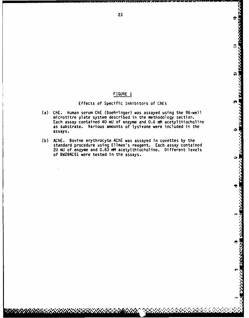
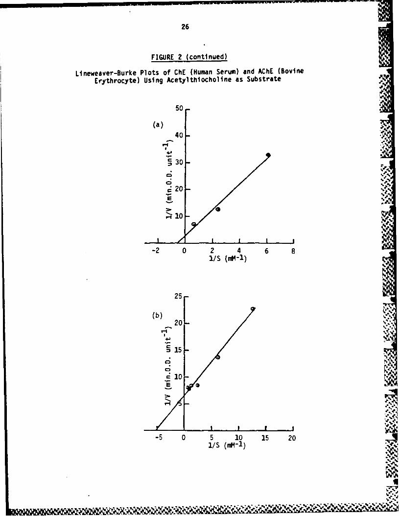
A comparison of the kinetics of hydrolysis of acetylthiocholine byAChE (as represented by bovine erythrocyte AChE) and ChE (asrepresented by human serum ChE) is described in Figure 2. Bovineerythrocyte AChE has a Km of 0.20 mM compared with 0.66 m forhuman ChE. The inhibitions of AChE by BW284C51 and of ChE bylysivane are described in Figure 1. On the basis of these data,8 PM BW284C51 and 11.4 vM lysivane were chosen as suitable inhibi-tory concentrations for routine studies.
That these two inhibitors do not cross-react and can be usedsuccessively in direct kinetic assays to distinguish the presenceof a mixture of AChE and ChE activities is demonstrated byFigure 3. It can be seen that human serum ChE is unaffected byaddition of 40 PM BW284C51, a concentration which completelyabolished AChE activity (Figure 2). Conversely, 70 vM lysivane,which abolished ChE activity (Figure 2), has no effect upon AChEactivity (Figure 3).
b) Polyacrylamide Gel Electrophoresis (PAGE)
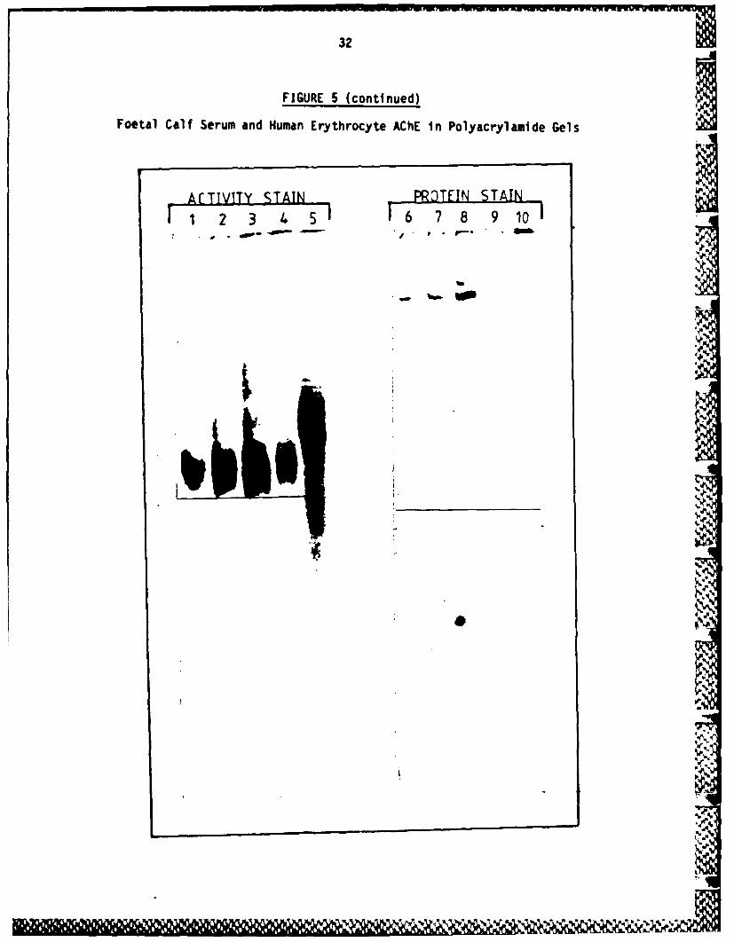
The above inhibitors have been used successfully in situ todistinguish AChE and ChE activities in polyacrylamide gels ofamniotic fluid (22). Confirmation of this finding is illustratedby Figure 4. While both inhibitors do give specific inhibition, aclear band separation of AChE and ChE activities is not obtainedwith commercial preparations of AChE and ChE. Rather, a smear ofactivities is obtained extending approximately 1 cm into the gel,much of the enzyme failing to penetrate the gel at all. This isconsistent with reports in the literature of multimeric forms ofAChE activity bound to various membranous components. Collagenasetreatment improved resolution to some extent (results notpresented). Modifications of the gel systems were examined and amodification based on the gel system of Clarke (16) gave improvedresolution. Each sample was prepared with 1% (v/v) Triton X-100prior to electrophoresis and 0.1% (v/v) Triton X-100 was includedin all gel buffers. One example is described in Figure 5 whichdemonstrates good resolution of AChE activities. This figure alsodemonstrates different migration of bovine and human ACHE.
Detection of AChE Activity in Neuroblastoma Cells
Preliminary studies were carried out in the absence of specificinhibitors and thereby measured both AChE and ChE activities. Table 1describes an example of the levels of enzyme activity in IMR-32 andCHP-126 after growth in complete and serumless media. CHP-126contains 3-4-fold higher levels of activity that IMR-32, confirming
a&&& 5&
13
the data of Glick et al. (23). These data also demonstrate that boththese cell lines possess both AChE and ChE enzymic species. Growthfor up to 7 days in serumless medium had no effect on AChE levels in
0 CHP-126, though IMR-32 cells seem less well suited to maintenance inthe serumless medium. It should be noted that the serumless mediumwas supplemented with appropriate growth factors as described in theMethods section.
PAGE of the extracts in gels containing 0.1% (w/v) polyoxyethyleneether W-1 helped confirm these results (data not presented). Activitystaining of the cell extracts resulted in a smear at the top of thegel. The level of activity in CHP-126 was greater than that inIMR-32, although equal protein was not added in the 2 wells.
Treatment with 1 PM BW284C51 inhibited the bovine erythrocyte AChEalmost completely and reduced the activity in human red blood cells.The farthest migrating activity in IMR-32 extracts was also removedand overall activity was reduced. CHP-126 cell extract activity wasnot substantially altered by treatment with BW284C51 because insuffi-cient inhibitor was used. Lysivane did reduce much of the leastmobile material and led to the appearance of a band of activity atapproximately the same position as that in the bovine erythrocyte AChEand human red blood cell enzyme.
Samples digested with DNase or glucuronidase before electrophoresisdid not alter behaviour upon gels but treatment with collagenaseincreased the mobility of material which tended to bind to the origin(results not presented).
Acetylc-holinesterase Activity in Animal Sera
During development of the assay systems, it was found that foetal calfserum, used to sustain the growth of neuroblastoma cells, possessedAChE activity. A survey of various commercial sera samples revealedthe presence of the following activities:
Foetal calf serum 100% AChE activityNew born calf serum 100% AChE activityAdult horse serum 50% AChE:50% ChE activityAdult human serum 10% AChE activity:90% ChE activityHuman red blood 100% AChE activity
cell extracts 100% AChE activity
Data from which these results were established are provided inFigure 3 along with data indicating that CHP-126 cell extracts possessboth AChE and ChE activity.
The level of enzyme activity detected in animal sera would besufficient to account for AChE activity detected in neuroblastoma cellextracts if the enzyme was absorbed from the growth media into thecells or attached to the outside of membranes. It has already been
.',
14 IfD
reported that AChE activity in A. aegypti cells correlates with theamount of foetal calf serum present in growth media (24).
An attempt was made to distinguish human and bovine AChE activityimmunologically. Extracts of CHP-126 cells, human erythrocytemembrane preparation and calf sera were mixed with varying amounts ofhuman erythrocyte membrane anti-sera, incubated for 10 min at 379C andanalysed on PAGE. The anti-sera retarded the migration of AChEactivity with all extracts, though the effect was clearly more markedwith the erythrocyte membrane preparation (results not presented). Asimilar result was obtained using crossed immunoelectrophoresis,though a precipitin band was obtained only with the erythrocytemembrane enzyme preparation. Presumably calf sera and neuroblastomaAChE preparations have enough common determinants to cross-react withthe anti-sera, but insufficient to form a precipitin band. Thus, theorigin of AChE activity in neuroblastoma cell extracts could not bedetermined immunologically.
Induction of Acetylcholinesterase Activity in Neuroblastoma Cells
Using mouse neuroblastoma cells it has been shown that cultureconditions which induce neurite outgrowth usually also induce AChEactivity (25), neurite outgrowth is induced by factors enhancinginteraction between the cell surface and the culture dish (passagefrom suspension to monolayer culture, serumless medium, 5-BrdU).Elevation of intracellular cyclic AMP in neuroblastoma cells alsoinduces many characteristic differentiated functions such as AChEactivity.
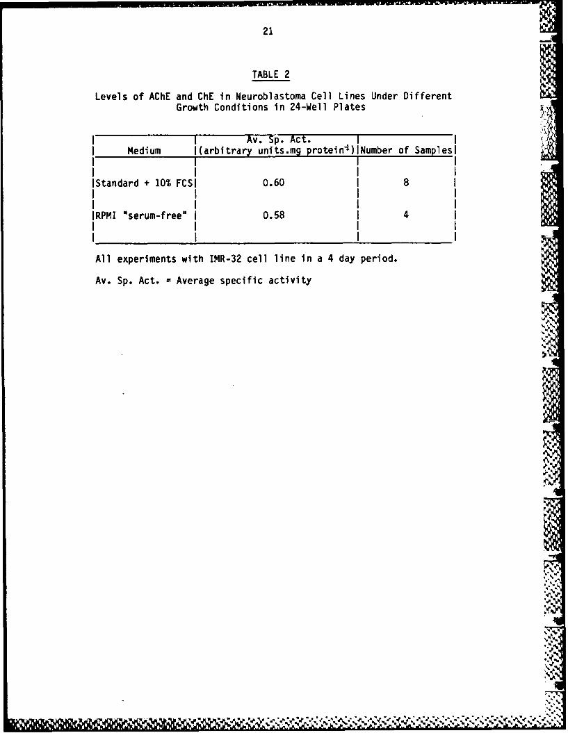
Some agents known to cause induction of enzyme activities and/ordifferentiation in cell lines was examined for their ability to induceAChE activity in IMR-32 and CHP-126 cells. Although previous studies(23) indicated that CHP-126 had higher basal levels of enzyme thanIMR-32, this does not suggest that induction should be less effectivein the latter cell line. Furthermore, IMR-32 cells grow better thanCHP-26 and hence study of induction was carried out with both celllines. In the first experiment, DMSO and 5-BrdU, papaverine andsoidum butyrate were compared, as well as the effect of growth ormaintenance of cells in serum-free or serumless medium. As can be %seen from Table 2, growth in serum-free or serumless medium did noteffect enzyme levels.
Visual examination of the cells revealed that only two treatmentscaused significant alteration in cellular morphology. Growth in thepresence of 25 or 50 pg.ml i papaverine markedly restricted growth andcaused large numbers of cells to round up and slough off the dish.Serum-free conditions led to extensive differentiation. Cells grew intight clumps with many long neurites and formation of an extensivenetwork.
;, -.
rEv-
15
Levels of total esterase activity (AChE and ChE) were measured usingthe 96 well plate assay technique described in Methods (Figure 7).DMSO at concentrations up to 2% (v/v) depressed the specific activityof the enzymes, while 5-BrdU up to 25 PM had very little effect onenzyme activities. Although papaverine treatment caused an apparentincrease in specific activity, the loss of material from thesecultures resulted in low levels of both enzyme activity and protein.These data must therefore be treated with caution. Exposure to sodiumbutyrate produced a steady increase in specific activity of AChE asthe concentration of inducer increased. Similar data generated aftertreatment of cells with nerve growth factor or TPA are presented inFigure 7. Visual inspection of cells did not reveal any morphologicalalterations to the cells, nor was there any significant increase inlevels of esterase activity.
Increasing the length of exposure to inducer to 10 days and assayingcell extracts in the presence of lysivane showed significant elevationof levels of AChE after treatment of cells with sodium butyrate and5-BrdU (Figure 9). Cells treated with these two agents assumed a morecobbled appearance and were more densely packed than those in controlcultures. At concentrations higher than 500 uM, sodium butyratecaused cell death (data not presented).
A summary of these and additional data is presented in Table 3, fromwhich it can be seen that similar data can be obtained with CHP-126cells.
The optimum concentration of sodium butyrate was chosen and timecourse of the effect upon AChE activity performed (Figure 9). Theincrease in AChE activity is maximal 4-6 days after induction.
-4
.I(
q
16
DISCUSSION
On the basis of available data, a number of conclusions can be drawn:
1. Both IMR-22 and CHP-126 synthesise both AChE and ChE enzymes.Evidence that AChE activity detected in cell extracts is not dueto contamination with enzyme absorbed from serum stems from twosources:
a) AChE activity accumulates in cells maintained for up to 10days in serum-free medium.
b) AChE activity can be induced by external agents such assodium butyrate and dB-cAMP.
Immunological evidence is still being sought.
2. CHP-126 has up to 10-fold (non-induced) and 28-fold (induced)higher levels of AChE activity than non-induced IMR-32 (Table 3).CHP-126 would seem, therefore, to be the cell line of choice forgene cloning studies.
3. The measurement of enzyme levels by catalytic activity is verysensitive. As shown in Figure 5, we have consistently failed todetect any protein-staining material at positions in PAGE thatstain for AChE activity.
Although AChE activity can be readily detected, this does not meanthat AChE mRNA will be easily found and isolated.
% %~
".
e-_-
q''a
17
FUTURE WORK
It is considered that the overall strategy outlined in the originalproposal is viable and will be pursued. It is suggested, however,that a number of minor modifications and additions be introduced, asdetailed below.
Rationale for Proposed Modifications
a) Change of Vector Systems
In the proposal it was suggested that initial cloning use pAT153and then a high expression vector based upon mp-7. However, newexpression vectors are being developed at such a rate that we andour advisors, Professor Burke and Professor Sherratt, proposedthat initial selection use puC8/9, developed by Messing (26).These vectors contain all the cloning sites from M13 and mp-8 andmp-9 with a portion of the lac 7 gene which complements in theJM103 host cell. Cloning into the vector inactivates the lac generendering the host cell lac-. These vectors are derived f'rompBr322, and Professor SheF-ratt is developing equivalent vectorsfrom his pAT153 which may be used subsequently. Furthermore, itis suggested that selection of a high expression system be delayeduntil required. New expression systems are being described at anincreasing rate and appropriate consideration should be given tobacterial, yeast and eukaryotic systems. No difficulty isenvisaged in selecting a suitable high expression system at theappropriate time.
b) Screening of Clones
In the proposal, primary screening of bacterial clones would qe byexpression of enzyme activity or immunological techniques or H-DFP binding, as well as competitive hybridisation of clones tomRNA prepared from induced and non-induced neuroblastoma cells.Recent studies by Dr. Doctor of the Walter Reed Institute forResearch, Washington, have revealed a part of the amino acidsequence of an active-site-containing peptide from AChE ofElectrophorus. This sequence is Trp-Asp-Pro-Asp-Arg-Glu-Met.Snce It Is possible that the region around the active site ofAChE has been preserved during evolution, it is possible thathuman enzyme will be similar to this sequence. We propose,therefore, in addition to primary screening as defined in theoriginal proposal, to prepare synthetic oligonucleotides relevantto this sequence as probes of primary clones. For example, onesuitable consensus oligonucleotide sequence in E. cola would bethe 12-mer TGGGACCCGGAT.
c) Gene Amplification Study
The primary aim of this project is to develop a system allowingthe preparation of large quantities of AChE. While it is likely
.,
Qf-

18 0
that this will be achieved by the above gene cloning technology,it would be expedient to consider alternative technologies. Ourexperience in handling the enzyme to date has emphasised itscomplex structure in vivo: association with collagen, isolationin membrane complexes, tendency to aggregate and hydrophobicnature. Even if the cloning project is successful, it would bevaluable to be able to compare the cloned product with unatural"material.
We propose, therefore, in parallel with present studies, toattempt to increase significantly the amount of enzyme produced byneuroblastoma cells. This will be achieved by selecting variantswhich have greatly amplified the AChE gene. Gene amplification isa well-established phenomenon whereby mammalian cells respond tostress by making multiple copies of appropriate genes to relievethe stress. Thus, methotrexate-resistant cells were selectedwhich had amplified the gene for dihydrofolate reductase manyhundreds of times. Similarly, Wilson (personal communication) hassucceeded in isolating variants of Chinese hamster ovary cellswhich have amplified the gene for glutamine synthetase some 1500-fold. In these cells, glutamine synthetase accounts for about 20%of the total cell protein. It may be possible by the techniquedescribed below to select variants of human neuroblastoma cellswhich have greatly amplified the number of gene copies of AChE sothat this enzyme will represent a considerable fraction of totalcellular protein.
r
.i
4.
4.

19
METHODOLOGY
Screening of Clones by Hybridisation to Synthetic Oligonucleotides
Oligonucleotides (12-15 mer) will be synthesised by the phospho-triester method (27). Such synthesis is likely to be carried outmanually, although Inveresk Research International Limited isconsidering the purchase of an automatic gene synthesiser. It islikely that 4-6 oligonucleotides containing appropriate code degener-iiies will be synthesised. These will be labelled at the 5'-end with'P using polynucleotide kinase (28) and used as hybridisation probes
by in situ hybridisation to bacterial colonies transferred tonitrocellulose filters (29). Positive colonies will be selected andcompared with those selected by other screening techniques.
Gene Amplification
Selection of cell variants with increased levels of AChE will be basedupon the essential requirement for choline in growth media of cellcultures. Choline is routinely added to cell culture growth media asan essential nutrient, and preliminary studies will establish thathuman neuroblastoma cells have such a requirement. The aim of thetechnique is to gradually subject cells to an increasingly depletedcholine environment so that variants are gradually selected from thepopulation. Choline-depleted growth media will be supplemented withacetylcholine, thereby requiring the presence of AChE or ChE forgrowth. AChE and ChE activities will be removed from animal sera,probably by dialysis against difluorophosphate, and lysivane will beadded to inhibit ChE produced by cell lines. The growth media contentof acetylcholine will be adjusted so that cell growth rate approxi-mates 70% of that in fully supplemented medium and growth andsubculture will be continued in this medium until a rapid growth rateis restored. Subsequently, the amount of acetylcholine supplementwill again be reduced and the process repeated through several cycles.It is envisaged that this selection procedure will take about 6months.
AChE is a highly active enzyme, and it may not be possible to add asufficiently low concentration of acetylcholine to restrict cellgrowth. If this is the case, restrictive growth conditions will beestablished by supplementation of growth medium with acetylcholine andan appropriate competitive inhibitor of choline kinase, the enzymeessential for choline utilisation.
Success by this procedure cannot be guaranteed or predicted. However,in view of the relatively small additional commitment in staff time,it is worthy of pursuit. N
This work will draw upon the advice and assistance of Dr. R.H. Wilson,Department of Genetics, University of Glasgow, who has successfullyamplified the gene for glutamine synthetase.
LO

20
TABLE 1
Levels of AChE and ChE in Neuroblastoma Cell Lines IMR-32 and CHP-126
ICell Linel Growth Conditions Igo innibitoir+ Lysivanel I~BWZB4CM
I CHP-126 IRPMI-1640 + 10% FCSI 14 I NM I NM II I I I I II CHP-126 ISerumless; 3 days 1 21 I NM I 9 I
1 CHP-126 ISerumless; 7 days 1 16 I 9 1 9 1
1 IMR-32 IRPMI-1640 + 10% FCSI 5 1 NM 1 2 1I I I I I I9I IMR-32 ISerumless; 3 days 1 5 1 NM I NM II I I I I II IMR-32 ISerumless; 7 days 1 0* 1 1 1I _ _ _ I. _ _ I. I_ _ _ I __
*=This preparation had low protein content. Possibly theactivity was below the level of detection.
NM =Not measuredSp. Act. =Specific activity
21
TABLE 2
Levels of AChE and ChE in Neuroblastoma Cell Lines Under DifferentGrowth Conditions in 24-Well Plates
I I Av. Sp. Act.1I Medium I(arbitrary units.mg protein-I)INumber of Samplesi
IStandard + 10% FCSI 0.60 I8 1
IRPMI "serum-free" I0.58 I4I
All experiments with IMR-32 cell line in a 4 day period.
Av. Sp. Act. =Average specific activity
% ,'
1111111 I'

22
TABLE 3
The Effects of Chemicals on AChE Levels in Neuroblastoma Cells
I I Av. Sp. Act. IICell Linel Treatment l(No. of Samples)1% of ControlI I I I II CHP-126 IControl 11.48 (8) 1 100 11 CHP-126 1+ 200 pg.mll dB-cAMP 26.25 (2) 2291 CHP-126 1+ 400 pg.ml dB-cAMP 28.46 (2) 248I CHP-126 1+ 800 pg.ml dB-cAMP 25.28 (2) 223I CHP-126 1+ 0.5 mg.ml i PGEs 12.00 (2) 104I CHP-126 1+ 1 vg.mll PGE1 I 7.75 (2) 68I CHP-126 1+ 2 vg.mll PGE1 8.85 (2) 77I CHP-126 1+ 4 vg.ml PGE1 7.94 (2) 69 II CHP-126 1+ 8 4g.mll PGE1 10.52 92I IMR-32 IControl 1.01 (5) 100 II IMR-32 1+ 200 vg.mli dB-cAMP 2.36 (1) 234 .I IMR-32 1+ 400 Pg.mli dB-cAMP 3.17 (2) 314I IMR-32 1+ 800 Pg.ml" dB-cAMP 3.95 (2) 3911 IMR-32 1+ 0.5 vg.ml' dB-cAMP 3.95 (2) 391 II IMR-32 1+ 0.5 vg.ml"1 PGE1 1.22 (1) 121I IMR-32 1+ 1 vg.ml4 PGE1 I 1.30 (1) 1291 IMR-32 1+ 2 vg.mli PGE1 1.17 (1) 116I IMR-32 1+ 4 vg.mll PGE1 1.51 (2) 1501 IMR-32 1+ 8 wg.ml"1 PGE1 1.45 (2) 1441 CHP-126 IControl 16.60 (4) 1 100 1I CHP-126 1+ 0.313 mM Na butyratel 38.19 (4) 1 230 11 CKP-126 1+ 0.625 mM Na butyratel 22.69 (4) 1 137 11 CHP-126 1+ 1.25 ,t4 Na butyrate 11.12 (4) 671 CHP-126 1+ 2.5 mM Na butyrate 7.54 (4) 451 CHP-126 1+ 5.0 nM Na butyrate 5.30 (4) 321 IMR-32 IControl 1.99 100 1l IMR-32 1+ 0.313 *1t Na butyratel 4.04 204 I1 IMR-32 1+0.625 mM Na butyrate 1 4.45 224I IMR-32 1+ 1.25 m01 Na butyrate 2.29 115IMR-32 1+ 2.5 nM Na butyrate 2.37 120
I IMR-32 1+ 5 mM Na butyrate 2.81 142
Av. Sp. Act. --Average specific activity
j,
-. 7-
23
FIGURE 1
Effects of Specific Inhibitors of ChEs
(a) ChE. Human serum ChE (Boehringer) was assayed using the 96-wellmicrotitre plate system described in the methodology section.Each assay contained 40 mU of enzyme and 0.6 mM acetylthiocholineas substrate. Various amounts of lysivane were included in theassays.
(b) ACHE. Bovine erythrocyte AChE was assayed in cuvettes by thestandard procedure using Ellman's reagent. Each assay contained20 mU of enzyme and 0.83 mW acetylthiocholine. Different levelsof BW284C51 were tested in the assays.
' ,

24
FIGURE I (continued)
Effects of Specific Inhibitors on ChEs
100 -(a) £"--
80 - . "
80
60 -A
4r-
-40
20
020 40 60 80 100
Lysivane Concentration (pM)
10.0- O 0 0
(b) 80
4, 0
20
0 i AI
1 2 3 4 SBW284C51 Concentration (1iM)
A-A AChE
o-o Serum ChE
. *. . . . . . . . .

25
FIGURE 2
Lineweaver-Burke Plots of ChE (Human Serum) and AChE (BovineErythrocyte) Using Acetylthiocholine as Substrate
(a) ChE. The reactions were carried out as described in themethodology section using Ellman's reagent. Assays were at roomtemperature (22C) and each contained 0.25 U of enzyme (humanserum ChE; Sigma).
(b) ACHE. The reactions were carried out as above except that eachcontained 20 mU of enzyme (bovine erythrocyte AChE; Sigma).
eV
26
FIGURE 2 (continued)
Lineweaver-Burke Plots of ChE (Human Serum) and AChE (BovineErythrocyte) Using Acetylthiocholine as Substrate
50 -
(a)
40
4o
25-
10 ",
I I I I
-2 0 2 4 6 8I/S (rM-l)
25
(b)
. .!S '
20 .
S15
C; 10
-5 0 5 10 15 20 41
1/S (nMw-l)
II
-- ----- -- -

27
FIGURE 3
Liquid Assay of AChE in Cell Extracts and Commercial Serum Samples
1 ml of assay mix containing 0.1 M sodium phosphate buffer, pH 7.4,0.2 mM DTNB, 0.05% v/v Triton X-100 and 0.05-0.2 ml of enzymepreparation was placed in a semi-microcuvette (path length 1 cm) andpre-incubated in a Unicam SP800 spectrophotometer at 24 C for 1-2 min.0.1 ml of substrate (1 mM acetylthiocholine, final concentration) wasthen added and the rate of reaction recorded at 412 nm. Aftersufficient time to record reaction rate, qither BW284C51 (4 x 10⁻⁷) M,final concentration) or lysivane (7 x 10⁻¹ M, final concentration) wasadded and the reaction rate recorded.
1: Foetal calf serum2: Human serum ChE (Sigma)3: Horse serum4: Bovine red blood cell AChE (Sigma)5: Human red blood cell extract6: Foetal celf serum7:. Extract of CHP-126 grown 7 days in serum-free medium
A: BW284C51; B: Lysivane added.
N':.
1-
... .>'A _ d ,,..,, ,,, .,,"_,, ,,,%",',.X,, ,..,'',,. . ,'.''. '.''.,-- .."_' , ," "'_,,; ".,,U
28
FIGURE 3 (continued)
Liquid Assay of AChE in Cell Extracts and Comercial Serum Samples
0
4f-I
E
C;C
wu I-
0 0
Wu Z~t e auqoq .
rq
U--''-,
29
FIGURE 4
Effects of Specific Inhibitors on ChE Activity in Polyacrylamide Gels
Gel electrophoresis was carried out using a composite acrylamide/agarose gel and Tris/glycine/Triton X-100 buffer system. A 3%acrylamide/0.5% agarose gel was run on an LKB 2117 Miltiphorapparatus. The gel buffer was 50 mM Tris/0.38 M glycine/0.2% TritonX-100 and electrophoresis buffer was a 1:1 dilution of this. Electro-phosesis was overnight at 5 mA plus 1 h at 30 mA. Cooling water wasused throughout.
Activity staining was as described in the methodology section, usingthe inhibitor concentrations detailed therein.
Tracks 1, 5, 9 4 0l of foetal calf serumTracks 2, 6, 10 4 I of human red blood cell extractTracks 3, 7, 11 40 mU of serum ChETracks 4, 8, 12 4 i of red blood cell extract + 40 mU
human ChE
Tracks 1-4 no inhibitorTracks 5-8 plus BW284C51Tracks 9-12 plus lysivane
N.I

30
FIGURE 4 (continued)
Effects of Specific Inhibitors on ChE Activity in Polyacrylamide Gels
Z:
I'- "-
I
Lfn
I" I
CD
%LiIP

31
FIGURE 5
Foetal Calf Serum and Human Erythrocyte AChE in Polyacrylamide Gels
Gel electrophoresis and staining were carried out as described in themethodology section. The gel system was based on that of Clarke (16).Electrophoresis was carried out in a Blo-Rad Protein 32 cm slab cellwith 1.5 mm spacers. Current was 3 mA for 18 h.
Tracks 1, 6 5 I foetal calf serumTracks 2, 7 10 o1 foetal calf serumTracks 3, 8 20 0d foetal calf serumTracks 4, 9 20 mU bovine erythrocyte AChETracks 5, 10 5 1 human red blood cell extract
Tracks 1-5 were activity stained.Tracks 6-10 were stained for protein.
10
elI
32
FIGURE 5 (continued)
Foetal Calf Serum and Human Erythrocyte ACHE In Polyacrylamide Gels
A[TIVITY STAIN PROTEIN STAIN -
i 2 3 4 5! 16 7 8 9 101
.I.- low
pp
* 'p.'
33
FIGURE 6
Effect of Inducers on ChEs in IMR-32 Neuroblastoma Cells .!After 4 Days of Exposure
Each of the inducers was added to confluent IMR-32 cells in a 24-wellLinbro plate at the concentrations indicated on the graphs. After 4days the cells were harvested and assayed using the 96-well microtitreplate assay described in the methodology section. No inhibitor wasincluded, so the results represent the sum of all ChEs in theextracts. The units of AChE are arbitrary. Protein was estimated bythe Lowry procedure (15). 9,
(a) Dimethylsulphoxide (DMSO)(b) 5'-Bromodeoxyurldine (5-BrdU)(c) Papaverine(d) Sodium butyrate
U'
,%
V- -w* ~ 9,*: ~U
34
FIGURE 6 (continued)
Effect of Inducers on ChEs in IMR-32 Neuroblastoia CellsAfter 4 Days of Exposure
Total ChE Activity (U.mg protein-1)
0a0
C,
r+r071.
~10
CIC
'C
CO N)Toa Ch ciit Ung rti
I-
r%3%

35
FIGURE 7
Effect of Nerve Growth Factor and 12-O-Tetradecanoyl-phorbol-13-acetate (TPA) on Cholmnesterases in IMR-32 Cells
(a) Nerve Growth Factor (NGF) (2.25 S form)Wb 12-0-tetradecanoyl -phorbol -13-acetate (TPA)

FIGURE 7 (continued)
Effect of Nerve Growth Factor and 12-0-Tetradecanoyl-phorbol-13-
acetate (TPA) on Cholinesterases in IMR-32 Cells
(a)
4J
4-'
0
4,0.5
V.'
0J
00 200 75J 100TPA Concentration (ng.ml-)

37
':"W
FIGURE 8
AChE Levels in IMR-32 Cells After 10 Days in Culture
Confluent IMR-32 cells in Linbro 24-well plates were incubated inmedium containing various levels of inducer for 10 days with freshmedium plus inducer every 2-3 days. The cells were harvested, washedand assayed as described in the methodology section. The assay mixcontaining 28.4 jjM lysivane.
(a) Sodium butyrate(b) 5'-Bromodeoxyuridine (5-BrdU)(c) Dimethylsulphoxide (DMSO)
1*
S.0
N
-~ ~ ~~~ P.~V%~.lo*.~

38-
FIGURE 8 (continued)
AChE Levels in IMR-32 Cells After 10 Days in Culture O
(a) .
0.
0 125 250 375 500 .SoimButyrate Concentration (pM)
CL
E
U
0 6.25 12.5 18.75 255-BudR Concentration (jIM)
CL
0.5
0
00 0.5 1 1.5 2DMSO Concentration ( /v) v/vr
~% 9% .0
. '
39 AS
FIGURE 9
Time Course of the Action of Sodium Butyrate on ChE Levelsi n Neurobl astoma Cel l s
Confluent IMR-32 cells in a Linbro 24-well plate were induced by0.625 mW sodium butyrate on day 0 and maintained on this supplementedmedium for a total of 14 days with fresh medium every 2-3 days. Atregular intervals (usually daily), 2 wells from the plate wereharvested, washed and stored in the vapour phase of a liquid N2 deepfreeze. Control cells were treated identically except that nobutyrate was added to the medium. Cells were assayed in a 96-wellmicroti-tre plate and lysivane was included at 28.4 iM in all assays.
9
1w
II-i
40
FIGURE 9
in Neuroblastoma Cells
'A
00
CDJ
C~0
T-' Uc-Ii 0wn jA4V33
-- - -- -
41
REFERENCES
(1) Rosenberry, T.L. (1975) Acetylcholinesterase. Adv. Enzymol., 43103-218. 1,
(2) Silk, E., King, J. and Whittaker, M. (1979) Assays ofcholinesterase in clinical chemistry. Annal. Clin. Biochem., 16,57-75.
(3) Massoulie, J. (1980) The polymorphism of cholinesterases and itsphysiological significance. Trends Biochem. Sci., 5, 160-164.
(4) Massoulie, J. and Bon, S. (1982) The molecular forms of cholin-esterase and acetylchoninesterase in vertabrates. Annu. Rev.Neurosci., 5, 57-106.
(5) Niday, E., Wang, C.S. and Alaupovic, P. (1977) Studies on thecharacterisation of human erythrocyte acetylcholinesterase andits interaction with antibodies. Biochim. Biophys. Acta, 469,180-193.
(6) Romer-Luthi, C.R., Ott, P. and Brodbeck, U. (1980) Reconstitution "
of human erythrocyte membrane acetylcholinesterase in phospho-lipid vesicles. Analysis of the molecular forms by cross-linkingstudies. Biochim. Biophys. Acta, 601, 126-133.
(7) Ott, P. and Brodbeck, U. (1978) Multiple molecular forms ofacetylcholinesterase from human erythrocyte membranes. Eur. J.Biochem., 88, 119-125.
(8) Smith, A.D., Wald, N.J., Cuckle, H.S., Stirrat, G.M., Bobrow, M.and Lagerceantz, H. (1979) Amniotic fluid acetylcholinesterase aspossible diagnostic test for neural tube defects in earlypregnancy. Lancet, 1, 685-688.
(9) Fambrough, D.M., Engel, A.G. and Rosenberry, T.L. (1982) Acetyl-cholinesterase of human arythrocytes and neuromuscular junctions:Homologies revealed by monoclonal antibodies. Proc. Natl. Acad.
Sci. USA, 79, 1078-1082.
(10) Harkins, J., Arsenault, M., Schlesinger, K. and Kates, J. (1972)Induction of neuronal functions: Acetylcholine induced acetyl-cholinesterase activity in mouse neuroblastoma cells. Proc. Nat.Acad. Sci. USA, 69, 3161-3164.
(11) Ellman, G.L., Courtney, K.D., Andres Jr., V. and Featherstone,R.M. (1961) A new and rapid colorimetric determination of acetyl-cholinesterase activity. Biochem. Pharmacol., 7, 88-95.
(12) Austin, L. and Berry, W.K. (1953) Two selective inhibitors ofcholinesterase. Biochem. J., 54, 695-700.
42
(13) Seller, M.J. and Cole, K.J. (1980) Polyacrylamide gel electro-phoresis of amniotic fluid cholinesterases: a good prenatal testfor neural tube defects. Br. J. Obs. Gynaecol., 87, 1103-1108.
(14) Kallow, W. and Lindsay, H.A. (1955) A comparison of optical andmanometric methods for the assay of human serum cholinesterase.Can. J. Blochem., 33, 568-574.
(15) Lowry, O.H., Rosebrough, N.J., Farr, A.L. and Randall, R.J.(1951) Protein measurement with the Folin phenol reagent. J.Biol. Chem., 193, 265-275.
(16) Clarke, J.T. (1964) Simplified "disc" (polyacrylamide gel)electrophoresis. Ann. N.Y. Acad. Sci., 121, 428-436.
(17) Gratzl, M., Krieger-Brauer, H. and Ekerdt, K. (1981) Latentacetylcholinesterase in secretory vesicles isolated from adrenalmedulla. Biochim. Biophys. Acta, 649, 355-366.
(18) Laemmli, U.K. (1970) Cleavage of structural proteins during theassembly of the head of bacteriophage T4. Nature, 227, 680-685.
(19) Koelle, G.B. (1951) The elimination of enzymatic diffusionartefacts in the histochemical localisation of cholinesterasesand a survey of their cellular distributions. J. Pharmac. exp.Ther., 103, 153-171.
(20) Chubb, I.W. and Smith, A.D. (1975) Isoenzymes of soluble andmembrane-bound acetylcholinesterase in bovine splanchnic nerveand adrenal medulla. Proc. R. Soc. Lond. B., 191, 245-261.
(21) Galbraith, D.A. and Watts, D.C. (1981) Human erythrocyteacetylcholinesterase in relation to cell age. Biochem. J., 195,221-228.
(22) Voigtlander, T., Friedl, W., Cremer, H., Schmidt, W. andSchroeder, T.M. (1981) Quantitative and Qualitative assay ofamniotic-fluid acetylcholinesterase in the prenatal diagnosis ofneural tube defects. Hum. Genet., 59, 227-231.
(23) Glick, M.C., Schlesinger, H. and Hummeler, K. (1976)Glycopeptides from the surface of human neuroblastoma cells.Cancer Res., 36, 4520-4524.
(24) Cohen, E. (1981) Acetylcholinesterase activity in an Aedesaegypti cell line. Experentia, 37, 429-431.
(25) Augusti-Tocco, G. (1976) Neuroblastoma culture: an experimentalsystem for the study of cellular differentiation. TIBS, 10, 151-154.
e IKLY A

43
(26) Messing, J., Crea, R. and Seeburg, P (1981) A system for shotgunDNA sequencing. Nucleic Acid Res., 9, 309.
(27 Seth, A.K. and Jay, E. (1980) A study of the efficiency and theproblem of sulphoration of several condensing reagents and theirmechanisms for the chemical synthesis of deoxyoligoribonucle-otides. Nucleic Acid Res., 8, 5445.
(28) Maxam, A.M. and Gilbert, N. (1980) Sequencing end-labelled DNAwith base specific chemical cleavages. Methods in Enzymology,65, 499-560.
(29) Grunstein, M. and Hogness, D.S. (1975) Colony hybridisation: Amethod for the isolation of cloned DNAs that contain a specificgene. Proc. Natl. Acad. Sci. USA, 72, 3961-3965.
w
"I
44
DISTRIBUTION LIST
4 copies CommanderU.S. Army Medical Research and Development CommandATTN: SGRD-RMSFort Detrick, Frederick, Maryland 21701-5012
5 copies CommanderU.S. Army Medical Research and Development CommandATTN: SGRD-PLEFort Detrick, Frederick, Maryland 21701-5012
12 copies Defense Technical Information Center (DTIC)ATTN: DTIC-DDACCameron StationAlexandria, VA 22304-6145
1 copy DeanSchool of MedicineUniformed Services University of theHealth Sciences
4301 Jones Bridge RoadBethesda, MD 20814-4799
1 copy CommandantAcademy of Health Sciences, U.S. ArmyATTN: AHS-CDMFort Sam Houston, TX 78234-6100
'
po-www--Ow '-W Aw _w -"W- im ... A . w Aw.