yeast kinetochores do not stabilize stu2p-dependent ...labs.bio.unc.edu/bloom/pdf/stu2.pdf · yeast...
TRANSCRIPT

Molecular Biology of the CellVol. 14, 4181–4195, October 2003
Yeast Kinetochores Do Not Stabilize Stu2p-dependentSpindle Microtubule Dynamics□V
Chad G. Pearson,* Paul S. Maddox, Ted R. Zarzar, E.D. Salmon, andKerry Bloom
Department of Biology, University of North Carolina at Chapel Hill, Chapel Hill, North Carolina27599-3280
Submitted March 27, 2003; Revised June 14, 2003; Accepted June 14, 2003Monitoring Editor: Trisha Davis
The interaction of kinetochores with dynamic microtubules during mitosis is essential for proper centromere motility,congression to the metaphase plate, and subsequent anaphase chromosome segregation. Budding yeast has been criticalin the discovery of proteins necessary for this interaction. However, the molecular mechanism for microtubule–kineto-chore interactions remains poorly understood. Using live cell imaging and mutations affecting microtubule bindingproteins and kinetochore function, we identify a regulatory mechanism for spindle microtubule dynamics involvingStu2p and the core kinetochore component, Ndc10p. Depleting cells of the microtubule binding protein Stu2p reduceskinetochore microtubule dynamics. Centromeres remain under tension but lack motility. Thus, normal microtubuledynamics are not required to maintain tension at the centromere. Loss of the kinetochore (ndc10-1, ndc10-2, and ctf13-30)does not drastically affect spindle microtubule turnover, indicating that Stu2p, not the kinetochore, is the foremostgovernor of microtubule dynamics. Disruption of kinetochore function with ndc10-1 does not affect the decrease inmicrotubule turnover in stu2 mutants, suggesting that the kinetochore is not required for microtubule stabilization.Remarkably, a partial kinetochore defect (ndc10-2) suppresses the decreased spindle microtubule turnover in the absenceof Stu2p. These results indicate that Stu2p and Ndc10p differentially function in controlling kinetochore microtubuledynamics necessary for centromere movements.
INTRODUCTION
Accurate segregation of the duplicated genome during celldivision requires the mechanical separation of chromosomesto the daughter cells. The microtubule-based mitotic spindleprovides the structural and mechanical framework, whereasprotein/DNA structures (kinetochores) on chromosomes fa-cilitate attachment to microtubules. Dynamically growingand shortening spindle microtubules nucleated from spindlepoles gain attachment of their plus-ends to the kinetochore(Rieder and Salmon, 1998; Kapoor and Compton, 2002).Kinetochores maintain “end-on” attachment to a bundle ofmicrotubules (kinetochore microtubules), producing tensionbetween sister chromatids. Dynamic microtubules produceforces necessary for chromosome movement to facilitatecongression to the spindle equator, oscillations at the meta-phase plate, and chromosome segregation (Inoue andSalmon, 1995; Dogterom and Yurke, 1997; Rieder andSalmon, 1998; McIntosh et al., 2002; Scholey et al., 2003).Microtubule–kinetochore interactions serve to produce theprimary force on chromosomes that, in concert with nonki-netochore forces (polar ejection forces and resistance be-tween sister chromatids), result in dynamic chromosomebehavior essential for proper cell division (Nicklas, 1989;Mitchison and Salmon, 1992; Skibbens et al., 1993; Murray
and Mitchison, 1994; Khodjakov et al., 1999). Furthermore,dynamic instability of microtubule plus-ends and polewardflux contribute to the turnover of kinetochore microtubulesin tissue cells (Mitchison, 1989). In vitro and in vivo studieshave shown that attachment of microtubules to kinetochoresgenerates stable microtubules compared with unattachedspindle microtubules (Mitchison et al., 1986; Zhai et al., 1995;Hunt and McIntosh, 1998). This difference in microtubulegrowth and shortening upon kinetochore attachment indi-cates that, in tissue cells, microtubule dynamics is stabilizedat the level of the kinetochore. The regulation of kinetochoremicrotubule dynamics is therefore critical for chromosomesegregation and likely to involve the coordination of inher-ent dynamic properties of the microtubule polymer andmicrotubule-associated proteins.
Saccharomyces cerevisiae is a genetically tractable organismto study this complex process (Winey and O’Toole, 2001).The mitotic spindle structure in haploid cells consists of �40microtubules: eight overlapping interpolar microtubulesand 32 kinetochore microtubules (Winey et al., 1995; O’Tooleet al., 1999). This ultrastructural information combined withlive cell analysis indicates that each kinetochore makes apersistent attachment to a single microtubule (Winey et al.,1995; O’Toole et al., 1999; Goshima and Yanagida, 2000;Goshima and Yanagida, 2001; Pearson et al., 2001). Beforeanaphase, kinetochores and spindle microtubules generateforces to separate sister centromeres toward their respectivespindle poles, whereas chromosome arms maintain cohesionalong their length. Centromere separation is dependentupon spindle microtubules and a functional kinetochore(Goshima and Yanagida, 2000; He et al., 2000; Tanaka et al.,
Article published online ahead of print. Mol. Biol. Cell 10.1091/mbc.E03–03–0180. Article and publication date are available atwww.molbiolcell.org/cgi/doi/10.1091/mbc.E03–03–0180.
□V Online version of this article contains video material for somefigures. Online version is available at www.molbiolcell.org.
* Corresponding author. E-mail address: [email protected].
© 2003 by The American Society for Cell Biology 4181

2000; Belmont, 2001; Pearson et al., 2001). Separated sistercentromeres align into two clusters equidistant from thespindle equator (Pearson et al., 2001). Furthermore, centro-meres oscillate along the spindle axis at rates similar tocytoplasmic microtubule plus-end dynamics (Pearson et al.,2001). Fluorescence redistribution after photobleaching(FRAP) studies have shown that microtubules turnoverwithin the mitotic spindle. However, unlike tissue culturecells, turnover seems to be restricted to growth and short-ening of kinetochore microtubule plus-ends (Maddox et al.,2000). Whether the kinetochore or microtubule binding pro-teins, or both, orchestrate chromosome dynamics can nowbe genetically dissected to identify the key regulatory path-ways governing kinetochore microtubule dynamics andchromosome movement.
The primary candidate for regulating kinetochore micro-tubule dynamics is the conserved TOG/XMAP215/Dis1/Stu2p family of proteins. Stu2p localizes along the length ofthe mitotic spindle and is essential for anaphase spindleelongation, microtubule dynamics, and centromere position-ing in S. cerevisiae (Kosco et al., 2001; Severin et al., 2001; Heet al., 2001) and efficient chromosome segregation in S. pombe(Nabeshima et al., 1998). In vitro and in vivo, Stu2p binds toand selectively promotes dynamicity of microtubule plus-ends (Van Breugal et al., 2003). In the absence of Stu2p orXMAP215 (Xenopus laevis), microtubule plus-ends spend anincreased time in a “paused” state identified by undetectablegrowth or shortening (Tran et al., 1997; Kosco et al., 2001;Shirasu-Hiza et al., 2003; Van Breugal et al., 2003). HowStu2p functions to create dynamic microtubule plus-ends is
critical to understanding the overall mechanisms of chromo-some motility.
We used live cell microscopy assays to dissect the role ofStu2p in regulating kinetochore microtubule dynamics andits effects on centromere motility and tension between sistercentromeres. We found that Stu2p promotes dynamics atmicrotubule plus-ends, that maintaining kinetochore attach-ment and tension does not require dynamic microtubuleplus-ends, and that kinetochores can promote microtubuledynamics independent of Stu2p.
MATERIALS AND METHODS
PlasmidsAll plasmids are defined in Table 1. pCP21 (CFP-NAT) was generated byreplacing the BglII/SacI fragment containing KAN of pDH3 with the BglII/SacI fragment containing natricin (NAT) from pAG31 (Dohmen et al., 1994).The KAN:pCUP:Arg:DHFRts:3HA (pBJ1212), CFP-KAN (pDH3), GFP-TUB1-URA3 (pAFS125), CSE4-GFP-TRP1 (pKK1), and TUB1-TUB1-GFP-TRP1(pTub1-GFP-Tub1) plasmids were generous gifts from Dr. Richard Heil-Chapdelaine (Washington University, St. Louis, MO), Dr. Trisha Davis (YeastResource Center, University of Washington, Seattle, WA), Dr. Aaron Straight(Harvard University, Boston, MA) (Straight et al., 1997), Dr. Richard Baker(University of Massachusetts Medical School, Worcester, MA) (Chen et al.,2000), and Drs. Karena Kosco and Tim Huffaker (Cornell University, Ithaca,NY) (Kosco et al., 2001), respectively.
Polymerase Chain Reaction (PCR) Fragments forIntegrationFluorescently labeled spindle poles were constructed using primers to pFA6-GFP-KANMX6 plasmid flanked by 50 bp of homology one codon upstream ofthe stop codon and one codon downstream of the stop codon of the spindle
Table 1. S. cerevisiae strains and plasmids
Strain name Relevant genotype Source/Reference
YEF473A MATa trp1-63 leu2-1 ura3-52 his3-200 lys2-801 PringleGT1 MATa trp1-63 leu2-1 ura3-52 his3-200 lys2-801 ura3-52::GFP-TUB1-URA3 Maddox et al (2000)KBY2012 MATa trp1-63 leu2-1 ura3-52 his3-200 lys2-801 cse4::HYG SPC29-CFP-KAN pKK1 This studyY170 MATa Ace1-UBR1 Ace1-ROX1 trp1� ade2-101 ura3-52 lys2-801
stu2�::URA3::Panb1UB-R-STU2 pTUB1-GFP-TUB1Kosco/Huffaker
KBY2660.291 MATa Ace1-UBR1 Ace1-ROX1 trp1� ade2-101 ura3-52 lys2-801stu2�::URA3::Panb1UB-R-STU2 cse4::HYG SPC29-CFP-KAN pKK1
This study
KBY2665 MATa trp1-63 leu2-1 ura3-52 his3-200 lys2-801 ura3-52::GFP-TUB1-URA3 KAN:pCUP:Arg:DHFRts:3HA:STU2
This study
KBY2155 MATa ndc10-2 his3-delta200 leu2-3, 112 ura3-52 Gal� ura3-52::GFP-TUBI-URA3pAFS125
This study
KBY2161 MATa ndc10-1 ura3 leu2 lys2 his3 trp1 ade2 pAFS125 This studyKBY2192 MATa ctf13-30 ura3 lys2 ade2 his3 leu2 pAFS125 This studyKBY2154 MATa ndc10-2 his3-delta200 leu2-3,112 ura3-52 Gal� trp::HYG cse4::KAN SPC29-CFP-
NAT pKK1This study
KBY2165 MATa ndc10-1 his3 leu2 ura3 lys2 trp1 ade2 cse4::HYG SPC29-CFP-KAN pKK1 This studyKBY2666 MATa ndc10-2 his3-delta200 leu2-3,112 ura3-52 Gal� ura3-52::GFP-TUB1-URA3
pAFS125 KAN:pCUP:Arg:DHFRts:3HA:STU2This study
KBY2164 MATa ndc10-1 ura3 leu2 lys2 his3 trp1 ade2 pAFS125 KAN:pCUP:Arg:DHFRts:3HA:STU2
This study
KBY2192 MATa ctf13-30 ura3 lys2 ade2 his3 leu2 pAFS125 KAN:pCUP:Arg:DHFRts:3HA:STU2 This study
Plasmid name Description/markers Source/Reference
pCP21 CFP-NAT in pFA6 This study.pBJ1212 pFA6a-KANMX6-pCUPts Degron-3HA R. Heil-ChapdelainepDH3 CFP-KAN in pFA6 Yeast Resource CenterpAFS125 ura3::TUB1-GFP-URA3 StraightpKK1 CSE4-GFP fusion, pRS314/TRP1 Keith/Baker/Fitzgerald-
HayespTub1GFP-Tub1 TUB1-GFP-TUB1-TRP1 Kosco/Huffaker
C.G. Pearson et al.
Molecular Biology of the Cell4182

pole component SPC29. Integration sequences were amplified from eitherpCP21 (SPC29-CFP-NAT) or pDH3 (SPC29-CFP-KAN) (Yeast Resource Cen-ter).
Gene knockouts were performed using PCR-based deletions by amplifyingheterologous drug-resistance marker cassettes flanked by 50 bp of homologyto the gene being deleted (Wach et al., 1994; Longtine et al., 1998; Goldsteinand McCusker, 1999). Deletions were confirmed by PCR amplification offlanking genomic DNA.
The stu2 temperature degron system (stu2td) was generated by PCR ampli-fication of the KAN:pCUP:Arg:DHFRts:3HA cassette by using primers to theplasmid, pBJ1212, flanked by 50 bp of homology to the site of insertion at thestart of STU2. pBJ1212 was adapted from a temperature-sensitive DHFRdegron (Dohmen et al., 1994) with a copper-inducible promoter (Heil-Chap-delaine, unpublished data). The product was transformed to generate a strainwith KAN:pCUP:Arg:DHFRts:3HA fused in frame to STU2. Proper integrationand function was confirmed by PCR, temperature sensitivity, CuSO4 sensi-tivity, and depletion of microtubule dynamics under repressing conditionssimilar to that previously shown for stu2cu (Kosco et al., 2001).
Yeast Strains and MediaS. cerevisiae strains are listed in Table 1. Strains were maintained on YCcomplete media (0.67% yeast nitrogen base, 2% glucose, 0.5% casamino acids,with 0.05% adenine, 0.2% tryptophan, and 0.2% uracil). Experiments usingcopper-induced depletion of Stu2p cells (stu2cu) were maintained on syntheticmedium containing dextrose (0.67% yeast nitrogen base, 2% glucose, and theappropriate amino acids). To deplete cells of Stu2p, copper was added asdescribed previously (Kosco et al., 2001). stu2td cells were maintained in 100�M CuSO4 to maintain transcription of stu2td. To deplete Stu2p, cells wereshifted to 37°C without CuSO4 for approximately 3 h. Conditional alleles ofndc10 and ctf13 were shifted to 37°C (from a 24°C permissive temperature) forapproximately 3 h before imaging with a stage heater set to 35–39°C.
Drug TreatmentFor azide treatment, midlogarithmic cells in YC complete media were washedwith sterile water and incubated for 10 min on imaging slabs containing 0.02%azide (NaN3; Sigma-Aldrich, St. Louis, MO) and 1.0 �M 2-deoxy-d-glucose asthe only carbon source (Marshall et al., 1997).
For benomyl treatment, midlogarithmic cells in YC complete media werewashed with sterile water and incubated for �45 min on slabs containing�15–30 �g/ml (35–50 �M) benomyl (DuPont, Wilmington, DE) in dimethylsulfoxide (DMSO). The drug concentration was variable due to incompletemixing of the benomyl solution with the slab media. We estimate that 35–50�M is a higher concentration than the actual drug concentration in the cells.
For hydroxyurea (HU) experiments, midlogarithmic cells in YC completemedia were treated with 200 mM HU for 1.5 h before shifting to 37°C for anadditional 3 h before imaging.
Fluorescence ImagingTechniques and equipment for fluorescent protein imaging have been de-scribed previously (Shaw et al., 1997; Maddox et al., 2000; Pearson et al., 2001).Static localization images were acquired in live cells by using 10 planeZ-series stacks at 0.5-�m steps. Fluorescence images with exposures of 250–750 ms were acquired for both green fluorescent protein (GFP) and cyanfluorescent protein (CFP). A single differential interference contrast imagewas acquired at the middle plane. Cse4-GFP centromere oscillations wereobserved by acquiring single GFP and CFP images at 5-s intervals. Epifluo-rescence acquisition settings and filter selection were described previously(Pearson et al., 2001). Centromere motility was judged based on the changesin distribution of Cse4-GFP. Changes in the compaction and fluorescencemovements from the fluorescence cluster were interpreted to be centromeremovements. Because spindle movement could contribute to the appearance ofcentromere movement, sequences where the spindle poles (labeled with CFP)
Figure 1. Microtubule turnover andcentromere motility. (A) FRAP of wild-type mitotic spindles labeled withGFP-Tub1.Theuppermicrotubulehalf-spindle (arrow) was photobleached bya short laser pulse (t � 0.0 min). Wild-type cells showed rapid, partial recov-ery (t1/2 � 49 � 18 s) of the bleachedhalf-spindle. Normalized recovery is aratio to equilibrate the percentage ofrecovery to the final wild-type fluores-cence recovery. Time, min. (B) Centro-meres, labeled with Cse4-GFP (green),and spindle poles labeled with Spc29-CFP (red) were imaged relative to eachother in the first panel. The average dis-tribution of spindle poles and centro-meres was quantitated and summarizedin Table 3. Cse4-GFP–labeled centro-meres in the subsequent five panels andin the kymograph show single focalplane images acquired at 5-s intervals.Indicative of centromere motility, Cse4plocalization in wild-type cells exhibitedchanges in the overall fluorescence dis-tribution. Refer to Movie 1. (C) lacOchromosome marker �1.1 kb fromCEN11 relative to Spc72-GFP–labeledspindle poles (Pearson et al., 2001). Asingle centromere proximal markershowed separation and movements rel-ative to its sister centromere and spindlepoles. Top, five selected frames of a time-lapse acquired at �1-s intervals. Bottom,kymograph of the entire time-course.Spindle poles, arrowheads. lacO spots,arrows. Time, sec. Bar, 2 �m.
Stu2p Regulation of Kinetochore Dynamics
Vol. 14, October 2003 4183

rotated out of the focal plane for greater than 15 s were not analyzed.Qualitative measurements of centromere movement were defined for eachcondition as �� (wild-type motility), �� (decreased motility), and � (noobservable movement). Each time lapse is representative of at least fiveindividual time lapses.
Centromere proximal lac operator (lacO) movements were followed rela-tive to GFP-labeled spindle poles at �1-s intervals as described previously(Pearson et al., 2001). Centromere motility with a single lacO marker wasjudged by observation of “directed movements.” A directed movement wasdefined as a movement in a continuous direction in five consecutive frames(Pearson et al., 2001). Each time-lapse is representative of at least five time-lapse sequences.
Fluorescence Recovery after PhotobleachingThe techniques and equipment used for FRAP were described previously(Maddox et al., 2000). Cells containing GFP-TUB1 were treated with a 35-ms,488-nm laser exposure focused to one-half of the mitotic spindle. Bleachedspindles were then monitored for fluorescence recovery at 30-s, 1-min, or2-min intervals by using a five plane optical Z-series acquisition. Images werethen compiled to a single plane maximum projection that was quantitated asdescribed by Maddox et al. (2000). Loss of fluorescence was also observed inthe opposing unbleached half-spindle (data not shown), indicating thatbleached GFP-Tub1 exchanged into the unbleached half-spindle with similarkinetics. Partial recovery of the half-spindle is due to the stable interpolarmicrotubules that do not recover and the limited pool of GFP-Tub1 (Maddoxet al., 2000). For each sequence, the half-time to recovery (t1/2) and normalizedrecovery of the bleach pole was calculated. The normalized recovery wascalculated as the fraction of the final fluorescence recovery relative to wild-type or permissive conditions (normalized recovery � Ffinal fluorescence/Fwild-type final fluorescence). No recovery was determined for cells showing 5% (16%normalized recovery) or less at the final time point (these data were notincluded in the recovery statistics).
Spindle Structure MeasurementsThe distance measurements for spindle length, centromere separation, andmetaphase localization were all measured and calculated as described previ-
ously (Pearson et al., 2001). Briefly, stacks were analyzed in three dimensionsto determine the localization of spindle poles and centromere clusters relativeto each other. The data were exported to an Excel spreadsheet for furtheranalyses (Excel; Microsoft, Redmond, WA).
Image PresentationImages and kymographs were prepared using both MetaMorph Imagingsoftware (Universal Imaging, Downingtown, PA) and Corel 11 (Corel, Ot-tawa, Ontario, Canada).
RESULTS
Spindle Microtubule Turnover Is Essential for CentromereMotility but Not SeparationTo investigate the role of microtubule dynamics in meta-phase sister centromere separation and motility, we ob-served centromeres when microtubules were experimentallystabilized. Microtubule dynamics in tissue cells is sup-pressed in vitro and in vivo by substoichiometric concentra-tions of the microtubule-destabilizing drugs vinblastine andnocadozole (Toso et al., 1993; Dhamodharan et al., 1995;Vasquez et al., 1997). To suppress microtubule dynamics inbudding yeast, cells expressing green fluorescent protein(GFP)-Tub1 were treated with low concentrations of a sim-ilar drug, benomyl, and spindle microtubule turnover wasassayed by FRAP. In wild-type cells, the half-spindle recov-ers 35 � 11% with a t1/2 of 49 � 18 s (Figure 1A and Table2) (Maddox et al., 2000). The incomplete recovery is likelydue to the stable class of interpolar microtubules and thelimited free pool of unbleached GFP-tubulin (Maddox et al.,
Table 2. Mitotic spindle fluorescence redistribution after photobleaching
Strain/Condition Recovery half-time (T1/2) Normalized recoverya No recoveryb P value n
Wild typeWT 49 � 18 1.0 � 0.3 0 7WT (�35–50 �M benomyl) 259 � 130 0.6–1.0 0 5WT (azide, deoxy-glucose) N/A N/A 5 5
Stu2p depletionWT (no CuSO4) 40 � 25 1.0 � 0.2 0 3Stu2p depleted (with CuSO4) 233 � 127 0.2 � 0.0 2 0.035 4stu2td Permissive temperature 59 � 31 1.0 � 0.4 0 8stu2td Restrictive temperature 302 � 200 0.9 � 0.4 6 0.003c 21
Defective kinetochore (ndc10-1)ndc10-1 Permissive temperature 59 � 20 1.0 � 0.1 0 7ndc10-1 Restrictive temperature 65 � 29 1.2 � 0.4 0 0.658c 6
Defective kinetochore (ctf13-30)ctf13-30 Permissive temperature 39 � 15 1.0 � 0.3 0 7ctf13-30 Restrictive temperature 51 � 21 1.2 � 0.6 0 0.237c 8
Defective kinetochore (ndc10-2)ndc10-2 Permissive temperature 40 � 14 1.0 � 0.4 0 15ndc10-2 Restrictive temperature bright 64 � 42 1.0 � 0.5 0 0.001c 13ndc10-2 Restrictive temperature dim 60 � 34 1.4 � 0.5 0 0.001c 10
Stu2p depletion; Defective kinetochorestu2td_ndc10-1 Permissive temperature 85 � 44 1.0 � 0.4 0 0.015c 10stu2td_ndc10-1 Restrictive temperature 205 � 109 0.6 � 0.3 10 0.159d 20stu2td_ctf13-30 Permissive temperature 72 � 38 1.0 � 0.3 0 �0.000c 10stu2td_ctf13-30 Restrictive temperature 330 � 144 0.7 � 0.3 4 0.730c 12stu2td_ndc10-2 Permissive temperature 124 � 72 1.0 � 0.4 1 0.085c 16stu2td_ndc10-2 Restrictive temperature 93 � 46 0.9 � 0.4 3 �0.000d 35
a Normalized recovery is the fraction of the final percentage of recovery compared to wild-type or permissive conditions (MATERIALS ANDMETHODS).b No measurable recovery of fluorescence intensity was observed in these cells.c P value (t1/2) compared with permissive conditions.d P value (t1/2) compared with stu2td alone at restrictive condition.
C.G. Pearson et al.
Molecular Biology of the Cell4184

2000). For comparison of the percentage of recovery, FRAPmeasurements were normalized to the average wild-typefluorescence recovery to produce “normalized FRAP” mea-surements (Figure 1A and Table 2). In cells treated with low
benomyl concentrations, spindle structure, and kinetochore-microtubule attachment was maintained; however, the rate offluorescence recovery decreased fivefold (259 � 130 s). Thefinal recovery of the bleached half-spindle was less than wild-
Figure 2. Decreased microtubuleturnover and centromere motility inlow concentrations of benomyl. (A)The rate of GFP-Tub1 FRAP was de-creased in low concentrations ofbenomyl. Microtubule tufts were stillevident, indicating that the decreasedturnover was not due to loss of mi-crotubule polymer. (B) Cells treatedwith low concentrations of benomylexhibited decreased centromere mo-tility but maintained metaphasealignment. The first panel shows nor-mal metaphase alignment. The fol-lowing frames and kymograph de-scribe severely decreased centromeremotility. Refer to Movie 2. (C) Cen-tromere proximal lacO markers incells treated with low concentrationsof benomyl showed decreased sepa-ration and frequency of oscillations.lacO markers did not oscillate rela-tive to spindle poles. Spindle poles,arrowheads. lacO spots, arrows.Time, sec. Bar, 2 �m.
Table 3. Mitotic centromere localization and spindle length
Strain/Condition description
Centromereseparationa
�m
Spindlelength
�m
Metaphasecells
% n
WT (Cse4-GFP, Spc29-CFP)WT 0.9 � 0.3 1.7 � 0.3 98 101WT (�35–50 �M benomyl) 0.7 � 0.4 1.4 � 0.4 90 101WT (azide, deoxy-glucose) 0.7 � 0.2 1.5 � 0.3 94 93
stu2td depletion(Cse4-GFP, Spc29-CFP)WT (no CuSO4) 0.9 � 0.3 1.6 � 0.3 90 100Stu2p depleted (with CuSO4) 0.4 � 0.4 1.6 � 0.4 84 69
ndc10-2 (Cse4-GFP, Spc29-CFP)Permissive temperature 0.6 � 0.4 1.4 � 0.4 N/A 42Restrictive temperature N/Ab 2.9 � 0.9 N/A 43
a Centroid to centroid distance of clustered centromeres.b The average centromere cluster length was not determined because Cse4-GFP signal was not detected.
Stu2p Regulation of Kinetochore Dynamics
Vol. 14, October 2003 4185

type levels (normalized recovery, 0.6–1.0; Figures 2A and 5 andTable 2). These data indicate that, similar to vinblastine- andnocodazole-treated tissue cells, spindle microtubules in S. cer-evisiae are stabilized by low concentrations of benomyl.
To determine whether microtubule dynamics contributedto tension on sister chromatids, we measured the metaphaseposition of the centromere-specific histone protein Cse4-GFP, relative to spindle poles labeled with Spc29-CFP. Theaverage separation of sister centromere clusters in largebudded wild-type cells before anaphase was 0.9 � 0.3 �mwith an average spindle length of 1.7 � 0.3 �m (Table 3 andFigure 1B). In cells treated with low concentrations of beno-myl, sister centromeres remained separated into two clus-ters, but the average separation (0.7 � 0.4 �m), spindlelength (1.4 � 0.4 �m), and percentage of population atmetaphase (90%) were all decreased. In low benomyl con-centrations, the overall spindle structure remained intact(Table 3 and Figure 2B). The decrease in separation of sistercentromeres in individual cells was proportional to the de-crease in spindle length. Thus, the average length of kinet-ochore microtubules did not change as drastically as spindlelength (our unpublished data).
Centromere oscillations were examined using either GFP-labeled kinetochores (Cse4-GFP; Figure 1B) or a centromereproximal lacO marker relative to fluorescently labeled spin-dle poles. Centromere oscillations were determined by
changes in the width and distribution of Cse4-GFP clustersalong the length of the spindle axis. Consistent with thedecrease in microtubule dynamics (Figure 2A), centromereoscillations were reduced in the presence of benomyl,whereas kinetochore microtubule attachment remained in-tact (Figure 2B). Figure 2B shows normal sister centromereseparation and clustering, indicating that there was no lossin kinetochore attachment. However, compared with wildtype (Figure 1B), centromere oscillations were decreased asindicated by the narrow clusters and decreased fluorescencebetween the clusters (Figure 2B). Oscillations of a singlecentromere pair observed by lacO spot movements relativeto each other and to GFP-labeled spindle poles (Figure 1C)were decreased in cells treated with low concentrations ofbenomyl (Figure 2C).
Metabolic inhibitors provide an alternative method to in-hibit spindle microtubule turnover in tissue cells (Wads-worth and Salmon, 1988). Azide and deoxy-glucose alsoinhibit microtubule turnover and centromere movements inyeast. No substantial half-spindle turnover (�16% normal-ized recovery) was observed in drug-treated cells after pho-tobleaching (Figures 3A and 5 and Table 2). Thus, cellsrequire ATP for growth and shortening of spindle microtu-bules. The spindle structure in azide-treated cells was sim-ilar to wild type (Figure 3B, first panel). The average sepa-ration of sister centromeres was 0.7 � 0.2 �m with a spindle
Figure 3. Decreased microtubuleturnover and centromere motility inazide-treated cells. (A) FRAP of GFP-Tub1p was severely retarded in cellstreated with metabolic inhibitors.This is observed by the lack of fluo-rescence recovery in the bleachedhalf-spindle. However, microtubuletufts are still evident, indicating thatthe overall structure of the spindle ismaintained. (B) Centromere localiza-tion relative to spindle poles displaysnormal kinetochore attachment, sis-ter separation, and spindle structure(first panel). Subsequent panels andkymograph show decreased changesin fluorescence distribution com-pared with wild type. Thus, reducedcentromere motility occurs in cellstreated with metabolic inhibitors. Re-fer to Movie 3. (C) Centromere prox-imal lacO markers were not as sepa-rated as wild-type and did notoscillate along the length of the spin-dle. Spindle poles, arrowheads. lacOspots, arrows. Time, sec. Bar, 2 �m.
C.G. Pearson et al.
Molecular Biology of the Cell4186

length of 1.5 � 0.3 �m (Table 3). Large budded cells showedmetaphase alignment in 94% of the cells indicating that mostcells maintained metaphase spindle organization in the ab-sence of microtubule dynamics (Table 3). Furthermore, cen-tromere oscillations were inhibited by ATP depletion (Fig-ure 3, B and C). Sister centromere markers were lessseparated (Figure 3C) compared with wild-type conditions(Figure 1C), likely due to a decrease in spindle length.
Importantly, loss of microtubule turnover inhibits centro-mere motility without breaking kinetochore-microtubule at-tachment. Thus, microtubule dynamics is coupled to centro-mere oscillations. Once sister centromeres are separated,maintenance of kinetochore tension does not require kinet-ochore microtubule dynamics or ATP.
Reduced Spindle Microtubule Turnover and CentromereMotility in the Absence of Stu2pTo understand the regulatory mechanisms for microtu-bule dynamics and centromere movements, we used ge-netic mutations that affect spindle microtubule turnover.Depletion of Stu2p (stu2cu) severely decreases microtu-bule turnover (Kosco et al., 2001). The rate (t1/2) wasapproximately sixfold slower (233 � 127 s) comparedwith wild type with a low normalized recovery (0.2 � 0.0;Figures 4A and 5 and Table 2) (Kosco et al., 2001). Two offour cells exhibited no measurable recovery after 20 min.To create a rapid Stu2p protein degradation system thatcould be easily moved to different strain backgrounds, weused a heat inducible degron (stu2td) under the control ofa conditional transcriptional promoter (Dohmen et al.,
1994; Heil-Chapdelaine; unpublished data). Stu2p deple-tion by using the degron (stu2td) was similar to the stu2cu
system. The rate of spindle microtubule turnover de-creased sixfold (302 � 200 s) compared with permissiveconditions (59 � 31 s; Figures 4A and 5 and Table 2). Thenormalized final recovery was greater than stu2cu mutants(0.9 � 0.4; Figures 4A and 5 and Table 2). This may be dueto incomplete depletion of Stu2p. Six of 21 cells exhibitedno measurable recovery (Table 2). Therefore, Stu2p pro-motes kinetochore microtubule turnover (Kosco et al.,2001) by relieving the paused state observed in the ab-sence of Stu2p (Kosco et al., 2001; Shirasu-Hiza et al., 2003;Van Breugal et al., 2003).
The photobleaching experiments do not distinguish be-tween defects in nucleation of new kinetochore microtu-bules and persistent attachment of existing kinetochoremicrotubules. To distinguish between these possibilities,we examined whether centromeres collapsed (loss of ki-netochore microtubules) or remained separated (persis-tent attachment) after depletion of Stu2p. The overallcentromere separation (Cse4-GFP) and spindle structurewas maintained in cells lacking Stu2p. The distance be-tween separated centromeres was decreased (0.4 � 0.4�m), and the spindle length unchanged (1.6 � 0.4 �m;Table 3), relative to wild type (0.9 � 0.3 �m and 1.6 � 0.3�m, respectively). The large SD in average separation isconsistent with the range of centromere distributions ob-served in Stu2p-depleted cells (Kosco et al., 2001). Greaterthan 40% of the Stu2p depleted cells have a single Cse4-GFP cluster compared with 0% for wild-type cells (our
Figure 4. Stu2p is essential for promoting spindle microtubule dynamics. (A) Cells depleted of Stu2p (stu2cu) showed severely decreasedmicrotubule turnover. In this example, the bleached half-spindle showed very low recovery after 24 min. Similar results were also found forthe stu2td system (Figure 9) Time, min. (B) Cells (stu2cu) treated with copper showed decreased centromere motility (panels and kymograph),while maintaining sister separation and metaphase alignment. Refer to Movie 4. Time, sec. Bar, 2 �m.
Stu2p Regulation of Kinetochore Dynamics
Vol. 14, October 2003 4187

unpublished data). Depletion of Stu2p requires approxi-mately 2 h, whereas azide and benomyl act within min-utes (Kosco et al., 2001). The relatively slow depletion timemay result in a mixed population; one that has enteredmitosis, but is unable to generate either bipolar attach-ments and/or force to produce tension, another in whichseparated centromeres are maintained even after Stu2pdepletion. Metaphase alignment was observed in 84% ofthe cells (Table 3). Centromere oscillations were dimin-ished in metaphase, indicative of decreased microtubuledynamics upon Stu2p depletion (Figure 4B). Thus, kinet-ochore microtubules persist in the paused state in theabsence of Stu2p and centromere separation does notrequire dynamic microtubule plus-ends.
Disruption of Kinetochore Function Does Not SeverelyAlter Microtubule DynamicsThe stabilization of microtubules bound to kinetochores intissue cells indicates there is a kinetochore-dependent regu-lation of microtubule dynamics (Mitchison et al., 1986; Zhaiet al., 1995; Hunt and McIntosh, 1998). To determine whetherthe kinetochore itself promotes microtubule dynamics, mi-crotubule turnover was measured in core kinetochore mu-
tants (NDC10 and CTF13) that fail to generate proper kinet-ochore-microtubule attachments (Doheny et al., 1993; Gohand Kilmartin, 1993; Jiang et al., 1993; Hyman and Sorger,1995; Kopski and Huffaker, 1997).
The ndc10-1 mutant at restrictive temperature displayedvariably longer, asymmetric spindles with turnover rates(65 � 29 s) indistinguishable from permissive temperature(59 � 20 s; p � 0.658; Table 2). Normalized recovery wasincreased compared with permissive conditions (1.2 � 0.4;Table 2). ctf13-30 mutants displayed normal spindle lengthswithout asymmetric half-spindles at restrictive temperature(our unpublished data). Microtubule turnover rates inctf13-30 (51 � 21 s) were not significantly different frompermissive temperature (39 � 15 s; p � 0.237) (Table 2).Similar to ndc10-1, ctf13-30 cells at restrictive temperaturehad a slight but measurable increased recovery comparedwith permissive conditions (1.2 � 0.6; Table 2). ndc10-2spindles were longer and asymmetric at restrictive temper-ature compared with permissive conditions (Table 3). Asym-metric distributions of GFP-Tub1 fluorescence with onebright half-spindle and an opposing half-spindle with de-creased fluorescence intensity were observed in �45% oflarge budded cells (Table 3 and Figure 6; our unpublished
Figure 5. Spindle microtubule dynamics driven by Stu2p are necessary for centromere motility. Comparison of t1/2 of bleached spindlemicrotubules for wild-type, azide/deoxy-glucose, low benomyl, stu2cu and stu2td. The experiments without recovery (normalized recovery�16%) were not included in the recovery measurements. Qualitative analysis of centromere motility is presented for comparison with themicrotubule turnover.
C.G. Pearson et al.
Molecular Biology of the Cell4188

data). The turnover of both halves of asymmetric spindlesfor ndc10-2 cells at restrictive temperature was measured.Each class of half-spindle recovered similarly (p � 0.955)with a t1/2 of 64 � 42 s and 60 � 34 s, respectively (Figure 6and Table 2), slightly slower than permissive temperature(40 � 14 s; p � 0.001 and 0.001, respectively). The normal-ized recovery was 1.0 � 0.5 and 1.4 � 0.5, respectively (Table2). Although the differences between permissive and restric-tive conditions were not statistically significant for ndc10-1or ctf13-30, the CBF3 components showed a reproducibledecrease in the rate of recovery. ndc10-2 cells displayed theonly statistically significant change in the rate of recoverycompared with permissive conditions.
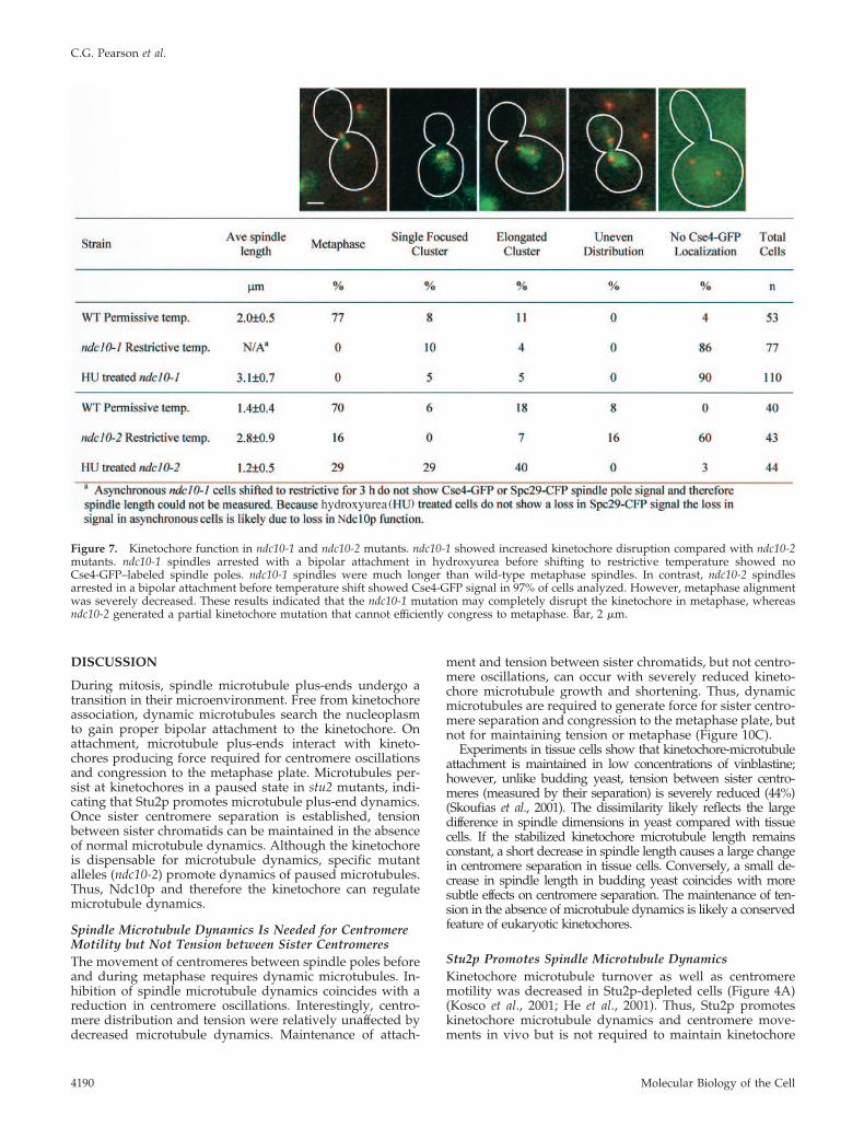
The difference in tubulin turnover of each kinetochoremutation is indicative of differential functional effects on thekinetochore leading to altered regulation of microtubuledynamics. To assay the severity of each kinetochore mutantallele on kinetochore function, we measured the localizationof Cse4-GFP in cells that achieved a bipolar attachment byarrest in hydroxyurea. Hydroxyurea arrests the cell cyclewith bioriented attachment of sister kinetochores to eachspindle pole (Goshima and Yanagida, 2000). Spindles inndc10-1 mutants arrested in hydroxyurea before tempera-ture shift showed longer spindles (3.1 � 0.7 �m) and punc-tate Cse4-GFP localization in only 10% of the cells (Figure 7).
The remaining cells had a diffuse Cse4-GFP signal through-out the nucleus. In contrast, spindles in ndc10-2 mutantsshifted to restrictive temperature after hydroxyurea arrestmaintained a normal metaphase length of 1.5 � 0.5 �m anddistinct Cse4-GFP localization at the kinetochore in 97% ofthe population. However, metaphase alignment was aber-rant in many cells (69%) with centromeres localized as asingle cluster or along the spindle axis (Figure 7). Theseresults indicate ndc10-1 severely disrupts the kinetochore aspreviously described (Doheny et al., 1993; Goh and Kilmar-tin, 1993). ndc10-2 generates partially functional kineto-chores unable to efficiently congress or separate sister cen-tromeres during metaphase (Figure 7). To determine themolecular difference between the ndc10 alleles, the ndc10alleles in each strain were sequenced and compared withwild type. ndc10-1 contains an ochre mutation (Q944*) gen-erating a carboxy terminal truncation of 12 amino acids;ndc10-2 contains a single transition mutation (A914T) in thecarboxy terminus.
These data indicate proper kinetochore function is notessential for promoting dynamic spindle microtubules. Lossof kinetochore structure and microtubule attachment(ndc10-1) did not severely affect spindle microtubule dynam-ics or turnover. However, a partial mutant (ndc10-2) pro-duces a measurable decrease in the rate of spindle microtu-bule turnover. Therefore, alterations in kinetochore functionindeed influence spindle microtubule dynamics.
Kinetochore Regulation of Microtubule Dynamics in theAbsence of Stu2pThe decreased microtubule turnover in the absence of Stu2pprovides the opportunity to determine kinetochore specificcontributions to generating dynamic microtubules. To dis-sect the role of kinetochores in regulating microtubule dy-namics, we depleted Stu2p (stu2td) in each of the previouslystudied CBF3 mutants (ndc10-1, ctf13-30, and ndc10-2).
The rate of spindle microtubule recovery did not increase inthe stu2td, ndc10-1, or stu2td, ctf13-30 double mutants. stu2td,ndc10-1 at restrictive temperature had an average recovery rateof 205 � 109 s compared with 85 � 44 s for permissive tem-perature (p � 0.015; Figure 8). Half of the cells showed nofluorescence recovery (Table 2). The rate of turnover was notsignificantly different from stu2td alone at restrictive tempera-ture (p � 0.159; Table 2). stu2td, ctf13-30 cells exhibited anaverage recovery half-time of 330 � 144 s at restrictive temper-ature and 72 � 38 s at permissive temperature. No significantdifference was observed between stu2td and stu2td, ctf13-30 atrestrictive temperature (p � 0.730). No recovery was observedin four cells at restrictive temperature (Table 2). Thus, muta-tions that completely disrupt kinetochore organization do notaffect microtubule turnover (Figure 9). Stabilization of micro-tubules in the absence of Stu2p is therefore independent ofkinetochore attachment.
To determine whether partially functional kinetochores(ndc10-2) affect the stable microtubule population in stu2td,photobleaching was performed in stu2td, ndc10-2 cells. Atpermissive temperature, stu2td, ndc10-2 double mutantsshowed a slower recovery compared with wild-type cells(124 � 72 s; Figures 8 and 9). Cells shifted to restrictivetemperature showed an increased rate of microtubule turn-over (93 � 46 s; Figures 8 and 9). Three of 21 cells exhibitedno recovery. This recovery was significantly faster thanstu2td alone (t1/2 � 302 � 200 s; p � 0.000). Thus, analysis ofmicrotubule dynamics in ndc10-2 revealed that kinetochoreshave the capacity to regulate kinetochore microtubule dy-namics.
Figure 6. Mutations in kinetochore function do not severely affectspindle microtubule dynamics. GFP-Tub1 turnover in mitotic spin-dles was measured in ndc10-1, ctf13-30, and ndc10-2 cells at restric-tive temperature. The overall spindle length was longer and approx-imately one-half of the cell population showed an asymmetricmicrotubule distribution with one microtubule tuft or half-spindlebrighter than the other. Figure describes a representative example ofFRAP of kinetochore mutants (example was ndc10-2). The averageturnover rate was slightly decreased compared with permissiveconditions (64 � 42 s [bright] and 60 � 34 s [dim] vs. 40 � 14 s).Similar results were found for ndc10-1 and ctf13-30 mutants (Table2) however ctf13-30 did not display longer, asymmetric spindles.Time, min. Bar, 2 �m.
Stu2p Regulation of Kinetochore Dynamics
Vol. 14, October 2003 4189
DISCUSSION
During mitosis, spindle microtubule plus-ends undergo atransition in their microenvironment. Free from kinetochoreassociation, dynamic microtubules search the nucleoplasmto gain proper bipolar attachment to the kinetochore. Onattachment, microtubule plus-ends interact with kineto-chores producing force required for centromere oscillationsand congression to the metaphase plate. Microtubules per-sist at kinetochores in a paused state in stu2 mutants, indi-cating that Stu2p promotes microtubule plus-end dynamics.Once sister centromere separation is established, tensionbetween sister chromatids can be maintained in the absenceof normal microtubule dynamics. Although the kinetochoreis dispensable for microtubule dynamics, specific mutantalleles (ndc10-2) promote dynamics of paused microtubules.Thus, Ndc10p and therefore the kinetochore can regulatemicrotubule dynamics.
Spindle Microtubule Dynamics Is Needed for CentromereMotility but Not Tension between Sister CentromeresThe movement of centromeres between spindle poles beforeand during metaphase requires dynamic microtubules. In-hibition of spindle microtubule dynamics coincides with areduction in centromere oscillations. Interestingly, centro-mere distribution and tension were relatively unaffected bydecreased microtubule dynamics. Maintenance of attach-
ment and tension between sister chromatids, but not centro-mere oscillations, can occur with severely reduced kineto-chore microtubule growth and shortening. Thus, dynamicmicrotubules are required to generate force for sister centro-mere separation and congression to the metaphase plate, butnot for maintaining tension or metaphase (Figure 10C).
Experiments in tissue cells show that kinetochore-microtubuleattachment is maintained in low concentrations of vinblastine;however, unlike budding yeast, tension between sister centro-meres (measured by their separation) is severely reduced (44%)(Skoufias et al., 2001). The dissimilarity likely reflects the largedifference in spindle dimensions in yeast compared with tissuecells. If the stabilized kinetochore microtubule length remainsconstant, a short decrease in spindle length causes a large changein centromere separation in tissue cells. Conversely, a small de-crease in spindle length in budding yeast coincides with moresubtle effects on centromere separation. The maintenance of ten-sion in the absence of microtubule dynamics is likely a conservedfeature of eukaryotic kinetochores.
Stu2p Promotes Spindle Microtubule DynamicsKinetochore microtubule turnover as well as centromeremotility was decreased in Stu2p-depleted cells (Figure 4A)(Kosco et al., 2001; He et al., 2001). Thus, Stu2p promoteskinetochore microtubule dynamics and centromere move-ments in vivo but is not required to maintain kinetochore
Figure 7. Kinetochore function in ndc10-1 and ndc10-2 mutants. ndc10-1 showed increased kinetochore disruption compared with ndc10-2mutants. ndc10-1 spindles arrested with a bipolar attachment in hydroxyurea before shifting to restrictive temperature showed noCse4-GFP–labeled spindle poles. ndc10-1 spindles were much longer than wild-type metaphase spindles. In contrast, ndc10-2 spindlesarrested in a bipolar attachment before temperature shift showed Cse4-GFP signal in 97% of cells analyzed. However, metaphase alignmentwas severely decreased. These results indicated that the ndc10-1 mutation may completely disrupt the kinetochore in metaphase, whereasndc10-2 generated a partial kinetochore mutation that cannot efficiently congress to metaphase. Bar, 2 �m.
C.G. Pearson et al.
Molecular Biology of the Cell4190

attachment to microtubules or sister centromere tension(Figure 10C). Therefore, Stu2p’s role is not in attachment;rather, it is in the transition from the paused state facilitatingmicrotubule shortening and/or growth (Figure 10C). Stu2pincreases catastrophe events during growth and rescueevents during shortening (Kosco et al., 2001). Stu2p likelyfunctions together with other microtubule binding proteinsto increase microtubule transition frequencies by the recruit-ment of growth factors to shortening ends and catastrophefactors to growing ends (Kinoshita et al., 2001, 2002;
Andersen and Wittmann, 2002). The dynamic microtubulestate is necessary to gain proper centromere attachment andpositioning, Stu2p promotes this by preventing prolongedpauses of microtubule plus-ends (Kosco et al., 2001; Shirasu-Hiza et al., 2003; Van Breugal et al., 2003).
Stu2p Promotes Spindle Microtubule DynamicsIndependent of the KinetochoreStu2p’s localization to the kinetochore (Kosco et al., 2001; Heet al., 2001) suggests the kinetochore may contribute toStu2p’s ability to promote spindle microtubule dynamics.We measured microtubule turnover in cells with aberrantkinetochores (ndc10-1, ctf13-30, and ndc10-2). Nonfunctionalkinetochores (ndc10-1) did not substantially affect the rate ofkinetochore microtubule turnover. Therefore, the interactionof microtubules with kinetochores is not essential for Stu2pto promote microtubule dynamics (Figure 10B). A functionaldistinction between ndc10-1 and ndc10-2 was observed in therates of microtubule turnover. The decreased turnover ratein ndc10-2 mutants may reflect kinetochore-dependentchanges in microtubule dynamics. Thus, kinetochore disrup-tion did not lead to stabilization of microtubules, as weobserved for stu2 mutants; however, small changes in turn-over occur in ndc10-2 mutants. These data extend studies onStu2p’s role in promoting cytoplasmic microtubule dynam-ics.
Three possible models for rapid recovery of spindle mi-crotubules are kinetochore microtubule plus-end growthand shortening, translocation of the microtubule lattice tothe spindle pole or poleward microtubule flux, and contin-ual detachment and reattachment of microtubules to kinet-ochores. Several lines of evidence suggest that Stu2p is re-sponsible for promoting kinetochore microtubule plus-enddynamics that enables GFP-Tub1 turnover in the mitoticspindle. Electron microscopy studies show yeast kineto-chore microtubule plus-ends exhibit open, “peeling or ramshorn” structure indicative of shortening microtubules,whereas minus-ends at the spindle pole display a closed,tapered structure consistent with stable microtubules (Byerset al., 1978; Mandelkow et al., 1991; O’Toole et al., 1999).Therefore, microtubule dynamics in yeast is likely restrictedto plus-ends. Fluorescence speckle microscopy studies findno evidence for minus-end assembly or disassembly of ei-ther polar microtubules in anaphase or astral microtubules(Maddox et al., 2000). Recent in vivo and in vitro studies findthat Stu2p and XMAP215 act specifically at microtubuleplus-ends to promote dynamicity (Kosco et al., 2001; Shirasu-Hiza et al., 2003; Van Breugal et al., 2003). Furthermore,Stu2p’s spindle pole localization (minus-end proximal) isnot surprising given that spindle pole components affectastral microtubule dynamics (Vogel et al., 2001). Therefore,we speculate that Stu2p can be regulated or stored by spin-dle poles before being shuttled to microtubule plus-ends topromote dynamics. Similar mechanisms have been de-scribed for loading Kar9p on cytoplasmic microtubules tofacilitate proper spindle positioning (Liakopoulos et al.,2003). XMAP215 displays both stabilizing and destabilizingactivity under varying experimental conditions (Gard andKirschner, 1987; Vasquez et al., 1994; Popov et al., 2001;Shirasu-Hiza et al., 2003; Van Breugal et al., 2003). Stu2p mayfunction differentially at plus-ends near the spindle pole topromote growth events and at plus-ends near the spindleequator to promote catastrophe events, thereby creating agradient of microtubule polymerization and depolymeriza-tion. Gradients of microtubule regulation have been pre-dicted by modeling experiments in budding yeast (Sprague
Figure 8. ndc10-2 kinetochore allele rescues spindle microtubuleturnover in stu2td mutants. (A) FRAP of GFP-Tub1–labeled mitotichalf-spindles in Stu2p depleted cells. Representative sequence andfluorescence intensity measurements showed slowed (t1/2 � 302 �200 s) and slightly decreased spindle microtubule turnover (normal-ized recovery � 0.9 � 0.4). (B) Microtubule turnover in stu2td andndc10-2 double mutants increased the turnover rate (t1/2 � 93 �46 s) with a similar normalized recovery compared with stu2td alone(0.9 � 0.4). (C) Example recovery curves of stu2td alone and stu2td,ndc10-2 double mutants. Time, min. Bar, 2 �m.
Stu2p Regulation of Kinetochore Dynamics
Vol. 14, October 2003 4191

et al., 2003) and experimental evidence in Xenopus extractsystems (Kalab et al., 2002). It is unlikely that microtubuleturnover is due to attachment and detachment of microtu-bules to kinetochores. Once sister centromeres separate,their reassociation is infrequent, indicating that a gain andloss of tension between sister chromatids does not occur aswould be expected for attachment and detachment (Gos-hima and Yanagida, 2001; Pearson et al., 2001). Thus, wefavor the model describing microtubule plus-end dynamicsto be responsible for spindle microtubule turnover. Thenature of Stu2p regulation of microtubule plus-end dynam-ics and the mechanism of spindle microtubule turnover willbe the focus of important future research.
Functional Differences between Kinetochore MutationsBoth ndc10-1 and ndc10-2 disrupt proper segregation ofthe genome (Goh and Kilmartin, 1993; Kopski and Huf-faker, 1997). Furthermore, ndc10-1 disrupts centromeretension, localization of all kinetochore proteins tested andspindle assembly checkpoint function (Tavormina andBurke, 1998; Goshima and Yanagida, 2000; He et al., 2000).We find that ndc10-2 mutants are not as severe in theirmislocalization of a core kinetochore component (Cse4p)compared with ndc10-1. Similarly, the two alleles werefound to have different effects on the spindle assemblycheckpoint. ndc10-2 mutants exhibit an increased cell cy-cle delay in microtubule destabilizing drugs compared
with ndc10-1, indicating that ndc10-2 mutants are morecompetent than ndc10-1 to activate the checkpoint (Burke,personal communication). Finally, our sequencing analy-sis shows that the ndc10-2 (A914T) mutation is a singleamino acid transition resulting in the substitution of ahydrophobic alanine residue to a hydrophilic threonineresidue. The more functionally severe mutation (ndc10-1;Q944*) is a 12-amino acid truncation also in the C termi-nus (our results, Burke and Kilmartin, personal commu-nication). Thus, a kinetochore mutation causing a subtlechange in microtubule dynamics (ndc10-2) is a partialkinetochore mutation, whereas the mutation without ameasurable effect on microtubule dynamics (ndc10-1) re-sults in complete kinetochore disruption. Furthermore,the close proximity of the two mutations suggests thecarboxy terminal domain of NDC10 is essential for regu-lation of kinetochore function.
Kinetochore Mutation That Promotes MicrotubuleDynamics in the Absence of Stu2p: Evidence for Kineto-chore Regulation of Microtubule Dynamicsstu2td /ndc10-2 double mutants exhibited spindle microtu-bule dynamics that were significantly faster than stu2td
alone. Thus, kinetochores can regulate microtubules inde-pendent of Stu2p. The increased microtubule dynamics maybe due to an altered state of microtubule attachment with apartially functional kinetochore. We infer the rescue of mi-
Figure 9. Kinetochore contribution to spindle microtubule dynamics. Comparison of t1/2 values of bleached spindle microtubule tufts forstu2td, ndc10-2, ndc10-1, ctf13-30, ndc10-2/stu2td, ndc10-1/stu2td, and ctf13-30/stu2td. Experiments with no recovery (normalized recovery�16%) were not included in the recovery analysis. Recovery of spindle microtubule turnover was observed in ndc10-2/stu2td double mutantsbut not in ndc10-1/stu2td or ctf13-30/stu2td cells.
C.G. Pearson et al.
Molecular Biology of the Cell4192

Figure 10. Model for regulation of spindle microtubule dynamics. (A) Dynamic spindle microtubules promoted by Stu2p are coupled to the forcenecessary for centromere oscillations. (B) Complete disruption of the kinetochore does not affect dynamics promoted by Stu2p. (C) Depletion of Stu2pstabilizes microtubules and centromere dynamics with separated sister centromeres indicating that kinetochores remain attached and under tension andStu2p prevents the paused state of microtubule dynamics. (D) Complete disruption of the kinetochore without Stu2p causes stable microtubules,indicating that microtubule stabilization does not require the kinetochore.
Stu2p Regulation of Kinetochore Dynamics
Vol. 14, October 2003 4193

crotubule turnover is due to a change in Ndc10p function atthe kinetochore. However, we cannot rule out the possibilitythat Ndc10p functions at unattached microtubule plus-ends.ndc10-2 kinetochores may generate dynamics by recruitingand/or allowing accessibility of other microtubule dynamicsregulators or by limiting access of stabilization factors to themicrotubule plus-end. In contrast to ndc10-1, ndc10-2 singlemutants seem to have partial kinetochore function to gener-ate an altered state of dynamic microtubules. The rate offluorescence recovery of stu2td /ndc10-2 spindles is not sig-nificantly different from ndc10-2 alone, indicating that Stu2pis not necessary for promoting microtubule dynamics inndc10-2.
These results provide evidence for a model where Stu2ppromotes dynamics of free or attached microtubule plus-ends within the cell (Figure 10, C and D). Kinetochores areable to maintain attachment to growing, shortening, orpaused microtubule plus-ends (evidenced by oscillationstoward and away from the spindle pole). However, pausedends may not allow capture, orientation, error-correctionmechanisms, or segregation. Therefore, dynamic plus-endsare the preferred physiological state; Stu2p’s role in theseprocesses is essential. In contrast, kinetochores are not re-quired for microtubule dynamics. However, kinetochoresmust maintain attachment to plus-ends to sustain sistercentromere oscillations. The discovery that ndc10-2 pro-motes microtubule dynamics in the absence of Stu2p revealskinetochore-specific regulation of microtubule dynamics tofacilitate proper centromere positioning. The kinetochorespecific mechanism may reflect local control of microtubuledynamics embedded in a kinetochore by limiting the accessor activity of microtubule-stabilizing factors and therebygenerating dynamic microtubules. Our results provide thefirst molecular evidence for separating the control of micro-tubule dynamics from kinetochore-specific regulation of in-dividual kinetochore microtubules during mitosis.
ACKNOWLEDGMENTS
We thank E. Yeh, J. Deluca, J.C. Labbe, M. Karthikeyan, A. Desai and A.Hyman for stimulating discussions and wonderful support; D. Burke and J.Kilmartin for communicating unpublished results; K. Kosco and T. Huffakerfor yeast strains; R. Heil-Chapdelaine, J. Cooper, T. Huffaker, A. Goldstein, J.McCusker, and the Yeast Resource Center for plasmids; and members of theBloom laboratory for helpful and critical comments on the manuscript. Thiswork was supported by National Institutes of Health grant GM-32238 to K.S.Band GM-24364 to E.D.S.
REFERENCES
Andersen, S.S., and Wittmann, T. (2002). Toward reconstitution of in vivomicrotubule dynamics in vitro. Bioessays 24, 305–307.
Belmont, A.S. (2001). Visualizing chromosome dynamics with GFP. TrendsCell Biol. 11, 250–257.
Byers, B., Shriver, K., and Goetsch, L. (1978). The role of spindle pole bodiesand modified microtubule ends in the initiation of microtubule assembly inSaccharomyces cerevisiae. J. Cell Sci. 30, 331–352.
Chen, Y., Baker, R.E., Keith, K.C., Harris, K., Stoler, S., and Fitzgerald-Hayes,M. (2000). The N terminus of the centromere H3-like protein Cse4p performsan essential function distinct from that of the histone fold domain. Mol. Cell.Biol. 20, 7037–7048.
Dhamodharan, R., Jordan, M., Thrower, D., Wilson, L., and Wadsworth, P.(1995). Vinblastine suppresses dynamics of individual microtubules in livingcells. Mol. Biol. Cell 6, 1215–1229.
Dogterom, M., and Yurke, B. (1997). Measurement of the force-velocity rela-tion for growing microtubules. Science 278, 856–860.
Doheny, K.F., Sorger, P.K., Hyman, A.A., Tugendreich, S., Spencer, F., andHieter, P. (1993). Identification of essential components of the S. cerevisiaekinetochore. Cell 73, 761–74.
Dohmen, R.J., Wu, P., and Varshavsky, A. (1994). Heat-inducible degron: amethod for constructing temperature-sensitive mutants. Science 263, 1273–1276.
Gard, D.L., and Kirschner, M.W. (1987). A microtubule-associated proteinfrom Xenopus eggs that specifically promote assembly at the plus-end. J. CellBiol. 105, 2203–2215.
Goh, P.Y., and Kilmartin, J.V. (1993). NDC10: a gene involved in chromosomesegregation in Saccharomyces cerevisiae. J. Cell Biol. 121 503–512.
Goldstein, A.L., and McCusker, J.H. (1999). Three new dominant drug resis-tance cassettes for gene disruption in Saccharomyces cerevisiae. Yeast 15, 1541–1553.
Goshima, G., and Yanagida, M. (2000). Establishing biorientation occurs withprecocious separation of the sister kinetochores, but not the arms, in the earlyspindle of budding yeast. Cell 100, 619–633.
Goshima, G., and Yanagida, M. (2001). Time course analysis of precociousseparation of sister centromeres in budding yeast: continuously separated orfrequently reassociated? Genes Cells 6, 765–773.
He, X., Asthana, S., and Sorger, P.K. (2000). Transient sister chromatid sepa-ration and elastic deformation of chromosomes during mitosis in buddingyeast. Cell 101, 763–775.
He, X., Rines, D.R., Espelin, C.W., Sorger, P.K. (2001). Molecular analysis ofkinetochore-microtubule attachment in budding yeast. Cell 106, 195–206.
Hunt, A.J., and McIntosh, J.R. (1998). The dynamic behavior of individualmicrotubules associated with chromosomes in vitro. Mol. Biol. Cell 9, 2857–2871.
Hyman, A.A., and Sorger, P.K. (1995). Structure and function of kinetochoresin budding yeast. Annu. Rev. Cell Dev. Biol. 11, 471–495.
Inoue, S., and Salmon, E.D. (1995). Force generation by microtubule assem-bly/disassembly in mitosis and related movements. Mol. Biol. Cell 6, 1619–1640.
Jiang, W., Lechner, J., and Carbon, J. (1993). Isolation and characterization ofa gene (CBF2) specifying a protein component of the budding yeast kineto-chore. J. Cell Biol. 121, 513–519.
Kalab, P., Weis, K., and Heald, R. (2002). Visualization of a Ran-GTP gradientin interphase and mitotic Xenopus egg extracts. Science 295, 2452–2456.
Kapoor, T.M., and Compton, D.A. (2002). Searching for the middle ground:mechanisms of chromosome alignment during mitosis J. Cell Biol. 157, 551–556.
Khodjakov,A.,Gabashvili,I.S.,andRieder,C.L.(1999).“Dumb”versus“smart”ki-netochore models for chromosome congression during mitosis in vertebratesomatic cells. Cell Motil. Cytoskeleton 43, 179–185.
Kinoshita, K., Arnal, I., Desai, A., Drechsel, D.N., and Hyman, A.A. (2001).Reconstitution of physiological microtubule dynamics using purified compo-nents. Science 294, 1340–1343.
Kinoshita, K., Habermann, B., and Hyman, A.A. (2002). XMAP 215, a keycomponent of the dynamic microtubule cytoskeleton. Trends Cell Biol. 12,267–273.
Kopski, K.M., and Huffaker, T.C. (1997). Suppressors of the ndc10–2 mutation:a role for the ubiquitin system in Saccharomyces cerevisiae kinetochore function.Genetics 147, 409–420.
Kosco, K.A., Pearson, C.G., Maddox, P.S., Wang, P.J., Adams, I.R., Salmon,E.D., Bloom, K., and Huffaker, T.C. (2001). Control of microtubule dynamicsby Stu2p is essential for spindle orientation and metaphase chromosomealignment in yeast. Mol. Biol. Cell 12, 2870–2880.
Liakopoulos, D., Kusch, J., Grava, S., Vogel, J., and Barral, Y. (2003). Asym-metric loading of Kar9 onto spindle poles and microtubules ensures properspindle alignment. Cell 112, 561–574.
Longtine, M.S., Mckenzie Iii, A., Demarini, D.J., Shah, N.G., Wach, A., Bra-chat, A., Philippsen, P., and Pringle, J.R. (1998). Additional modules forversatile and economical PCR-based gene deletion and modification in Sac-charomyces cerevisiae. Yeast 14, 953–961.
Maddox, P., Bloom, K., and Salmon, E.D. (2000). Polarity and dynamics ofmicrotubule assembly in the budding yeast Saccharomyces cerevisiae. Nat. CellBiol. 2, 36–41.
Mandelkow, E.M., Mandelkow, E., and Milligan, R.A. (1991). Microtubuledynamics and microtubule caps: a time-resolved cryo-electron microscopystudy. J. Cell Biol. 114, 977–991.
C.G. Pearson et al.
Molecular Biology of the Cell4194

Marshall, W.F., Straight, A., Marko, J.F., Swedlow, J., Dernberg, A., Belmont,L., Murray, A.W., Agard, D.A., and Sedat, J.W. (1997). Interphase chromo-somes undergo constrained diffusional motion in living cells. Curr. Biol. 7,930–939.
McIntosh, J.R., Grishchuk, E.L., and West, R.R. (2002). Chromosome-micro-tubule interactions during mitosis. Annu. Rev. Cell Dev. Biol. 18, 193–219.
Mitchison, T., Evans, L., Schulze, E., and Kirschner, M. (1986). Sites of micro-tubule assembly and disassembly in the mitotic spindle. Cell 45, 515–527.
Mitchison, T.J., and Salmon, E.D. (1992). Poleward kinetochore fiber move-ment occurs during both metaphase and anaphase-A in newt lung-cell mito-sis. J. Cell Biol. 119, 569–582.
Mitchison, T.J. (1989). Polewards microtubule flux in the mitotic spindle:evidence from photoactivation of fluorescence. J. Cell Biol. 109, 637–652.
Murray, A.W., and Mitchison, T.J. (1994). Mitosis. Kinetochores pass the IQtest. Curr. Biol. 4, 38–41.
Nabeshima, K., Nakagawa, T., Straight, A.F., Murray, A., Chikashige, Y.,Yamashita, Y.M., Hiraoka, Y., and Yanagida, M. (1998). Dynamics of centro-meres during metaphase-anaphase transition in fission yeast: Dis1 is impli-cated in force balance in metaphase bipolar spindle. Mol. Biol. Cell 9, 3211–3225.
Nicklas, R.B. (1989). The motor for poleward chromosome movement inanaphase is in or near the kinetochore. J. Cell Biol. 109, 2245–2255.
O’Toole, E.T., Winey, M., and McIntosh, J.R. (1999). High-voltage electrontomography of spindle pole bodies and early mitotic spindles in the yeastSaccharomyces cerevisiae. Mol. Biol. Cell 10, 2017–2031.
Pearson, C.G., Maddox, P.S., Salmon, E.D., and Bloom, K. (2001). Buddingyeast chromosome structure and dynamics during mitosis. J. Cell Biol. 152,1255–1266.
Popov, A.V., Pozniakovsky, A., Arnal, I., Antony, C., Ashford, A.J., Kinoshita,K., Tournebize, R., Hyman, A.A., and Karsenti, E. (2001). XMAP215 regulatesmicrotubule dynamics through two distinct domains. EMBO J. 20, 397–410.
Rieder, C.L., and Salmon, E.D. (1998). The vertebrate cell kinetochore and itsroles during mitosis. Trends Cell Biol. 8, 310–318.
Scholey, J.M., Brust-Mascher, I., and Mogilner, A. (2003). Cell division. Nature422, 746–752.
Severin, F., Habermann, B., Huffaker, T., and Hyman, T. (2001). Stu2 promotesmitotic spindle elongation in anaphase. J. Cell Biol. 153, 435–442.
Shaw, S.L., Yeh, E., Bloom, K., and Salmon, E.D. (1997). Imaging greenfluorescent protein fusion proteins in Saccharomyces cerevisiae. Curr. Biol. 7,701–704.
Shirasu-Hiza, M., Coughlin, P., and Mitchison, T. (2003). Identification ofXMAP215 as a microtubule-destabilizing factor in Xenopus egg extract bybiochemical purification. J. Cell Biol. 161, 349–358.
Skibbens, R.V., Skeen, V.P., and Salmon, E.D. (1993). Directional instability ofkinetochore motility during chromosome congression and segregation inmitotic newt lung cells: a push-pull mechanism. J. Cell Biol. 122, 859–875.
Skoufias, D.A., Andreassen, P.R., Lacroix, F.B., Wilson, L., and Margolis, R.L.(2001). Mammalian mad2 and bub1/bubR1 recognize distinct spindle-attach-ment and kinetochore-tension checkpoints. Proc. Natl. Acad. Sci. USA 98,4492–4497.
Sprague, B.L., Pearson, C.G., Maddox, P.S., Bloom, K.S., Salmon, E.D., andOdde, D.J. (2003). Mechanisms of microtubule-based kinetochore positioningin the yeast metaphase spindle. Biophys. J. 84, 3529–3546.
Straight, A.F., Marshall, W.F., and Murray, A.W. (1997). Mitosis in livingbudding yeast: anaphase A but no metaphase plate. Science 277, 574–578.
Tanaka, T., Fuchs, J., Loidl, J., and Nasmyth, K. (2000). Cohesin ensuresbipolar attachment of microtubules to sister centromeres and resists theirprecocious separation. Nat. Cell Biol. 2, 492–499.
Tavormina, P.A., and Burke, D.J. (1998). Cell cycle arrest in cdc20 mutants ofSaccharomyces cerevisiae is independent of Ndc10p and kinetochore functionbut requires a subset of spindle checkpoint genes. Genetics 148, 1701–1713.
Toso, R.J., Jordan, M.A., Farrell, K.W., Matsumoto, B., and Wilson, L. (1993).Kinetic stabilization of microtubule dynamic instability in vitro by vinblas-tine. Biochemistry 32, 1285–1293.
Tran, P.T., Walker, R.A., and Salmon, E.D. (1997). A metastable intermediatestate of microtubule dynamic instability that differs significantly between plusand minus ends. J. Cell Biol. 138, 105–117.
Van Breugal, M., Drechsel, D., and Hyman, A. (2003). Stu2p, the buddingyeast member of the conserved Dis1/XMAP215 family of microtubule-asso-ciated proteins is a plus end-binding microtubule destabilizer. J. Cell Biol. 161,359–369.
Vasquez, R.J., Gard, D.L., and Cassimeris, L. (1994). XMAP from Xenopus eggspromotes rapid plus end assembly of microtubules and rapid microtubulepolymer turnover. J. Cell Biol. 127, 985–993.
Vasquez, R.J., Howell, B., Yvon, A.M., Wadsworth, P., and Cassimeris, L.(1997). Nanomolar concentrations of nocodazole alter microtubule dynamicinstability in vivo and in vitro. Mol. Biol. Cell 8, 973–985.
Vogel, J., Drapkin, B., Oomen, J., Beach, D., Bloom, K., and Snyder, M. (2001).Phosphorylation of gamma-tubulin regulates microtubule organization inbudding yeast. Dev. Cell 1, 621–631.
Wach, A., Brachat, A., Pohlmann, R., and Philippsen, P. (1994). New heterol-ogous modules for classical or PCR-based gene disruptions in Saccharomycescerevisiae. Yeast 10, 1793–1808.
Wadsworth, P., and Salmon, E.D. (1988). Spindle microtubule dynamics:modulation by metabolic inhibitors. Cell Motil. Cytoskeleton 11, 97–105.
Winey, M., Mamay, C.L., O’Toole, E.T., Mastronarde, D.N., Giddings, T.H.,Jr., McDonald, K.L., and McIntosh, J.R. (1995). Three-dimensional ultrastruc-tural analysis of the Saccharomyces cerevisiae mitotic spindle. J. Cell Biol. 129,1601–1615.
Winey, M., and O’Toole, E.T. (2001). The spindle cycle in budding yeast. Nat.Cell Biol. 3, E23–E27.
Zhai, Y., Kronebusch, P.J., and Borisy, G.G. (1995). Kinetochore microtubuledynamics and the metaphase-anaphase transition. J. Cell Biol. 131, 721–734.
Stu2p Regulation of Kinetochore Dynamics
Vol. 14, October 2003 4195