year 2 water+electrolytes - imperial college unionelectrolytes.pdf ·...
TRANSCRIPT
Science & Patient WATER & ELECTROLYTES Alexandra Burke-‐Smith
1. Water and Dehydration Jeremy Levy
NORMAL WATER BALANCE + SOLUTE DISTRIBUTION Physiological control of water and circulating blood volume Water and solutes
• 60% of the body is water. 2/3 of this I is intracellular • The total amount of solute in the water is the osmolarity • Water moves down osmolar gradients
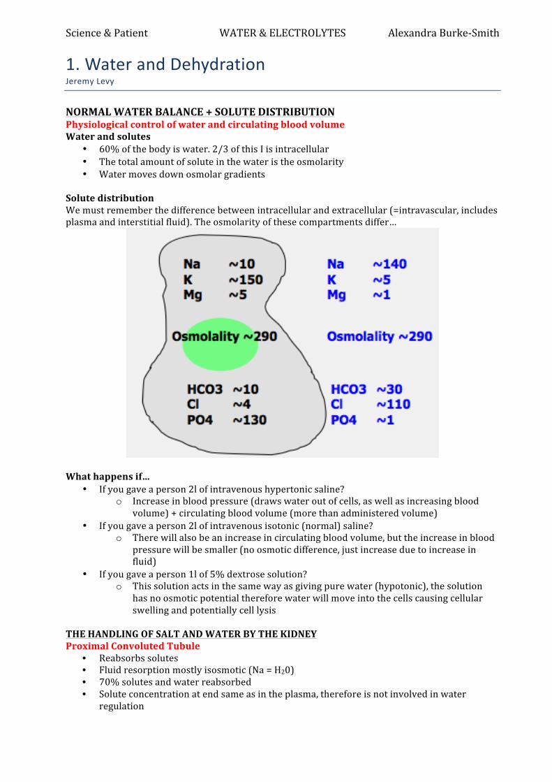
Solute distribution We must remember the difference between intracellular and extracellular (=intravascular, includes plasma and interstitial fluid). The osmolarity of these compartments differ…
What happens if…
• If you gave a person 2l of intravenous hypertonic saline? o Increase in blood pressure (draws water out of cells, as well as increasing blood
volume) + circulating blood volume (more than administered volume) • If you gave a person 2l of intravenous isotonic (normal) saline?
o There will also be an increase in circulating blood volume, but the increase in blood pressure will be smaller (no osmotic difference, just increase due to increase in fluid)
• If you gave a person 1l of 5% dextrose solution? o This solution acts in the same way as giving pure water (hypotonic), the solution
has no osmotic potential therefore water will move into the cells causing cellular swelling and potentially cell lysis
THE HANDLING OF SALT AND WATER BY THE KIDNEY Proximal Convoluted Tubule
• Reabsorbs solutes • Fluid resorption mostly isosmotic (Na = H20) • 70% solutes and water reabsorbed • Solute concentration at end same as in the plasma, therefore is not involved in water
regulation

Science & Patient WATER & ELECTROLYTES Alexandra Burke-‐Smith Loop of Henle
• Descending limb water permeable -‐ salt stays in • Thin ascending limb water impermeable -‐ salt diffuses out • Thick ascending limb reabsorbs Na (NaKCl2 transporter)
• This is targeted by Furosemide (diuretic) • Countercurrent mechanism for urinary concentration
Countercurrent principle
If we consider our starting point to be a loop filled with isotonic fluid (tubular fluid = interstitial fluid)
• The ascending limb pumps ions into the interstitial fluid, therefore decreasing the osmolarity of the tubular fluid and increasing the osmolarity of the interstitial fluid
• In the descending limb, water will move to counteract this, resulting in a higher concentration of tubular fluid in the descending limb compared to the ascending limb
• This process repeats continuously, as fluid is constantly flowing. By doing this, we progressively create a very concentrated interstitial fluid surrounding the loop of Henle (concentrates the medulla)
• As water movement is prevented in the ascending limb, the loop of Henle is responsible for producing a dilute urine by reabsorbing salt
We therefore require a mechanism to reabsorb water to concentrate our urine… Distal tubule and collecting duct Distal Tubule
• Active solute reabsorption – last 2–3% • Urine maximally dilute (50 mosmol/kg)
Collecting Duct • ADH sensitive • If no ADH – CD is water insensitive
hence dilute urine • In presence of ADH – water is reabsorbed via insertion of AQP2 channels into luminal
membrane HORMONAL REGULATION ADH (anti-‐diuretic hormone)
• Also known as arginine vasopressin (AVP) • Nonapeptide synthesised by hypothalamus and secreted from posterior pituitary • Osmoceptors sense change in serum osmolality NOT Na • 1% ↑ ECF osmolality ⊕ ADH release (eg fluid deprivation) • 1% ↓ ECF osmolality ADH release (eg water ingestion) • Non-‐osmotic stimuli include stress, hypoxia, pain, volume depletion
• This happens post-‐operatively

Science & Patient WATER & ELECTROLYTES Alexandra Burke-‐Smith Mechanism of action:
• Direct vasoconstrictor • NaCl reabsorption in thin ascending limb of loop of Henle • Water retention in collecting ducts
o Receptor binding activates cAMP which stimulates water channel (aquaporin 2) incorporation into apical membrane
§ This could be altered in a number of ways, absence of ADH, ineffective AQP2, inability of AQP2 insertion into cell membrane, ineffective ADH receptor
§ This is a protective mechanism for maintaining circulating blood volume in the face of dehydration, in order to maintain adequate tissue perfusion
Angiotensin II Renin-‐angiotensin aldosterone system drives Angiotensin II production in times of dehydrationn via Renin. Actions include:
• Vasoconstriction • Increased Na/water retention • Aldosterone secretion
Drugs that interfere with this include ACE inhibitors, ARBs, direct renin inhibitors.
• Complications of these drugs include an inability to overcome blockade in the setting of extreme volume depletion, eg during D+V
Atrial Natriuretic Peptide (ANP) This polypeptides released from cardiac myocytes has the opposite effect, and is important in times of water overloading. Its actions include:
• Increases urinary excretion of Na and water • Inhibits Na resorption by collecting duct • Inhibits renin production and aldosterone secretion
Only one drugs interfere with this – Vaptans

Science & Patient WATER & ELECTROLYTES Alexandra Burke-‐Smith Summary of Water Balance
• ↑ fluid ingestion (eg 5 pints beer) → ↓ serum osmolality → thirst suppression
→ ↓ ADH + Aldosterone, ↑ ANP + → ↑ water excretion (to make max dilute urine)
• Water deprivation → ↑ serum osmolality → ↑ thirst → ↑ADH + Aldosterone, ↓ ANP release → ↑ water reabsorption (to concentrate urine)
ABNORMALITIES OF WATER BALANCE How could you lose too much water through the kidney?
• No ADH • Insensitive to ADH • TAL nakcl2 channel in TAL blocked • Other solute carrying water through the kidney especially glucose • Alcohol, caffeine • Drugs inhibiting ADH • Excess ANP
These will push too much salt and water through your kidney, causing excess water loss > dehydration Dehydration
• 1% weight loss – thirst • 2% weight loss – more thirst, vague discomfort, loss of appetite • 3-‐4% weight loss – increased blood red cell concentration, lethargy, apathy, nausea,
emotional instability • 6% weight loss – tingling limbs, heat exhaustion, increased body temp • 8% weight loss – dizziness, confusion, delerium • 20% weight loss -‐ death
Causes
• Vomiting • Diarrhoea • Alcohol • Diabetes (Mellitus + Insipidus) • Burns – assessed by weight loss • Iatrogenic – diuretic, chemotherapy, any nephrotoxic drugs • Bleeding • Earthquakes • Brain injury to the posterior pituitary • Diabetic ketoacidosis • Diabetic hyperosmolar coma • Post-‐operative • Sepsis • Anorexia
Clinical features First you need to distinguish intracellular/interstitial from intravascular fluid depletion, ie is it diuresis/vomiting/diarrhoea or blood loss?
• Postural hypotension, tachycardia • Low skin turgor • Sunken eyes • Dry mouth, thirst
Science & Patient WATER & ELECTROLYTES Alexandra Burke-‐Smith Biochemical features Assessing hydration is a clinical thing, but there are some biochemical clues.
• Serum osmolality high • Serum Na usually high • Serum K / Mg / Ca – high/low or normal • Serum urea high • Hb high
Treatment Replace fluid loss. This depends on the cause…
• If true dehydration > water • If salt and water loss > saline • If blood loss > blood
Hypernatraemia Causes
• Pure water deficit -‐ diabetes insipidus (central or nephrogenic) • Hypotonic fluid deficit – diuretics, osmotic diuresis, polyuric acute renal failure, post-‐
obstruction, vomiting, diarrhoea, fistulae, burns, excess sweating • Hypertonic sodium gain – salt ingestion, hypertonic saline, hypertonic sodium bicarbonate,
parenteral nutrition Symptoms
• Acute hypernatraemia > brain shrinkage > venous sinus bleeding • Chronic hypernatraemia > nausea, weakness, fasciculations, lethargy, coma
Treatment
• Water correction over 24-‐48 hours • Then treat the cause…
Diabetes insipidus
• Central (lack of secretion) or nephrogenic (failure to respond) to ADH (AVP) • Obligately dilute urine despite elevated serum osmolality • Patients may produce 10 –15 litres urine per day • Differential diagnosis is psychogenic polydipsia • Diagnose by water deprivation test under observation
Water deprivation test
• Deprive patient of all water. Weigh them. Baseline serum Na, osmolality, urine osmolality • Weigh hourly, monitor urine • What are you looking for in DI? No ability to concentrate urine
Diabetes mellitus
• Glucose is a potent osmolite, therefore hyperglycaemia can cause polyuria and severe dehydration
• Typical biochemical features of a DM patient are: Na 162 mmol/l K 2.2 mmol/l Urea 15 mmol/l Glucose 50 mmol/l

Science & Patient WATER & ELECTROLYTES Alexandra Burke-‐Smith
2. Clinical Scenarios of Water & Dehydration Dr Jeremy Levy
CASE 1 You foolishly decide to take part in the Marathon des Sables – running 150 km across the Sahara in 6 days, carrying your food and emergency kit weighing 9 Kg. On the third day you lose you water bottle soon after starting, but continue running all day. You crawl into camp that afternoon rather the worse for wear.
1. If you were to have a blood test done, what would be your serum sodium and osmolality? Give a rough guess.
• Dehydration leads to solute concentration e.g. hypernatraemia (high sodium) à 145mmol/l (max 160 mmol/l) would lead to severe symptoms – not very high as tightly controlled
• Some salt has been lost in sweat, but not as much as water (you are losing a hypotonic solution, roughly 30mmol/l) – so serum osmolality will ALSO go up (>290 mosmol/l e.g. 310mosmol/l))
• This is a problem seen in CF, as more chloride is moved into sweat, affecting the way they become dehydrated.
2. Would your blood pressure be low, normal or high?
• Dehydration leads to reduction of both extracellular and intracellular volume, thus your blood pressure would be low (100/60mmHg)
• Water and salt are lost intravascularly (water then moves from cells to blood, and is further lost, leading to total body water depletion – intracellular and intracerebral)
3. Briefly explain the physiological processes that have kept you more or less alive, especially
the link between brain and kidneys. Dehydration is a very potent stimulus of ADH release, which should minimise further water loss through the kidneys by increasing the reabsorption of water in the distal tubules – less water is excreted as urine The renin-‐angiotensin system is also crucial in control of water balance via the effects of angiotensin II and aldosterone on sodium handling and vasoconstriction Correct physiological responses to dehydration • Hypotonic fluid losses + Increased serum osmolality • Stimulate ADH, renin and angiotensin • Inhibit ANP • à Vasoconstriction • à Salt and water retention • à Low urine volume, low urine Na, high osmolality • This is NOT renal failure, but the normal renal physiological response!!

Science & Patient WATER & ELECTROLYTES Alexandra Burke-‐Smith CASE 2 A 25 year old woman is admitted to hospital for the birth of her first baby. After a prolonged labour she requires a caesarian section, and the baby is safely delivered. Blood test on the next day show: Serum Na 149 mmol/l (135-‐145) Slightly high Serum potassium 5.0 mmol/l (3.5-‐5) Upper limit Serum urea 8 mmol/l (2.5 – 6.7) High
1. Can you explain why these results are abnormal? If you wanted to correct them, how would you do it?
Dehydration – long labour, nothing to drink, nil by mouth, then in pain • Serum sodium and serum osmolality tend to track one another, unless patient has diabetes due
to presence of glucose • Potassium levels are difficult to interpret. Finely controlled by renin-‐angiotensin-‐aldosterone
system. Stimulation of RAAS leads to increased loss of potassium, but to a lesser extent than reduced urine output. Furthermore, potassium in an intracellular ion, thus potassium levels do not typically become depleted in dehydration and the latter two factors are more powerful than the loss of potassium due to RAAS.
Encourage woman to drink. If she loses consciousness, can be given fluids via naso-‐gastric tube or using an IV saline drip 48 hours later she is noted to be semi conscious, barely rousable, with a new divergent squint of her eyes. Blood tests show: Serum Na 105 mmol/l (135-‐145) low Serum potassium 3 mmol/l (3.5-‐5) low Serum urea 3 mmol/l (2.5 – 6.7) low
2. What has happened to her? She has developed hyponatraemia
3. Can you think of possible causes? DDs: 1. Hypervolaemic hyponatraemia e.g. medical negligence (giving too much saline). Many other
causes e.g. cirrhosis, congestive heart failure, nephritic syndrome, massive oedema of any cause 2. Euvolemic hyponatraemia e.g. severe pain/nausea, trauma/brain damage, SIADH,
hypothyroidism, glucocorticoid deficiency 3. Hypovolaemic hyponatraemia e.g. vomiting, decreased oral intake, severe diarrhoea, or diuretic
use 4. Miscellaneous causes e.g. facticious hyponatraemia, hypothyroidism + adrenal insufficiency, beer
potomania and other malnourished states, primary polydipsia
4. Why is she unconscious? Neurological manifestations due to hyponatraemia
• When sodium levels in blood become too low, excess water enters cells and causes the cells to swell. Swelling in the brain is especially dangerous because the brain is confined by the skull and is unable to expand.
• The disorder in the brain caused by hyponatremia is called hyponatremic encephalopathy, and accounts for symptoms such as headache, nausea, vomiting and confusion, but can also present with seizures, respiratory arrest and non-‐cardiogenic pulmonary edema.
• Neurological symptoms most often are due to very low serum sodium levels (usually <115 mEq/L), resulting in intracerebral osmotic fluid shifts and brain edema. This neurological symptom complex can lead to tentorial herniation with subsequent brain stem compression and respiratory arrest, resulting in death in the most severe cases.

Science & Patient WATER & ELECTROLYTES Alexandra Burke-‐Smith
5. Now see if you can find the name of this condition and any facts about it – you will need to look this up.
Central pontine myelinolysis caused by rapid fall in sodium, mostly occurs from the too rapid correction of mild hypernatraemia
• The whole centre of the pons has lost its myelin • This is why eye signs are seen, as cranial nerves controlling eye movements originate here. • 5-‐10 women die from this every year. Mostly iatrogenic.
CASE 3
1. Can you think how you might distinguish between a patient with primary polydipsia (drinking too much from psychiatric illness) causing polyuria, and a patient with true polyuria from diabetes insipidus.
Water deprivation test Including hourly weight, hourly urine volumes, serum and urine Na & osmolality.
• With DI, they cannot concentrate their urine, so weight will fall, urine volume will remain high, and serum sodium/osmolality will increase. They rapidly become dehydrated.
• Caveat: if you have chronic psychogenic polydipsia, urine flows so fast through the tubules that you no longer have a medullary concentration gradient, and water cannot be reabsorbed as well as a healthy individual.
How to tell if patient is a cheating psychogenic polydiptic – measure their weight, which will stay the same, in comparison to a patient with DI.
2. How could you prove this (describe any test which might be helpful). Injection of hypertonic saline differentiates central from nephrogenic DI CASE 4 Cholera is uncommon now (although outbreaks occur regularly in developing countries). Cholera causes terrible diarrhoea leading to dehydration and death.
1. How does cholera cause diarrhoea? • Toxin mediated • Binds irreversibly to small bowel epithelium • Taken up into cells and stimulates camp production • Inhibits absorption of NaCl in villus cells • Stimulates chloride secretion in crypt cells • Massive loss of isotonic electrolyte solution Production of the toxins that interact with host cell mechanisms to pump chloride ions into the small intestine, creating an ionic pressure that prevents sodium ions from entering the cell. The chloride and sodium ions create a salt-‐water environment in the small intestines, which through osmosis can pull up to six litres of water per day through the intestinal cells, creating the massive amounts of diarrhoea. DD: shigella dysentery, more likely in developed countries. Diarrhoea is bloody. Often affects the elderly and the young.
2. Why do people die from dehydration? What actually kills them? After you have lost 20% of your body weight, you die • Intravascular hypotension • Acidosis • Intracellular volume contraction • Cerebral shrinkage and venous sinus bleeding Increased blood sodium + osmolality – cells lose water + shrink, and integrity is lost. Patient becomes acidotic as pH is disturbed, further contributing to cellular failure

Science & Patient WATER & ELECTROLYTES Alexandra Burke-‐Smith Brain: shrinks due to cell water loss, venous sinuses are stretched and start to bleed leading to intracerebral bleeding. Hypotension + poor organ perfusion – intravascular loss Cell death? Thick blood – thrombi? CASE 5 A patient with cholera is found to have severe renal failure with a serum creatinine of 500 mcmol/l (70-‐130).
1. Why do patients with dehydration sometimes get renal failure? Often know as pre-‐renal renal failure. If left, it becomes tubular necrosis, which takes longer to recover from Kidney injury as a result of dehydration: low blood volume leads to decrease effective blood flow to the kidney. CASE 6 A 21 year old woman has become tired and lethargic and gone to her GP. She is found to have a BP of 110/60 and weighs 50 Kg. She has weak muscles globally. Some investigations show: Serum Na 138 mmol/l Normal Serum K 2.2 mmol/l Low Serum Urea 10 mmol/l High
1. How would you decide if she was intravascular volume depleted? Blood pressure may give you a clue. However, as a young woman, 110/60 may be normal. However, postural change is the key. If you stand her up, she will not be able to maintain her BP. Signs + symptoms; • a fast pulse • infrequent and low volume urination • dry mucous membranes (e.g. a dry tongue) • poor capillary refill (e.g. when the patient's fingertip is pressed, the skin turns white, but upon
release, the skin does not return to pink as fast as it should -‐ usually >2 seconds) • decreased skin turgor (e.g. the skin remains "tented" when it is pinched) • a weak pulse • orthostatic hypotension (dizziness upon standing up from a seated or reclining position, due to a
drop in cerebral blood pressure) • orthostatic increase in pulse rate • cool extremities (e.g. cool fingers)
2. Assuming she was, give a list of possible causes of this scenario, and for each one explain why the sodium is normal, and the potassium low.
DD: diuretic abuse confirmed by urine test DD: vomiting (bulimia), although unlikely, unless accompanied by self-‐starvation, and difficult to explain low potassium due to vomiting alone. DD: low potassium may also be accounted for by Conn’s syndrome (primary hyperaldosteronism), however, Conn’s causes hypertension, so it can’t be this. DD: Sheehan’s syndrome due to infarction of pituitary. Often occurs in the setting of shock, or post-‐partum haemorrhage. Patients become hypotensive with altered ion concentrations

Science & Patient WATER & ELECTROLYTES Alexandra Burke-‐Smith
3. Blood Gases and Acid-‐Base Balance Doris Doberenz
NB: Blood gas analysers can analyse oxygen status, electrolyes and metabolites INTRODUCTION Common acid-‐base distrubances Disturbance Causes Metabolic acidosis Lactic acidosis (shock)
Ketoacidosis (starvation, diabetes) Decreased in sodium/bicarbonate (gut, kidneys, hyperchloraemia)
Metabolic alkalosis Hypochloraemia Hypokalaemia
Respiratory acidosis High production/reduced respiratory elimination of CO2 May be brain, NS, muscle, airway or lung
Respiratory alkalosis Reduced production/hyperventilation (hypoxia, high altitude) Systemic approach to evaluating acid-‐base abnormalities
• History, Drugs, fluids • Examination • Blood pH? acidosis/?alkalosis • pCO2? respiratory cause or compensation • Bicarbonate/base excess/deficit? metabolic cause/compensation • Other basic blood and urine tests (Na, K, Cl, renal function, lactate, ketones, urine dipsticks)
HOMEOSTATIC PRINCIPLES Maintenance of normal hydrogen ion concentration/pH is important for optimal cellular function (membrane potentials and transports/transcellular ion pumps, enzymatic reactions etc)
• However, this is not all. It is not only important for the body at a cellular level, but in a systemic way. The pathophysiological consequences of severe acid-‐base distrubances include acidaemia and alkalaemia
Acidaemia Alkalaemia • Systemic vasodilation • Sympathoadrenal activation:
tachycardia, increased cardiac output • More O2 release from Hb • Increased tissue O2 delivery • High blood potassium as it moves out of
cells in exchange for H+ ions moving into cells
• Direct negative inotropic effect
• Systemic + cerebral vasoconstriction • Reduced cardiac output • Less O2 release from Hb (shift of
dissociation curve to the left) • Decreased O2 delivery • Ionized hypocalcaemia, as calcium
binding to plasma proteins increased • Low blood potassium as it moves into
cells in exchange for H+ ions moving out of cells
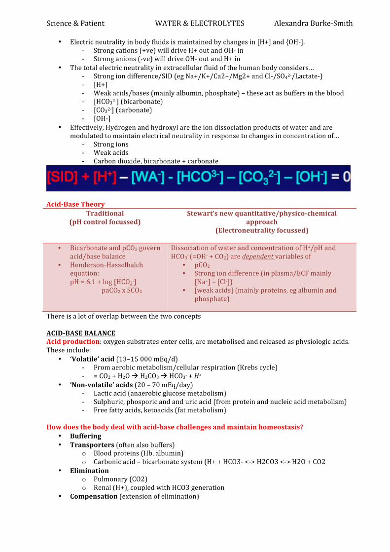
Remember: There is an independent electric neutrality priniciple between hydrogen and potassium ions, which is the reason for hyperkalaemia/hypokalaemia Maintenance of electric neutrality In any watery solution or compartment, the sum of all positively charged ions must equal the sum of all negatively charged ions.
• This is in fact the more prevalent principle: the relative ratio of [H+] and [OH-‐] must always be constant, e.g. [Na+] + [H+] = [Cl-‐] + [OH-‐]
Science & Patient WATER & ELECTROLYTES Alexandra Burke-‐Smith
• Electric neutrality in body fluids is maintained by changes in [H+] and {OH-‐]. -‐ Strong cations (+ve) will drive H+ out and OH-‐ in -‐ Strong anions (-‐ve) will drive OH-‐ out and H+ in
• The total electric neutrality in extracellular fluid of the human body considers… -‐ Strong ion difference/SID (eg Na+/K+/Ca2+/Mg2+ and Cl-‐/SO42-‐/Lactate-‐) -‐ [H+] -‐ Weak acids/bases (mainly albumin, phosphate) – these act as buffers in the blood -‐ [HCO32-‐] (bicarbonate) -‐ [CO32-‐] (carbonate) -‐ [OH-‐]
• Effectively, Hydrogen and hydroxyl are the ion dissociation products of water and are modulated to maintain electrical neutrality in response to changes in concentration of…
-‐ Strong ions -‐ Weak acids -‐ Carbon dioxide, bicarbonate + carbonate
Acid-‐Base Theory
Traditional (pH control focussed)
Stewart’s new quantitative/physico-‐chemical approach
(Electroneutrality focussed)
• Bicarbonate and pCO2 govern acid/base balance
• Henderson-‐Hasselbalch equation:
pH = 6.1 + log [HCO3-‐] paCO2 x SCO2
Dissociation of water and concentration of H+/pH and HCO3-‐ (=OH-‐ + CO2) are dependent variables of
• pCO2 • Strong ion difference (in plasma/ECF mainly
[Na+] – [Cl-‐]) • [weak acids] (mainly proteins, eg albumin and
phosphate)
There is a lot of overlap between the two concepts ACID-‐BASE BALANCE Acid production: oxygen substrates enter cells, are metabolised and released as physiologic acids. These include:
• ‘Volatile’ acid (13–15 000 mEq/d) -‐ From aerobic metabolism/cellular respiration (Krebs cycle) -‐ = CO2 + H2O à H2CO3 à HCO3-‐ + H+
• ‘Non-‐volatile’ acids (20 – 70 mEq/day) -‐ Lactic acid (anaerobic glucose metabolism) -‐ Sulphuric, phosporic and and uric acid (from protein and nucleic acid metabolism) -‐ Free fatty acids, ketoacids (fat metabolism)
How does the body deal with acid-‐base challenges and maintain homeostasis?
• Buffering • Transporters (often also buffers)
o Blood proteins (Hb, albumin) o Carbonic acid – bicarbonate system (H+ + HCO3-‐ <-‐> H2CO3 <-‐> H2O + CO2
• Elimination o Pulmonary (CO2) o Renal (H+), coupled with HCO3 generation
• Compensation (extension of elimination)

Science & Patient WATER & ELECTROLYTES Alexandra Burke-‐Smith
o Mainly a link between the respiratory and metabolic link via carbonic-‐acid bicarbonate system
o H+ + HCO3-‐ <-‐> H2CO3 <-‐> H2O + CO2 Carbonic-‐acid Bicarbonate System = Metabolic-‐respiratory link for buffering, transport, elimination and compensation of acid-‐base state
• This is in a state of dynamic balance between pulmonary elimination and renal elimination/generation
• Maintenance of normal physiological pH ~ 7.4 requires maintenance of 20:1 relationship
between bicarbonate and carbon dioxide component (Henderson-‐Hasselbalch equation)
Removal of Acid/Base
Lungs Kidneys Normal Removal of normal CO2
production by ventilation Excretion of H+ and generation of HCO3-‐ for removal and titration of normal non-‐volatile acid load
Metabolic acidosis Increased CO2 removal by hyperventilation
Increase excretion of H+ (&Cl-‐) and generation of HCO3-‐ for removal [also in respiratory acidosis]
Metabolic alkalosis Decreased removal of CO2 by hypoventilation
Decreased excretion of H+ > net urinary loss of filtered HCO3-‐ Decreased generation of HCO3-‐ [also in respiratory alkalosis]
As the kidneys excrete H+ etc in the urine, the urine pH range =4.5-‐8.5, allowing the blood range to be maintained. This is very easy to test Metabolic “balancing” The liver is also highly important for metabolic balancing and removal of acid/base
Liver Kidney • Lactate metabolism and gluconeogenesis
using H+ and generating HCO3-‐ for titration of peripheral H+ from lactic acid
• Urea formation generating H+ (titrating HCO3-‐ from metabolism of neutral and alkaline amino acids, but excess H+ from acid amino acids. (decreased in acidosis)
• Glutamine generation using H+ (increased in acidosis)
• Lactate metabolism to CO2 and H2O using H+ and generating HCO3-‐
• Excretion of H+ and generation HCO3-‐ in acidosis (or net excretion of HCO3-‐ in alkalosis incl. reduced urea generation, e.g. liver dysfunction)
• Glutamine metabolism > NH4+ excretion > HCO3-‐ generation
The balance between urea and glutamine in the liver is also key in acid base balance.

Science & Patient WATER & ELECTROLYTES Alexandra Burke-‐Smith INTERPRETING RESULTS AND COMMON ABNORMALITIES pH
• Definition = negative logarithm to the base 10 of hydrogen ion concentration [H+] • Implications
o Increase in pH ~ decrease in [H+], decrease in pH ~ increase in [H+] o pH change by 1 = hydrogen ion concentration change by factor 10 o Small decimal changes in pH mean relatively large changes in hydrogen ion
concentration e.g. pH change 7.4 to 7.1: doubling of hydrogen ion concentration from 40 to 80 nmol
o pH range compatible with life: 6.8 – 7.8 = hydrogen ion concentration 160 – 16 nmol § 6.8 is relatively common in diabetic ketoacidosis
• Normal pH o arterial = 7.35-‐7.45 (average = 7.4) o venous blood = 7.3-‐7.4 (average =7.36) o intracellular = 6.8-‐7.7 (average =7.0)
• Decreased pH o acidaemia: Increase in blood [H+] = decrease in pH (<7.36) o acidosis: Abnormal physiological process leading to blood pH < 7.36 o here, the Concentration of hydrogen ions exceeds that of hydroxyl/bicarbonate ions
• Increased pH o alkalaemia: Decrease in blood [H+] = increase in pH (>7.45) o alkalosis: Abnormal physiological process leading to blood pH > 7.45 o Hydroxyl ion/bicarbonate ion concentration exceeds hydrogen ion concentration
Standard bicarbonate Concentration of bicarbonate in a sample of blood kept under ‘standard’ conditions:
• Temp = 37˚ and pCO2 of 5.3 kPa (= 40 mmHg) • This is normalized respiratory conditions to eliminate changes in bicarbonate resulting
from potentially abnormal respiration, thus this assesses only the metabolic component of acid-‐base balance (either metabolic cause or compensation)
• Normal range 21 – 27 mmol/l • < 21 mmol/l: metabolic acidosis • > 27 mmol/l: metabolic alkalosis
Base excess and deficit
• Blood sample put under standard conditions in blood gas machine (37˚ and pCO2 of 5.3 kPa = 40 mmHg) to assess how much excess base or acid is in the blood sample due to metabolic alkalosis/acidosis or metabolic compensation of respiratory acidosis/alkalosis
• Compared to Standard Bicarbonate, takes not only carbonic-‐acid-‐bicarbonate but all blood buffer systems into account (depends on haemoglobin and albumin concentration in blood)
• more accurate assessment of metabolic component of acid-‐base status • Dependence on Haemoglobin and Albumin, which are often abnormal in sick
patients • Base excess = Amount of strong acid that would have to be added to produce normal pH
7.4 in sample • Base excess > + 2 mmol/l = metabolic alkalosis (or metabolic compensation of a
respiratory acidosis) • Base deficit: Amount of strong base that would have to be added to produce normal pH 7.4
in sample • Base deficit more negative than -‐ 2 mmol/l = metabolic acidosis (or metabolic
compensation of respiratory alkalosis) • You need to look at these AFTER looking at the pH

Science & Patient WATER & ELECTROLYTES Alexandra Burke-‐Smith Normal physiological values
• pH 7.36 – 7.44 • Actual Bicarbonate 21 – 28 mmol/l • Standard Bicarbonate: 21 – 27 mmol/l • Base excess/deficit: 0 +/-‐ 2 mmol/l • Lactate < 2mmol/l
COMMON ABNORMALITIES Metabolic acidosis
• Mechanisms: o Increase production metabolic acids o Decreased metabolism/elimination of metabolic acids o Decreased buffer capacity o Decrease in/loss of [HCO3] (+ rise in [Cl-‐])
• This overwhelms our buffer capacity, which results in Increased production and decreased metabolism and elimination of acid (eg. reduced lactate metabolism by liver, heart, kidney, renal elimination of H+ and generation of HCO3-‐)
• Respiratory compensation = increased removal of CO2 by hyperventilation (Kussmaul breathing) à maintenance of ~ 20:1 relationship between bicarbonate and carbon dioxide
• Common causes: o Addition of acid (overwhelming buffers and metabolic elimination or addition of
acid anions leading to rise in H+ to maintain electric neutrality_ § Lactic acidosis (caused by reduced blood flow and oxygen delivery) –
reduced oxygen delivery to tissues > increased anaerobic metabolism > increased production of lactate and reduced blood flow and oxygen delivery to liver, heart and kidney > decreased metabolism (to CO2 and H2O + glucogeogenesis) in liver, heart, kidney > increased lactate levels
§ Ketoacidosis (either beta-‐hydroxybutyrate, acetoacetate) caused by increased oxidation fo free fatty acids as opposed to glucose. This occurs in diabetes mellitus and starvation (think in pre-‐op nil-‐by-‐mouth patients)
§ Poisoning with external acid substances and their metabolites, eg salicylate, anti-‐freeze, methanol, ethanol
o Loss of Sodium bicarbonate ions § Gastrointestinal (diarrhoa, fistulae) § Renal (renal tubular acidosis)
o Hyperchloraemia – as blood chloride rises, blood hydrogen ion rises and bicarbonate falls (to maintain electric neutrality)
§ Normal = 154mmol/l o Renal failure – reduced net renal H+ removal and HCO3-‐ regeneration, or decreased
elimination of “fixed renal acids” and their anions sulphate and phosphate • Diagnosis (history, clinical examination, blood/urine tests)
o Blood lactate o Blood chloride o Urinanalysis (dipstick)
§ Ketoacidosis (starvation/diabetes) – ketones, glucose (high in diabetes, absent in starvation)
§ Real failure – urine pH <5, proteinuria, haematouria, low specific gravity Metabolic alkalosis
• Mechanism: Raised pH from raised HCO3-‐ • Causes:
o H+ loss -‐ Loss of gastric acid (and Na+) from severe vomiting or excessive gastric tube drainage
o Increased HCO3-‐ -‐ commonly due to hypochloraemia from diuretics (loss of Cl-‐ compensated by increased Bicarbonate anion to maintain electric neutrality
o Bicarbonate administration

Science & Patient WATER & ELECTROLYTES Alexandra Burke-‐Smith
o H+ shift into cells -‐ (Hypokalaemia) • Compensation: Pulmonary CO2 retention by hypoventilation (gain in volatile acid) -‐
(maintenance of ~20:1 relationship between HCO3-‐ and CO2) Chloride Changes in [Cl-‐] are associated with opposite changes in [HCO3 -‐] (to preserve electric neutrality)
• High blood chloride -‐ commonly from ‘Normal Saline’ administration (154 mmol/ Cl vs. normal blood Cl ~ 100 mmol/l) > low [HCO3-‐] > metabolic acidosis
• Low chloride -‐ commonly due to renal chloride loss from diuretics (+ also K+ and H+ loss) > high [HCO3-‐] > metabolic alkalosis
Respiratory Acidosis
• Mechanism: increase in CO2 (volatile acid) • Causes
o tissue acidosis -‐ increased production and decreased removal by poor perfusion § occurs with sepsis, hyperthermia, fever, hyperalimentation
o blood acidosis/acidaemia -‐ decreased removal by ventilation > ↑ CO2 + H2O è H2CO3 è HCO3-‐ + H+ ↑
§ occurs with hypoventilation due to pathology in CNS, PNS, respiratory muscles, airways, lungs/CVS
• Metabolic/renal compensation: Increased excretion of H+ (e.g. as NH4+Cl-‐) + retention/production of HCO3-‐
• Clinical correlate: Anaesthetized or critically ill patients on controlled mechanical ventilation lose the capacity to regulate their own pCO2
o This can cause drastic changes in their acid base status, e.g. metabolic acidosis exacerbation by too little mechanical ventilation
Respiratory Alkalosis
• Mechanism: Increased pH from excessive respiratory loss of CO2 (volatile acid) • Cause: hyperventilation
o Primary: anxiety, pain, stress o Response to hypoxia, eg. high altitude, pathological hypoxia o Mechanical hyperventilation
• Compensation: Increased renal HCO3-‐ excretion and H+ retention (gain of metabolic acid), i.e. reduced standard bicarbonate and base deficit

Science & Patient WATER & ELECTROLYTES Alexandra Burke-‐Smith SUMMARY
Key points
• H+ generated by metabolism (anaerobic metabolism of glucose + dissociation of CO2 from aerobic metabolism)
• H+ concentration controlled by: o Blood/tissue buffering – plasma proteins, Hb
(illustrated Fig 6) o Excretion of CO2 by the lungs o Renal excretion of H+ o Renal regeneration of HCO3-‐ (most important
buffer of H+, illustrated fig 7)

Science & Patient WATER & ELECTROLYTES Alexandra Burke-‐Smith
• pH addresses whether high or low [H+] o acidaemia = pH <7.35 o alkalaemia = pH > 7.44
• CO2 addresses respiratory physiology o High indicates inadequate CO2 excretion
(causes fig 10) or excess CO2 production =(rare, maybe severe lung disease or hyperpyrexia)
• Base excess addresses metabolic physiology o If positive, means acid needs to be added to
blood to remain normal = metabolic alkalosis o If negative, means acid needs to be removed
from the blood to remain normal = metabolic acidosis
• Compensation: dysfunction in one system (resp or metabolic) will result in compensatory
changes in the other. o Rapid chemical buffering occurs instantly but buffers rapidly depleted o Respiratory compensation:
§ Metabolic acidosis > hyperventilation (low PaCO2) § Metabolic alkalosis > hypoventilation (high PaCO2)
o Renal compensation: respond to disturbances by altering H+ excretion + HCO3-‐ reabsorption/regeneration. This always occurs with metabolic disturbances, so we tend to think of renal compensation as occurring with respiratory compensation
§ Respiratory acidosis > increase H+ excretion + increased HCO3-‐ reabsorption/regeneration > positive base excess
§ Respiratory alkalosis > decreased H+ excretion + decreased HCO3-‐ reabsorption/regeneration > negative base excess
• Mixed acidosis occurs when both PCO2 is high and base excess is negative (ie too much acid)
• Mixed alkalosis occurs when both PCO2 is low and base excess is positive (ie too little acid) http://www.nda.ox.ac.uk/wfsa/html/u13/u1312_01.htm

Science & Patient WATER & ELECTROLYTES Alexandra Burke-‐Smith
4. Acid-‐Base Balance: Clinical Cases Doris Doberenz
CASE 1 • 28 year old lady • 3 day history of abdominal pain, vomiting, lack of appetite and constipation 3 months after
appendicectomy • Admitted to surgical ward for observation and “drip and suck”: nil by mouth, intravenous
normal saline drip and nasogastric tube for gastric aspiration for presumed subacute bowel obstruction (due to adhesions after appendicectomy)
• Third night in hospital: increasing pain and abnormal blood acid base status • Abdominal operation: no surgical abnormality found
What kind of acid base abnormality could be present? • This patient has metabolic ketoacidosis due to starvation. This could be confirmed by
checking ketones in urine/blood. • Pre/intra-‐operative blood acid base status…
pH 7.3 (7.19 post-‐op) paCO2 3.3 kPa (4.1 post-‐op) standard bicarbonate 12 mmol/l (11.5 post-‐op) Base deficit -‐13 mmol/l (-‐15 post-‐op)
Thus, operation lead to deterioration. She didn’t need an operation! • If diabetes is suspected, they could have checked blood glucose. Although, this was normal.
The only reason for her acid base disturbance was starvation. • Ventilation eliminates carbon dioxide
CASE 2
• Elderly heavy smoker with severe chronic obstructive lung disease What acid base abnormality and compensation would you expect?
• Respiratory acidosis with metabolic compensation pH 7.34 PaCO2 7 kPa PaO2 7 kPa (on air) Standard Bicarbonate 30 mmol/l Base Excess + 5 mmol/l
CASE 3
• Recent onset acute asthma (bronchospasm, difficulty breathing) What acid base abnormalities could be expected in relation to the severity and duration?
• Respiratory alkalosis due to hyperventilation because of breathing difficulties. NO compensation, as acute. This is a type 1 respiratory failure
pH 7.46 PaCO2 4.1 kPa PaO2 8 kPa Standard Bicarbonate 25 mmol/l Base deficit 0 Lactate 1 mmol/l
• Eventually, carbon dioxide retention will occur, which leads to respiratory acidosis… pH 7.3 PaCO2 9 kPa PaO2 10 kPa (on 60% oxygen) Standard Bicarbonate 23 mmol/l Base deficit -‐ 1.5 Lactate 1.5 mmol/l

Science & Patient WATER & ELECTROLYTES Alexandra Burke-‐Smith
• If an asthmatic runs into this scenario, alarm bells ring. We know it is going to get worse and worse, and they may be intubated and over-‐ventilated, and go into cardiac arrest… mixed respiratory and metabolic acidosis
pH 7.1 PaCO2 8 kPa Standard Bicarbonate 10 mmol/l Base deficit -‐ 16 Lactate 10 mmol/l
CASE 4
• 43 year old gentleman • History of alcohol abuse • Upper abdominal pain • Persistent vomiting, unable to tolerate food • Very anxious/nervous, breathing fast
Which acid-‐base abnormality is likely? • Alkalosis: respiratory due to anxiety, and metabolic due to vomiting
pH 7.484 paO2 20.7 kPa (on 2l/min oxygen) paCO 4.77 kPa Bicarbonate 28.9 mmol/l Base excess + 4.8 mmol/l [Na] 116 mmol/l [K] 3.1 mmol/l Urine pH 7.5
CASE 5
• 60 year old lady • alcohol abuse • heavy smoking and chronic lung disease • possible ischaemic heart disease • previous stroke, associated with fits • found collapsed with no cardiac output • low blood glucose (1 mmol/l). • Cardiopulmonary resuscitation (including intubation and ventilation) • cardiac output re-‐established
Which acid base abnormality(ies) is/are likely? • Acidosis due to alcohol abuse and no cardiac output and diabetes and possible chronic lung
problems and poor ventilation. She also may have aspirated, or have pulmonary oedema. Many reasons!
pH 7.125 paCO2 7.93 kPa Standard bicarbonate 15.9 mmol/l Base deficit -‐11mmol/l Glucose 1.7 mmol/l (normal range 4 – 6 mmol/l)

Science & Patient WATER & ELECTROLYTES Alexandra Burke-‐Smith CASE 6
• 38 year old lady • Brain haemorrhage and difficult brain surgery • In Intensive Care on mechanical ventilation with aim to keep arterial carbon dioxide low
normal (to help control brain blood volume and brain pressure) Which (respiratory) acid base abnormality is to be expected and how would it be compensated? Respiratory alkalosis due to low paCO2 with a slight metabolic compensation by kidneys
pH 7.46 paCO2 4.3 kPa standard bicarbonate 20 mmol/l Base deficit -‐ 5 mmol/l
Later, after coming off ventilation and after prolonged IV normal saline, her results look like this… pH 7.33 paCO2 4.3 kPa standard bicarbonate 20 mmol/l Base deficit -‐ 5 mmol/l Na 145 mmol/l Chloride 115 mmol/l (normal 98 – 108)
She now has a metabolic acidosis with respiratory compensation CASE 7
• 46 y.o. lady in Emergency Dept • Chest pain • Nausea • Very anxious and distressed
Has respiratory alkalosis with no compensation due to acute onset pH 7.67 paCO2 2.3 kPa paO2 15.6 kPa Standard Bicarbonate 26 mmol/l Base Excess 2.2 mmol/l [Na+] 135 mmol/l [Cl-‐] 103 mmol/l [K+] 3.3 mmol/l Lactate 2.2 mmol/l
Some time later, she re-‐presented with severe vomiting. She therefore has metabolic alkalosis due to vomiting.
pH 7.53 PaCO2 5.3 kPa PaO2 11.2 kPa [Na+] 127 mmol/l [Cl-‐] 85 mmol/l [K+] 2.3 mmol/l Lactate 1.1 mmol/l

Science & Patient WATER & ELECTROLYTES Alexandra Burke-‐Smith
5. Sodium and Potassium Handling Dr EJ Clutterbuck
SODIUM HANDLING Sodium
• Mainly extracellular ion • Main component of ECF – most of osmotic activity,
depending on concentration not amount, sodium and water balance are closely linked
• Na is mainly regulated by the kidney, thus the kidney is seen as the regulator of ECF volume
Regulation of sodium
• Sodium content of body o 70kg wt -‐ 45L water -‐ 3000mmol Na
• Sodium intake o Food, drinks, medication ~60-‐200mmol / day
• Sodium loss o GI Tract -‐ vomit, diarrhoea ~100-‐130mmol/litre (same as ECF)
§ (10L) 1500mmol/day most reabsorbed. This leaves 15mmol o Skin -‐ insensible losses -‐ 900ml /30mmol Na o Urine -‐ Na excretion regulates Na balance (3-‐200mm/day excreted)
Renal regulation of sodium
• There is no barrier during glomerular filtration of sodium. Therefore with GFR of 125ml/min, we get a filtrate of 140mm/l of sodium.
• Tubular reabsorption is key in controlling extracellular volume
• With reduced sodium intake, urine losses of sodium can be reduced to 3mmol/day
Sites of sodium reabsorption:
• Filtrate -‐ 25,000 mmol/day • Proximal tubule (65%) -‐ 9,000 mmol remain • Loop of Henle (25%) -‐ 3,000 mmol remain • Distal tubule (9-‐10%) -‐ 3-‐200 mmol remain
Proximal tubule Na reabsorption
• Sodium transport from the tubular lumen to the peritubular capillary is controlled mainly by the basolateral membrane-‐bound ATPase
ICF ECF Na 10 140 Cl <10 100 HCO3 <10 25 K 100 3.5 Volume 30 litres 15 litres
(/day) filtration Excretion % reabsorbed
Water 180litres 1 litre 99.4
Sodium 25,000mm 100mm 99.6
Chloride 18,000mm 100mm 99.5
Bicarbonate 5,000mm Nil 100
Potassium 700mm 50mm 93.0

Science & Patient WATER & ELECTROLYTES Alexandra Burke-‐Smith
• This active NA/K exchange pump pumps Na out of cell in exchange for K, keeping intracellular [Na] low
• Na in the tubular lumen can then enter the cell passively down its concentration gradient • Other ways in which sodium is transported into the cell includes: co-‐transport with amino
acids/glucose/phosphate, and Na/H antiporter o The result of sodium transport is water movement along its osmotic gradient.
• Exchange transport: Na/H antiporter o H is generated in the cell from CO2 and H2O (action of carbonic anhydrase) o Filtrate bicarbonate is then converted back into CO2 (CA action), and the CO2
diffuses back into the cell o HCO3 in the cell will diffuse into the capillary. This is the mechanism for bicarbonate
reabsorption • As other solutes and water leave the tubular lumen, the relative chloride concentration
increases. This drives the paracellular diffusion of chloride ions into the peritubular capillary
Summary: the PCT is responsible for 65% of total filtrate sodium reabsorption Loop of Henle
• This is responsible for absorbing 25% of filtered sodium. This is a constant amount! • Sodium reabsorption occurs in the thick ascending limb, involving the sodium potassium
chloride co-‐transporter o The transporter is rate limited by chloride ion concentration, and the K+ transport
generates a positive potential difference • There is also passive paracellular Na (+Ca and Mg) movement • Loop diuretics (eg Frusemide) competes with Cl-‐, therefore blocking the action of the co-‐
transporter and reducing the corresponding water movement Distal Convoluted Tubule
• Responsible for 5% of Na reabsorption • Transport:
o Baselolateral Na/K pump (provides energy +keeps intracellular Na concentration low)
o Na/Cl co-‐transport • Thiazides diuretics inhibit NaCl reabsorption
Cortical collecting duct
• In the CT, there are 2 types of cells: o Intercalated cells (1/3) are responsible for H ion transport o Principle cells (2/3) are responsible for Na and Cl reabsorption, and K secretion
• Again, the basolateral Na/K pump maintains a low intracellular Na concentration, which drives passeive sodium entry into the cell via lumen channels. This creates a negative lumen potential.
• Aldosterone increases the number of sodium channels Regulation of Distal Na reabsorption Renin
• Allows almost complete reabsorption of Na -‐ Controlled finally by aldosterone • Juxtaglomerular apparatus (Afferent arteriole of glomerulus adjacent to its own distal
tubule (macula densa)). Renin released from JGA on stimulation by: o stretch receptors in afferent arteriole wall (ie blood volume low renin release) o cardiac/arterial baroreceptors -‐ adrenergic nerves o fall in lumen Cl in macula densa
• Renin converts plasma angiotensinogen to Ang I, which is converted to Ang II by ACE

Science & Patient WATER & ELECTROLYTES Alexandra Burke-‐Smith Angiotensin II
• increases systemic arteriolar resistance > rise in Blood pressure • stimulates aldosterone release from adrenal gland
o increases tubular sodium reabsorption (increases number of Na channels in distal cortical collecting duct)
o increases activity of Na -‐ H antiporter (proximal tubule Na reabsorption) Aldosterone
• High aldosterone states : low urinary sodium, sodium and water retention, hypokalaemia, alkalosis
Non-‐aldosterone factors affecting sodium excretion
• Changes in glomerular filtration rate o V Low GFR decreases Na excretion o Low GFR reduces sodium presented to prox tubule, ie Nearly all reabsorbed
• Changes in renal blood flow o Low tubular flow increases prox tubule reabsorption
• Changes in plasma oncotic pressure in tubule blood vessels o Haemoconcentration increases prox tubule reabsorption
Sodium and water handling These are linked…
Disorders of sodium handling Loss/gain of sodium is usually accompanied by H2O and indicated by a change in ECF volume NOT ECF [Na]
• Hypernatraemia (high Na) is usually a water deficit NOT sodium excess > cellular dehydration
o Presents with thirst, confusion and coma • Hyponatraemia (low Na) is usually water overload NOT sodium loss
o Sodium loss is usually isotonic, but water overload causes cellular overhydration o Presents with headache, nausea/vomiting, cramps, confusion, fits
• Pseudo-‐hyponatraemia is due to other osmotic agents attracting water and riluting sodium, eg glucose (diabetes) and urea (renal failure)
• Factitious hyponatraemia occurs when hyperlipidaemia/hyperproteinaemia replaces plasma water, and sodium is measured mmol/l of plasma

Science & Patient WATER & ELECTROLYTES Alexandra Burke-‐Smith Predominant sodium depletion is caused by…
• GI Fluid loss with water replacement o diarrhoea, vomit, sweat, burns -‐ drink water o iatrogenic -‐ excess dextrose replacement fluids
• Failure of homeostasis o Addisons disease (absence of aldosterone)
§ Normal homeostasis – aldosterone acts to maintain plasma osmolarity at the expense of Na loss
§ Addisons results in an inability to retain sodium, resulting in a high urinary sodium concentration and worsening of hyponatraemia
Summary of Hyponatraemia Serum [Na] is influence by the amount of Na and the total body water volume. This means we have to assess volume status before correction! Volume status Causes Urine Na conc Hypovolaemia Na-‐losing nephritis
Diuretic excess Addisons
>20
Vomit, diarrhoea Burns sweat
<10
Normovolaemia SIADH Hypo T4 Steroid deficiency Excess IV fluids
>20
Hypervolaemia Acute/chronic/nephrotic renal failure >20 Cardiac failure Cirrhosis Excess IV fluids
<10
Causes of sodium loss Direct causes:
• Vomiting, Diarrhoea, GI Fistula, Burns • Kidney tubule dysfunction
o pyelonephritis, analgesic nephropathy o recovery from ATN, post-‐obstructive uropathy
Associated with volume loss • plasma volume reduced -‐ dizzy, faint, tachycardia, low BP (postural drop) • reduced interstitial volume
o thirsty, nausea o no oedema, sunken eyes, dry mucosa
Treatment
• Treat underlying cause , eg: GI losses, hydrocortisone, fludrocortisone • Hypovolaemic Hyponatraemia:
o Replace salt and water (increase [Na]<6mm/l/day) • Normal saline (140mm/l) • Dioralyte (60mm/l Na, 20mm/l K, 90mm/l glucose)
• Normovolaemic Hyponatraemia: (SIADH) • Hypervolaemic Hyponatraemia :
o diuretics (to remove extracellular sodium), o water restrict (1l), ACEI

Science & Patient WATER & ELECTROLYTES Alexandra Burke-‐Smith Things to remember: you very rarely need to give hypertonic saline. Never correct more than 6mmol/l in 6 hours (ideally no more than this in first 24 hours). Overcorrection may cause osmotic demyelination syndrome Predominant Excess of Sodium Causes
• Inappropriate secretion of excess aldosterone • Primary aldosteronism (Conn’s syndrome)
o benign adenoma of adrenal cortex o urinary sodium retention
• Secondary aldosteronism o liver disease, Nephrotic, Protein malnutrition o reduced renal blood flow: hypertension, renal artery stenosis o cardiac failure
Primary aldosteronism = Conn’s Syndrome
o Presentation: water excess, high BP, hypokalaemia, high plasma bicarbonate o Early: low urinary sodium o Late: tubular damage -‐ rise urinary Na
Summary of sodium reabsorption
• Filtrate 25,000 mmol/day • Proximal tubule 65% 9,000 mmol remain • Loop of Henle 25% 3,000 mmol remain
– frusemide competes NA2ClK transporter • Distal tubule 9-‐10% 3-‐200 mmol remain
– thiazides compete NaCl transporter – aldosterone is main regulator (Na channels)
Summary of Sodium Handling
• Sodium and water homeostasis are interlinked • Potassium and Hydrogen take part in exchange • Fluid distribution between cells and ECF
o depends on osmotic differences o which are usually due to sodium concentration
• Aldosterone o is most important factor in Na regulation o controlled by RAS -‐ responds to renal blood flow
POTASSIUM HANDLING Regulation of potassium Potassium is the main intracellular ion’ it has a low concentration in the ECF, which does not vary much with changes in total body water
• Total body K ~ 3000mmol • Changes in Plasma [K] -‐ due to change in total body K or shift in K in / out of large
intracellular pool • Rise in ECF [K] -‐ fall in membrane potential, lower threshold for excitation (nerve, muscles)
Potassium balance • Intake -‐ foods: 60-‐80mmol / day • Output
o colonic fluid (rich in K) -‐ 10 mmol o sweat -‐ 10 mmol o primarily urine
Potassium and the kidney • Glomerular filtrate = 700 mmol / day • proximal tubule ~100% reabsorption

Science & Patient WATER & ELECTROLYTES Alexandra Burke-‐Smith
• distal tubule -‐ re-‐secreted (Na exchange) • urine losses ~ 60 mmol / day
• depends upon… • amount of Na available for exchange • amount of H and K in distal tubule / collecting ducts (H competes with K) • aldosterone level (causes Na retention / K loss)
Tubular secretion in the distal tubule (principle cells)
• Na/K ATPase o maintains high ICF K o K diffusion to lumen o Na diffusion from lumen
• Aldosterone is stimulated by the increase in plasma potassium. The effects of aldosterone are to…
o Increase Na and K channels o Drives Na/K ATPase
Tubular secretion in the medullary collecting duct (intercalated cells)
• Acidify urine -‐ Pump H against conc gradient (makes fresh bicarbonate) • Luminal Active K pump -‐ Reabsorb K
Remember, potassium secretion is determined by priority of Na reabsorption and H secretion. However, increased potassium concentration stimulates aldosterone secretion which in turn stimulates potassium secretion Potassium and Hydrogen handling
• Potassium depletion results in o K shift from cells to ECF o extracellular alkalosis o acid urine o high plasma bicarbonate (may lag behind a few days)
• This is useful when correcting acidotic states. ALKalotic states are associated with a Low K (potassium), therefore correcting acidosis also corrects hyperkalemia
Hypokalaemia Causes
• Loss of total body K o GI losses -‐ vomit, diarrhoea, surgical fistula o Kidney -‐ diuretics, renal disease, Cushings syndrome / steroids increased
aldosterone production • Shift from ECF into cells
o primary alkalosis (H leaves cells to correct alkalosis, K shifts into cells in exchange) o use of insulin (drives glucose into cells, takes K)
• Symptoms: o weakness, tetany (alkalosis reduces Ca ionisation) o ECG changes: ST depression, flat T; arrythmia
Therapeutic Potassium Repletion • Don’t overshoot • Not too fast (arrhythmias) -‐ no more than 40mmol per hour • If not fluid overloaded / normal kidney function -‐ give in normal saline 20-‐40mmol KClper
litre • Remember correcting acidosis will induce hypokalemia, so consider K replacement. In
diabetic ketoacidosis, fluids/insulin will also cause a fall in K • An example of a post-‐op patient with normal kidney function, give…
• 2 litres Nsaline (140mm/l) • 1litre dextrose 5%

Science & Patient WATER & ELECTROLYTES Alexandra Burke-‐Smith
• 20mmol KCl per litre • 1litre NSaline with KCL for GI losses
Hyperkalaemia Mechanism: Increased total body K Causes:
• increased intake -‐ food, drugs, IV fluids • renal disease (unable to secrete) • Aldosterone deficiency
o adrenal dysfunction o Blockage of renin -‐ angiotensin stimulus to aldosterone, eg ACEInhibitors
(captopril); ARBlockers (losartan) o Inhibition of action on distal tubule -‐ Competitive inhibitor: Spironolactone
• Inhibition of Na transport in distal tubule, eg Amiloride , triamterene • Shift of K from Cells to ECF
o severe systemic acidosis -‐ K exchanges for H • insulin deficiency (reduced uptake by cells) • cellular damage (release of K) -‐ rhabdomyolysis • False hyperkalaemia: haemolysis, high platelets • May be multifactorial -‐ Diabetic, hypertensive, heart failure, renal failure
o Treatment induced: Captopril, spironolactone Consequences:
• Potentially life-‐threatening • weak, irritable, paraesthesiae • ECG changes (tall T, wide QRS, flat P) • Arrhythmia àcardiac arrest if >7.5-‐8mmol/l
Treatment
• Shift into cells: iv Glucose / insulin (50ml 50% glucose / 10u actrapid) • Stabilise membrane potential: iv Calcium (10ml 10% Calcium gluconate iv) • Correct acidosis: Sodium Bicarbonate (oral, iv: 500ml 1.26%,100ml 8.4%-‐ watch Na load) • Chelate: Calcium resonium (PR 30g, PO 15g tds+lactulose) • If Renal failure -‐ call dialysis unit!

Science & Patient WATER & ELECTROLYTES Alexandra Burke-‐Smith
6. When the kidneys fail… Damian Ashby
INTRODUCTION Illustrative Case History Initial Presentation…
• HS, 26 male, Overweight, Collapsed at work, BP 100/40 • Drugs: enalapril, aspiring, glicazide (HTN, diabetes) • Blood: high glucose, low Na, high K • Why is K high? Cocaine, medication, diet, renal failure, diabetes? • Hyperkalaemia > loss of P waves, broad QRs, bradycardia (+ tall T waves) • Treatment: nothing
Returns 3 week later…
• Breathlessness • BP 160/85, R 24, swollen feat • FBC: Hb 113, Na 129, k 5.1, creat 460, gluc 7.4 • CXR shows pulmonary oedema • OE: pitting peripheral oedema • Diagnosis: renal impairement • How should he be treated? Frusemide (loop diuretic)
Returns 3 months later…
• 3 months • losing weight • confused, vomiting • BP 105/50 • Urine P++, B-‐, K-‐ • Clear chest x ray • Na 132, K 5.4, Urea 32.4, creat 560, gluc 12.2 • Diagnosis: kidney failure • Treatment: dialysis
Kidney Functions Regulation of…
• Fluid compartment volumes (ECF/ICF, Na/H2O) o Dysregulation > oedema (peripheral or pulmonary)
• Electrolyte balance (K, pH) o Dysregulation > hyperkalemia
§ ECG changes seen with hyperkalaemia = tall T waves, loss of P waves, broad QRS, bradycardia
• Excretion of metabolic wastes (urea, etc) o Dysregulation > nausea, pericarditis, encephalopathy
• Hormones (erythropoietin, vitamin D) o Dysregulation > Vit D deficiency/hypocalcaemia, anaemia
§ Vit D deficiency > bone disease > fractures and softening of bone § Anaemia > ventricular hypertrophy and iron overload
• Epo can be given IV

Science & Patient WATER & ELECTROLYTES Alexandra Burke-‐Smith RENAL REPLACEMENT THERAPY The indications for continuous renal replacement therapy include volume overload, electrolyte imbalance, uremia, acid-‐base disturbances, drugs. There are in fact 3 treatment options for renal failure:
• Filtration • Dialysis • Transplant
Haemofiltration + Haemodialysis Haemodialysis removes solutes by diffusion. The haemodialysis membrane contains long, tortuous, interconnecting channels that result in high resistance to blood flow
• During dialysis, blood flows along one side of a semi-‐permeable membrane as a solution of crystalloids (dialysate) is pumped along the other side of the membrane against the direction of blood flow.
• Small molecules diffuse across the membrane from regions of greater concentration to regions of lesser concentration, and the composition of the dialysis fluid is designed to produce as near normalization of the plasma as possible
o Therefore the sodium concentration of the dialysis fluid is physiologic but the potassium concentration is lower than that of normal plasma in order to establish a gradient from plasma to the fluid that promotes the removal of potassium ions from the patient’s blood
• The removal of salt and water is achieved by the creation of a transmembrane pressure gradient
There are 3 primary methods used to gain access to the blood in haemodialysis:
• Central venous catheter (CVC): plastic catheter with two lumens inserted into a large vein (vena cava, IJV or femoral) to allow large flows of blood to be withdrawn from one lumen and returned via the other. This is usually used for rapid access for immediate dialysis.
• AV (arteriovenous) fistulas are recognized as the preferred access method, and involves a surgical joining of an artery and vein to create an anastomosis. Blood flow rapidly flows through this, and can be felt (“thrill”) placing your finger over it, or heard (“bruit”) using a stethoscope. During treatment, two needles are inserted into the fistula. There are fewer infections, higher blood rates and lower incidence of thrombosis associated with the fistula, but lack of blood flow through the capillaries can result in cold extremities, and risk of aneurysm is higher.
• AV graft is like a fistula, but a synthetic graft joins the artery and vein. The advantage of a graft is that more locations are available as the graft can be made to different lengths
There are three types of dialysis: • Conventional – 3 times/week, 3-‐4 hours • Daily – at home • Nocturnal

Science & Patient WATER & ELECTROLYTES Alexandra Burke-‐Smith The advantages of haemodialysis include:
• Less expensive • Technically easier • Toxicity of molecules of higher molecular weight has yet to be demonstrated • Haemofiltration can only reduce, not normalize, the concentration of larger solutes
Haemofiltration removes solutes by convection. The haemofiltration membrane consists of relatively straight channels of increasing diameter that offer little resistance to fluid flow.
• This is used almost exclusively in the ICU for acute renal failure. • It is a slow continuous therapy in which sessions usually last between 12 to 24 hours
(performed daily) • During haemofiltration, a patient’s blood is passed through a set of tubing (filtration circuit)
via a machine to a semipermeable membrane (the filter) where waste products and water are removed. Replacement fluid is assed and the blood is returned to the patient
• With haemofiltration, dialysate is not used. Instead, a positive hydrostatic pressure drives water and solutes across the filter membrane from the from the blood compartment to the filtrate compartment, from which it is drained. Solutes, both small and large, get dragged through the membrane at a similar rate by the flow of water that has been engendered by the hydrostatic pressure. Thus convection overcomes the reduced removal rate of larger solutes (due to their slow speed of diffusion) seen in hemodialysis.
o An isotonic replacement fluid is then added to the blood to replace fluid volume and electrolytes. This is infused directly into the bloodline.
• Advantages of haemofiltration include:
o Near-‐complete control of the rate of fluid removal (i.e the ultrafiltration rate) o Precision and stability o Electrolytes or any formed element of the circulation, eg platelets or WBC, can be
removed/added independently of changes in the volume of total body water. This gives a better control of blood pressure
o There is also less risk of hyperlipidaemia associated with treatment Peritoneal dialysis
• Here, blood filtering occurs inside the body. This process uses the patient’s peritoneum in the abdomen as a membrane across which fluids and dissolved substances (electrolytes, urea, glucose, albumin and other small molecules) are exchanged from the blood.
• In PD, a soft tube called a catheter is used to fill your abdomen with dialysate. The fluid is drained regularly (4 times a day)

Science & Patient WATER & ELECTROLYTES Alexandra Burke-‐Smith Transplant Immune suppression is required for pre-‐ and post-‐operative treatment following renal transplant to prevent rejection. (examples opposite). This increases the likelihood for infection by opportunistic pathogens Complications
• Early complications o Vein thrombosis o Ureteric obstruction o Acute Rejection
• Late complications o Infections – CMV, fungal o Cancer – skin, lymphoma o Chronic graft failure
NB: life-‐expectancy is still reduced despite successful transplantation DOS AND DONTS!
• Don't prescribe IV fluids to dialysis patients • Don't use dialysis access devices for anything • Do discuss patients with their renal units • Do check drug dose with your pharmacist
Drug class Examples Corticosteroids Prednisolone Calcineurin inhibitors Cyclosporine
Tacrolimus Antiproliferative agents Azathioprine
Mycophenolate Monoclonal antibodies Daclizumab (IL-‐2R)
Alemtuzumab (CD52)

Science & Patient WATER & ELECTROLYTES Alexandra Burke-‐Smith
7. Drinking yourself to death: Ecstasy and water loading status Dr Peter Choi
Summary • Ecstasy may cause death by acute water overload, hyponatraemia and cerebral
oedema • Water balance is tightly regulated via thirst and aquaresis in order to prevent
osmotic water shifts across brain cells • Inappropriately elevated AVP levels are a feature of chronic water loaded-‐states. • Elevated AVP levels can now be treated by V2R antagonists, although long-‐term
survival benefit has not yet been defined CASE HISTORY Presentation
• 17 yo female student, First visit to nightclub • Vomitting, progression to semi-‐consciousness
and multiple seizures • Intubated • Cardiac arrest and death • Investigations showed:
o Very low serum sodium o Cerebral oedema
• Friends revealed… o Few alcoholic drinks o She had taken two ecstasy tablets o “Careful” to take lots of water
Ecstasy
• 3,4-‐Methylenedioxy-‐methamphetamine (MDMA} • Developed in 1914 as an appetite suppressant • The second most commonly abuse recreational drug in Europe. In the US, 39% college
students have used ecstasy • Ecstasy effects
o Sympathomimetic – tachycardic, sweating o Induces serotonin release – psychological effect (energy, empathy + euphoria)
• Pharmacology: o metabolisim via…
§ N-‐dealkylation § O-‐demethylation (COMT), P450 isoenzyme CYP2D6
o Metabolism is non linear o Genetic variability in metabolic status correlates with risk of toxicity o Drugs which inhibit CYP2D6 increase risk of toxicity
• Clinical syndromes associated with ecstasy are usually associated with the unpredictable side-‐effects (not dose dependent)
o Death – risk of death with first use 1:2000-‐1:50,000 o Neurological – acute vs chronic
§ Neurones release pre-‐formed serotonin § If exposure is chronic, neurons lose their ability to do so, therefore become
sad and lethargic o Cardiovascular -‐ hypertension, tachycardia, arrhythmia, sudden death (extreme) o Gastrointestinal – hepatotoxicity (also rare)

Science & Patient WATER & ELECTROLYTES Alexandra Burke-‐Smith
• Acute kidney injury from ecstasy. Ways in which it does this… o Non-‐traumatic rhabdomyolysis: extreme exertion, seizure, hyperpyrexia + possible
direct toxicity o Malignant hypertension o Volume depletion o Acute tubular injury (direct effect)
• Ecstasy and hyponatraemia is commonly described on first exposure (>85% in young women). Why?
o Direct effect on secretion of AVP (ADH) § Elevated urine osmolarity in face of serum hypo-‐osmolality § Controlled administration of MDMA to volunteers induces AVP § Serotonin dependent effect
o Direct effect on thirst centre o GI stasis o Club behaviour
Why did the girl die?
• MDMA led to inappropriate secretion of ADH • SIADH results in failure to excrete water • Water overload and dilute serum (hyponatramia) results in intracellular shift in water • Cellular swelling leads to cerebral oedema and brain stem herniation
WATER + OSMOLARITY BALANCE Water homeostasis
• 50% of body mass is made up of water. In normal steady state, there is <1% variation • Control is via regulation of…
o Thirst and water intake o Urinary loss of water
• Sodium is the most important ion with regards to water regulation
• Water regulation can
be thought of as being due to salt as the affector arm, with ADH forming the effector arm
• The functional role of volume regulation is during bleeding

Science & Patient WATER & ELECTROLYTES Alexandra Burke-‐Smith Regulation of water balance
• Normal plasma osmolality is 275-‐290mOsmol/kg • Variation of >1% initiates compensatory mechanism • Water overloading leads to a reduced plasma osmolarity and sodium, which results in
suppression of ADH • In conditions of water loss, there is an increase in plasma osmolarity and plasma sodium.
This results in an increase in ADH and thirst Hypothalamic osmoreceptors
• In the AV3V region, hypothalamic osmoreceptors initiate the effect mechanisms after sensing changes in osmolarity. This has been shown through infusion and ablation studies in rats.
• In response to the basic sensing mechanisms, higher order neurons are activated, with complex neural pathways regulating thirst. These are poorly defined.
o This regulation dysregulation is seen in psychiatric disease and extremes of age. Regulation of water excretion (acquaresis)
• During aquaresis, 90% of filtered water is reabsorbed (constitutively, PCT and descending loop of Henle)
• 10% of filtered water is then reabsorbed in the collecting ducts… o subject to regulation o ADH (AVP) dependent o Involves aquaporins o Also ADH independent processes involving secretin and oxytocin
• Arginine Vasopressin (AVP) inhibits acquaresis [aka antidiuretic hormone (ADH)]
o This is a cyclic octapeptide synthesized and secreted by the posterior pituitary gland (supraoptic nucleus + paraventricular magnocellular nuclei)
• Stimuli for AVP secretion include…
o Increasing plasma osmolarity (linear relationship shown on graph)
o Decreased intravascular volume (non-‐linear response that has protective effects during blood loss)
o Vomiting Arginine Vasopressin (AVP) This is the primary determinant of “free water” excretion, resulting in an increased permeability of luminal membrane of collecting duct tubules
• There are a number of different AVP receptors, binding at which have the following effects… o V1R – vasoconstriction, renal prostaglandin release (limits the action of AVP) o V2R– mediates anti-‐diuresis

Science & Patient WATER & ELECTROLYTES Alexandra Burke-‐Smith
o V3R – induces release of ACTH from pituitary • Half life of ADH is 15 minutes
o It is metabolized by the liver and kidneys • AVP binds V2r at the basolateral membrane, resulting in…
o Adenylate cyclase activation o Increased cAMP o Activation of PKA o AQP2 phosphorylation and apical translocation
• AQP2 insertion results in a net movement of water from the urine to the blood
• Aquaporins (AQP) are small hydrophobic molecules which form water channels. o There are 13 aquaporins identified o If you have a familial defect of AQP function, you have hereditary nephrogenic
diabetes insipidus PATHOLOGICAL WATER OVERLOAD Inappropriate ADH
• This results in an impairment of water excretion, with a mismatch water intake and loss leading to water retention and hyponatraemia
• Clinical characteristics for diagnosis of the syndrome of inappropriate ADH (SIADH): o Hyponatraemia o Hypo-‐osmolarity o Urine osmolarity of >100mOsmol/kg o Urine sodium >40mmol/l (usually) o Low plasma uric acid level (often)
urine
blood

Science & Patient WATER & ELECTROLYTES Alexandra Burke-‐Smith
• Determinants of severity of SIADH o Balance of intake and
excretion. If you have SIADH, but you control how much you drink, you can maintain your water balance
o Solute excretion (how significant is your osmolar water excretion?) If you are very salt loaded, you may be able to pee more, and thus are less likely to become water overloaded
o Escape from ADH: we do not understand why some people can escape, but some people with SIADH to be less responsive to ADH treatment
• Causes of SIADH o Drugs
§ Chlorpropamide, Carbamazepine, Cyclophosphamide § therapeutic ddAVP
o Malignancy § SCLC, Mesothelioma, Duodenal, Gastric
o ectopic secretion § CNS Disorders § Infection, CVA, Head Trauma
o Pulmonary Disorders § Pneumonia, TB, Asthma, Acute Respiratory Failure
o Surgery o Hereditary SIADH
§ V2R mutation The common clinical episodes we associate with water overload are… Heart Failure
• Hyponatraemia common in congestive heart failure (CHF)
• Severity of the hyponatraemia correlates with survival
o a 3mmol/L decrease in Na results in 10% increase in mortality
• The patients have inappropriately elevated AVP levels, and increased AQP2 expression (resulting from poor cardiac output and chronically “low” circulating volume). This in fact worsens oedema.

Science & Patient WATER & ELECTROLYTES Alexandra Burke-‐Smith
• Cirrhosis (liver failure) o Hyponatraemia also correlates with survival o AVP levels inappropriately elevated o AVP not suppressed by acute water load o Increased renal AQP2 expression
• Water overload and renal failure is not associated with SIADH as a result of reduced glomerular function. It is far more likely they will have a diabetes-‐like syndrome with a failure of urine concentration. They eventually become water overloaded as glomerular filtration falls
o However, underlying renal disease may exacerbate SIADH NEW THERAPEUTIC OPTIONS The dangers of hyponatraemia: what we are trying to avoid!
This diagram illustrates how the brain undergoes a process of adaptation to the hypo-‐osmolar state.
• The danger of sudden correction of the tonicity of the volume is the shunting of water out of the neuron cells, causing osmotic demylination. This is common in the pons, and may result in death
Treatment of hyponatraemia
• Fluid restriction (classic treatment) -‐ Aim <1L/day • Caution in pre-‐existing renal impairment, SAH • Intravenous saline – hypertonic to the urine osmolarity in order to increase solute excretion • Demeclocyline diminishes response to AVP at distal tubule (potential nephrotoxicity) • Vasopressin receptor antagonists (VAPTANS) are used preferentially to demeclocyline

Science & Patient WATER & ELECTROLYTES Alexandra Burke-‐Smith
o These are non-‐peptide V2R antagonists (conivaptan, tolvaptan, mozavaptan, lixivaptan) are licensed for euvolaemic/hypervolaemic (normal/high circulating volume) hyponatraemia
§ Not used in dehydration! o They increase acquaresis, raising serum sodium with no compensatory activation of
RAAS o Studies of short-‐term Tolvaptan use result in an increased Na retention and reduced
weight § Studies have also shown long-‐term use of Tolvaptan shows the effect on Na
is maintained, showing their efficacy o Limitations of V2R antagonists….
§ Thirst is increased, which may limit rise in sodium therefore patients are told not to drink
§ Excessive rate of correction is also possible (2% in clinical trials) § Cost therefore limited to acute life threatening hyponatraemia
Rate of correction of hyponatraemia is very important…
• The rate of correction of hyponatraemia is very important, balancing the risk of neurological complications with severe hyponatraemia (seizures, coma, confusion, dizziness, lethargy) and the risk of pontine demyelination with inappropriately rapid elevation of sodium…
o Max 2-‐4mmol/L over first 2-‐4hours in acute situations o Max 10mmol/L over 24 hours in chronic situations
• Example: Severe Hyponatraemia (<120mmol/L, seizure, coma) o 100mL bolus 3%saline o repeated after 10min if required o NEVER normal saline
Could she have been saved after the first seizure? YES
• Three boluses of 100ml 3% saline • Plasma sodium rises to 125 after 2hrs • Remains drowsy for 24hours • Rapid recovery thereafter • No requirement for Tolvaptan > FULL RECOVERY

Science & Patient WATER & ELECTROLYTES Alexandra Burke-‐Smith
8. The Renin-‐Angiotensin System Professor K Meeran
Look at year 1 notes! Causes of hypertension:
1. Essential 2. Adrenaline/Noradrenaline (physiological response to 3. Pheochromocytoma 4. Iatrogenic 5. Conn’s Syndrome 6. Stress (adrenaline) 7. Fluid overloading 8. Excess cortisol (Cushing’s) 9. SIADH
THE KIDNEY Roles of the kidney:
1. Excretion of metabolic waste products (urea, uric acid, drugs) 2. Excretion of water 3. Regulation electrolyte balance 4. Acid-‐base balance 5. Arterial BP regulation 6. Hormone secretion (erythropoietin, renin), metabolism (Vitamin D activation via 1-‐alpha
hydroxylase) + excretion 7. Gluconeogenesis
If your kidneys fail, you become acidotic and hyperkalemic Regulation Regulation of 1-‐5 is achieved through coupling of:
• Renal blood flow of renal vasculature o There are 2 capillary networks: the proximal afferent and distal efferent o The high pressure in the afferent arteriole drives glomerular filtration o The low pressure distal network includes peritubular capillaries (Vasa recta) which
is important in the function of the nephron • Fluid manipulation in renal tubular system
o There are 2 tubular systems in series: proximal and distal o The proximal tubular system is the Bowmans capsule, which is responsible for
production of primary urine o The distal tubular system includes the PCT, loop of Henle, DCT and collecting duct.
This is responsible for production of final urine • Renal blood flow is mainly regulated through the effects on the proximal capillary system
BLOOD PRESSURE Blood pressure is the force exerted by circulating blood on the walls of blood vessels.
• This force is due to the fact that the fluid (blood) is incompressible, but the vessel wall is distensible
• The result of the distensible vessel wall results in a cycle of accumulation of potential energy and its release > pulsatile flow
General concepts of blood flow regulation apply to any vessel in the body. • Blood flow depends on the pressure difference (gradient) and the obstruction (vascular
resistance) that it is meant to pass • Blood flow is also dependent on vessel diameter; reducing the vessel diameter reduces
blood flow (think of turning off a tap)

Science & Patient WATER & ELECTROLYTES Alexandra Burke-‐Smith Renal Blood Flow Total renal vascular resistance If we consider blood flow regulation in the kidneys, we look at renal blood flow = blood flow through the glomerulus (not entire kidney). There are 3 major segments of the blood flow through the glomerulus that are responsible for total renal vascular resistance:
• Interlobular arteries • Afferent arterioles • Efferent arterioles
Resistance (diameter) in the kidneys is controlled by: • Sympathetic nervous system • Local intrarenal control (autoregulation) • Hormones
In general, decreased diameter and increased resistance reduces renal blood flow. Decreased resistance with increased diameter increases renal blood flow.
• If renal artery and vein pressures remain constant, there is a conservation of energy = Bernoulli principle.
Glomerular filtration In the glomerulus, renal blood flow is roughly proportional to glomerular filtration. It is a balance of local pressures; hydrostatic and oncotic
• Total hydrostatic pressure is proportional to renal blood flow, and is the pressure responsible for pushing fluid out of the artery into tissues
• Oncotic pressure is caused by plasma proteins, and is responsible for retaining fluid within the intravascular space
We can only manipulate the hydrostatic pressure, via changes in the afferent and efferent arteriole, while oncotic pressure remains fixed by the blood protein content.
Afferent arteriole Efferent arteriole Increased resistance > decreased RBF > decreased total hydrostatic pressure + decreased glomerular filtration rate
Increased resistance > decreased RBF > increased total hydrostatic pressure + increased glomerular filtration rate
Decreased resistance > increased RBF > increased total hydrostatic pressure and increased glomerular filtration rate
Decreased resistance > decreased RBF > decreased total hydrostatic pressure + decreased glomerular filtration rate
Systemic Pressure
• For each heartbeat, blood pressure varies between systolic and diastolic pressures.. • Diastolic pressure is minimum pressure in the arteries, which occurs near the end of the
cardiac cycle when the ventricles are filled with blood • Systolic pressure is the peak pressure in the arteries immediately after the beginning of
the cardiac systole (ejection/ventricular contraction) • An example of normal measured values for a resting, healthy adult human is 115 mmHg
systolic and 75 mmHg diastolic (written as 115/75 mmHg, and spoken (in US) as "one fifteen over seventy-‐five").
• Pulse pressure is the difference between systolic and diastolic pressures. • Systolic and diastolic arterial blood pressures are not static but undergo natural variations
from one heartbeat to another and throughout the day (in a circadian rhythm). o They also change in response to stress, nutritional factors, drugs, disease, exercise,
and momentarily from standing up. o Sometimes the variations are large.
• Hypertension refers to arterial pressure being abnormally high, as opposed to hypotension, when it is abnormally low. Along with body temperature, blood pressure measurements are the most commonly measured physiological parameters.
• Systemic BP = arterial BP (mmHg) o Measures peripherally in every day clinical practice (brachial, radial or femoral
artery) using either a brachial cuff (non-‐invasive) or an arterial line (invasive)

Science & Patient WATER & ELECTROLYTES Alexandra Burke-‐Smith
• Brachial cuff measurement… o As soon as pressure in cuff = systolic BP, some blood flow will enter the arm =
marking the beginning of the Korachoff sounds o The cuff is then let down, where you hear a pulse. When the pressure in the artery is
lower than the cuff, the flow is continuous and the sounds disappear = diastolic BP Normal values
Regulation
• Short-‐term regulation is achieved by the sympathetic nervous system, controlling… o Total peripheral vascular resistance o Capacitance o Cardiac pumping ability o This is important in hypovolaemia, haemorrhage and septic shock
• Long-‐term regulation is achieved by the kidneys, which control… o Body fluid balance o Sodium balance o This is important in hypertension, pregnancy, diabetes and drugs
RENIN-‐ANGIOTENSIN SYSTEM Basic Principle
NB: Hypertension is the result of a sequence of 2 events…Volume loading and subsequent increased TPR

Science & Patient WATER & ELECTROLYTES Alexandra Burke-‐Smith BP control The renin-‐angiotensin system takes 20 minutes to become fully active following a decrease in BP. Ang II has two important effects with regards to BP control…
• Short-‐term vasoconstrictor effect (important in haemorrhage, shock etc) • Long-‐term volume loading effect (salt/water retention through kidneys)
Angiotensin II induced hypertension Chronically elevated angiotensin II levels (occurs in renal artery stenosis and intrinsic kidney disease resulting from diabetes, hypertension or glomerulonephritis) will result in hypertension
• Renal artery stenosis: constricted kidney with poor perfusion à ischaemia à RAS activation à salt/water retention/vasoconstriction by BOTH affected and unaffected kidneys à hypertension
o NB: ACE inhibitors will remove the rescue perfusion in the affected kidney, therefore are contraindicated
• Intrinsic kidney disease à patchy areas of local tissue ischaemia à chronic renin secretion à RAS activation à salt/water retention + vasoconstriction à hypertension
PHARMACOLOGICAL INTERVENTIONS A(B)CD guidelines These incorporate two aspects of the renal function…
• Renal UO capacity (RAS) • Salt intake
There are differences in the treatment choices within the population • Low renin HTN (salt-‐sensitive) -‐ In hypertensive patients aged 55 or older or black
patients of any age, the first choice for initial therapy should be either a calcium-‐channel blocker or a thiazide-‐type diuretic. For this recommendation, black patients are considered to be those of African or Caribbean descent, not mixed-‐race, Asian or Chinese.
• High renin HTN (salt-‐insensitive) -‐ In hypertensive patients younger than 55, white or Asian, the first choice for initial therapy should be an angiotensin-‐converting enzyme (ACE) inhibitor (or an angiotensin-‐II receptor antagonist if an ACE inhibitor is not tolerated).

Science & Patient WATER & ELECTROLYTES Alexandra Burke-‐Smith
CLINICAL CORRELATES Adrenocortical failure In response to angiotensin II, aldosterone is synthesized and secreted by the adrenal cortex.
• Raised aldosterone causes the tubules of the kidneys to increase the reabsorption of sodium and water, with concurrent secretion of potassium. This increases the blood volume and pressure, thus acts like a negative feedback system on the RAS
• In adrenocortical failure, this does not happen; therefore renin may continue to be secreted leading to hypertension
Heart failure
• What happens in heart failure? The heart’s pumping action becomes weak, with a resulting drop in BP from 120/70 to 119/69.
• In response, the body increases sodium retention, with a raised central venous pressure and a consequential improvement in BP
• As the failure worsens, the system increases CVP until the pressure is so high until the pressure is so high that fluid leaks into the lungs à pulmonary oedema (left-‐sided HF)