xylella fastidiosa: host range and advance in molecular ... · paolo baldi [email protected]...
TRANSCRIPT

REVIEWpublished: 08 June 2017
doi: 10.3389/fpls.2017.00944
Frontiers in Plant Science | www.frontiersin.org 1 June 2017 | Volume 8 | Article 944
Edited by:
Jesús Mercado-Blanco,
Consejo Superior de Investigaciones
Científicas (CSIC), Spain
Reviewed by:
Leonardo De La Fuente,
Auburn University, United States
Joao Lucio Azevedo,
University of São Paulo, Brazil
*Correspondence:
Paolo Baldi
Specialty section:
This article was submitted to
Plant Microbe Interactions,
a section of the journal
Frontiers in Plant Science
Received: 21 March 2017
Accepted: 22 May 2017
Published: 08 June 2017
Citation:
Baldi P and La Porta N (2017) Xylella
fastidiosa: Host Range and Advance
in Molecular Identification Techniques.
Front. Plant Sci. 8:944.
doi: 10.3389/fpls.2017.00944
Xylella fastidiosa: Host Range andAdvance in Molecular IdentificationTechniquesPaolo Baldi 1* and Nicola La Porta 1, 2
1 IASMA Research and Innovation Centre, Fondazione Edmund Mach, Trento, Italy, 2MOUNTFOR Project Centre, European
Forest Institute, Trento, Italy
In the never ending struggle against plant pathogenic bacteria, a major goal is the
early identification and classification of infecting microorganisms. Xylella fastidiosa, a
Gram-negative bacterium belonging to the family Xanthmonadaceae, is no exception
as this pathogen showed a broad range of vectors and host plants, many of which
may carry the pathogen for a long time without showing any symptom. Till the last
years, most of the diseases caused by X. fastidiosa have been reported from North
and South America, but recently a widespread infection of olive quick decline syndrome
caused by this fastidious pathogen appeared in Apulia (south-eastern Italy), and several
cases of X. fastidiosa infection have been reported in other European Countries. At least
five different subspecies of X. fastidiosa have been reported and classified: fastidiosa,
multiplex, pauca, sandyi, and tashke. A sixth subspecies (morus) has been recently
proposed. Therefore, it is vital to develop fast and reliable methods that allow the
pathogen detection during the very early stages of infection, in order to prevent further
spreading of this dangerous bacterium. To this purpose, the classical immunological
methods such as ELISA and immunofluorescence are not always sensitive enough.
However, PCR-based methods exploiting specific primers for the amplification of target
regions of genomic DNA have been developed and are becoming a powerful tool for
the detection and identification of many species of bacteria. The aim of this review is to
illustrate the application of the most commonly used PCR approaches to X. fastidiosa
study, ranging from classical PCR, to several PCR-based detection methods: random
amplified polymorphic DNA (RAPD), quantitative real-time PCR (qRT-PCR), nested-
PCR (N-PCR), immunocapture PCR (IC-PCR), short sequence repeats (SSRs, also
called VNTR), single nucleotide polymorphisms (SNPs) and multilocus sequence typing
(MLST). Amplification and sequence analysis of specific targets is also mentioned. The
fast progresses achieved during the last years in the DNA-based classification of this
pathogen are described and discussed and specific primers designed for the different
methods are listed, in order to provide a concise and useful tool to all the researchers
working in the field.
Keywords: grape pierce’s disease, citrus variegated chlorosis, OQDS, CoDiRO, Xylella diagnosis, asymptomatic,
leaf scorch disease, quarantine organism

Baldi and La Porta Xylella fastidiosa: PCR-Based Identification Approaches
INTRODUCTION
Xylella fastidiosa is a Gram-negative, slow growing and strictlyaerobic bacterium in the family Xanthmonadaceae. It is a widelydistributed plant pathogen as it can colonize the xylem of manydifferent species, causing a variety of diseases such as Pierce’sdisease (PD) in grape (Vitis vinifera) or citrus variegated chlorosis(CVC) (Purcell, 2013). X. fastidiosa can move upstream anddownstream along plant xylem, thanks to the presence of longtype IV pili (Li et al., 2007). The bacteria actively multiply until,in the later stages of infection, they block the plant xylem byforming a biofilm. As a consequence, water stress and nutritionaldeficiencies can occur in the host plant, causing the appearanceof disease symptoms (Hopkins, 1989). The first report abouta disease caused by X. fastidiosa dates back to the end of thenineteenth century, when the so called “California vine disease”destroyed about 14,000 ha of grapes in the Los Angeles area (CA,USA). Newton Pierce (1856–1916), a bacteriologist, was assignedto study the epidemic and even though he was not able to identifythe causal agent, he came to the conclusion that the disease waslikely caused by a microscopic infectious agent (Pierce, 1892).The disease was named Pierce’s disease in 1939, in a bulletin of theCalifornia Department of Agriculture (Gardner, 1974), but for along time the etiological agent was thought to be a virus, untilit was recognized as a bacterium in 1973 (Goheen et al., 1973;Hopkins and Mollenhauer, 1973). Pure cultures of the bacteriumwere isolated from grape in 1978 (Davis et al., 1978) and finally,in 1987, the causal agent of PD was properly classified and namedXylella fastidiosa (Wells et al., 1987).
Since the first report in grape, X. fastidiosa was isolated andidentified from an increasingly large number of plant hosts, withor without symptoms, and recognized to be the causal agentof different diseases(Moller et al., 1974; Hearon et al., 1980;Chang et al., 1993; Grebus et al., 1996; McElrone et al., 1999;Hopkins and Purcell, 2002; Montero-Astua et al., 2008). In manycases also wild plant species were found to carry this pathogen,but often in a latent stage only (Freitag, 1951; Raju et al.,1983; Hopkins and Adlerz, 1988; Blake, 1993; Hill and Purcell,1997; Li et al., 2001). The distribution range of X. fastidiosa isusually limited to tropical and subtropical areas, being its optimalgrowing temperature 26–28◦C (Feil and Purcell, 2001). In somecases strains of X. fastidiosa have been found in much coldercountries, such as Canada (Goodwin and Zhang, 1997), even ifusually this pathogen does not occur in areas with low wintertemperatures, such as New York and the Pacific Northwest ofUSA, or at high altitudes (Hopkins and Purcell, 2002). To date,most of the diseases caused by X. fastidiosa have been reportedfrom North and South America. According to the EPPO GlobalDatabase (https://gd.eppo.int/taxon/XYLEFA/distribution), onlyfew cases have been reported outside this area, like in Yugoslavia(Berisha et al., 1998), Switzerland (EPPO, 2015a), France (EPPO,2015b; Marcelletti and Scortichini, 2016b; Denancé et al., 2017),Germany (EPPO, 2016), Iran (Amanifar et al., 2014), and Taiwan(Leu and Su, 1993). In Europe X. fastidiosa was first recordedin Puglia region (southern Italy, province of Lecce), where itwas recognized to be the causal agent of a dangerous disease ofolive trees, the so-called olive quick decline syndrome (OQDS)(Elbeaino et al., 2014; Loconsole et al., 2014).
In the beginning X. fastidiosa was regarded as an extendedgroup of bacteria capable of infecting a wide range of host plantsand it was only in the early nineties, with the introductionof DNA-based genotyping techniques, that the researchersstarted to divide the species into different genetic groups (Chenet al., 1992). To date, at least five different subspecies ofX. fastidiosa have been reported and classified: X. fastidiosasubsp. fastidiosa, in one report called subsp. piercei and thencorrected to fastidiosa according to the rules (Rule 13d) ofthe International Code of Nomenclature of Bacteria (Schaadet al., 2004); X. fastidiosa subsp. multiplex; X. fastidiosa subsp.pauca; X. fastidiosa subsp. Sandyi, and X. fastidiosa subsp. tashke(Schaad et al., 2004; Randall et al., 2009; Janse and Obradovic,2010). The subsp. sandyi is associated with disease in oleander,Jacaranda spp., daylily and magnolia (Schuenzel et al., 2005;Hernandez-Martinez et al., 2007). Recently, a new subspecies, X.fastidiosa subsp.morus, has been proposed (Nunney et al., 2014c).Moreover, it cannot be ruled out that other subspecies still exist,as till now most of the studies about the genetic diversity of X.fastidiosa have been performed on cultivated crops of relevanteconomic importance, while little is known about the strainsthat colonize wild grasses, sedges and forest trees. Therefore, thedevelopment and application of molecular methods to the studyofX. fastidiosa’s genetic diversity can be of primary importance tofill such gap and extend our knowledge about the true diversity ofthis organism.
Many sap-feeding insects can function as vectors forthe transmission of X. fastidiosa to host plants, especiallysharpshooters and froghoppers or spittlebugs (Cicadellidae)(Janse and Obradovic, 2010; Bhowmick et al., 2016). Afteracquisition from the source plant, the bacterium is persistentin the vector (Severin, 1949) and can multiply in the foregut(Brlansky et al., 1983; Hill and Purcell, 1995). The process ofacquisition and transmission of X. fastidiosa by the vector isvery complex and can be dependent on many variables such ashost plant, vector species and bacterium subspecies in interactionenvironmental variables, first at all climate. Nevertheless, oneimportant factor has been proven to influence the efficiency ofacquisition, that is X. fastidiosa population size (number of livecells per gram of plant tissue) (Hill and Purcell, 1997). In fact,the feeding apparatus of vectors is not an easy environment tobe colonized, due to the fast flow of sap, that was estimatedto reach an average speed of 8 cm/s (Purcell et al., 1979) andthe possible turbulence caused by the fast contractions of themuscles allowing the insect to pump sap from the plant, thatare contracted and relaxed approximately once every second(Dugravot et al., 2008). Therefore, it is likely that only few cells,out of the thousands acquired by the vector when feeding from aninfected plant, actually succeed to colonize the vector’s foregut.Another factor that can influence the efficiency of X. fastidiosatransmission, and particularly inoculation, is a long access periodof the vector to the plant, as probably longer periods allow theinsects to deliver a greater number of bacteria, having time togenerate a large number of inoculation events (Almeida andPurcell, 2003).
The initial belief that X. fastidiosa was a generalist pathogencapable of infecting a very large range of host plants hasgradually changed with the discovery and characterization of
Frontiers in Plant Science | www.frontiersin.org 2 June 2017 | Volume 8 | Article 944

Baldi and La Porta Xylella fastidiosa: PCR-Based Identification Approaches
genetically different strains of X. fastidiosa, each capable ofinfecting distinct hosts. Also the infection characteristics canvary considerably in different species. In some hosts the bacteriamultiply locally but cannot move and eventually the infectioncan regress spontaneously (Purcell and Saunders, 1999). In thepast, reciprocal transmission tests have been conducted forX. fastidiosa strains. For example, the strains infecting grapecannot infect peach (Prunus persica) and peach strains cannotinfect grape (Hopkins, 1989). Similarly, grape strains cannotinfect oleander (Nerium oleander) and vice-versa (Purcell et al.,1999), while a CVC strain produced leaf scorch disease in coffee(Coffea arabica) (Li et al., 2001) and CVC and coffee strainscan infect grape (Hopkins and Purcell, 2002). Natural isolatesproduced leaf scorch disease in American elm tree but failed inreciprocal transmission in sycamore (Sherald, 1993). However,in this puzzling host/pathogen interaction the host specificity ofX. fastidiosa is probably a genetically-controlled character, eventhough in the genome of this bacterium no genes coding foreffector proteins were found, nor it is present a type III secretionsystem (Van Sluys et al., 2003). Pathogenicity factors have beenfound in X. fastidiosa under the control of a cell-cell signalingsystem, similar but not equal to the one found in the sistergenus Xanthomonas (Chatterjee et al., 2008). By altering suchcell-cell signaling system it is possible to modify X. fastidiosa hostspecificity (Killiny and Almeida, 2011).
HOSTS AND DISEASES
As already mentioned above, X. fastidiosa can infect a greatnumber of plant species. A partial list of the main hosts is shownin Table 1 (EFSA, 2016). At present, according to the EuropeanFood Safety Authority (EFSA), the updated list of X. fastidiosahosts consists of 359 plant species (including hybrids) from 75different plant families (EFSA, 2016). Even if the infection processis always the same, the symptoms and the diseases caused by X.fastidiosamay vary among species. In addition to PD (Stevensonet al., 2005), CVC and ALS, the most known and dangerousdiseases are phony peach disease (PPD) in peach and a numberof leaf scorch diseases such as oleander leaf scorch (OLS), coffeeleaf scorch (CLS) and plum (Prunus domestica) leaf scald (PLS).
Grape (PD)
Symptoms of Pierce’s disease may vary according to thecharacteristics of the infected cultivar as well as to the time ofinfection and seasonal factors (Goheen, 1988). Plants that wereinfected the previous growing season will generally show moresevere symptoms when compared to those infected only in thecurrent season. The first signs of the disease are a sudden dryingand yellowing of the leaf margins (a red band can be presentin red cultivars), due to the occlusion of leaf veins by bacterialinfestation (Hopkins, 1981; Newman et al., 2003). Eventually,the affected (scorched) leaves will fall, usually from the distalpart of the petiole, leaving the leaf stems attached to the cane(matchsticks) (Stevenson et al., 2005). Severely infected plantscan be completely defoliated within late summer. During thefirst year of infection, the symptoms are limited to one or fewshoots, but get worse over time. The seasonal concentration of
bacteria in infected tissues is variable, being at a maximum in latespring and early summer. Usually, X. fastidiosa is not detectablein shoots from current season during the first 4 weeks of growth,but it’s detectable in old wood (Hopkins, 1981). New woodwill mature irregularly, producing patches of brown and greenbark (the so called green islands), especially in the intermediatezone of a shoot, between the green tip and the browned basalpart (Stevenson et al., 2005). The tips of canes may eventuallydie back and the new shoots will be shorter and stunted. Alsofruit production will be progressively reduced, with most of thefruit clusters drying and wilting. In 1–5 years, depending onthe susceptibility of the different genotypes, the infected plantsusually die, even if differences may be due also to the climate andparticularly to water stress (Thorne et al., 2006). Indeed, for manyyears most of the symptoms of PD were attributed to the limitedwater transport due to vessels occlusion (Hopkins, 1989) butrecently it was shown that plants infected with X. fastidiosa showsymptoms that are characteristic of PD and cannot be reproducedby water deficit (Thorne et al., 2006).
Citrus (CVC)
X. fastidiosa can infect any type of citrus species and hybrids butthe severity of symptoms may vary according to the genotypeof the host. Sweet oranges are the most susceptible, whilegrapefruit, mandarins, lemons, limes and trifoliate orange areonly moderately susceptible (Garcia et al., 2012; Gmitter et al.,2012; Casais et al., 2014; Fadel et al., 2014). In most cases CVCis not lethal but infected trees always display a reduced vigorand growth rate and a decreased productive lifespan. Symptomsdevelop faster in young trees, that are also more susceptibleto new infections (Garcia et al., 2012) Symptoms can be easilyconfused with zinc deficiency, as plants show chlorotic spotson the upper surface of leaves, especially in the interveinal area(Beretta et al., 1997). Deficiencies of P and K have been reportedin leaves of CVC affected trees, together with high concentrationsof Fe, Mn, and Zn (Silva-Stenico et al., 2009). On the leaves,chlorotic lesions appear on the upper side, while on the lowerleaf side gummy lesions may appear, due to the production byX. fastidiosa of fastidian gum, an exopolysaccharide that wasproposed to be involved in the formation of biofilms that allowthe attachment and survival of bacteria inside the xylem vessels(da Silva et al., 2001). While the leaf matures, the gummy lesionscan enlarge and become necrotic. Fruits are also affected andthis represents one of the major problems of CVC, especiallyfrom an economical point of view. Affected fruits remain smaller(thinning does not occur on infected branches), become hard andripen earlier. The color is the same as healthy fruits but the juicecontent is reduced, while acidity is higher (Gonçalves et al., 2014).Bacteria can be found also in roots of infected plants (Hopkinset al., 1991).
Peach (PPD)
Symptoms of PPD are not immediately apparent on host plantsuntil 1 year or more from the first infection. Infected plantsusually show a reduced internode length of new growth, giving tothe tree a rather bushy aspect (Hutchins, 1933). Leaves becomeflattened and dark-green. In spring, trees infected by X. fastidiosa
Frontiers in Plant Science | www.frontiersin.org 3 June 2017 | Volume 8 | Article 944

Baldi and La Porta Xylella fastidiosa: PCR-Based Identification Approaches
TABLE 1 | Partial list of the main plant hosts of Xilella fastidiosa and their X. fastidiosa subspecies.
Host scientific name Type of infection EPPOCode Subspecies
Acacia saligna Incidental ACASA pauca
Acer rubrum Incidental ACRRB multiplex
Carya illinoinensis Minor CYAIL multiplex
Citrofortunella microcarpa Minor CJFMI nd
Citroncirus Minor 1CJCG nd
Citrus Minor 1CIDG pauca, fastidiosa
Citrus sinensis Major CIDSI pauca
Coffea sp. Major COFSS pauca (BRA)
Coffea sp. Major COFSS fastidiosa (C.Rica)
Cyperaceae Wild/Weed 1CYPF nd
Fortunella Minor 1FOLG nd
Liquidambar styraciflua Incidental LIQST multiplex
Medicago sativa Minor MEDSA fastidiosa
Morus alba Incidental MORAL morus, (former multiplex, sandyi)
Morus rubra Incidental MORRU fastidiosa
Nerium oleander Major NEROL sandyi
Olea europaea Major OLVEU pauca (ITA, ARG, BRA)
Olea europaea Major OLVEU multiplex (USA, FRA)
Persea americana Incidental PEBAM nd
Platanus occidentalis Minor PLTOC multiplex
Poaceae Wild/Weed 1GRAF nd
Polygala myrtifolia Major POGMY pauca (ITA)
Polygala myrtifolia Major POGMY multiplex FRA)
Poncirus trifoliata Minor PMITR nd
Prunus angustifolia Incidental PRNAN nd
Prunus armeniaca Minor PRNAR multiplex
Prunus avium Minor PRNAV pauca (ITA)
Prunus avium Minor PRNAV fastidiosa (USA)
Prunus cerasifera Incidental PRNCF multiplex
Prunus domestica Minor PRNDO multiplex
Prunus dulcis Minor PRNDU multiplex-fastidiosa (USA)
Prunus dulcis Minor PRNDU pauca (ITA)
Prunus persica Major PRNPS multiplex, fastidiosa
Prunus salicina Minor PRNSC multiplex
Quercus palustris Minor QUEPA multiplex
Quercus rubra Minor QUERU multiplex
Sorghum halepense Wild/Weed SORHA nd
Spartium junceum Incidental SPUJU fastidiosa (USA)
Spartium junceum Incidental SPUJU multiplex (FRA), pauca (ITA)
Ulmus americana Minor ULMAM multiplex
Vaccinium corymbosum Minor VACCO multiplex
Vaccinium virgatum Minor VACVG nd
Vinca minor Incidental VINMI pauca (ITA)
Vitis Minor 1VITG fastidiosa
Vitis labrusca Minor VITLA fastidiosa
Vitis vinifera Major VITVI fastidiosa
Westringia fruticosa Incidental WESRO pauca (ITA)
woody plants Wild/Weed 2WOOP multiplex
Source: EPPO Global Database (https://gd.eppo.int/taxon/XYLEFA/hosts), and EFSA Journal database (EFSA, 2016). Major infections are indicated in bold.
Frontiers in Plant Science | www.frontiersin.org 4 June 2017 | Volume 8 | Article 944

Baldi and La Porta Xylella fastidiosa: PCR-Based Identification Approaches
flower and leaf earlier than normal and hold their foliage longerin the fall (Wells et al., 1981; Hopkins and Purcell, 2002). Unlikeother diseases caused by X. fastidiosa, PPD is not lethal to theinfected plants. Nevertheless, PPD may cause significant damageto orchards as one of the main consequences of the infectionis loss of fruit production. If trees become infected prior toproduction age, they will never produce fruits, while plantsinfected after production age will exhibit reduced productionand smaller and highly colored fruits that are not suitable forthe market (Evert, 1985). After 2–4 years after first infection,diseased trees may stop producing fruit at all. In peach, X.fastidiosa was detected in roots xylem fluid both in symptomaticor asymptomatic trees (Aldrich et al., 1992).
Unlike other Prunus species such as almond and plum,peach does not show leaf scorch symptoms when infected by X.fastidiosa (Ledbetter and Rogers, 2009).
Other Hosts (Scorch Diseases)
Many plant species, when infected by X. fastidiosa, show verysimilar symptoms, especially on the leaves, and namely: earlyafter infection a slight chlorosis appears, usually along themargins of leaves. In some species (e.g., coffee, olive), thesesymptoms may affect initially younger shoots, while in others(e.g., elm) may progress from older to younger leaves (Sherald,1993) and this may confound the early diagnosis. Symptomson leaf petioles of infected plants colonized by X. fastidiosavaried from coffee, plum, and sweet orange in clear decreasingorder of severity (Alves et al., 2004). In all cases symptoms getworse with time, even if differences may exist depending onthe genetic background of the host plant, timing of inoculationand overwinter survival of the pathogen (Cao et al., 2011). Thedisease gradually extend from one or few branches to the entirecrown and the leaves may desiccate completely and eventuallyfall. One of the major problems caused by X. fastidiosa infectionsis the decreased fruit quality and yield in commercially importantcrops such as coffee and olive (Rocha et al., 2010; Della Colettaet al., 2016) Plants affected by scorch disease usually show weakand stunt growth, may be more susceptible to environmentalstresses, such as water or heat stress, but do not always die.Instead, they can be removed to reduce the risk of new infectionsor because the weakened plant has become dangerous (e.g.,ornamental trees). The problem of X. fastidiosa infection isgetting particularly serious in the south-eastern part of Italy onolive trees as a strain of X. fastidiosa subsp. pauca was stronglyassociated to a severe burst of OQDS (Saponari et al., 2013).Only few years ago (2008–2010) the first cases where reported.Nowadays, according to the last surveys, an area of at least10,000 ha in the province of Lecce (Salento) could be infectedby X. fastidiosa (Martelli et al., 2016). Only recently two olivecultivars, Leccino and Favolosa FS-17 were selected, that appearto have some degree of tolerance to the disease (Giampetruzziet al., 2016). The introduction in Puglia of X. fastidiosa could bedue to infected plant material imported from Central America(Giampetruzzi et al., 2015a; Marcelletti and Scortichini, 2016b).Other recent reports describe the presence of X. fastidiosa in olivetrees showing leaf scorch symptoms in Argentina (Haeltermanet al., 2015) and Brazil (Della Coletta et al., 2016). The two South
American strains resulted different from each other and fromthe Italian strain but were both classified as belonging to thesubsp. pauca. Over the last few years, the presence of X. fastidiosaoutside the American continent is becoming more and morecommon. In October 2015, the bacterium (X. fastidiosa subsp.multiplex) was discovered in France, initially on the island ofCorsica, on Polygala myrtifolia plants (ornamentals) and lateron the mainland (X. fastidiosa subsp. multiplex) (EPPO, 2015b),with almost 300 foci found and nearly 30 host plant speciesdeclared contaminated (Denancé et al., 2017). In order to prevententry and spread of X. fastidiosa within the European Unionterritory, specific EU phytosanitary measures have been taken(PM 7/24 (2) 2016. Xylella fastidiosa. EPPO Bull, 46: 463–500.doi:10.1111/epp.12327).
Detection and IdentificationDetection and identification of X. fastidiosa is not an easytask. First of all, these bacteria are slow-growing (up to 2–3 weeks can be necessary to obtain colonies on agar media)and secondly many common culture media are not suitable togrow X. fastidiosa strains. Instead, selective media (PD2, PW,CS20) should be used (Schaad et al., 2001). Direct identificationof bacteria is possible only using dark field or phase contrastmicroscopy, due to the dimensions of X. fastidiosa cells (0.2–0.4 µm radius and 0.9–3.5 µm length) (Wells et al., 1987).In addition, microscopy identification requires a quite specificprofessional training to be efficient. Immunological methods,such as enzyme-linked immunosorbent assay (ELISA) can beused (Chang et al., 1993; Leu et al., 1998). More recently,detection of X. fastidiosa by immunofluorescence technologyhas been described (Carbajal et al., 2004; Buzkan et al., 2005).Anyway, most of the antibody-based detection assay are effectiveonly at the species level, while at our knowledge a single reportis available describing the development of single chain variablefragment (scFv) antibodies specific for X. fastidiosa subsp. pauca(Yuan et al., 2015). Another detection method that since thenineties has become more and more popular relies on PCRamplification of bacterial DNA. Several protocols have beendeveloped employing different techniques, all with the use ofspecific primers capable of recognizing target sequences ongenomic DNA. The PCR-based detection methods are usuallyfaster and cheaper than standard plating methods and morespecific than immunological methods as it is often possible todesign primers specific for the desired genus, species or sub-species. Despite all these advantages, some drawbacks are stillpresent in the use of DNA amplification methods. First of allspecific equipment is required and often the protocols must beoptimized in order to work properly with the specific samplesand conditions of a given laboratory. Then, even when all thetechnical requirement are met, there is still the possibility todetect false-positive or false-negative for several reasons. Forexample, the PCR-based methods all allow detection of bacterialDNA, that in some cases can still persist in the environmenteven when the bacterial cells are no longer viable (Willerslevand Cooper, 2005). Therefore these methods alone cannot beused to distinguish between viable and non-viable bacteria, withexception of few cases (Gedalanga and Olson, 2009). The genetic
Frontiers in Plant Science | www.frontiersin.org 5 June 2017 | Volume 8 | Article 944
Baldi and La Porta Xylella fastidiosa: PCR-Based Identification Approaches
material for PCR amplification can be isolated with standardprotocols, which may be optimized according to the user’s needs,or using several commercial kits, many of which are suitable forrobotized DNA extraction and allow the simultaneous processingof up 96 samples (Smit et al., 2001). In order to avoid thelaborious and time-consuming step of DNA purification, severalattempts were made using directly plant sap as a template, notalways with reliable results (Minsavage et al., 1994; Banks et al.,1999).
The DNA-based methods are rapidly becoming the mostwidely used in all modern laboratories for the detection andidentification of X. fastidiosa strains infecting a number of plantspecies and new protocols and primers are continuously beingdeveloped. Here follows a review of the most commonly usedPCR approaches (summarized in Table 2).
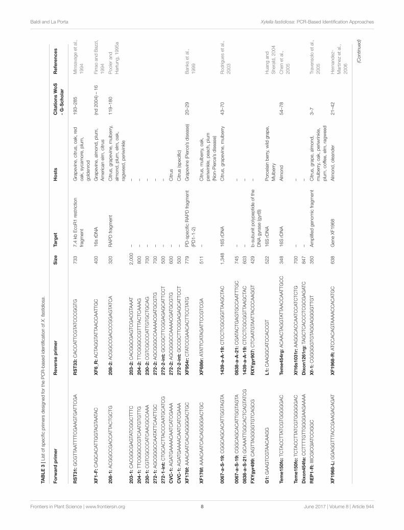
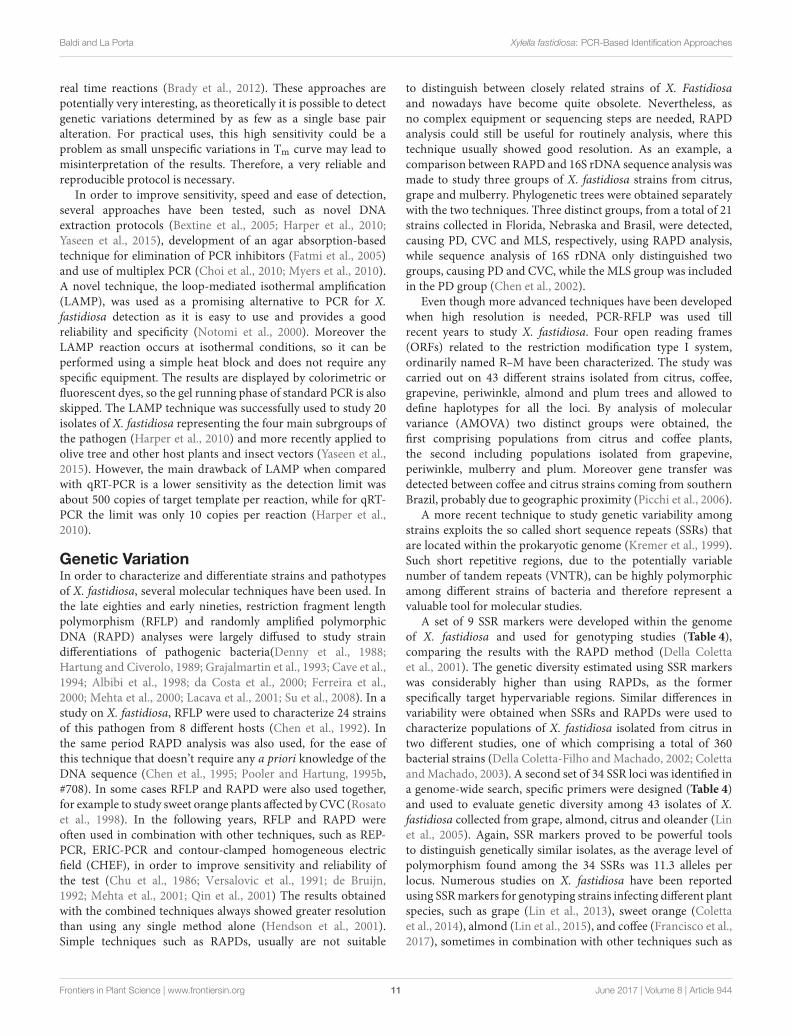
PCR Detection with Specific PrimersSince the first half of the nineties specific PCR primers havebeen used in order to identify X. fastidiosa from infected plants.In the first reports, the genomic region amplified could belongto a defined gene, such as 16S rDNA (Firrao and Bazzi, 1994),but also to selected fragments of bacterial DNA of unknownfunction (Minsavage et al., 1994). In Pooler and Hartung (1995a)developed a series of specific primers capable to detect X.fastidiosa in general and X. fastidiosa strains that cause CVCby cloning and sequencing randomly amplified polymorphicDNA (RAPD) products (Pooler and Hartung, 1995a). Sincethen a number of reports have been published describing thedevelopment of specific PCR primers for the identification ofX. fastidiosa (Table 3) and if the 16S rDNA and the 16S-23Sintergenic spacer region (ITS) are the most common targets(Rodrigues et al., 2003; Chen et al., 2005; Martinati et al.,2007), also several other genomic sequences, with or without aknown function, have been used (Travensolo et al., 2005; Huang,2009). The use of different genes, alone or in combinations,can be exploited in order to increase the level of sensitivity andspecificity of the test. As a matter of fact, the early detection ofpathogens before the development of any symptom by the planthost is crucial for the development of a correct defense strategybut when used for field analysis the detection test must be alsoas easy and fast as possible. Therefore, it is difficult to developa single test that can be used in all conditions, but combiningdifferent approaches the researchers can find a balance amongsensitivity, specificity and ease of use. As an example, the geneencoding the b-subunit polypeptide of the DNA gyrase (gyrB)was used in combination with the 16S rDNA in order to increasethe specificity of the test, because gyrB is thought to evolve muchfaster than 16S rDNA (Yamamoto et al., 1999), therefore allowinga higher resolution when comparing closely related strains ofbacteria (Rodrigues et al., 2003). For an initial genetic analysis ofthe population of X. fastidiosa in Texas, gyrB was used togetherwith mopB, the latter coding for an outer membrane proteinof the OmpA family with a fairly conserved sequence (Moranoet al., 2008). When using a single primer pair to amplify aspecific target, there is a low but non-zero possibility of false-positives due to primer recognition of non-target DNA with
high sequence similarity to the target. On the contrary, false-negatives can be generated due to variations in bacterial genome,particularly to mutations in the recognition site of the primers(Scally et al., 2005). To avoid such problems, more complexapproaches can be used. In a recent study, gyrB was combinedwith a set of housekeeping genes for the development of an array-PCR protocol that allowed to analyze a large number of samplesavoiding the occurrence of false negatives due to the failure ofPCR amplification of a single primer (Livingston et al., 2010).
Based on the target genomic region or selected bacterialgene, the specificity of the PCR primers used for the analysiscan vary, from genus to sub-species and in some cases even todifferent strains belonging to the same sub-species, so it is cruciala correct primer choice in order to avoid misinterpretationof results. For X. fastidiosa analysis, a number of explicativeexamples can be found in literature. Starting from a pool ofX. fastidiosa-specific RAPDs, Pooler and Hartung (Pooler andHartung, 1995a) analyzed a group of 21 bacterial strains collectedin different regions of USA and Brazil and developed a pair ofprimers capable to distinguish X. fastidiosa strains causing CVCfrom all the others. Similarly, Banks and colleagues designed twospecific sets of primers from a PD strain collected in Florida:the first could be used to distinguish between X. fastidiosa andXanthomonas campestris and the second to amplify a 511 bpfragment from 98 PD strains but not from CVC strains of X.fastidiosa (Banks et al., 1999). Therefore, both sets of primerscan be exploited to identify X. fastidiosa but the second showed agreater specificity and can be used in more detailed studies. Morerecently a set of primers specific for OLS strains of X. fastidiosawas developed and tested successfully on cultured bacteria,infected plant samples and insect vectors capable of transmittingOLS (Huang, 2009). A particular case was reported when a setof primers specific for mulberry-infecting strains of X. fastidiosawas developed starting from the nucleotide sequence of a uniqueopen reading frame identified only in mulberry-infecting strainsamong all the North and South American strains of X. fastidiosasequenced at the date of that work (Guan et al., 2015). Suchprimers could distinguish between mulberry-infecting strainsand those infecting other species, such as sycamore, elm, oak,plum, maple, and grape. Surprisingly, with the same set of primera specific amplification could be obtained also from two isolatesof X. fastidiosa belonging the recently sequenced CoDiRO straininfecting olive trees in Italy (Guan et al., 2015). This means thatsometimes it can be difficult to identify unambiguously a givenstrain of X. fastidiosa by the use of a single approach. Even theuse of advanced techniques such as multilocus sequence typing(described in more details in one of the next sections) is notalways enough to distinguish among closely related strains, asfor the characterization of a new X. fastidiosa strain (Salento-1)infecting olive trees in Italy (Bleve et al., 2016). In this case, theanalysis of two additional genes, the polymerase sigma 70 factor(rpoD) and the chromosomal replication initiator protein DnaA(dnaA) was necessary to separate Salento-1 from closely relatedstrains ofX. fastidiosa subsp. pauca isolated from citrus and coffeein Brazil (Bleve et al., 2016).
A multiprimer combination with three different targets wasdeveloped in order to differentiate strains of X. fastidiosa
Frontiers in Plant Science | www.frontiersin.org 6 June 2017 | Volume 8 | Article 944

Baldi and La Porta Xylella fastidiosa: PCR-Based Identification Approaches
TABLE2|Most
commonlyuse
dPCR-base
dtechniquesforX.fastidiosaidentificatio
n.
Molecular
method
Advantages
Disadvantages
References
ClassicPCR
Highse
nsitivity,sp
ecificity
andaccurate
resu
ltsforthedetectio
nof
Xylella
andits
subsp
eciesalsoinnon-axe
nicconditions.
Many
applicatio
nsin
molecularanalysis.
Easy
diagnosticinterpretatio
n
Unableto
quantifythetargetDNA,onlyqualitativetest.Some
metabolitesorcontaminants
inthesa
mplecaninterfere
with
PCRperform
ance.PCRconditionsmust
beoptim
izedin
eachhost
andenvironmentforbetterperform
ance
FirraoandBazzi,1994;Minsa
vageetal.,
1994;Poolerand
Hartung,1995a;Banks
etal.,
1999;Rodrig
uesetal.,
2003;
HuangandSherald,2004;Chenetal.,
2005;Travenso
lo
etal.,
2005;Hernandez-Martinezetal.,
2006;Martinatietal.,
2007;Moranoetal.,
2008;Huang,2009;Livingstonetal.,
2010;Melanso
netal.,
2012;Guanetal.,
2015;Bleve
etal.,
2016
RAPD
Use
fultostudyunkn
ownsp
ecieswhere
there
isnotapriori.
knowledgeofse
quencing.Quite
use
fultodetecthigh
polymorphisms.
Lim
itedcost,Sim
pleandrapid.Smallamountof
template
DNArequire
d.Theamplificatio
nproducts
canbefurther
characterized
Markers
are
dominant.Reproducibility
canbelowamonglabs
andwith
differentpolymerase
sandfacilities,
esp
ecially
when
notusingrandom
prim
ers
with
highannealingtemperature.In
manycase
sstandardizatio
noftheprotocolforeachlabis
require
d.Nowadays
thistechniqueisconsideredobso
lete
Dennyetal.,
1988;HartungandCiverolo,1989;Grajalm
artin
etal.,
1993;Chenetal.,
1995,2002;PoolerandHartung,
1995a;Albibietal.,
1998;Rosa
toetal.,
1998;Banks
etal.,
1999;Hendso
netal.,
2001;Lacava
etal.,
2001;Qin
etal.,
2001;Suetal.,
2008
RFLP
Itwasoneofthefirst
methodsuse
dforgenetic
fingerprin
ting,The
basicRFLPanalysisisnolongeruse
d.Varia
tionsexist
suchas
term
inalrestric
tionfragmentlength
polymorphism
(TRFLP),which
maystillhave
applicatio
nsrelatedto
thecharacterizatio
nof
bacteria
.ByPCR-R
FLP,
thehyb
ridizatio
nstepcanbeskipped
Obso
lete
technique.RelativelylargeamountofDNAis
require
d.RFLPapproachistediousandrequire
snumerous
stepsthatmaytake
weeks
toyield
resu
lts.Relativelyhigh
cost
andlow
polymorphism
Chenetal.,
1992;Rosa
toetal.,
1998;Mehta
etal.,
2001;Qin
etal.,
2001;Picchietal.,
2006
qRT-PCR
Itallowsnotonlytheidentificatio
n,butalsothequantificatio
nof
bacteria
inrealtim
e.Highse
nsitivity,sp
ecificity
andreproducibility.
Relativelyfast
method.Itispossibleto
use
alsovaria
tionsin
meltingtemperature
todifferentiate
strainsofbacteria
Exp
ensive
equipmentandreagents
are
require
d.Settingup
andoptim
izatio
noftheprotocolrequire
specifictechnical
skillsaswelltheinterpretatio
nofresu
lts
Oliveira
etal.,
2002;Schaadetal.,
2002;Bextineetal.,
2005;
Francisetal.,
2006;BextineandChild,2007;Choietal.,
2010;Harperetal.,
2010;Bradyetal.,
2012;Guanetal.,
2013;Lietal.,
2013;Ionesc
uetal.,
2016
SSR
Sim
plelabprocedure,relativelylow
coststo
start,base
donPCR
term
ocycler.Highlevelo
fpolymorphism
andrelativelylow
amount
oftargetDNArequire
d.Co-dominantmarkers.Thereproducibility
isquite
good
Previouskn
owledgeofthegenomicse
quenceisrequire
dto
designsp
ecificprim
ers,thusSSRsare
limitedprim
arilyto
economically
importantsp
ecies.
Pointmutatio
nsatthesite
of
prim
erannealingcould
leadto
occurrenceofnullalleles
Della
Coletta-Filhoetal.,
2001;ColettaandMachado,2003;
Lin
etal.,
2005,2013,2015;Montero-A
stuaetal.,
2007;
Montes-Borregoetal.,
2015;Franciscoetal.,
2017
MLST
Highlydiscrim
inatory
nucleotid
ese
quencebase
dmethodof
characterizatio
nbase
donthese
quencingofapproximately
450-bpinternalfragments
ofse
venhouse
keepinggenesamplified
byPCR.Thisapproachisparticularly
helpfulforthetypingof
bacteria
lpathogens.
Thesystem
isvery
sensitiveto
discrim
inate
X.fastidiosasu
bsp
eciesandstrainsin
rapid
realtim
ereactio
ns.
ThemajoradvantageofMLSTisthepossibility
tocompare
the
resu
ltsobtainedin
differentstudies.
Itmayalsobeuse
dto
address
basicquestionsaboutevo
lutio
nary
andpopulatio
n
biologyofbacteria
lspp.
Theanalysisofonlyse
venlocim
aylim
itthese
nsitivity,
esp
ecially
whenclose
strainsare
analyzed.Sequencingofthe
PCRproducts
usinganautomatedse
quencerisrequire
d.For
that,MLSTisnotalways
suitableforroutin
einfectio
ncontrols
oroutbreakinvestigatio
ndueto
relativelyhighcost
andlack
ofbroadaccess
tohigh-throughputDNAse
quencing
Scally
etal.,
2005;Schuenzeletal.,
2005;Alm
eidaetal.,
2008;Yuanetal.,
2010;Bradyetal.,
2012;Nunneyetal.,
2012,2013,2014a,b,c;Parkeretal.,
2012;Elbeainoetal.,
2014;HarrisandBalci,2015;MarcellettiandScortichini,
2016a;Bergsm
a-Vlamietal.,
2017;Coletta-Filhoetal.,
2017;
Denancéetal.,
2017;Kandeletal.,
2017
MultiplexPCR
Costsare
reducedwhencomparedto
standard
PCRaswellas
reactio
nvo
lumes.
Itallowsrapid
detectio
nalsoofmultiplestrains
simultaneously.Close
tubesystem
limits
theriskofcontaminatio
n
Prim
erdesignisthecriticalp
oint,theycaninterfere
each
othergivingfalsenegative(genesorbacteria
undetected).
Skilledpersonnelisrequire
dto
perform
thetest
Rodrig
uesetal.,
2003;Choietal.,
2010;Myers
etal.,
2010;
Lopesetal.,
2014;Ja
cquesetal.,
2016
NestedPCR
Improvedse
nsitivity
andsp
ecificity
whencomparedwith
classical
PCRmethodology.Use
fultechniqueforstudyingmolecular
epidemiologyin
thefield
Theprotocolm
aybealittle
more
difficultto
optim
izethanfor
standard
PCR.More
timeconsu
mingandexp
ensive
than
norm
alP
CR.Unableto
quantifythetargetDNA
Pooleretal.,
1997;Buzkanetal.,
2003;Ciapinaetal.,
2004;
Huang,2007;Silvaetal.,
2007;Lopesetal.,
2014
Frontiers in Plant Science | www.frontiersin.org 7 June 2017 | Volume 8 | Article 944
Baldi and La Porta Xylella fastidiosa: PCR-Based Identification Approaches
TABLE3|Listofsp
ecificprim
ers
designedforthePCR-base
didentificatio
nofX.fastidiosa.
Forw
ard
primer
Reverseprimer
Size
Target
Hosts
CitationsWoS
-G-S
cholar
References
RST31:GCGTTA
ATTTTCGAAGTGATTCGA
RST33:CACCATTCGTA
TCCCGGTG
733
7.4
kbEcoR1restric
tion
fragment
Grapevine,citrus,
oak,
red
oak,
sycamore,plum,
goldenrod
193–2
85
Minsa
vageetal.,
1994
XF1-F:CAGCACATTGGTA
GTA
ATA
CXF6_R
:ACTA
GGTA
TTA
ACCAATTGC
400
16srD
NA
Grapevine,alm
ond,plum,
Americ
anelm
,citrus
(nd2004)–16
FirraoandBazzi,
1994
208-1:ACGGCCGACCATTA
CTGCTG
208-2:ACGGCCGACCCGGAGTA
TCA
320
RAPDfragment
Citrus,
grapevine,mulberry,
alm
ond,plum,elm
,oak,
ragweed,periw
inkle
119–1
80
Poolerand
Hartung,1995a
203-1:CACGGCGAGTA
TCGGCTTTC
203-2:CACGGCGAGTCGACGTA
AAT
2,000
––
204-1:TTCGGGCCGTCAATGTGTTG
204-2:TTCGGGCCGTTTA
CTCAAAG
800
––
230-1:CGTCGCCCATCAACGCCAAA
230-2:CGTCGCCCATTGTGCTGCAG
700
––
272-1:AGCGGGCCAATA
TTCAATTGC
272-2:AGCGGGCCAAAACGATGCGTG
700
––
272-1-int:CTGCACTTA
CCCAATGCATCG
272-2-int:GCCGCTTCGGAGAGCATTCCT
500
––
CVC-1:AGATGAAAACAATCATCGAAA
272-2:AGCGGGCCAAAACGATGCGTG
600
–Citrus
CVC-1:AGATGAAAACAATCATCGAAA
272-2-int:GCCGCTTCGGAGAGCATTCCT
500
–Citrus(specific)
XF176f:AAACAATCACAGGGGACTGC
XF954r:CTA
TCCGAACACTTCCTA
TG
779
PD-specificRAPDfragment
(PD1-1-2)
Grapevine(Pierce’sdisease
)20–2
9Banks
etal.,
1999
XF176f:AAACAATCACAGGGGACTGC
XF686r:ATA
TTCATA
GATTCCGTCGA
511
–Citrus,
mulberry,oak,
periw
inkle,peach,plum
(Non-P
ierce’sdisease
)
0067-a-S
-19:CGGCAGCACATTGGTA
GTA
1439-a-A
-19:CTCCTCGCGGTTA
AGCTA
C1,348
16SrD
NA
Citrus,
grapevine,mulberry
43–7
0Rodrig
uesetal.,
2003
0067-a-S
-19:CGGCAGCACATTGGTA
GTA
0838-a-A
-21:CGATA
CTGAGTGCCAATTTGC
745
––
0838-a-S
-21:GCAAATTGGCACTCAGTA
TCG
1439-a-A
-19:CTCCTCGCGGTTA
AGCTA
C603
––
FXYgyr499:CAGTTA
GGGGTGTCAGCG
RXYgyr907:CTCAATGTA
ATTA
CCCAAGGT
429
b-subunitpolypeptid
eofthe
DNAgyrase
(gyrB)
–
G1:GAAGTCGTA
ACAAGG
L1:CAAGGCATCCACCGT
522
16SrD
NA
Porcelain
berry,wild
grape,
Mulberry
Huangand
Sherald,2004
Teme150fc:TCTA
CCTTA
TCGTGGGGGAC
Teme454rg:ACAACTA
GGTA
TTA
ACCAATTGCC
348
16SrD
NA
Alm
ond
54–7
8Chenetal.,
2005
Teme150fc:TCTA
CCTTA
TCGTGGGGGAC
Xf16s1031r:AAGGCACCAATCCATCTCTG
700
––
Dixon454fa:CCTTTTGTTGGGGAAGAAAA
Dixon1261rg:TA
GCTCACCCTCGCGAGATC
847
––
REP1-R
:IIIICGICGIATCCIGGC
Xf-1:CGGGGGTGTA
GGAGGGGTTGT
350
Amplifiedgenomicfragment
Citrus,
grape,alm
ond,
mulberry,oak,
periw
rinkle,
plum,coffee,elm
,ragweed
3–7
Travenso
loetal.,
2005
XF1968-L:GGAGGTTTA
CCGAAGACAGAT
XF1968-R
:ATCCACAGTA
AAACCACATGC
638
GeneXF1968
Alm
ond,oleander
21–4
2Hernandez-
Martinezetal.,
2006
(Continued)
Frontiers in Plant Science | www.frontiersin.org 8 June 2017 | Volume 8 | Article 944

Baldi and La Porta Xylella fastidiosa: PCR-Based Identification Approaches
TABLE3|Contin
ued
Forw
ard
primer
Reverseprimer
Size
Target
Hosts
CitationsWoS
-G-S
cholar
References
XF2542-L:TTGATCGAGCTGATGATCG
XF2542-R
:CAGTA
CAGCCTGCTGGAGTTA
412
GeneXF2542
Grape,alm
ond,Spanish
broom,Brassicasp
p.
ALM1:CTGCAGAAATTGGAAACTTCAG
ALM2:GCCACACGTGATCTA
TGAA
521
GeneALM1
Alm
ond(specific)
16S-23SF:GATGACTGGGGTGAAGTCGT
16S-23SR:GACACTTTTCGCAGGCTA
CC
650
16S-23Sintergenicsp
acer
Citrus,
coffee,grapevine,
mulberry,alm
ondelm
,
ragweed,periw
inkle
1–2
Martinatietal.,
2007
307BBF:GCAAGTCAGGGTA
GCGTCTC
943BBR:GGCTTCTCTGTCGATTTTCG
nd
mopB
Grape,Seamyrtle,
Redsp
ikeMexicanhat,
others
5–1
0Moranoetal.,
2008
HQ-O
LS08:TGTA
CGTCCTGAAACCATCTTG
HQ-O
LS05:TTCTGGAAGCTTTGAGTA
AGGG
274
RAPDfragment
Oleander(specific)
7–1
0Huang,2009
D056L:AACAAGGGACCTTCCATGC
D858R:AGCAATCGCTGCACCTA
AAT
842
Succinyl-C
oAsynthetase
alphasu
bunit(sucD)
Alm
ond
0–0
Livingstonetal.,
2010
G151f:GATCCGGAAAGTGGGGAGATTA
CTA
TC
G368r:GCCATTGCAAAAGCAGTA
CGCTCA
217
DNApolymerase
IIIsu
bunit
beta
(dnaN)
–
A151f:GATGCGGAAAGCGGGGAGATTA
CTA
TT
A518r:GCCTTTA
CGCGGCAAAATA
ATCTGA
367
DNApolymerase
IIIsu
bunit
beta
(dnaN)
–
A97f:CATGCTGGTGGTA
AGTTCGACGATA
AC
A476r:CAACAATGCCGTTGTGCTCACCG
379
b-subunitpolypeptid
eofthe
DNAgyrase
(gyrB)
–
G223f:CGGTGGCGAAACGGTA
ATCC
G705r:GGAGAAATGTTTGGCAAAGACAGGC
482
mdhMalate
dehyd
rogenase
–
A345f:TTTTTCAATGTTGGCGACAGGCTTA
CT
A705r:GGAGAAATGTTTGGCAAAGACAGGT
360
mdhMalate
dehyd
rogenase
–
B001L:TTA
GGTGGCAAGGATCGAAT
B462R:GGGCCGATCAAAATCAATCT
491
ppiB
Peptid
yl-prolyl
cis-transisomerase
–
A284f:
GACGAGTTTGCCAAGTTTGATGATGAAATC
A680r:GCCAGTCGAACCCACCAAG
396
gltA
Citrate
synthase
–
I068L:CGTGGGTCACGAGTCATA
AA
I386R:TCACACAAAACTA
CGGCACTG
358
rpsI30Srib
oso
malp
rotein
S9
–
PspB-256f:TGAGTGCCTGCGGTGGTA
PspB-256r:CGAAACTTGGCAGCTA
ACG
256
psp
BSerin
eprotease
–
XFPglA_F
w:GCCTCCGGTGCGACTGCTTC
XFPglA_R
v:GCTGCGATTGGACACACATTG
nd
PglA
Pecan,Grapevine,
Oleander,sycamore
10–1
4Melanso
netal.,
2012
Mul-15040-F:ATTTTCGCGATTTTGGAGTT
Mul-15040-R
:TTCTTGTGTA
CTCCGCCTCA
312
Hyp
otheticalp
rotein
with
putativebacillith
iolsystem
oxidoreductase
,YpdAfamily
Mulberryandolive(specific)
2–2
Guanetal.,
2015
Xfa-rpod-F4:ACTGAGGTTGTCGTTGGCTT
Xfa-rpod-R
4:CCTCAGGCATGTCCATTTCC
988
RNApolymerase
sigma-70
factor(rpoD)
Olive,citrus,
coffee
1–2
Bleve
etal.,
2016
Xfa-d
naA-2F:TTCCATCAAATTGACGCGCT
Xfa-d
naA-2R:CGGCAAGCATGTA
ACACTGT
650
Chromoso
malreplicatio
n
initiatorprotein
DnaA(dnaA)
–
Frontiers in Plant Science | www.frontiersin.org 9 June 2017 | Volume 8 | Article 944

Baldi and La Porta Xylella fastidiosa: PCR-Based Identification Approaches
infecting grape, almonds and oleander (Hernandez-Martinezet al., 2006). In this way, strains belonging to different subspecies(multiplex, fastidiosa, and sandyi) could be distinguished.Moreover, two different strains (ALSI and ALSII) belonging tosubspecies multiplex and infecting almonds could be separatedon the basis of amplification patterns. When the aim of thestudy is to characterize a new strain or a group of strainsof X. fastidiosa rather than the fast and efficient identificationof the pathogen under field conditions, the combination ofmultiple molecular techniques can greatly enhance the resolutionpower of the test. A good example is a study performed onpecan, where a multiprimer PCR assay was used in combinationwith other PCR-based techniques, such as amplification andsequence analysis of the 16S-23S ITS and pglA gene as well asenterobacterial repetitive intergenic consensus (ERIC)-PCR andrepetitive extragenic palindromic (REP)-PCR. In this way, theauthors were able to classify the X. fastidiosa strains infectingpecan as belonging to the subsp. multiplex (Melanson et al.,2012).
A different way to increase PCR sensitivity is the so-callednested-PCR (N-PCR) that involves the use of two different sets ofprimers specific for the same region in two successive runs. Thesecond set of primers is designed to recognize a secondary targetwithin the PCR product obtained with the first set. In Pooleret al. (1997) used N-PCR in combination with immunomagneticseparation to screen a population of 16 different species ofleafhoppers, putative X. fastidiosa vectors, living on Americanelm (Ulmus americana L.) (Pooler et al., 1997). Two of thesespecies regularly tested positive with this technique, with asensitivity of as few as five bacteria per sample. Similarly, N-PCRwas used in combination with immunocapture (IC) for detectionof X. fastidiosa in grapevine tissue (Buzkan et al., 2003). Whencompared with standard non-IC-PCR, a 10,000-fold increase ofsensitivity was obtained thanks to the IC procedure and a further1,000-fold increase with N-PCR primers, achieving a maximumsensitivity of 2 cfu/ml of bacteria concentration in grape leafextract. In another work, three different bacterial extraction andpurification protocols were combined with N-PCR and use toidentify alternative hosts of X. fastidiosa in the Washington D.C.area (McElrone et al., 1999). By the optimization of bacterialDNA extraction method using ionic exchange resin (Chelex 100)and N-PCR, it was possible to increase X. fastidiosa detectionsensitivity from citrus plants and sharpshooter leafhoppers up totwo bacteria per reaction (Ciapina et al., 2004). Therefore, oncethat primer pairs and PCR conditions have been optimized, N-PCR can be considered a very efficient way to increase detectionsensitivity and could be used for the early detection of X.fastidiosa.
Real-Time PCRA further improvement in PCR-based techniques of bacteriadetection can be obtained by the use of quantitative real-timePCR (qRT-PCR), that allows not only the identification of thepathogen but also its quantification (Heid et al., 1996; Ionescuet al., 2016). Therefore, by qRT-PCR it is possible to study inmore details the temporal and spatial distribution of X. fastidiosain infected plants. The primers used for qRT-PCR are similar to
those used for conventional PCR, while the amplification productis usually shorter that in normal PCR. Moreover, dependingon the technique used (SYBR green or TaqMan), a fluorescent-labeled probe can be necessary (Heid et al., 1996; Wittwer et al.,1997) (see Supplementary Materials for a list of primers andprobes used for X. fastidiosa detection). In one of the firstreports published, qRT-PCR was used to quantify X. fastidiosain naturally and artificially infected citrus (Oliveira et al., 2002).Temporal differences in bacterial cell number were detected,increasing with the age of the examined leaves. Spatial differenceswere also found, with no bacteria detected in the upper midribsection of young leaves. In the same work qRT-PCR was usedto compare a resistant and a susceptible citrus cultivar. Thisis a good example of how qRT-PCR can be used to study thedevelopment of bacterial diseases in plants. By qRT-PCR it ispossible for example to quantify bacteria in different plant organsat different time points after infection, correlate a given amountof bacteria to the appearance of the first symptoms and finddifferences between plants showing variable degrees of resistance.Nevertheless, qRT-PCR can be used also for field applications. Aportable Smart Cycler for 1-h on-site diagnosis was used to detectX. fastidiosa in grape (Schaad et al., 2002). Using sap and samplesof macerated chips of secondary xylem from trunks of grape treesin a direct qRT-PCR without extraction of DNA, the authorswere able to positively detect X. fastidiosa in about 26% of theexamined asymptomatic plants. The results were then confirmedby other techniques, such as direct isolation of bacteria. Even ifthese results suggest that qRT-PCR can be used effectively insteadof standard PCR, it is always up to the researcher to choose themore suitable approach for each study.
As for conventional PCR methods, also for qRT-PCR the levelof specificity can vary according to the genomic target and primerdesign. As an example, primers HL5 and HL6 (Table S1) weredesigned to amplify a unique region common to the sequencedgenomes of four X. fastidiosa strains causing PD, ALS, OLS, andCVC. Such primers can be effectively used to distinguish betweeninfections of X. fastidiosa and other plant pathogens and havebeen tested in several plant and insect species (Francis et al.,2006). In other works, primer sets were specifically developedin order to recognize oleander-infecting strains (Guan et al.,2013) or strains causing CVC (Li et al., 2013). In all, qRT-PCR showed the same advantages as conventional PCR, that arehigh sensitivity and specificity. Moreover, additional informationabout temporal and spatial distribution of the pathogen can beobtained. One of the drawbacks of this technique is that it canbe more difficult to optimize the protocol, especially when usingTaqMan probes. When using qRT-PCR, additional strategies canbe used to distinguish among different genotypes. A protocolwas developed, based on SYBR green qRT-PCT, for X. fastidiosagenotype differentiation using a single primer pair and exploitingdifferences in melting temperature (Tm) due to small differencesof target sequence. Such protocol was tested on eight PD, sixOLS, and six ALS strains that could be successfully placed intothe respective strain group by the analysis of Tm curve (Bextineand Child, 2007). Similarly but using a set of fluorescent probes,Brady et al. developed a multilocus melt typing (MLMT) systemto discriminate X. fastidiosa subspecies and strains in rapid
Frontiers in Plant Science | www.frontiersin.org 10 June 2017 | Volume 8 | Article 944
Baldi and La Porta Xylella fastidiosa: PCR-Based Identification Approaches
real time reactions (Brady et al., 2012). These approaches arepotentially very interesting, as theoretically it is possible to detectgenetic variations determined by as few as a single base pairalteration. For practical uses, this high sensitivity could be aproblem as small unspecific variations in Tm curve may lead tomisinterpretation of the results. Therefore, a very reliable andreproducible protocol is necessary.
In order to improve sensitivity, speed and ease of detection,several approaches have been tested, such as novel DNAextraction protocols (Bextine et al., 2005; Harper et al., 2010;Yaseen et al., 2015), development of an agar absorption-basedtechnique for elimination of PCR inhibitors (Fatmi et al., 2005)and use of multiplex PCR (Choi et al., 2010; Myers et al., 2010).A novel technique, the loop-mediated isothermal amplification(LAMP), was used as a promising alternative to PCR for X.fastidiosa detection as it is easy to use and provides a goodreliability and specificity (Notomi et al., 2000). Moreover theLAMP reaction occurs at isothermal conditions, so it can beperformed using a simple heat block and does not require anyspecific equipment. The results are displayed by colorimetric orfluorescent dyes, so the gel running phase of standard PCR is alsoskipped. The LAMP technique was successfully used to study 20isolates of X. fastidiosa representing the four main subrgroups ofthe pathogen (Harper et al., 2010) and more recently applied toolive tree and other host plants and insect vectors (Yaseen et al.,2015). However, the main drawback of LAMP when comparedwith qRT-PCR is a lower sensitivity as the detection limit wasabout 500 copies of target template per reaction, while for qRT-PCR the limit was only 10 copies per reaction (Harper et al.,2010).
Genetic VariationIn order to characterize and differentiate strains and pathotypesof X. fastidiosa, several molecular techniques have been used. Inthe late eighties and early nineties, restriction fragment lengthpolymorphism (RFLP) and randomly amplified polymorphicDNA (RAPD) analyses were largely diffused to study straindifferentiations of pathogenic bacteria(Denny et al., 1988;Hartung and Civerolo, 1989; Grajalmartin et al., 1993; Cave et al.,1994; Albibi et al., 1998; da Costa et al., 2000; Ferreira et al.,2000; Mehta et al., 2000; Lacava et al., 2001; Su et al., 2008). In astudy on X. fastidiosa, RFLP were used to characterize 24 strainsof this pathogen from 8 different hosts (Chen et al., 1992). Inthe same period RAPD analysis was also used, for the ease ofthis technique that doesn’t require any a priori knowledge of theDNA sequence (Chen et al., 1995; Pooler and Hartung, 1995b,#708). In some cases RFLP and RAPD were also used together,for example to study sweet orange plants affected by CVC (Rosatoet al., 1998). In the following years, RFLP and RAPD wereoften used in combination with other techniques, such as REP-PCR, ERIC-PCR and contour-clamped homogeneous electricfield (CHEF), in order to improve sensitivity and reliability ofthe test (Chu et al., 1986; Versalovic et al., 1991; de Bruijn,1992; Mehta et al., 2001; Qin et al., 2001) The results obtainedwith the combined techniques always showed greater resolutionthan using any single method alone (Hendson et al., 2001).Simple techniques such as RAPDs, usually are not suitable
to distinguish between closely related strains of X. Fastidiosaand nowadays have become quite obsolete. Nevertheless, asno complex equipment or sequencing steps are needed, RAPDanalysis could still be useful for routinely analysis, where thistechnique usually showed good resolution. As an example, acomparison between RAPD and 16S rDNA sequence analysis wasmade to study three groups of X. fastidiosa strains from citrus,grape and mulberry. Phylogenetic trees were obtained separatelywith the two techniques. Three distinct groups, from a total of 21strains collected in Florida, Nebraska and Brasil, were detected,causing PD, CVC and MLS, respectively, using RAPD analysis,while sequence analysis of 16S rDNA only distinguished twogroups, causing PD and CVC, while the MLS group was includedin the PD group (Chen et al., 2002).
Even though more advanced techniques have been developedwhen high resolution is needed, PCR-RFLP was used tillrecent years to study X. fastidiosa. Four open reading frames(ORFs) related to the restriction modification type I system,ordinarily named R–M have been characterized. The study wascarried out on 43 different strains isolated from citrus, coffee,grapevine, periwinkle, almond and plum trees and allowed todefine haplotypes for all the loci. By analysis of molecularvariance (AMOVA) two distinct groups were obtained, thefirst comprising populations from citrus and coffee plants,the second including populations isolated from grapevine,periwinkle, mulberry and plum. Moreover gene transfer wasdetected between coffee and citrus strains coming from southernBrazil, probably due to geographic proximity (Picchi et al., 2006).
A more recent technique to study genetic variability amongstrains exploits the so called short sequence repeats (SSRs) thatare located within the prokaryotic genome (Kremer et al., 1999).Such short repetitive regions, due to the potentially variablenumber of tandem repeats (VNTR), can be highly polymorphicamong different strains of bacteria and therefore represent avaluable tool for molecular studies.
A set of 9 SSR markers were developed within the genomeof X. fastidiosa and used for genotyping studies (Table 4),comparing the results with the RAPD method (Della Colettaet al., 2001). The genetic diversity estimated using SSR markerswas considerably higher than using RAPDs, as the formerspecifically target hypervariable regions. Similar differences invariability were obtained when SSRs and RAPDs were used tocharacterize populations of X. fastidiosa isolated from citrus intwo different studies, one of which comprising a total of 360bacterial strains (Della Coletta-Filho and Machado, 2002; ColettaandMachado, 2003). A second set of 34 SSR loci was identified ina genome-wide search, specific primers were designed (Table 4)and used to evaluate genetic diversity among 43 isolates of X.fastidiosa collected from grape, almond, citrus and oleander (Linet al., 2005). Again, SSR markers proved to be powerful toolsto distinguish genetically similar isolates, as the average level ofpolymorphism found among the 34 SSRs was 11.3 alleles perlocus. Numerous studies on X. fastidiosa have been reportedusing SSRmarkers for genotyping strains infecting different plantspecies, such as grape (Lin et al., 2013), sweet orange (Colettaet al., 2014), almond (Lin et al., 2015), and coffee (Francisco et al.,2017), sometimes in combination with other techniques such as
Frontiers in Plant Science | www.frontiersin.org 11 June 2017 | Volume 8 | Article 944

Baldi and La Porta Xylella fastidiosa: PCR-Based Identification Approaches
TABLE 4 | List of SSR markers specific for X. fastidiosa.
Marker Forward primer Reverse primer Motif Hosts Citations WoS
- G-Scholar
SSR20a ATGAAGAAGCCAGGATACAT GCTACACGTGCAACAAC (ATTGCTG)13 Citrus, coffee,
grapevine, plum,
Japanese Lantern,
periwinkle
8–98
SSR21a AACACGGATCAAGCTCATG GGAACACGCAATAGTAAGA (TGTTATC)21 –
SSR26a CTGTGATCGGTGAATTGA TCAAGCACACTTCCTACG (GTGTGTGA)37 –
SSR28a GCAACGCTGTTATCTCAAT ATTACGCTTCTTATCGCTGT (GTGTGCCT)11 –
SSR30a TACGCTGCACCTGTCTG CTGTGAACTTCCATCAATCC (TGATCCTG)15 –
SSR36a ATGTCACTCAGGTCAGG CAGAACCACCGACTG (TGTTGGGG)10 –
SSR40a ACCTTGACGACGGATG TAGGAACTGCTGCTACTGAT (GAAGGCGTA)27 –
SSR32a AGATGAACCTCGCCAC GTACTCATCTGCGATGG (CTGATGTG)9 –
SSR34a TGATAGAACTGTTTGACGCATTTG TCGGGAAGTTTGGGGTGAC (TTGGGTAG)22/(TTGGGTAA)35 –
OSSR-2b TTGCTTCACCATTAGCCTTATC GGCCGTACAGGACCGATC (ATG)9 Grape, citrus,
almond, oleander
22–37
OSSR-9b TAGGAATCGTGTTCAAACTG TTACTATCGGCAGCAGAC (TTTCCGT)13 –
OSSR-12b ACAGTCTGTGTCCGCAATTTG CAGGCGCAGATAGCATTGATC (AGAGGGTAT)9 –
OSSR-14b GGCGTAACGGAGGAAACG ATGAACACCCGTACCTGG (TGATCCATCCCTGTG)11 – –
OSSR-16b GCAAATAGCATGTACGAC GTGTTGTGTATGTGTTGG (CTGCTA)12 –
OSSR-17b AGTACAGCGAACAGGCATTG AGCAACCAGGACGGGAAC (TGCCTG)10 –
OSSR-19b GCTGTGAACTTCCATCAATCC GCAAGTAGGGGTAAATGTGAC (CAGGATCA)10 –
OSSR-20b ATCTGTGCGGCGGTTCTG CACTTGCGGCGTAGATACTTC (AGGATGCTA)20 –
CSSR-4b AACCCAATTCTTTTAATATGTG TTGCAGCATTAGATATTTGAG (TGCC)7 –
CSSR-6b CGCACTGTCATCCATTTAATC GCTGCTTCATCTAGACGTG (GCTGTA)7 –
CSSR-7b CACAGCGAACAGGCATTG AGCAACCAAGACGGGAAC (CTGTGC)14 –
CSSR-10b GCAACCACAAAGCCGCAG AGCACCTCTTAGCATCACTGG (CAATGA)10 –
CSSR-12b TAAGTCCATCACCGAGAAG AAACGGATTTAGGAACACTC (GAAGGCGTA)27 –
CSSR-13b CAATGTCACTCAGGTCAG TTCTGGAATACATCAAATGC (TGTTGGGG)10 –
CSSR-16b CGATCAACCCATTCACTG GCTCCTATTTGCATGATATTG (GTGGTGGCA)6 –
CSSR-17b AGAAGTATTCGCTACGCTACG GGTGATGATTCAGTTGGTGTTG (CTGATGTG)9 –
CSSR-18b GTGCTTCCAGAAGTTGTG GACTGTTCTCTTCGTTCAG (GCCAA)12 –
CSSR-19b TGCTGTGATTGGAGTTTTGC TCAAACGAATCTGTCCATCAAG (TGGTGAG)7 –
CSSR-20b GGTATCGCCTTTGGTTCTGG GACAACCGACATCCTCATGG (GTAGCA)8 –
ASSR-9b GGTTGTCGGGCTCATTCC TTGTCACAGCATCACTATTCTC (CAAGTAC)11 –
ASSR-11b AGAGGCAACGCAGGAACAG GTGAGTTATATCGGTGCAGCAG (ACGCATC)10 –
ASSR-12b TGCTCATTGTGGCGAAGG CGCAACGTGCATTCATCG (GATTCAG)14 –
ASSR-14b TTGACTCAAGGAATAAAAC GAAAAGAGTGTCAATACG (CTGCGTGC)11 –
ASSR-16b TTAATCAACAACGCTTATCC TCGCAGTAGCCAGTATAC (GCTCCGGTTCTA)26 –
ASSR-19b CGCCGACTGTCTATGTGAC TTCCTAGCAATGGCAATGTTG (ACAACG)10 –
ASSR-20b TTACTATCGGCAGCAGACG TGAAGCAATGGTGGATTTAGG (ACAGAAA)10 –
GSSR-4b GCGTTACTGGCGACAAAC GCTCGTTCCTGACCTGTG (ATCC)7 –
GSSR-6b TGTTCTCTTCGTTCAGCCAAGC CGCAGCAGAGCAGCAGTG (CTTGT)12 –
GSSR-7b ATCATGTCGTGTCGTTTC CAATAAAGCACCGAATTAGC (GGCAAC)24 –
GSSR-12b TTACGCTGATTGGCTGCATTG GTCAAACACTGCCTATAGAGCG (TATCTGT)20 –
GSSR-14b TTGATGTGCTTTTGCGGTAAG GACAGGTCCTCTCATTGCG (TCCCGTA)24 –
GSSR-15b CCGCAGAGTCCGTTGTAAC AGCCGACGCACGGTATATC (AGCCTGC)17 –
GSSR-19b GCCGATGCAGAACAAGAAC TCAACTTCGCCACACCTG (GAAAACAAG)19 –
GSSR-20b TGGATGGATAGATGATTCAGCC CGATCAGTGGAGGATGTCTTG (GAACCACTA)7 –
COSS1c GAAACAAGATGGCGGTTGC CATTTAAACGGGCGGCATA (ATTGCTG)15 Coffee, citrus 0–0
COSSR6c TGCTGCGCGATAACCAAGT CATCCAATCAGCCCTAACCT (GTGATGCG)10 –
CSSR45c ACAGACATCACCGGCATTG AATGTCGCTGCCAATCCAT (CACACCGAGATGGAC)8 –
(Continued)
Frontiers in Plant Science | www.frontiersin.org 12 June 2017 | Volume 8 | Article 944

Baldi and La Porta Xylella fastidiosa: PCR-Based Identification Approaches
TABLE 4 | Continued
Marker Forward primer Reverse primer Motif Hosts Citations WoS
- G-Scholar
COSSR4c CAAGGTGACCGCTAGCCTAT GCTGTCATTGGGTGATGC (CAATACAC)13 –
COSSR5c ACACTGACACAACAGCCACCA AATGGTGGGTGTGATGGTTTC (CATACAGA)9 –
COSSR3c AAGTATTCGCTACGCTACGC GTGTGTTATGTGTGCCATTCGT (CTGATGTG)10 –
CSSR42c ATTACGCTGATTGGCTGCAT GTTTCATTACGCGGAACAC (TGTTATC)21 –
aDella Coletta-Filho et al. (2001).bLin et al. (2005).cFrancisco et al. (2017).
RFLP (Montero-Astua et al., 2007) (Table 4). In all cases, by theanalysis of multiple SSR loci, it was possible to study temporaland spatial differences between closely related populations of X.fastidiosa, as well as population structure.
A less common but useful technique to characterizeX. fastidiosa from a molecular point of view, especiallyconsidering the increasing amount of genomic sequencesavailable, is represented by single nucleotide polymorphisms(SNPs) (Stoneking, 2001). A genome-wide search for SNPsand insertion/deletions (INDELs) using genome sequenceinformation from four X. fastidiosa strains was performed(Doddapaneni et al., 2006). A total of 12,754 SNPs and 14,449INDELs in the 1528 common genes and 20,779 SNPs and10,075 INDELs in the 194 non-coding sequences were found.SNP markers were developed from 16 distinct genomic regionsof 24 strains of X. fastidiosa isolated from coffee and citrusand positively used to discriminate among genetically closegenotypes (Wickert et al., 2007). Combined use of SNPs and othertechniques allowed to assess genetic diversity of X. fastidiosastrains from citrus and coffee plants (Montes-Borrego et al., 2015)and olive trees (Mang et al., 2016).
Another molecular approach that can be useful to inferphylogenetic relationships in bacteria is the characterization ofthe rDNA genetic locus. Such locus is of extreme importancefor all organisms and moreover it is conserved enough to allowa universal classification of evolutionary relationships amongspecies (Cedergren et al., 1988; LeblondBourget et al., 1996).16S rDNA sequence analysis was often used to study inter- andintraspecific phylogenetic relationships in X. fastidiosa. The 16SrDNA sequences from 16 strains of X. fastidiosa isolated from 9different hosts were amplified by PCR, cloned and sequenced.The results indicated that the strains could be divided intothree groups, one including PD and MLS strains, the secondincluding PLS, PPD, OLS, ELS, and perwinkle wilt strains andthe third CVC and CLS strains (Chen et al., 2000a). A 20-bp oligonucleotide from the same sequence was also identifiedto be highly characteristic of X. fastidiosa and different fromother bacteria, including the closely related Xanthomonas genus.Therefore, 16S rDNA was proposed as a signature character forthe identification of X. fastidiosa (Chen et al., 2000b). In all, 16SrDNA sequence analysis can be considered a fast and reliablemethod for the identification of bacteria at genus or species level.Nevertheless, when genetic distances decrease under the specieslevel, the sequence differences found in the 16S rDNA aren’t
always enough to distinguish between closely related strains. Agood way to partially overcome this problemis to analyze also the16S-23S ITS.This intergenic region presents a higher variationin length and sequence than 16S rDNA and therefore can beused to increase the sensitivity of the analysis (Garcia-Martinezet al., 1999; Jeng et al., 2001). In X. fastidiosa, combined sequenceanalysis of 16S-23S ITS and 16S rDNA was performed in strainsfrom grape, citrus, coffee, plum, and pear (Mehta and Rosato,2001). The level of similarity was indeed higher for 16S rDNA(97.1–100%) than in 16S-23S ITS (79.8–100%). Phylogeneticrelationships based on 16S-23S ITS sequence analysis amongstrains of X. fastidiosa isolated from a number of different hostshave been studied (Huang and Sherald, 2004; Martinati et al.,2005). Randall et al. (2009) have analyzed several strains of X.fastidiosa, collected from New Mexico, California and Arizonaand infecting Chitalpa tashkentensis, a common ornamentallandscape plant used throughout the southwestern USA. Bysequence analysis of 16S rDNA and 16S-23S ITS, a differentiationof chitalpa strains from all the known X. fastidiosa subspecieswas highlighted and therefore a new subspecies (subsp. tashke)was proposed. The differentiation of chitalpa strains from otherknown strains was further analyzed by several approaches (suchas analysis of gyrB, SSRs and the virulence-associated proteinVapD) that sometimes gave ambiguous results, such as forexample the VapD analysis, showing that the chitalpa isolatesfrom New Mexico were more similar to the CVC strain than tothe Arizona isolates. Since the first report, the newly proposedsubsp. tashke has not been reported anymore and remains poorlycharacterized. Moreover, it must be noted that the distinctionbetween subspecies is not always clear (see below), depending onthe method used for the characterization and the fast evolutionof bacterial populations.
X. fastidiosa was the first plant bacterium to have its completegenome sequenced (Simpson et al., 2000). Since then, a lotof efforts have been made to elucidate the complete genomesequence of several X. fastidiosa strains infecting different planthosts, such as grape (Van Sluys et al., 2003), almond (Chenet al., 2010), mulberry (Guan et al., 2014b), sycamore (Guanet al., 2014a), pear (Su et al., 2014), coffee (Giampetruzzi et al.,2015b), and olive (Giampetruzzi et al., 2015a). This huge amountof data can be used for extensive in silico analysis in order toidentify similarities and differences among strains at a wholegenome scale. Such approach allowed the researchers to studyX. fastidiosa strains with unprecedented resolution. By whole
Frontiers in Plant Science | www.frontiersin.org 13 June 2017 | Volume 8 | Article 944

Baldi and La Porta Xylella fastidiosa: PCR-Based Identification Approaches
genome approach, the complete set of unique genes present ineach strain can be characterized, together with those that arecommon to all strains (Bhattacharyya et al., 2002; Barbosa et al.,2015). Sets of genes involved in important functions can becompared and studied (da Silva et al., 2007; Barbosa et al., 2015),SNPs can be identified and PCR primers can be designed forthe identification of specific strains (Doddapaneni et al., 2006;Marcelletti and Scortichini, 2016b) and detailed phylogeneticanalysis can be performed. As an example, by genome-widecomparison of 21 X. fastidiosa strains, Marcelletti and Scortichini(2016a) constructed a phylogenetic tree analyzing 820,088nucleotides, ∼30% of the entire X. fastidiosa genome. Accordingto their results, three different, clearly defined subspecies of X:fastidiosa can be identified, while the two subsp. sandyi andmorus are actually members of the subsp. fastidiosa (Marcellettiand Scortichini, 2016a). Although extremely powerful, thewhole genome analysis is still a time-consuming and relativelyexpensive technique that cannot be considered an applicablemethod for a fast identification of pathogens, especially in fieldapplications. Instead, it could be very useful to perform a wholegenome study when a new site of infection is found, especially inthose Countries where X. fastidiosa was never reported before, inorder to efficiently classify the strain and its origin (Marcellettiand Scortichini, 2016b).
A relatively new method for bacteria identification ismultilocus sequence typing (MLST). It was developed at theend of the nineties and the characterization of different strainsis based on nucleotide sequence differences in a small numberof housekeeping genes, typically seven (Maiden et al., 1998).Briefly, in MLST each allele of a given gene is assigned a number,so different strains of bacteria can be characterized by a seriesof numbers, representing one allele for each locus analyzed.The combination of the allele numbers at each locus determinethe so called sequence type (ST) for each analyzed strain. Theadvantages of MLST are high resolution and reproducibility, fastanalysis and easy interpretation of data that usually are madeavailable in a public database (http://pubmlst.org/xfastidiosa/).Moreover MLST datasets can be used to estimate the relativecontributions of recombination and point mutations in theformation of new alleles within a closely related group of strainsthat is defined as clonal complex (Enright and Spratt, 1998;Feil et al., 2000, 2001, 2004). MLST was applied to 25 strainsof X. fastidiosa from five different host plants: grape, oleander,oak, almond and peach. An initial set of 10 sequences was used(Table 5) and all the bacterial strains were grouped into six clonalcomplexes corresponding to previously identified phylogeneticclades (Scally et al., 2005). The same approach was used tostudy the evolutionary relationships, geographic variation, anddivergence times among 26 X. fastidiosa isolates from grape,oleander, almond oak, peach, plum and citrus (Schuenzel et al.,2005). MLST approach was compared to SSRs in a study on26 CVC and 20 CLS strains (Almeida et al., 2008). Althoughboth SSRs and MLST provided similar results, the authorsconcluded that SSRs are more useful to study X. fastidiosaat population level, while MLST may be more suitable forstrain/subspecies studies. In the last years, MLST was largelyemployed in population studies of X. fastidiosa, for example
to better understand the genetic diversity of subsp. fastidiosaand subsp. sandyi (Yuan et al., 2010) or to clarify the role ofmutation and homologous recombination in bacterial evolution(Nunney et al., 2012). The introduction of two new taxa of X.fastidiosa into Central America was documented, together withtheir introgression into the native subspecies (Nunney et al.,2014b). Recently, MLST was used to better understand theepidemiology of leaf scorch disease in a urban environment, bythe analysis of 101 samples isolated from 84 trees of 7 differentspecies located in Washington D.C. (Harris and Balci, 2015). Animportant evolutionary issue studied by MLST is intersubspecifichomologous recombination (IHR) and how it can affect X.fastidiosa host specialization and host shifts (Nunney et al., 2013,2014a,c). As an example, by MLST analysis of 21 X. fastidiosaisolates from mulberry, it was determined that such strainsrepresent a distinct group, different from all the others describedin literature. Therefore a new subspecies was proposed (subsp.morus), originating by IHR from X. fastidiosa subsp. fastidiosaand X. fastidiosa subsp. multiplex (Nunney et al., 2014c). Asmentioned above, in a study combiningMLST andwhole genomeanalysis, Marcelletti and Scortichini (2016a) proposed that thesubsp.morus is included in the subsp. fastidiosa, thus contrastingwith the results from Nunney et al. (2014c). It is thereforeconceivable that, due to the continuous gene flow by IHRamong different X. fastidiosa subspecies, in the future it willbecome more and more difficult to determine a clear distinctionbetween subspecies. As a matter of fact, a new subspecies ofX. fastidiosa was recently described in pears and grapevine inTaiwan (Su et al., 2012, 2013, 2016; Almeida and Nunney, 2015).However, additional genetic typing is needed to fully supportthe recognition of this new subspecies and to further study theisolates in Taiwan (Nunney et al., 2012; Retchless et al., 2014).
Moreover, new variants of X. fastidiosa infecting coffee wereisolated and characterized in South America (Jacques et al., 2016;Bergsma-Vlami et al., 2017). In any case, the genetic diversity ofX. fastidiosa is probably broad and still poorly characterized assuggested by a very recent work, studying genetic diversity ofsubsp. pauca and multiplex in several locations and plant hostsin South America (Coletta-Filho et al., 2017). By MLST analysis,the authors could identify 7 new ST for subsp. pauca and 2 newST for subsp. multiplex. Also in this work, IHR between the twosubspecies was observed, suggesting that in X. fastidiosa geneflow can occur in short time, as the subsp. multiplex is thoughtto be introduced in South America from North America and itwas first reported only in the seventies (French, 1978). Despite allthe recent works suggest that MLST is a very effective techniquethat can be used to successfully study X. fastidiosa geneticdiversity, especially at subspecies level, one possible limitationis due to the fact that the housekeeping genes used for theanalysis are usually relatively conserved among strains, limitingthe resolution power when studying closely related genotypes.One possible way to overcome this problem is by multilocussequence analysis (MLSA) of genes influenced by environmentalfactors, termed environmentally mediated genes. Such genesare supposed to be subject to positive selection pressure andtherefore to have a greater sequence variability than conservedhousekeeping genes (Hoffmann and Willi, 2008). Multilocus
Frontiers in Plant Science | www.frontiersin.org 14 June 2017 | Volume 8 | Article 944

Baldi and La Porta Xylella fastidiosa: PCR-Based Identification Approaches
TABLE 5 | Housekeeping genes and specific primers used for multilocus sequence typing (MLST) in X. fastidiosa.
Gene Position Forward primer Reverse primer Gene function
holCa XF0136 GATTTCCAAACCGCGCTTTC TCATGTGCAGGCCGCGTCTCT DNA polymerase III holoenzyme, chi subunit
rfbDa XF0257 TTTGGTGATTGAGCCGAGGGT CCATAAACGGCCGCTTTC dTDP-4-dehydrorhamnose-3, 5-epimerase
nuoLa XF0316 CATTATTGCCGGATTGTTAGG GCGGGAAACATTACCAAGC NADH-ubiquinone oxidoreductase, NQO12 subunit
nuoNa XF0318 GGGTTAAACATTGCCGATCT CGGGTTCCAAAGGATTCCTAA NADH-ubiquinone oxidoreductase, NQO14 subunit
gltTa XF0656 TTGGGTGTGGGTACGTTGCTG CGCTGCCTCGTAAACCGTTGT Glutamate symport protein
cysGa XF0832 GGCGGCGGTAAGGTTG GCGTATGTCTGTGCGGTGTGC cysG Siroheme synthase
petCa XF0910 CTGCCATTCGTTGAAGTACCT CGTCCTCCCAATAAGCCT Ubiquinol cytochrome c oxidoreductase
pilUa XF1632 CAATGAAGATTCACGGCAATA ATAGTTAATGGCTCCGCTATG Twitching motility protein
leuAa XF18188 GGGCGTAGACATTATCGAGAC GTATCGTTGTGGCGTACACTG 2-Isopropylmalate synthase
lacFa XF2447 TTGCTGGTCCTGCGGTGTTG CCTCGGGTCATCACATAAGGC ABC transporter sugar permease
rfbDb XF0257 TTTGGTGATTGAGCCGAGGGTa TCCATAAACGGCGCCTTC dTDP-4-dehydrorhamnose-3, 5-epimerase
cysGb XF0832 GGCGGCGGTAAGGTTGa GCATATGTCTGTGCGGTGTGC cysG Siroheme synthase
holCc XF0136 ATGGCACGCGCCGACTTCT ATGTCGTGTTTGTTCATGTGCAGG DNA polymerase III holoenzyme, chi subunit
nuoLc XF0316 TAGCGACTTACGGTTACTGGGC ACCACCGATCCACAACGCAT NADH-ubiquinone oxidoreductase, NQO12 subunit
gltTc XF0656 TCATGATCCAAATCACTCGCTT ACTGGACGCTGCCTCGTAAACC Glutamate symport protein
cysGc XF0832 GCCGAAGCAGTGCTGGAAG GCCATTTTCGATCAGTGCAAAAG cysG Siroheme synthase
petCc XF0910 GCTGCCATTCGTTGAAGTACCT GCACGTCCTCCCAATAAGCCT Ubiquinol cytochrome c oxidoreductase
leuAc XF18188 GGTGCACGCCAAATCGAATG GTATCGTTGTGGCGTACACTG 2-Isopropylmalate synthase
malFc XF2447 TTGCTGGTCCTGCGGTGTTG GACAGCAGAAGCACGTCCCAGAT ABC transporter sugar permease
pilUc XF1632 CCGTAATCACAACTCAACAGGACA CTGCGAATCAGCATGGCGTA Twitching motility protein
acvBd PD_1902 ACAGTATCGCCGTCGAAGTGATGA CATGCATACRGCGATGYTTCCGAT Virulence protein: suggested to regulate pathogenicity
copBd PD_0101 ATGAACACCCGTACCTGGTTCGTA ATTTAGTCTCCACCATGAGCCGCA Copper resistance protein B precursor
cvaCad PD_0215 TGCGTGAATTRACATTGACCG CCTAGTCTGCGGCTTAAGCAGATT Colicin V precursor
fimAd PD_0062 CCCAGTGCGTCGTTATCGATTATTGGT TTTYGYACTCTCAAGCATCGCATC Fimbrial subunit precursor
gaad PD_0315 TGAGAGCTGCYGATGTTCCAATGA ACAGCTTCTGGCAAGAACAAGCAC Glutaryl-7-aminocephalosporanic acid acylase precursor
pglAad PD_1485 TAGTGCTGGCCTAACGATGTYGGT CCGTATCAGCAACCACATGGAAGT Polygalacturonase precursor
pilAd PD_1924 ATCGCKCTGCCYATGTACCAAA CAGCATTGATCGTRTTGCTGTRTG Fimbrial protein
rpfFd PD_0407 GCGCTCCATAGTTCGGAGTGATTT ATGTCCGCTGTACATCCCATTCCT Regulator of pathogenicity factors
xadAad PD_0731 TGGGAGGTCAAAGYACTGCCATCA GCATTGGCAGCAACACTCGAATCA Outer membrane afimbrial adhesin
gltTe PD1516 TTTTTCAGGGGTGTCGCGC TTCCAACGTTACTGGACGCT Glutamate symport protein
cysGe PD1840 CCAAACATAGAAGCACGCCG CGTATGTCTGTGCGGTGTG Siroheme synthase
leuAe PD1047 GGCCAGTGCTGTGTTTTGTT GGGCTACTTGCTGGAGGAAG 2-Isopropylmalate synthase
lacFe PD1465 TTCTTTGGTGGGTTGGGTGT CACACAGCATCAACGTCGTC ABC transporter sugar permease
aSchuenzel et al. (2005).bAlmeida et al. (2008).cYuan et al. (2010).dParker et al. (2012).eHarris and Balci (2015).
sequence analysis of environmentally mediated genes (MLSA-E)was applied to 54 X. fastidiosa strains isolated from differentspecies (Parker et al., 2012). Nine genes that resulted fromprevious studies to have a role in the infection process and tobe environmentally mediated were selected, amplified by specificprimers (Table 5) and sequenced. Phylogenetic results indicatedthat MLSA-E can increase differentiation of X. fastidiosa isolateswhen compared with MLST and identify novel variations withinthe same subspecies. In another work, MLST was combined withthe analysis of 6 genes involved in different biochemical functionsin order to increase the genetic information and therefore theresolution power of the experiment (Elbeaino et al., 2014). Suchapproach was used for characterizing X. fastidiosa isolated from
olive trees affected by OQDS in Italy and the results showed thatolive strain is distinct from the four previously defined speciesfastidiosa, multiplex, sandyi and pauca, showing the highest levelof similarity with the latter. In conclusion, at present MLST isprobably the best PCR-based approach to identify and classifyX. fastidiosa, as testified by the wide use of this technique inanalyzing the new strains identified in the recent outbreaks ofthis pathogen in Europe (Elbeaino et al., 2014; Marcelletti andScortichini, 2016a; Denancé et al., 2017). The diffusion of MLSTwill be probably even wider in the next years as the drawbacksof the method, that are mainly due to costs and availability of anautomated DNA sequencer, will be overcome very quickly by thefast evolution and cost reduction that this kind of technology is
Frontiers in Plant Science | www.frontiersin.org 15 June 2017 | Volume 8 | Article 944

Baldi and La Porta Xylella fastidiosa: PCR-Based Identification Approaches
undergoing in the last years. Nevertheless, for the same reasons, itis conceivable that in the close future whole genome approacheswill gradually become the classification method of choice, due tothe superior level of sensitivity and amount of information thatcan be provided by such techniques.
CONCLUSION
X. fastidiosa has recently emerged as a potential threat toEuropean agriculture after its outbreak in southern Italy were itwas found associated to OQDS (Saponari et al., 2013; Cariddiet al., 2014; Elbeaino et al., 2014; Martelli et al., 2016). Amongthe most dangerous features of this pathogen are: (i) the hostrange is very wide, as X. fastidiosa is capable to infect more than300 plant species; (ii) the initial stages of infection are oftensymptomless, so there’s a high risk of contaminating new areasby importation of diseased plants; (iii) bacteria can be irregularlydistributed in host tissues making the pathogen difficult to bedetected; (iv) many insects of the Cicadellidae family are capableof transmitting X. fastidiosa, including some European speciessuch as Cicadella viridis and Philaenus spumarius (Saponariet al., 2014). Therefore, it’s become mandatory to strengthenthe surveillance, looking for infected plant material, in orderto limit the spread of this pathogen. Moreover, the geographicexpansion of the olive epidemic area in Italy should encouragenew studies to identify possible vectors and hosts of X. fastidiosain this newly colonized environment. DNA-based molecularapproaches have proven to be a valuable tool for detectionand characterization of plant pathogens and in the last yearsthey are gradually substituting the traditional serological and
biochemical methods. Even though some limitations still exist,the fast development of new and more sensitive technologies, aconcomitant reduction of costs and a greater ease of use, togetherwith the increasing amount of Xylella’s genomic sequencesavailable (Simpson et al., 2000; Bhattacharyya et al., 2002; VanSluys et al., 2003; Chen et al., 2010; Barbosa et al., 2015;Giampetruzzi et al., 2015a) will allow a further diffusion ofthe DNA-based methods for studying the ecology, epidemiologyand taxonomy of X. fastidiosa. New researches on topics suchas bacteria distribution, identification and classification of newstrains, host specificity and transmission vectors will be ofprimary importance in order to efficiently fight this fastidiouspathogen.
AUTHOR CONTRIBUTIONS
Both authors have made substantial, direct and intellectualcontribution to the work, and approved it for publication.
ACKNOWLEDGMENTS
The study was carried out by the Grant P1611034I providedby the Reseacrh and Innovation Centre, Fondazione EdmundMach.
SUPPLEMENTARY MATERIAL
The Supplementary Material for this article can be foundonline at: http://journal.frontiersin.org/article/10.3389/fpls.2017.00944/full#supplementary-material
REFERENCES
Albibi, R., Chen, J., Lamikanra, O., Banks, D., Jarret, R. L., and Smith, B. J.
(1998). RAPD fingerprinting Xylella fastidiosa Pierce’s disease strains isolated
from a vineyard in north Florida. FEMS Microbiol. Lett. 165, 347–352.
doi: 10.1111/j.1574-6968.1998.tb13168.x
Aldrich, J. H., Gould, A. B., and Martin, F. G. (1992). Distribution
of Xylella fastidiosa within roots of peach. Plant Dis. 76, 885–888.
doi: 10.1094/PD-76-0885
Almeida, R. P. P., Nascimento, F. E., Chau, J., Prado, S. S., Tsai, C. W., Lopes, S. A.,
et al. (2008). Genetic structure and biology of Xylella fastidiosa strains causing
disease in citrus and coffee in Brazil. Appl. Environ. Microbiol. 74, 3690–3701.
doi: 10.1128/AEM.02388-07
Almeida, R. P. P. and Nunney, L. (2015). How do plant diseases caused by Xylella
fastidiosa emerge? Plant Dis. 99, 1457–1467. doi: 10.1094/PDIS-02-15-0159-FE
Almeida, R. P. P., and Purcell, A. H. (2003). Transmission of Xylella fastidiosa
to grapevines by Homalodisca coagulata (Hemiptera: Cicadellidae). J. Econ.
Entomol. 96, 264–271. doi: 10.1093/jee/96.2.264
Alves, E., Marucci, C. R., Lopes, J. R. S., and Leite, B. (2004). Leaf symptoms
on plum, coffee and citrus and the relationship with the extent of
xylem vessels colonized by Xylella fastidiosa. J. Phytopathol. 152, 291–297.
doi: 10.1111/j.1439-0434.2004.00843.x
Amanifar, N., Taghavi, M., Izadpanah, K., and Babaei, G. (2014).
Isolation and pathogenicity of Xylella fastidiosa from grapevine
and almond in Iran. Phytopathol. Mediterr. 53, 318–327.
doi: 10.14601/Phytopathol_Mediterr-12647
Banks, D., Albibi, R., Chen, J. C., Lamikanra, O., Jarret, R. L., and Smith, B.
J. (1999). Specific detection of Xylella fastidiosa Pierce’s disease strains. Curr.
Microbiol. 39, 85–88. doi: 10.1007/s002849900423
Barbosa, D., Alencar, V. C., Santos, D. S., de Freitas Oliveira, A. C., de Souza,
A. A., Coletta-Filho, H. D., et al. (2015). Comparative genomic analysis of
coffee-infecting Xylella fastidiosa strains isolated from Brazil.Microbiology 161,
1018–1033. doi: 10.1099/mic.0.000068
Beretta, M. J. G., Barthe, G. A., Ceccardi, T. L., Lee, R. F., and Derrick, K. S.
(1997). A survey for strains of Xylella fastidiosa in citrus affected by citrus
variegated chlorosis and citrus blight in Brazil. Plant Dis. 81, 1196–1198.
doi: 10.1094/PDIS.1997.81.10.1196
Bergsma-Vlami, M., Bilt, J. L. J., Tjou-Tam-Sin, N. N. A., Helderman, C. M.,
Gorkink-Smits, P. P. M. A., Landman, N. M., et al. (2017). Assessment of the
genetic diversity of Xylella fastidiosa in imported ornamental Coffea arabica
plants. Plant Pathol. doi: 10.1111/ppa.12696
Berisha, B., Chen, Y. D., Zhang, G. Y., Xu, B. Y., and Chen, T. A. (1998). Isolation
of Peirce’s disease bacteria from grapevines in Europe. Eur. J. Plant Pathol. 104,
427–433. doi: 10.1023/A:1008655621235
Bextine, B., Blua, M., Harshman, D., and Miller, T. A. (2005). A SYBR green-
based real-time polymerase chain reaction protocol and novel DNA extraction
technique to detect Xylella fastidiosa in Homalodisca coagulata. J. Econ.
Entomol. 98, 667–672. doi: 10.1603/0022-0493-98.3.667
Bextine, B., and Child, B. (2007). Xylella fastidiosa genotype differentiation
by SYBR Green-based QRT-PCR. FEMS Microbiol. Lett. 276, 48–54.
doi: 10.1111/j.1574-6968.2007.00910.x
Bhattacharyya, A., Stilwagen, S., Ivanova, N., D’Souza, M., Bernal, A., Lykidis, A.,
et al. (2002). Whole-genome comparative analysis of three phytopathogenic
Xylella fastidiosa strains. Proc. Natl. Acad. Sci. U.S.A. 99, 12403–12408.
doi: 10.1073/pnas.132393999
Bhowmick, T. S., Das, M., Heinz, K. M., Krauter, P. C., and Gonzalez, C. F. (2016).
Transmission of phage by glassy-winged sharpshooters, a vector of Xylella
fastidiosa. Bacteriophage 6:e1218411. doi: 10.1080/21597081.2016.1218411
Frontiers in Plant Science | www.frontiersin.org 16 June 2017 | Volume 8 | Article 944

Baldi and La Porta Xylella fastidiosa: PCR-Based Identification Approaches
Blake, J. H. (1993). Distribution of Xylella fastidiosa in Oak, Maple and Sycamore
in South-Carolina. Plant Dis. 77, 1262–1262. doi: 10.1094/PD-77-1262B
Bleve, G., Marchi, G., Ranaldi, F., Gallo, A., Cimaglia, F., Logrieco, A.
F., et al. (2016). Molecular characteristics of a strain (Salento-1) of
Xylella fastidiosa isolated in Apulia (Italy) from an olive plant with
the quick decline syndrome. Phytopathol. Mediterr. 55, 139–146.
doi: 10.14601/Phytopathol_Mediterr-17867
Brady, J. A., Faske, J. B., Ator, R. A., Castaneda-Gill, J. M., and Mitchell,
F. L. (2012). Probe-based real-time PCR method for multilocus melt
typing of Xylella fastidiosa strains. J. Microbiol. Methods 89, 12–17.
doi: 10.1016/j.mimet.2012.02.002
Brlansky, R. H., Timmer, L. W., French, W. J., and McCoy, R. E. (1983).
Colonization of the sharpshooter vectors Oncometopia-nigricans and
Homalodisca-coagulata by xylem-limited bacteria. Phytopathology 73, 530–535.
doi: 10.1094/Phyto-73-530
Buzkan, N., Kocsis, L., and Walker, M. A. (2005). Detection of Xylella
fastidiosa from resistant and susceptible grapevine by tissue sectioning and
membrane entrapment immunofluorescence. Microbiol. Res. 160, 225–231.
doi: 10.1016/j.micres.2004.05.006
Buzkan, N., Krivanek, A. F., Eskalen, A., and Walker, M. A. (2003). Improvements
in sample preparation and polymerase chain reaction techniques for detection
of Xylella fastidiosa in grapevine tissue. Am. J. Enol. Vitic. 54, 307–312.
Cao, T. S., Connell, J. H., Wilhelm, M., and Kirkpatrick, B. C. (2011). Influence
of inoculation date on the colonization of Xylella fastidiosa and the persistence
of almond leaf scorch disease among almond cultivars. Plant Dis. 95, 158–165.
doi: 10.1094/PDIS-05-10-0327
Carbajal, D., Morano, K. A., and Morano, L. D. (2004). Indirect
immunofluorescence microscopy for direct detection of Xylella fastidiosa
in xylem sap. Curr. Microbiol. 49, 372–375. doi: 10.1007/s00284-004-4369-5
Cariddi, C., Saponari, M., Boscia, D., De Stradis, A., Loconsole, G., Nigro, F., et al.
(2014). Isolation of a Xylella fastidiosa strain infecting olive and oleander in
Apulia, Italy. J. Plant Pathol. 96, 425–429. doi: 10.4454/JPP.V96I2.024
Casais, V. O., Patrocínio, E. D., Oliveira, S. A. S. D., Schnadelbach, A. S., Barbosa,
C. D. J., and Barbosa, L. V. (2014). Genetic diversity ofXylella fastidiosa in citrus
producing regions in the state of Bahia, Brazil. Pesquisa Agropecuária Brasileira
49, 26–33. doi: 10.1590/S0100-204X2014000100004
Cave, H., Bingen, E., Elion, J., and Denamur, E. (1994). Differentiation of
Escherichia coli strains using randomly amplified polymorphic DNA analysis.
Res. Microbiol. 145, 141–150. doi: 10.1016/0923-2508(94)90007-8
Cedergren, R., Gray, M. W., Abel, Y., and Sankoff, D. (1988). The evolutionary
relationships among known life forms. J. Mol. Evol. 28, 98–112.
doi: 10.1007/BF02143501
Chang, C. J., Garnier, M., Zreik, L., Rossetti, V., and Bove, J. M. (1993). Culture and
serological detection of the xylem-limited bacterium causing citrus variegated
chlorosis and its identification as a strain of Xylella fastidiosa. Curr. Microbiol.
27, 137–142. doi: 10.1007/BF01576010
Chatterjee, S., Almeida, R. P. P., and Lindow, S. (2008). Living in two worlds:
the plant and insect lifestyles of Xylella fastidiosa. Ann. Rev. Phytopathol. 46,
243–271. doi: 10.1146/annurev.phyto.45.062806.094342
Chen, J. C., Banks, D., Jarret, R. L., Chang, C. J., and Smith, B. J. (2000b). Use of
16S rDNA sequences as signature characters to identify Xylella fastidiosa. Curr.
Microbiol. 40, 29–33. doi: 10.1007/s002849910006
Chen, J., Chang, C. J., Jarret, R. L., and Gawel, N. (1992). Genetic
variations among Xylella fastidiosa strains. Phytopathology 82, 973–977.
doi: 10.1094/Phyto-82-973
Chen, J. C., Hartung, J. S., Chang, C. J., and Vidaver, A. K. (2002). An
evolutionary perspective of Pierce’s disease of grapevine, citrus variegated
chlorosis, and mulberry leaf scorch diseases. Curr. Microbiol. 45, 423–428.
doi: 10.1007/s00284-002-3785-7
Chen, J., Groves, R., Civerolo, E. L., Viveros, A., Freeman, A., and Zheng, Y.
(2005). Two Xylella fastidiosa genotypes associated with almond leaf scorch
disease on the same location in California. Phytopathology 95, 708–714.
doi: 10.1094/PHYTO-95-0708
Chen, J., Jarret, R. L., Qin, X., Hartung, J. S., Banks, D., Chang, C. J., et al. (2000a).
16S rDNA sequence analysis of Xylella fastidiosa strains. Syst. Appl. Microbiol.
23, 349–354. doi: 10.1016/S0723-2020(00)80064-8
Chen, J., Lamikanra, O., Chang, C. J., and Hopkins, D. L. (1995). Randomly
amplified polymorphic DNA analysis of Xylella fastidiosa Pierce’s
disease and oak leaf scorch pathotypes. Appl. Environ. Microbiol. 61,
1688–1690.
Chen, J., Xie, G., Han, S., Chertkov, O., Sims, D., and Civerolo, E. L. (2010).
Whole genome sequences of two Xylella fastidiosa strains (M12 and M23)
causing almond leaf scorch disease in California. J. Bacteriol. 192, 4534–4534.
doi: 10.1128/JB.00651-10
Choi, H. K., Goes da Silva, F., Lim, H. J., Iandolino, A., Seo, Y. S., Lee, S.
W., et al. (2010). Diagnosis of Pierce’s disease using biomarkers specific to
Xylella fastidiosa rRNA and Vitis vinifera gene expression. Phytopathology 100,
1089–1099. doi: 10.1094/PHYTO-01-10-0014
Chu, G., Vollrath, D., and Davis, R. W. (1986). Separation of large DNA-molecules
by contour-clamped homogeneous electric-fields. Science 234, 1582–1585.
doi: 10.1126/science.3538420
Ciapina, L. P., Carareto Alves, L. M. C., and Lemos, E. G. M. (2004).
A nested-PCR assay for detection of Xylella fastidiosa in citrus
plants and sharpshooter leafhoppers. J. Appl. Microbiol. 96, 546–551.
doi: 10.1111/j.1365-2672.2004.02176.x
Coletta, H. D., Francisco, C. S., and Almeida, R. P. P. (2014). Temporal and
spatial scaling of the genetic structure of a vector-borne plant pathogen.
Phytopathology 104, 120–125. doi: 10.1094/PHYTO-06-13-0154-R
Coletta, H. D., and Machado, M. A. (2003). Geographical genetic structure of
Xylella fastidiosa from citrus in São Paulo State, Brazil. Phytopathology 93,
28–34. doi: 10.1094/PHYTO.2003.93.1.28
Coletta-Filho, H. D., Francisco, C. S., Lopes, J. R., Muller, C., and Almeida,
R. P. (2017). Homologous recombination and Xylella fastidiosa host-
pathogen associations in South America. Phytopathology 107, 305–312.
doi: 10.1094/PHYTO-09-16-0321-R
da Costa, P. I., Franco, C. F., Miranda, V. S., Teixeira, D. C., and Hartung,
J. S. (2000). Strains of Xylella fastidiosa rapidly distinguished by arbitrarily
primed-PCR. Curr. Microbiol. 40, 279–282. doi: 10.1007/s002849910055
da Silva, F. R., Vettore, A. L., Kemper, E. L., Leite, A., and Arruda, P.
(2001). Fastidian gum: the Xylella fastidiosa exopolysaccharide possibly
involved in bacterial pathogenicity. FEMS Microbiol. Lett. 203, 165–171.
doi: 10.1111/j.1574-6968.2001.tb10836.x
da Silva, V. S., Shida, C. S., Rodrigues, F. B., Ribeiro, D. C., de Souza, A. A.,
Coletta-Filho, H. D., et al. (2007). Comparative genomic characterization
of citrus-associated Xylella fastidiosa strains. BMC Genomics 8:474.
doi: 10.1186/1471-2164-8-474
Davis, M. J., Purcell, A. H., and Thomson, S. V. (1978). Pierce’s disease
of grapevines - Isolation of causal bacterium. Science 199, 75–77.
doi: 10.1126/science.199.4324.75
de Bruijn, F. J. (1992). Use of repetitive (repetitive extragenic palindromic
and enterobacterial repetitive intergeneric consensus) sequences and the
polymerase chain reaction to fingerprint the genomes of Rhizobium meliloti
isolates and other soil bacteria. Appl. Environ. Microbiol. 58, 2180–2187.
Della Coletta-Filho, H., and Machado, M. A. (2002). Evaluation of the
genetic structure of Xylella fastidiosa populations from different
Citrus sinensis varieties. Appl. Environ. Microbiol. 68, 3731–3736.
doi: 10.1128/AEM.68.8.3731-3736.2002
Della Coletta, H., Francisco, C. S., Lopes, J. R. S., De Oliveira, A. F., and Da
Silva, L. F. D. (2016). First report of olive leaf scorch in Brazil, associated
with Xylella fastidiosa subsp pauca. Phytopathol. Mediterr. 55, 130–135.
doi: 10.14601/Phytopathol_Mediterr-17259
Della Coletta, H., Takita, M. A., de Souza, A. A., Aguilar-Vildoso, C. I., and
Machado, M. A. (2001). Differentiation of strains of Xylella fastidiosa by
a variable number of tandem repeat analysis. Appl. Environ. Microbiol. 67,
4091–4095. doi: 10.1128/AEM.67.9.4091-4095.2001
Della Coletta-Filho, H., Takita, M. A., de Souza, A. A., Aguilar-Vildoso, C. I. and
Machado, M. A. (2001). Differentiation of strains of Xylella fastidiosa by a
variable number of tandem repeat analysis. Appl. Env. Microbiol. 6, 4091–4095.
doi: 10.1128/AEM.67.9.4091-4095.2001
Denancé, N., Legendre, B., Briand, M., Olivier, V., de Boisseson, C., Poliakoff,
F., et al. (2017). Several subspecies and sequence types are associated with
the emergence of Xylella fastidiosa in natural settings in France. Plant Pathol.
doi: 10.1111/ppa.12695
Denny, T. P., Gilmour, M. N., and Selander, R. K. (1988). Genetic diversity and
relationships of 2 pathovars of Pseudomonas-syringae. J. Gen. Microbiol. 134,
1949–1960. doi: 10.1099/00221287-134-7-1949
Frontiers in Plant Science | www.frontiersin.org 17 June 2017 | Volume 8 | Article 944

Baldi and La Porta Xylella fastidiosa: PCR-Based Identification Approaches
Doddapaneni, H., Yao, J., Lin, H., Walker, M. A., and Civerolo, E. L.
(2006). Analysis of the genome-wide variations among multiple strains of
the plant pathogenic bacterium Xylella fastidiosa. BMC Genomics 7:225.
doi: 10.1186/1471-2164-7-225
Dugravot, S., Backus, E. A., Reardon, B. J., and Miller, T. A. (2008). Correlations
of cibarial muscle activities of Homalodisca spp. sharpshooters (Hemiptera:
Cicadellidae) with EPG ingestion waveform and excretion. J. Insect Physiol. 54,
1467–1478. doi: 10.1016/j.jinsphys.2008.05.008
EFSA (2016). Scientific report on the update of a database of host plants of Xylella
fastidiosa: 20 November 2015. EFSA J. 14, 4378–4418. doi: 10.2903/j.efsa.2016.
4378
Elbeaino, T., Valentini, F., Abou Kubaa, R., Moubarak, P., Yaseen, T., and Digiaro,
M. (2014). Multilocus sequence typing of Xylella fastidiosa isolated from olive
affected by “olive quick decline syndrome” in Italy. Phytopathol. Mediterr. 53,
533–542. doi: 10.14601/Phytopathol_Mediterr-15000
Enright, M. C., and Spratt, B. G. (1998). A multilocus sequence typing
scheme for Streptococcus pneumoniae: identification of clones
associated with serious invasive disease. Microbiology 144, 3049–3060.
doi: 10.1099/00221287-144-11-3049
EPPO (2015a). EPPO Reporting Service 2015/181 Xylella fastidiosa Detected in
Coffea spp. Plants Imported into Switzerland. Available online at: https://gd.
eppo.int/reporting/article-5128 (Accessed May 28, 2017).
EPPO (2015b). EPPO Global Database. First Report of Xylella fastidiosa in France.
EPPO Reporting Service no. 08-2015. Available online at: https://gd.eppo.int/
reporting/article-4942 (Accessed May 28, 2017).
EPPO (2016). Normes OEPP EPPO Standards - PM 7/24 (2) Xylella fastidiosa.
EPPO Bull. 46, 463–500. doi: 10.1111/epp.12327
Evert, D. R. (1985). Influence of phony disease of peach on the relationship between
fruit weight and firmness near harvest. Hortscience 20, 87–88.
Fadel, A. L., Stuchi, E. S., de Carvalho, S. A., Federici, M. T., and Della Coletta, H.
(2014). Navelina ISA 315: a cultivar resistant to citrus variegated chlorosis.Crop
Protect. 64, 115–121. doi: 10.1016/j.cropro.2014.06.014
Fatmi, M., Damsteegt, V. D., and Schaad, N. W. (2005). A combined
agar-absorption and BIO-PCR assay for rapid, sensitive detection
of Xylella fastidiosa in grape and citrus. Plant Pathol. 54, 1–7.
doi: 10.1111/j.1365-3059.2005.01114.x
Feil, E. J., Enright, M. C., and Spratt, B. G. (2000). Estimating the relative
contributions of mutation and recombination to clonal diversification: a
comparison betweenNeisseria meningitidis and Streptococcus pneumoniae. Res.
Microbiol. 151, 465–469. doi: 10.1016/S0923-2508(00)00168-6
Feil, E. J., Holmes, E. C., Bessen, D. E., Chan, M. S., Day, N. P. J., Enright, M. C.,
et al. (2001). Recombination within natural populations of pathogenic bacteria:
short-term empirical estimates and long-term phylogenetic consequences.
Proc. Natl. Acad. Sci. U.S.A. 98, 182–187. doi: 10.1073/pnas.98.1.182
Feil, E. J., Li, B. C., Aanensen, D. M., Hanage, W. P., and Spratt, B. G. (2004).
eBURST: inferring patterns of evolutionary descent among clusters of related
bacterial genotypes from multilocus sequence typing data. J. Bacteriol. 186,
1518–1530. doi: 10.1128/JB.186.5.1518-1530.2004
Feil, H., and Purcell, A. H. (2001). Temperature-dependent growth and survival of
Xylella fastidiosa in vitro and in potted grapevines. Plant Dis. 85, 1230–1234.
doi: 10.1094/PDIS.2001.85.12.1230
Ferreira, H., Gonçalves, E. R., Rodrigues Neto, J., and Rosato, Y. B. (2000). Primers
specific for Xylella fastidiosa based on RAPD differential fragments. Summa
Phytopathol. 26, 15–20.
Firrao, G., and Bazzi, C. (1994). Specific identification of Xylella fastidiosa using
the polymerase chain reaction. Phytopathol. Mediterr. 33, 90–92.
Francis, M., Lin, H., Cabrera-La Rosa, J., Doddapaneni, H., and Civerolo, E.
L. (2006). Genome-based PCR primers for specific and sensitive detection
and quantification of Xylella fastidiosa. Eur. J. Plant Pathol. 115, 203–213.
doi: 10.1007/s10658-006-9009-4
Francisco, C. S., Ceresini, P. C., Almeida, R. P. P., and Coletta-Filho, H. D. (2017).
Spatial genetic structure of coffee-associated Xylella fastidiosa populations
indicates that cross infection does not occur with sympatric citrus orchards.
Phytopathology. 107, 395–402. doi: 10.1094/phyto-08-16-0300-r
Freitag, J. H. (1951). Host range of the Pierce’s disease virus of Grapes as
determined by insect transmission. Phytopathology 41, 920–934.
French,W. J. K. (1978). Occurrence of plum leaf scald in Brazil and Paraguay. Plant
Dis. Rep. 62, 1035–1038.
Garcia, A. L., Torres, S. C. Z., Heredia, M., and Lopes, S. A. (2012).
Citrus Responses to Xylella fastidiosa Infection. Plant Dis. 96, 1245–1249.
doi: 10.1094/PDIS-10-11-0868-RE
Garcia-Martinez, J., Acinas, S. G., Anton, A. I., and Rodriguez-Valera, F. (1999).
Use of the 16S-23S ribosomal genes spacer region in studies of prokaryotic
diversity. J. Microbiol. Methods 36, 55–64. doi: 10.1016/S0167-7012(99)00011-1
Gardner, M.W. (1974). Pierce’s Disease of Grapevine: The Anaheim Disease and the
California Vine Disease. Berkeley, CA: University of California Press.
Gedalanga, P. B., and Olson, B. H. (2009). Development of a quantitative
PCR method to differentiate between viable and nonviable bacteria in
environmental water samples. Appl. Microbiol. Biotechnol. 82, 587–596.
doi: 10.1007/s00253-008-1846-y
Giampetruzzi, A., Chiumenti, M., Saponari, M., Donvito, G., Italiano,
A., Loconsole, G., et al. (2015a). Draft genome sequence of the
Xylella fastidiosa CoDiRO strain. Genome Announc. 3, e01538–e01514.
doi: 10.1128/genomea.01538-14
Giampetruzzi, A., Loconsole, G., Boscia, D., Calzolari, A., Chiumenti, M.,
Martelli, G. P., et al. (2015b). Draft genome sequence of CO33, a
coffee-infecting isolate of Xylella fastidiosa. Genome Announc. 3:e01472-15.
doi: 10.1128/genomeA.01472-15
Giampetruzzi, A., Morelli, M., Saponari, M., Loconsole, G., Chiumenti, M., Boscia,
D., et al. (2016). Transcriptome profiling of two olive cultivars in response
to infection by the CoDiRO strain of Xylella fastidiosa subsp pauca. BMC
Genomics 17:475. doi: 10.1186/s12864-016-2833-9
Gmitter, F. G., Chen, C., Machado, M. A., de Souza, A. A., Ollitrault, P.,
Froehlicher, Y., et al. (2012). Citrus genomics. Tree Genet. Genomes 8, 611–626.
doi: 10.1007/s11295-012-0499-2
Goheen, A. C. H. (1988). “Pierce’s disease,” in Compendium of Grape Diseases,
eds R. C. Pearson and A. C. Goheen (St Paul, MN: Am. Phytopatol. Soc.),
44–45.
Goheen, A. C., Nyland, G., and Lowe, S. K. (1973). Association of a
rickettsialike organism with Pierce’s disease of grapevines and alfalfa dwarf
and heat therapy of the disease in grapevines. Phytopathology 63, 341–345.
doi: 10.1094/Phyto-63-341
Gonçalves, F. P., Stuchi, E. S., Lourenço, S. A., Kriss, A. B., Gottwald, T. R., and
Amorim, L. (2014). The effect of irrigation on development of citrus variegated
chlorosis symptoms. Crop Protect. 57, 8–14. doi: 10.1016/j.cropro.2013.
11.016
Goodwin, P. H., and Zhang, S. (1997). Distribution of Xylella fastidiosa in southern
Ontario as determined by the polymerase chain reaction. Can. J. Plant Pathol.
19, 13–18. doi: 10.1080/07060669709500564
Grajalmartin, M. J., Simon, C. J., and Muehlbauer, F. J. (1993). Use of random
amplified polymorphic DNA (RAPD) to characterize Race 2 of Fusarium-
oxysporum F sp pisi. Phytopathology 83, 612–614. doi: 10.1094/Phyto-83-612
Grebus, M. E., Henry, J. M., Hartin, J. E., and Wilen, C. A. (1996). Bacterial leaf
scorch of oleander: a new disease in southern California. Phytopathology 86(11
Suppl.), S110–S110.
Guan, W., Shao, J., Davis, R. E., Zhao, T., and Huang, Q. (2014a). Genome
sequence of a Xylella fastidiosa strain causing sycamore leaf scorch
disease in Virginia. Genome Announc. 2:e00773-14. doi: 10.1128/genomeA.00
773-14
Guan, W., Shao, J., Elbeaino, T., Davis, R. E., Zhao, T. C., and Huang, Q.
(2015). Specific detection and identification of American Mulberry-infecting
and Italian olive-associated strains of Xylella fastidiosa by polymerase chain
reaction. PLoS ONE 10:e0129330. doi: 10.1371/journal.pone.0129330
Guan, W., Shao, J., Singh, R., Davis, R. E., Zhao, T. C., and Huang, Q. (2013). A
TaqMan-based real time PCR assay for specific detection and quantification of
Xylella fastidiosa strains causing bacterial leaf scorch in oleander. J. Microbiol.
Methods 92, 108–112. doi: 10.1016/j.mimet.2012.11.008
Guan,W., Shao, J., Zhao, T., and Huang, Q. (2014b). Genome sequence of a Xylella
fastidiosa strain causing mulberry leaf scorch disease in Maryland. Genome
Announc. 2:e00916-13. doi: 10.1128/genomeA.00916-13
Haelterman, R. M., Tolocka, P. A., Roca, M. E., Guzman, F. A., Fernandez,
F. D., and Otero, M. L. (2015). First presumptive diagnosis of Xylella
fastidiosa causing olive scorch in Argentina. J. Plant Pathol. 97:393.
doi: 10.4454/JPP.V97I2.023
Harper, S. J., Ward, L. I., and Clover, G. R. G. (2010). Development of
LAMP and real-time PCR methods for the rapid detection of Xylella
Frontiers in Plant Science | www.frontiersin.org 18 June 2017 | Volume 8 | Article 944

Baldi and La Porta Xylella fastidiosa: PCR-Based Identification Approaches
fastidiosa for quarantine and field applications. Phytopathology 100, 1282–1288.
doi: 10.1094/PHYTO-06-10-0168
Harris, J. L., and Balci, Y. (2015). Population structure of the bacterial
pathogen Xylella fastidiosa among street trees in Washington DC. PLoS ONE
10:e0121297. doi: 10.1371/journal.pone.0121297
Hartung, J. S., and Civerolo, E. L. (1989). Restriction fragent length
polymorphisms distinguish Xanthomonas-campestris strains isolated from
Florida citrus nurseries from Xc pv-citri. Phytopathology 79, 793–799.
doi: 10.1094/Phyto-79-793
Hearon, S. S., Sherald, J. L., and Kostka, S. J. (1980). Association of xylem-limited
bacteria with elm, sycamore and oak leaf scorch. Can. J. Bot. 58, 1986–1993.
doi: 10.1139/b80-228
Heid, C. A., Stevens, J., Livak, K. J., and Williams, P. M. (1996). Real time
quantitative PCR. Genome Res. 6, 986–994. doi: 10.1101/gr.6.10.986
Hendson, M., Purcell, A. H., Chen, D. Q., Smart, C., Guilhabert, M., and
Kirkpatrick, B. (2001). Genetic diversity of Pierce’s disease strains and
other pathotypes of Xylella fastidiosa. Appl. Environ. Microbiol. 67, 895–903.
doi: 10.1128/AEM.67.2.895-903.2001
Hernandez-Martinez, R., Costa, H. S., Dumenyo, C. K., and Cooksey, D. A.
(2006). Differentiation of strains of Xylella fastidiosa infecting grape, almonds,
and oleander using a multiprimer PCR assay. Plant Dis. 90, 1382–1388.
doi: 10.1094/PD-90-1382
Hernandez-Martinez, R., de la Cerda, K. A., Costa, H. S., Cooksey, D. A. andWong,
F. P. (2007). Phylogenetic relationships ofXylella fastidiosa strains isolated from
landscape ornamentals in southern California. Phytopathology 97, 857–864.
doi: 10.1094/PHYTO-97-7-0857
Hill, B. L., and Purcell, A. H. (1995). Acquisition and retention of Xylella fastidiosa
by an efficient vector,Graphocephala atropunctata. Phytopathology 85, 209–212.
doi: 10.1094/Phyto-85-209
Hill, B. L., and Purcell, A. H. (1997). Populations of Xylella fastidiosa in plants
required for transmission by an efficient vector. Phytopathology 87, 1197–1201.
doi: 10.1094/PHYTO.1997.87.12.1197
Hoffmann, A. A., and Willi, Y. (2008). Detecting genetic responses to
environmental change. Nat. Rev. Genet. 9, 421–432. doi: 10.1038/nrg2339
Hopkins, D. L. (1981). Seasonal concentration of the Pierce’s disease bacterium
in grapevine stems, petioles, and leaf veins. Phytopathology 71, 415–418.
doi: 10.1094/Phyto-71-415
Hopkins, D. L. (1989). Xylella fastidiosa. Xylem-limited bacterial
pathogen of plants. Annu. Rev. Phytopathol. 27, 271–290.
doi: 10.1146/annurev.py.27.090189.001415
Hopkins, D. L., and Adlerz, W. C. (1988). Natural hosts of Xylella fastidiosa in
Florida. Plant Dis. 72, 429–431. doi: 10.1094/PD-72-0429
Hopkins, D. L., Bistline, F.W., Russo, L.W., and Thompson, C.M. (1991). Seasonal
fluctuation in the occurrence ofXylella fastidiosa in root and stem extracts from
citru with blight. Plant Dis. 75, 145–147. doi: 10.1094/PD-75-0145
Hopkins, D. L., and Mollenhauer, H. H. (1973). Rickettsia-like bacterium
associated with Pierce’s disease of grapes. Science 179, 298–300.
doi: 10.1126/science.179.4070.298
Hopkins, D. L., and Purcell, A. H. (2002). Xylella fastidiosa: cause of Pierce’s
disease of grapevine and other emergent diseases. Plant Dis. 86, 1056–1066.
doi: 10.1094/PDIS.2002.86.10.1056
Huang, Q. (2007). Natural occurrence of Xylella fastidiosa in a
commercial nursery in Maryland. Can. J. Plant Pathol. 29, 299–303.
doi: 10.1080/07060660709507473
Huang, Q. (2009). Specific detection and identification of Xylella fastidiosa strains
causing oleander leaf scorch using polymerase chain reaction. Curr. Microbiol.
58, 393–398. doi: 10.1007/s00284-008-9324-4
Huang, Q., and Sherald, J. L. (2004). Isolation and phylogenetic analysis of Xylella
fastidiosa from its invasive alternative host, porcelain berry. Curr. Microbiol. 48,
73–76. doi: 10.1007/s00284-003-4109-2
Hutchins, L. M. (1933). Identification and control of the phony disease of the
peach. U.S. Dep. Agric. Agric. Bull. 78:55.
Ionescu, M., Yokota, K., Antonova, E., Garcia, A., Beaulieu, E., Hayes,
T., et al. (2016). Promiscuous diffusible signal factor production and
responsiveness of the Xylella fastidiosa Rpf system. MBio 7, e01054–16.
doi: 10.1128/mBio.01054-16
Jacques, M. A., Denance, N., Legendre, B., Morel, E., Briand, M., Mississipi, S.,
et al. (2016). New coffee plant-infecting Xylella fastidiosa variants derived
via homologous recombination. Appl. Environ. Microbiol. 82, 1556–1568.
doi: 10.1128/AEM.03299-15
Janse, J. D., and Obradovic, A. (2010). Xylella fastidiosa. Its biology, diagnosis,
control and risks. J. Plant Pathol. 92, S35–S48.
Jeng, R. S., Svircev, A. M., Myers, A. L., Beliaeva, L., Hunter, D. M., and Hubbes,
M. (2001). The use of 16S and 16S-23S rDNA to easily detect and differentiate
common Gram-negative orchard epiphytes. J. Microbiol. Methods 44, 69–77.
doi: 10.1016/S0167-7012(00)00230-X
Kandel, P. P., Almeida, R. P., Cobine, P. A., and De La Fuente, L. (2017).
Natural competence rates are variable among Xylella fastidiosa strains and
homologous recombination occurs in vitro between subspecies fastidiosa
and multiplex. Mol. Plant-Microbe Interact. doi: 10.1094/MPMI-02-17-
0053-R
Killiny, N., and Almeida, R. P. P. (2011). Gene regulation mediates host
specificity of a bacterial pathogen. Environ. Microbiol. Rep. 3, 791–797.
doi: 10.1111/j.1758-2229.2011.00288.x
Kremer, K., van Soolingen, D., Frothingham, R., Haas, W. H., Hermans, P. W. M.,
Martin, C., et al. (1999). Comparison of methods based on different molecular
epidemiological markers for typing of Mycobacterium tuberculosis complex
strains: interlaboratory study of discriminatory power and reproducibility. J.
Clin. Microbiol. 37, 2607–2618.
Lacava, P. T., Araujo, W. L., Maccheroni, W., and Azevedo, J. L. (2001).
RAPD profile and antibiotic susceptibility of Xylella fastidiosa, causal
agent of citrus variegated chlorosis. Lett. Appl. Microbiol. 33, 302–306.
doi: 10.1046/j.1472-765X.2001.01000.x
LeblondBourget, N., Philippe, H., Mangin, I., and Decaris, B. (1996). 16S rRNA
and 16S to 23S internal transcribed spacer sequence analyses reveal inter- and
intraspecific Bifidobacterium phylogeny. Int. J. Syst. Bacteriol. 46, 102–111.
doi: 10.1099/00207713-46-1-102
Ledbetter, C. A., and Rogers, E. E. (2009). Differential susceptibility of prunus
germplasm (Subgenus Amygdalus) to a California isolate of Xylella fastidiosa.
HortScience 44, 1928–1931.
Leu, H. H., Leu, L. S., and Lin, C. P. (1998). Development and application of
monoclonal antibodies against Xylella fastidiosa, the causal bacterium of pear
leaf scorch. J. Phytopathol. 146, 31–37. doi: 10.1111/j.1439-0434.1998.tb04747.x
Leu, L. S., and Su, C. C. (1993). Isolation, cultivation, and pathogenicity of Xylella
fastidiosa, the causal bacterium of pear leaf scald in Taiwan. Plant Dis. 77,
642–646. doi: 10.1094/PD-77-0642
Li, W. B., Pria, W. D., Teixeira, C., Miranda, V. S., Ayres, A. J., Franco, C. F., et al.
(2001). Coffee leaf scorch caused by a strain of Xylella fastidiosa from citrus.
Plant Dis. 85, 501–505. doi: 10.1094/PDIS.2001.85.5.501
Li, W. B., Teixeira, D. C., Hartung, J. S., Huang, Q., Duan, Y. P., Zhou, L. J.,
et al. (2013). Development and systematic validation of qPCR assays for rapid
and reliable differentiation of Xylella fastidiosa strains causing citrus variegated
chlorosis. J. Microbiol. Methods 92, 79–89. doi: 10.1016/j.mimet.2012.10.008
Li, Y., Hao, G., Galvani, C. D., Meng, Y., De La Fuente, L., Hoch, H. C., et al.
(2007). Type I and type IV pili of Xylella fastidiosa affect twitching motility,
biofilm formation and cell-cell aggregation. Microbiology 153(Pt 3), 719–726.
doi: 10.1099/mic.0.2006/002311-0
Lin, H., Civerolo, E. L., Hu, R., Barros, S., Francis, M., and Walker, M. A.
(2005). Multilocus simple sequence repeat markers for differentiating strains
and evaluating genetic diversity of Xylella fastidiosa. Appl. Environ. Microbiol.
71, 4888–4892. doi: 10.1128/AEM.71.8.4888-4892.2005
Lin, H., Islam, M. S., Cabrera-La Rosa, J. C., Civerolo, E. L., and Groves, R. L.
(2015). Population structure of Xylella fastidiosa associated with almond leaf
scorch disease in the San Joaquin Valley of California. Phytopathology 105,
825–832. doi: 10.1094/PHYTO-09-14-0254-R
Lin, H., Islam, M. S., Morano, L., Groves, R., Bextine, B., Civerolo, E., et al. (2013).
Genetic variation of Xylella fastidiosa associated with grapevines in two major
viticultural regions in the United States California and Texas. J. Plant Pathol.
95, 329–337. doi: 10.4454/JPP.V95I2.028
Livingston, S., Chen, J. C., and Civerolo, E. L. (2010). Seasonal behavior of
Xylella fastidiosa causing almond leafscorch disease under field conditions and
improved detection of the bacteria by means of array-PCR. J. Phytopathol. 158,
40–45. doi: 10.1111/j.1439-0434.2009.01577.x
Loconsole, G., Potere, O., Boscia, D., Altamura, G., Djelouah, K., Elbeaino, T., et al.
(2014). Detection ofXylella fastidiosa in olive trees bymolecular and serological
methods. J. Plant Pathol. 96, 7–14. doi: 10.4454/JPP.V96I1.041
Frontiers in Plant Science | www.frontiersin.org 19 June 2017 | Volume 8 | Article 944

Baldi and La Porta Xylella fastidiosa: PCR-Based Identification Approaches
Lopes, J. R., Landa, B. B., and Fereres, A. (2014). Short communication.
A survey of potential insect vectors of the plant pathogenic bacterium
Xylella fastidiosa in three regions of Spain. Span. J. Agric. Res. 12, 795–800.
doi: 10.5424/sjar/2014123-5613
Maiden, M. C. J., Bygraves, J. A., Feil, E., Morelli, G., Russell, J. E., Urwin, R., et al.
(1998). Multilocus sequence typing: a portable approach to the identification of
clones within populations of pathogenic microorganisms. Proc. Natl. Acad. Sci.
U.S.A. 95, 3140–3145. doi: 10.1073/pnas.95.6.3140
Mang, S. M., Frisullo, S., Elshafie, H. S., and Camele, I. (2016). Diversity evaluation
of Xylella fastidiosa from infected olive trees in Apulia (Southern Italy). Plant
Pathol. J. 32, 102–111. doi: 10.5423/PPJ.OA.08.2015.0153
Marcelletti, S., and Scortichini, M. (2016a). Genome-wide comparison and
taxonomic relatedness of multiple Xylella fastidiosa strains reveal the
occurrence of three subspecies and a new Xylella species. Arch. Microbiol. 198,
803–812. doi: 10.1007/s00203-016-1245-1
Marcelletti, S., and Scortichini, M. (2016b). Xylella fastidiosa CoDiRO strain
associated with the olive quick decline syndrome in southern Italy belongs
to a clonal complex of the subspecies pauca that evolved in Central America.
Microbiology 162, 2087–2098. doi: 10.1099/mic.0.000388
Martelli, G. P., Boscia, D., Porcelli, F., and Saponari, M. (2016). The olive
quick decline syndrome in south-east Italy: a threatening phytosanitary
emergency. Eur. J. Plant Pathol. 144, 235–243. doi: 10.1007/s10658-015-
0784-7
Martinati, J. C., Pacheco, F. T. H., de Miranda, V. F. O., and Tsai, S. M.
(2005). Phylogenetic relationships of Xylella fastidiosa strains based on 16S-
23S rDNA sequences. Curr. Microbiol. 50, 190–195. doi: 10.1007/s00284-004-
4405-5
Martinati, J. C., Pacheco, F. T. H., de Miranda, V. F. O., and Tsai, S. M.
(2007). 16S-23S RDNA: polymorphisms and their use for detection and
identification of Xylella fastidiosa strains. Braz. J. Microbiol. 38, 159–165.
doi: 10.1590/S1517-83822007000100033
McElrone, A. J., Sherald, J. L., and Pooler, M. R. (1999). Identification of
alternative hosts of Xylella fastidiosa in theWashington, D.C., area using nested
polymerase chain reaction (PCR). J. Arboricult. 25, 258–263.
Mehta, A., Leite, R. P., and Rosato, Y. B. (2000). Polymorphism of Xylella fastidiosa
strains by RAPD-PCR and SDS-PAGE of proteins. Fitopatol. Bras. 25, 651–656.
Mehta, A., Leite, R. P., and Rosato, Y. B. (2001). Assessment of the genetic
diversity of Xylella fastidiosa isolated from citrus in Brazil by PCR-
RFLP of the 16S rDNA and 16S-23S intergenic spacer and rep-PCR
fingerprinting. Antonie Van Leeuwenhoek Int. J. Gen. Mol. Microbiol. 79, 53–59.
doi: 10.1023/A:1010219811555
Mehta, A., and Rosato, Y. B. (2001). Phylogenetic relationships of Xylella
fastidiosa strains from different hosts, based on 16S rDNA and 16S-
23S intergenic spacer sequences. Int. J. Syst. Evol. Microbiol. 51, 311–318.
doi: 10.1099/00207713-51-2-311
Melanson, R. A., Sanderlin, R. S., McTaggart, A. R., and Ham, J. H.
(2012). A systematic study reveals that Xylella fastidiosa strains from
pecan are part of X. fastidiosa subsp multiplex. Plant Dis. 96, 1123–1134.
doi: 10.1094/PDIS-09-11-0730-RE
Minsavage, G. V., Thompson, C. M., Hopkins, D. L., Leite, R., and Stall,
R. E. (1994). Development of a polymerase chain-reaction protocol for
detection of Xylella fstidiosa in plant tissue. Phytopathology 84, 456–461.
doi: 10.1094/Phyto-84-456
Moller, W. J., Sanborn, R. R., Mircetich, S. M., Williams, H. E., and Beutel, J. A.
(1974). A newly recognized and serious leaf scorch disease of almond. Plant
Dis. Report. 58, 99–101.
Montero-Astua, M., Chacon-Diaz, C., Aguilar, E., Mario Rodriguez, C., Garita,
L., Villalobos, W., et al. (2008). Isolation and molecular characterization of
Xylella fastidiosa from coffee plants in costa rica. J. Microbiol. 46, 482–490.
doi: 10.1007/s12275-008-0072-8
Montero-Astua, M., Hartung, J. S., Aguilar, E., Chacon, C., Li, W., Albertazzi,
F. J., et al. (2007). Genetic diversity of Xylella fastidiosa strains from Costa
Rica, Sao Paulo, Brazil, and United States. Phytopathology 97, 1338–1347.
doi: 10.1094/PHYTO-97-10-1338
Montes-Borrego, M., Lopes, J. R. S., Jimenez-Diaz, R. M., and Landa, B. B. (2015).
Combined use of a new SNP-based assay and multilocus SSR markers to assess
genetic diversity of Xylella fastidiosa subsp pauca infecting citrus and coffee
plants. Int. Microbiol. 18, 13–24. doi: 10.2436/20.1501.01.230
Morano, L. D., Bextine, B. R., Garcia, D. A., Maddox, S. V., Gunawan, S., Vitovsky,
N. J., et al. (2008). Initial genetic analysis of Xylella fastidiosa in Texas. Curr.
Microbiol. 56, 346–351. doi: 10.1007/s00284-007-9088-2
Myers, B. A., Brady, J. A., Mitchell, F. L., and Rathburn, H. B. (2010). Genotyping
Xylella fastidiosa strains using multiplex PCR. Phytopathology 100:S88.
Newman, K. L., Almeida, R. P., Purcell, A. H., and Lindow, S. E. (2003).
Use of a green fluorescent strain for analysis of Xylella fastidiosa
colonization of Vitis vinifera. Appl. Environ. Microbiol. 69, 7319–7327.
doi: 10.1128/AEM.69.12.7319-7327.2003
Notomi, T., Okayama, H., Masubuchi, H., Yonekawa, T.,Watanabe, K., Amino, N.,
et al. (2000). Loop-mediated isothermal amplification of DNA. Nucleic Acids
Res. 28:E63. doi: 10.1093/nar/28.12.e63
Nunney, L., Hopkins, D. L., Morano, L. D., Russell, S. E., and Stouthamer, R.
(2014a). Intersubspecific recombination in Xylella fastidiosa strains native to
the United States: infection of novel hosts associated with an unsuccessful
invasion. Appl. Environ. Microbiol. 80, 1159–1169. doi: 10.1128/AEM.02
920-13
Nunney, L., Ortiz, B., Russell, S. A., Ruiz Sanchez, R., and Stouthamer, R. (2014b).
The complex biogeography of the plant pathogen Xylella fastidiosa: genetic
evidence of introductions and Subspecific introgression in Central America.
PLoS ONE 9:e112463. doi: 10.1371/journal.pone.0112463
Nunney, L., Schuenzel, E. L., Scally, M., Bromley, R. E., and Stouthamer, R. (2014c).
Large-scale intersubspecific recombination in the plant-pathogenic bacterium
Xylella fastidiosa is associated with the host shift to mulberry. Appl. Environ.
Microbiol. 80, 3025–3033. doi: 10.1128/AEM.04112-13
Nunney, L., Vickerman, D. B., Bromley, R. E., Russell, S. A., Hartman, J. R.,
Morano, L. D., et al. (2013). Recent evolutionary radiation and host plant
specialization in the Xylella fastidiosa subspecies native to the United States.
Appl. Environ. Microbiol. 79, 2189–2200. doi: 10.1128/AEM.03208-12
Nunney, L., Yuan, X., Bromley, R. E., and Stouthamer, R. (2012). Detecting
genetic introgression: high levels of intersubspecific recombination found
in Xylella fastidiosa in Brazil. Appl. Environ. Microbiol. 78, 4702–4714.
doi: 10.1128/AEM.01126-12
Oliveira, A. C., Vallim, M. A., Semighini, C. P., Araujo, W. L., Goldman, G. H., and
Machado, M. A. (2002). Quantification of Xylella fastidiosa from citrus trees
by real-time polymerase chain reaction assay. Phytopathology 92, 1048–1054.
doi: 10.1094/PHYTO.2002.92.10.1048
Parker, J. K., Havird, J. C., and De La Fuente, L. (2012). Differentiation of Xylella
fastidiosa strains via multilocus sequence analysis of environmentally
mediated genes (MLSA-E). Appl. Environ. Microbiol. 78, 1385–1396.
doi: 10.1128/AEM.06679-11
Picchi, S. C., Vilas-Boas, L. A., Ceresini, P. U., Lemos, E. G. D., and
Lemos, M. V. F. (2006). Strain variability in the DNA immigration
control region (ICR) of Xylella fastidiosa. Res. Microbiol. 157, 254–262.
doi: 10.1016/j.resmic.2005.07.001
Pierce, N. (1892). The California vine disease. U.S. Dep. Agric. Div.Veg. Pathol.
Bull. 2:222.
Pooler, M. R., and Hartung, J. S. (1995a). Specific PCR detection and identification
of Xylella fastidiosa strains causing citru variegated chlorosis. Curr. Microbiol.
31, 377–381. doi: 10.1007/BF00294703
Pooler, M. R. and Hartung, J. S. (1995b). Genetic relationships among strains of
Xylella fastidiosa from RAPD-PCR data. Curr. Microbiol. 31, 134–137. doi: 10.
1007/BF00294290
Pooler, M. R., Myung, I. S., Bentz, J., Sherald, J., andHartung, J. S. (1997). Detection
of Xylella fastidiosa in potential insect vectors by immunomagnetic separation
and nested polymerase chain reaction. Lett. Appl. Microbiol. 25, 123–126.
doi: 10.1046/j.1472-765X.1997.00188.x
Purcell, A. (2013). Paradigms: examples from the bacteriumXylella fastidiosa.Ann.
Rev. Phytopathol. 51, 339–356. doi: 10.1146/annurev-phyto-082712-102325
Purcell, A. H., Finlay, A. H., and McLean, D. L. (1979). Pierce’s disease bacterium.
Mechanism of transmission by leafhopper vectors. Science 206, 839–841.
doi: 10.1126/science.206.4420.839
Purcell, A. H., and Saunders, S. R. (1999). Fate of Pierce’s disease strains of Xylella
fastidiosa in common riparian plants in California. Plant Dis. 83, 825–830.
doi: 10.1094/PDIS.1999.83.9.825
Purcell, A. H., Saunders, S. R., Hendson,M., Grebus,M. E., andHenry,M. J. (1999).
Causal role of Xylella fastidiosa in oleander leaf scorch disease. Phytopathology
89, 53–58. doi: 10.1094/PHYTO.1999.89.1.53
Frontiers in Plant Science | www.frontiersin.org 20 June 2017 | Volume 8 | Article 944

Baldi and La Porta Xylella fastidiosa: PCR-Based Identification Approaches
Qin, X. T., Miranda, V. S., Machado, M. A., Lemos, E. G. M., and Hartung, J. S.
(2001). An evaluation of the genetic diversity of Xylella fastidiosa isolated from
diseased citrus and coffee in Sao Paulo, Brazil. Phytopathology 91, 599–605.
doi: 10.1094/PHYTO.2001.91.6.599
Raju, B. C., Goheen, A. C., and Frazier, N.W. (1983). Occurrence of Pierce’s disease
bacteria in plants and vectorsin California. Phytopathology 73, 1309–1313.
doi: 10.1094/Phyto-73-1309
Randall, J. J., Goldberg, N. P., Kemp, J. D., Radionenko,M., French, J.M., Olsen,M.
W., et al. (2009). Genetic analysis of a novel Xylella fastidiosa subspecies found
in the southwestern United States. Appl. Environ. Microbiol. 75, 5631–5638.
doi: 10.1128/AEM.00609-09
Retchless, A. C., Labroussaa, F., Shapiro, L., Stenger, D. C., Lindow, S. E. and
Almeida, R. P. (2014). “Genomic insights into Xylella fastidiosa interactions
with plant and insect hosts,” in Genomics of Plant-Associated Bacteria, eds D.
C. Gross, A. Lichens-Park, and C. Kole (Berlin; Heidelberg: Springer), 177–202.
Rocha, J. G., Zambolim, L., Zambolim, E. M., do Vale, F. X. R., Junior, W. C. J., and
Bergamin, A. (2010). Quantification of yield loss due to coffee leaf scorch. Crop
Protect. 29, 1100–1104. doi: 10.1016/j.cropro.2010.04.011
Rodrigues, J. L. M., Silva-Stenico, M. E., Gomes, J. E., Lopes, J. R. S., and Tsai, S. M.
(2003). Detection and diversity assessment ofXylella fastidiosa in field-collected
plant and insect samples by using 16S rRNA and gyrB sequences.Appl. Environ.
Microbiol. 69, 4249–4255. doi: 10.1128/AEM.69.7.4249-4255.2003
Rosato, Y., Rodrigues Neto, J., Miranda, V., Carlos, E., and Manfio, G. (1998).
Diversity of aXylella fastidiosa Population Isolated fromCitrus sinensisAffected
by Citrus Variegated Chlorosis in Brazil. Syst. Appl. Microbiol. 21, 593–598.
doi: 10.1016/S0723-2020(98)80072-6
Saponari, M., Boscia, D., Nigro, F., and Martelli, G. P. (2013). Identification of
DNA sequences related to Xylella fastidiosa in oleander, almond and olive trees
exhibiting leaf scorch symptoms in Apulia (Southern Italy). J. Plant Pathol.
95:668. doi: 10.4454/JPP.V95I3.035
Saponari, M., Loconsole, G., Cornara, D., Yokomi, R. K., De Stradis, A., Boscia,
D., et al. (2014). Infectivity and Transmission of Xylella fastidiosa by Philaenus
spumarius (Hemiptera: Aphrophoridae) in Apulia, Italy. J. Econ. Entomol. 107,
1316–1319. doi: 10.1603/EC14142
Scally, M., Schuenzel, E. L., Stouthamer, R., and Nunney, L. (2005). Multilocus
sequence type system for the plant pathogen Xylella fastidiosa and relative
contributions of recombination and point mutation to clonal diversity.
Appl. Environ. Microbiol. 71, 8491–8499. doi: 10.1128/AEM.71.12.8491-849
9.2005
Schaad, N. W., Jones, J. B., and Chun, W. (eds.). (2001). Laboratory Guide for the
Identification of Plant Pathogenic Bacteria, 3rd Edn. St. Paul, MN: American
Phytopathological Society Press.
Schaad, N. W., Opgenorth, D., and Gaush, P. (2002). Real-time polymerase
chain reaction for one-hour on-site diagnosis of Pierce’s disease of
grape in early season asymptomatic vines. Phytopathology 92, 721–728.
doi: 10.1094/PHYTO.2002.92.7.721
Schaad, N. W., Postnikova, E., Lacy, G., Fatmi, M. B., and Chang, C. J. (2004).
Xylella fastidiosa subspecies: X-fastidiosa subsp piercei, subsp nov., X-fastidiosa
subspmultiplex subsp. nov., andX-fastidiosa subsp pauca subsp nov. Syst. Appl.
Microbiol. 27, 290–300. doi: 10.1078/0723-2020-00263
Schuenzel, E. L., Scally, M., Stouthamer, R., and Nunney, L. (2005). A multigene
phylogenetic study of clonal diversity and divergence in North American
strains of the plant pathogen Xylella fastidiosa. Appl. Environ. Microbiol. 71,
3832–3839. doi: 10.1128/AEM.71.7.3832-3839.2005
Severin, H. (1949). Transmission of the virus of Pierce’s disease of grapevines by
leafhoppers. Hilgardia 19, 190–206. doi: 10.3733/hilg.v19n06p190
Sherald, J. L. (1993). Pathogenicity of Xylella fastidiosa in american elm and failure
of reciprocal transmission between strains from elm and sycamore. Plant Dis.
77, 190–193. doi: 10.1094/PD-77-0190
Silva, M. R., Meneguim, A. M., Paião, F. G., Meneguim, L., Canteri,
M. G., and Leite,R. P. Jr. (2007). Natural infectivity of Xylella
fastidiosa Wells et al. in sharpshooters (Hemiptera: Cicadellidae) from
coffee plantations of Parana, Brazil. Neotrop. Entomol. 36, 274–281.
doi: 10.1590/S1519-566X2007000200015
Silva-Stenico, M. E., Pacheco, F. T. H., Pereira, E. R., Rodrigues, J. L. M., Souza,
A. N., Etchegaray, A., et al. (2009). Nutritional deficiency in citrus with
symptoms of citrus variegated chlorosis disease. Braz. J. Biol. 69, 859–864.
doi: 10.1590/S1519-69842009000400013
Simpson, A. J. G., Reinach, F. C., Arruda, P., Abreu, F. A., Acencio, M., Alvarenga,
R., et al. (2000). The genome sequence of the plant pathogen Xylella fastidiosa.
Nature 406, 151–157. doi: 10.1038/35018003
Smit, M. L., Giesendorf, B. A. J., Vet, J. A. M., Trijbels, F. J. M., and Blom, H.
J. (2001). Semiautomated DNA mutation analysis using a robotic workstation
and molecular beacons. Clin. Chem. 47, 739–744.
Stevenson, J. F., Matthews, M. A., and Rost, T. L. (2005). The
developmental anatomy of Pierce’s disease symptoms in grapevines:
green islands and matchsticks. Plant Dis. 89, 543–548. doi: 10.1094/PD-
89-0543
Stoneking, M. (2001). Single nucleotide polymorphisms - From the evolutionary
past. Nature 409, 821–822. doi: 10.1038/35057279
Su, C. C., Chang, C. J., Yang, W. J., Hsu, S. T., Tzeng, K. C., Jan, F. J., et al. (2012).
Specific characters of 16S rRNA gene and 16S-23S rRNA internal transcribed
spacer sequences of Xylella fastidiosa pear leaf scorch strains. Eur. J. Plant
Pathol. 132, 203–216. doi: 10.1007/s10658-011-9863-6
Su, C. C., Chang, C. J., Chang, C. M., Shih, H. T., Tzeng, K. C., Jan,
F. J., et al. (2013). Pierce’s disease of grapevines in Taiwan: isolation,
cultivation, and pathogenicity ofXylella fastidiosa. J. Phytopathol. 161, 389–396.
doi: 10.1111/jph.12075
Su, C. C., Deng, W. L., Jan, F. J., Chang, C. J., Huang, H., and Chen, J. (2014). Draft
genome sequence ofXylella fastidiosa pear leaf scorch strain in Taiwan.Genome
Announc. 2:e00166-14. doi: 10.1128/genomeA.00166-14
Su, C. C., Deng, W. L., Jan, F. J., Chang, C. J., Huang, H., Shih, H. T., et al. (2016).
Xylella taiwanensis sp. nov., causing pear leaf scorch disease. Int. J. Syst. Evol.
Microbiol. 66, 4766–4771. doi: 10.1099/ijsem.0.001426
Su, C. C., Yang, W. J., Feng, C. Y., Hsu, S. T., and Tzeng, K. C. (2008).
The application of DNA fingerprintings amplified by arbitrary primers in
differentiating pear leaf scorch bacterium from other Xylella fastidiosa strains.
Plant Pathol. Bull. 17, 261–269.
Thorne, E. T., Stevenson, J. F., Rost, T. L., Labavitch, J. M., and Matthews, M. A.
(2006). Pierce’s disease symptoms: comparison with symptoms of water deficit
and the impact of water deficits. Am. J. Enol. Vitic. 57, 1–11.
Travensolo, R. F., Ciapina, L. P., and Lemos, E. C. M. (2005). Development of a
scar marker for pierce’s disease strains of Xylella fastidiosa. Fitopatol. Bras. 30,
115–120. doi: 10.1590/S0100-41582005000200002
Van Sluys, M. A., de Oliveira, M. C., Monteiro-Vitorello, C. B., Miyaki,
C. Y., Furlan, L. R., Camargo, L. E. A., et al. (2003). Comparative
analyses of the complete genome sequences of Pierce’s disease and citrus
variegated chlorosis strains of Xylella fastidiosa. J. Bacteriol. 185, 1018–1026.
doi: 10.1128/JB.185.3.1018-1026.2003
Versalovic, J., Koeuth, T., and Lupski, J. R. (1991). Distribution of repetitive
DNA sequences in eubacteria and application to fingerprinting of
bacterial genomes. Nucleic Acids Res. 19, 6823–6831. doi: 10.1093/nar/19.
24.6823
Wells, J. M., Raju, B. C., Hung, H. Y., Weisburg, W. G., Mandelcopaul, L., and
Brenner, D. J. (1987). Xylella fastidiosa gen-nov. sp-nov. - Gram negative,
xylem-limited, fastidious plant bacteria related toHanthomonas spp. Int. J. Syst.
Bacteriol. 37, 136–143. doi: 10.1099/00207713-37-2-136
Wells, J. M. R., Thompson, J. M., and Lowe, S. K. (1981). Ethiology of
phony peach and plum leaf scald diseases. Phytopathology 71, 1156–1161.
doi: 10.1094/Phyto-71-1156
Wickert, E., Machado, M. A., and Lemos, E. G. M. (2007). Evaluation of the
genetic diversity of Xylella fastidiosa strains from citrus and coffee hosts
by single-nucleotide polymorphism markers. Phytopathology 97, 1543–1549.
doi: 10.1094/PHYTO-97-12-1543
Willerslev, E., and Cooper, A. (2005). Ancient DNA. Proc. R. Soc. B Biol. Sci. 272,
3–16. doi: 10.1098/rspb.2004.2813
Wittwer, C. T., Herrmann, M. G., Moss, A. A., and Rasmussen, R. P. (1997).
Continuous fluorescence monitoring of rapid cycle DNA amplification.
Biotechniques 22:130.
Yamamoto, S., Bouvet, P. J. M., and Harayama, S. (1999). Phylogenetic structures
of the genus Acinetobacter based on gyrB sequences: comparison with the
grouping by DNA-DNA hybridization. Int. J. Syst. Bacteriol. 49, 87–95.
doi: 10.1099/00207713-49-1-87
Yaseen, T., Drago, S., Valentini, F., Elbeaino, T., Stampone, G., Digiaro,
M., et al. (2015). On-site detection of Xylella fastidiosa in host
plants and in “spy insects” using the real-time loop-mediated
Frontiers in Plant Science | www.frontiersin.org 21 June 2017 | Volume 8 | Article 944

Baldi and La Porta Xylella fastidiosa: PCR-Based Identification Approaches
isothermal amplification method. Phytopathol. Mediterr. 54, 488–496.
doi: 10.14601/Phytopathol_Mediterr-15250
Yuan, Q., Jordan, R., Brlansky, R. H., Istomina, O., and Hartung, J. (2015).
Development of single chain variable fragment (scFv) antibodies againstXylella
fastidiosa subsp pauca by phage display. J. Microbiol. Methods 117, 148–154.
doi: 10.1016/j.mimet.2015.07.020
Yuan, X., Morano, L., Bromley, R., Spring-Pearson, S., Stouthamer, R., and
Nunney, L. (2010). Multilocus sequence typing of Xylella fastidiosa causing
Pierce’s disease and oleander leaf scorch in the United States. Phytopathology
100, 601–611. doi: 10.1094/PHYTO-100-6-0601
Conflict of Interest Statement: The authors declare that the research was
conducted in the absence of any commercial or financial relationships that could
be construed as a potential conflict of interest.
Copyright © 2017 Baldi and La Porta. This is an open-access article distributed
under the terms of the Creative Commons Attribution License (CC BY). The use,
distribution or reproduction in other forums is permitted, provided the original
author(s) or licensor are credited and that the original publication in this journal
is cited, in accordance with accepted academic practice. No use, distribution or
reproduction is permitted which does not comply with these terms.
Frontiers in Plant Science | www.frontiersin.org 22 June 2017 | Volume 8 | Article 944