wound care: the basics - university of virginia - uva · pdf filewound care: the basics suzann...
TRANSCRIPT

Wound Care: The Basics
Suzann Williams-Rosenthal, RN, MSN, WOC, GNPNorma Branham, RN, MSN, WOC, GNP
University of VirginiaMay, 2010
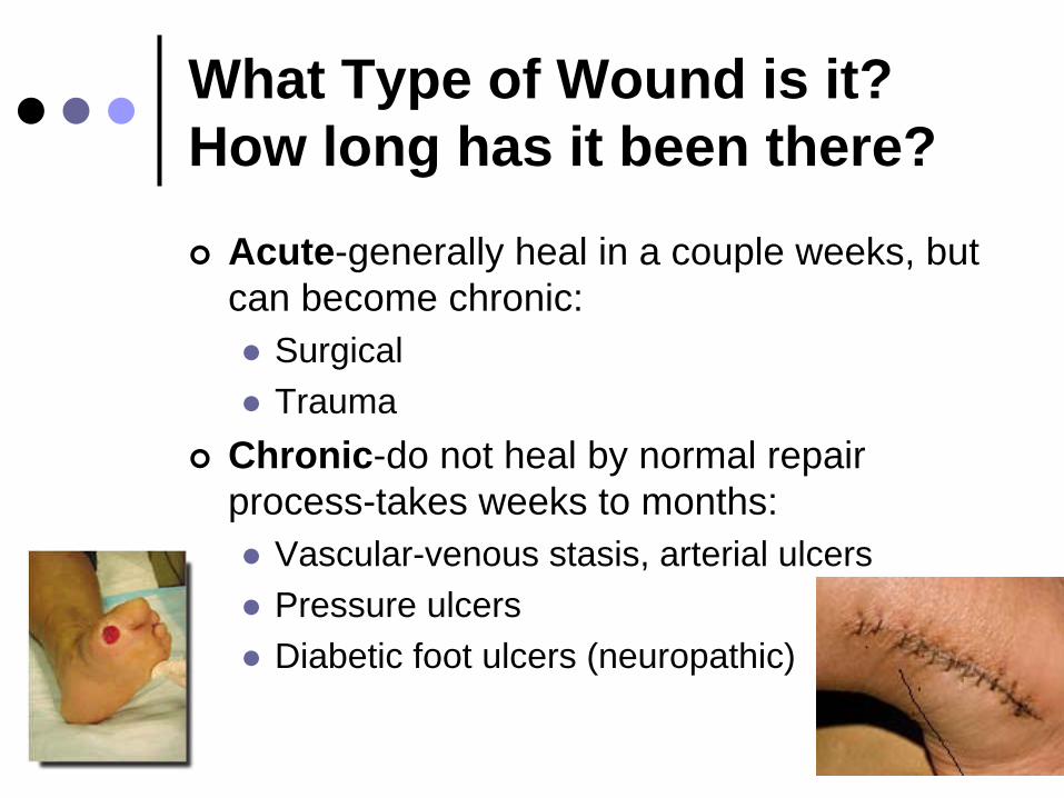
What Type of Wound is it? How long has it been there?
Acute-generally heal in a couple weeks, but can become chronic:
Surgical
Trauma
Chronic-do not heal by normal repair process-takes weeks to months:
Vascular-venous stasis, arterial ulcers
Pressure ulcers
Diabetic foot ulcers (neuropathic)
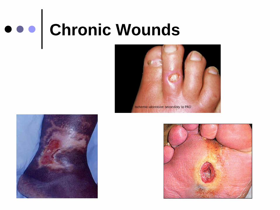
Chronic Wounds

Pressure Ulcer Staging

Where is it?
Where is it located?
Use anatomical location-heel, ankle, sacrum, coccyx, etc.
Measurements-in centimeters
Length X Width X Depth• Length = greatest length (head to toe)• Width = greatest width (side to side)• Depth = measure by marking the depth with a Q-
Tip and then hold to a ruler


Wound Characteristics:
Describe by percentage of each type of tissue:
Granulation tissue:• red, cobblestone appearance (healing,
filling in)
Necrotic:• Slough-yellow, tan dead tissue
(devitalized)• Eschar-black/brown necrotic tissue, can
be hard or soft

Evaluating additional tissue damage:
Undermining
Separation of tissue from the surface under the edge of the wound
• Describe by clock face with patients head at 12 (“undermining is 1 cm from 12 to 4 o’clock”)
Tunneling
Channel that runs from the wound edge through to other tissue
• “tunneling at 9 o’clock, measuring 3 cm long”

Wound Drainage and Odor
Exudate
Fluid from wound• Document the amount, type and odor
• Light, moderate, heavy• Drainage can be clear, sanguineous (bloody),
serosanguineous (blood-tinged), purulent (cloudy, pus-yellow, green)
Odor
Most wounds have an odor
Be sure to clean wound well first before assessing odor (wound cleanser, saline)
• Describe as faint, moderate, strong
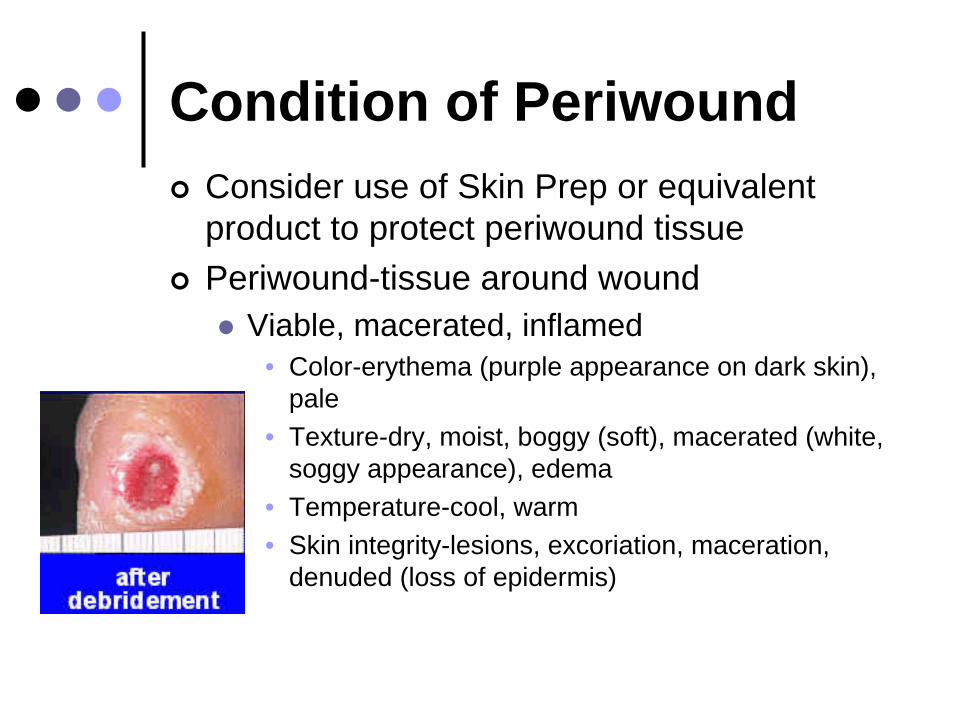
Condition of Periwound
Consider use of Skin Prep or equivalent product to protect periwound tissue
Periwound-tissue around wound
Viable, macerated, inflamed • Color-erythema (purple appearance on dark skin),
pale • Texture-dry, moist, boggy (soft), macerated (white,
soggy appearance), edema• Temperature-cool, warm• Skin integrity-lesions, excoriation, maceration,
denuded (loss of epidermis)

Is the wound infected?
All wounds are contaminated, but not necessarily infected:
Contamination-microorganisms on wound surface
Colonization-bacteria growing in wound bed without signs or symptoms of infection
Critical colonization-bacterial growth causes delayed wound healing, but has not invaded the tissue
Infection-bacteria invades soft tissue, causes systemic response
• Inflammation, pus, increase/change in exudate, fever, pain, delirium in elderly

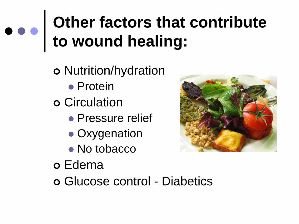
Other factors that contribute to wound healing:
Nutrition/hydration
Protein
Circulation
Pressure relief
Oxygenation
No tobacco
Edema
Glucose control - Diabetics

PUP-the highpoints
Minimize friction, sheer, and pressure
Repositioning every 1-2 hours• Necessary even when using specialty beds, in chair
HOB <30 degrees
Elevate heels
Incontinence
Scheduled toileting
Frequent changing, skin barrier
Nutrition
R.D. assessment
Calories, protein, supplements
Education
Staff, resident, families

Dressings-The Basics
DO:
Relieve pain, especially prior to dressing change
RELIEVE PRESSURE! • TURN AT LEAST EVERY 1-2 HOURS!• Consider specialty support surfaces for bed/chair
Fill in dead space if wound is deep
Protect skin from incontinence by using barrier cream
Protect periwound tissue by using Skin Prep
DO NOT:
DO NOT use wet-to-dry dressings!
DO NOT wrap tape completely around an extremity! • Tourniquet effect
DO NOT pull dressing off a wound• Can cause further tissue damage
• Soak to remove

Dressing selection
Determined by condition of the wound bed
Determine dressing according to amount of exudate (drainage)
Consider cost and availability of dressings at your institution $$$$
Assess wound at least every 2 weeks and change treatment if not improved
If not healing or questions about dressing selection, consult WOC nurse

Cleansing the wound bed:
Be gentle!
Saline or wound cleanser
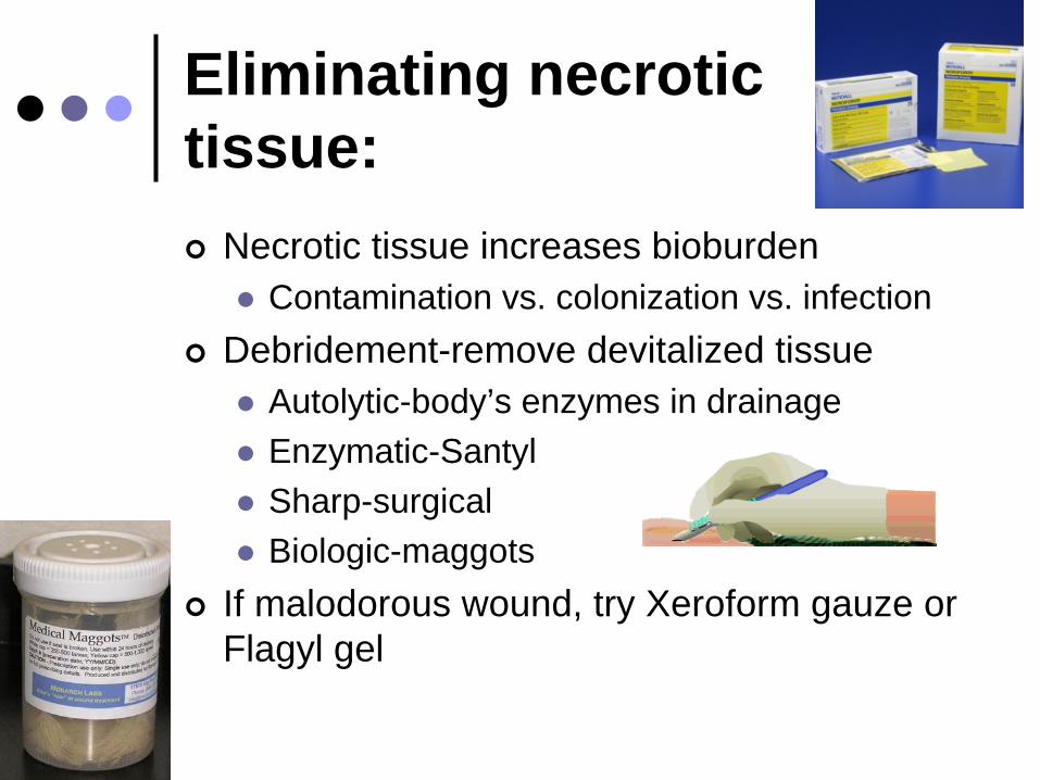
Eliminating necrotic tissue:
Necrotic tissue increases bioburden
Contamination vs. colonization vs. infection
Debridement-remove devitalized tissue
Autolytic-body’s enzymes in drainage
Enzymatic-Santyl
Sharp-surgical
Biologic-maggots
If malodorous wound, try Xeroform gauze or Flagyl gel

Management of devitalized tissue
Eschar-black necrotic tissue
Slough-soft, moist,avascular tissue
Firm, dry, stable eschar should not be debrided from heels
May not have adequate circulation to heal wound
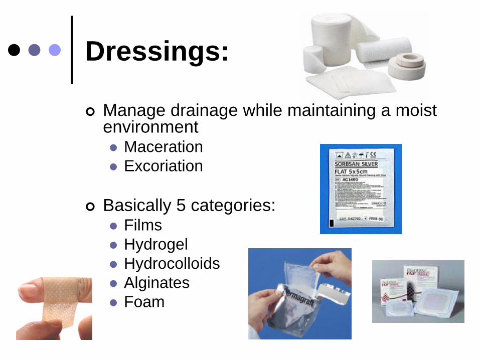
Dressings:
Manage drainage while maintaining a moist environment
Maceration
Excoriation
Basically 5 categories:
Films
Hydrogel
Hydrocolloids
Alginates
Foam
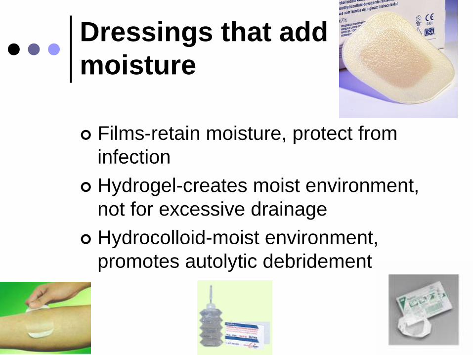
Dressings that add moisture
Films-retain moisture, protect from infection
Hydrogel-creates moist environment, not for excessive drainage
Hydrocolloid-moist environment, promotes autolytic debridement

Dressings that absorb moisture
Foams for moderate drainage
Calcium alginate for moderate to heavy drainage, hemostasis
Control of wound bioburden:
Antimicrobial dressings for wound contamination
Antibiotics only for infected wounds (not just colonized/contaminated)
Cultures not generally recommended because all wounds are contaminated
If culture indicated, cleanse wound bed with saline, then express drainage from wound bed.

Specialty Dressings
Antimicrobial dressings
Silver
Cadexomer iodine
Specialty Treatments
Vacuum-assisted wound treatments
Hyperbaric oxygen treatment

Websites
John A. Hartford Foundation, Institute for Geriatric Nursing:http://www.hartfordign.org/index.html
How to Try This: Braden scale video/article/CEU’s:http://www.nursingcenter.com/prodev/ce_article.asp?tid=7514 31
National Pressure Ulcer Advisory Panel:http://npuap.org/
Braden Scale:http://www.bradenscale.com/default.htm
Websites
Agency for Healthcare Research and Quality:Clinical Practice Guidelines:http://www.ahrq.gov/clinic/cpgonline.htm
National Guideline Clearinghouse:Guideline for prevention and management of pressure ulcers:
http://www.guideline.gov/summary/summary.aspx?ss= 15&doc_id=3860&nbr=3071