wound assessment and management guideline
TRANSCRIPT

WAHT-NUR-090 It is the responsibility of every individual to ensure this is the latest version as published on the Trust Intranet
Wound Assessment and Management Guideline
WAHT-NUR-090 Page 0 of 25 Version 1
Wound Assessment and Management
Guideline
This guidance does not override the individual responsibility of health professionals to make appropriate decision according to the circumstances of the individual patient in
consultation with the patient and /or carer. Health care professionals must be prepared to justify any deviation from this guidance.
Introduction This guideline is To ensure safe practice and maintain core standards of evidence based practice in Wound Management.
This guideline is for use by the following staff groups: Worcestershire Acute Hospital Trust (WAHT) Staff who are involved in any way in wound assessment and management.
Lead Clinician(s)
Elaine Bethell Lead Tissue Viability Nurse Lisa Martin Tissue Viability Nurse Approved by Clinical Policies Group, SKIN Matters Group on: 22nd June 2015 This guideline should not be used after end of: 22nd June 2017
Key amendments to this guideline
Date Amendment Approved By:

WAHT-NUR-090 It is the responsibility of every individual to ensure this is the latest version as published on the Trust Intranet
Wound Assessment and Management Guideline
WAHT-NUR-090 Page 1 of 25 Version 1
Contents
1. Introduction ........................................................................................................................ 2
2. Competencies required ...................................................................................................... 3
3. Patients covered ................................................................................................................ 3
4. Wound assessment and documentation ............................................................................ 3
5. Nutritional assessment & support ..................................................................................... 4
6. Psychological and Social Assessment ................................................................................ 5
7. Documentation ................................................................................................................. 5
8. Wound type & aetiology ................................................................................................... 6
9. Wound measurement ....................................................................................................... 6
10. Photography ..................................................................................................................... 7
11. Type of wound tissue ........................................................................................................ 7
12. Debridement ..................................................................................................................... 8
13. Exudate ............................................................................................................................ 9
14. Wound cleansing ............................................................................................................ 10
15. Infection control principles in relation to wound care ....................................................... 10
16. Topical antimicrobial agents ............................................................................................ 11
17. Potassium permanganate ............................................................................................... 11
18 Wound infection .............................................................................................................. 11
19 Taking a wound swab for microscopy .............................................................................. 12
20 Wound dressing collection .............................................................................................. 12
21 Wound re-assessment .................................................................................................... 13
22. Pain assessment ............................................................................................................ 13
23. Surgical/ plastics referral.................................................................................................. 14
24. Individual self-centred care .............................................................................................. 14
25. Transfer of individuals between care settings ................................................................. 14
26. Clinical audit and monitoring ........................................................................................... 14
27. Monitoring Tool ............................................................................................................... 15
28. Contributions .................................................................................................................. 16
29. References ...................................................................................................................... 17
Appendix 1 Wound assessment chart .............................................................................. 21
Appendix 2 Equality Impact Assessment Tool ................................................................. 24
Appendix 3 Financial Impact Assessment ....................................................................... 25
WAHT-NUR-090 It is the responsibility of every individual to ensure this is the latest version as published on the Trust Intranet
Wound Assessment and Management Guideline
WAHT-NUR-090 Page 2 of 25 Version 1
1. Introduction
a. The effective care and management of individuals with wounds is dependent upon a systematic and holistic approach. It should be based upon the knowledge of anatomy and physiology, wound healing, holistic assessment, specific wound management and appropriate selection of wound management products. For the individual, having a wound may cause them pain, sleeplessness, immobility, depression and isolation. For the health care professional it is essential that they understand the fundamentals necessary to manage the variety of wounds to individuals they care for. Wound care can be the cause of frustration, whilst also a resource burden for the Health Service in terms of time and costs incurred (Palmer 1999, Posnett and Franks 2008). There are reported to be 200,000 individuals in the UK at any one time who are suffering with a chronic wound. The cost to the NHS of caring for these individuals is a conservative estimate of £2.3bn to £3.1bn per year, which is around 3% of total NHS spend. (Posnett and Franks 2008, Posnett et al 2009).
b. This guideline replaces the previous Wound care guidance published in 2007. National and international guidelines have been considered and reviewed (EPUAP & NPUAP 2014, NICE 2014, EWMA 2005, EWMA 2004, EWMA 2008, DH 2010a, DH 2010b, DH2010c, RCN 2006, WUWHS 2008a, DH 2010d, NICE 2004, NICE 2008).
c. This guideline should be used to guide and direct staff on the evidence based best practice in the management of primary, secondary and tertiary wounds across a primary care setting.
d. Where individuals are failing to respond to treatment or their needs are beyond the scope of the clinician, it is advised that a referral is made to the Tissue Viability Service.
e. The principles upon which this guideline is based include:
i. An individualised holistic patient assessment undertaken and evidence-based treatment plans commenced. These should take into account the wound aetiology, the individuals circumstances and wishes, the overall aims of therapy, the practitioner’s clinical experience, available resources and evidenced research findings.
ii. Those who undertake assessment, planning, implementation and evaluations of care should be trained, educated and competent in wound management.
iii The individual or their carers, if appropriate, should be informed and involved in the decision making process and consent to treatment.
iv To ensure continuity, the care should be clearly documented in the individuals records and made accessible to the individual and, where appropriate all those involved in their care.
v A collaborative, multi-disciplinary, inter agency approach should be adopted to address all the needs of the individual where appropriate.
vi, Individuals, staff and carers should have access to the equipment and resources necessary to deliver quality care.
iv Monitoring and development of quality initiatives should be undertaken regularly through a clinical audit process.

WAHT-NUR-090 It is the responsibility of every individual to ensure this is the latest version as published on the Trust Intranet
Wound Assessment and Management Guideline
WAHT-NUR-090 Page 3 of 25 Version 1
2. Competencies required
a. Health professionals must have a sufficient level of knowledge in wound management. They must have achieved the required level of competency to be able to assess and treat patients with a wound, develop their skills to enable the application of new technologies and therapies. Competency based training is available from Worcestershire Acute Hospital Trust. Educational programmes, incorporating internal courses and accredited degree level study. This is available to all staff groups, for further information please contact Tissue Viability.
3. Patients covered
a. This guideline outlines the best practice recommendations to prevent, assess and manage wounds and applies to all individual groups including adults and children. The guideline is to be used by all staff employed across the Worcestershire Acute and Hospital Trust, engaged in the prevention assessment and management of wounds. Whilst not mandatory it is recommended for use by Care Homes and general practice staff across Worcestershire Health Economy.
b. N.B When assessing a child clinicians as well as appropriate consent clinicians must consider, in the first instance consulting with the community children’s nursing team and/or the responsible medical practitioner i.e. GP/hospital consultant.
4. Wound assessment and documentation
a. Wound management practice can vary greatly, therefore, local guidance is required to aid clinical decision-making, standardise documentation and recommend best practice in wound care.
b. This guideline offers practical guidance on the general principles of wound management, to include; describing tissue type, dressing selection, measuring wound size, detecting infection and bacterial sampling.
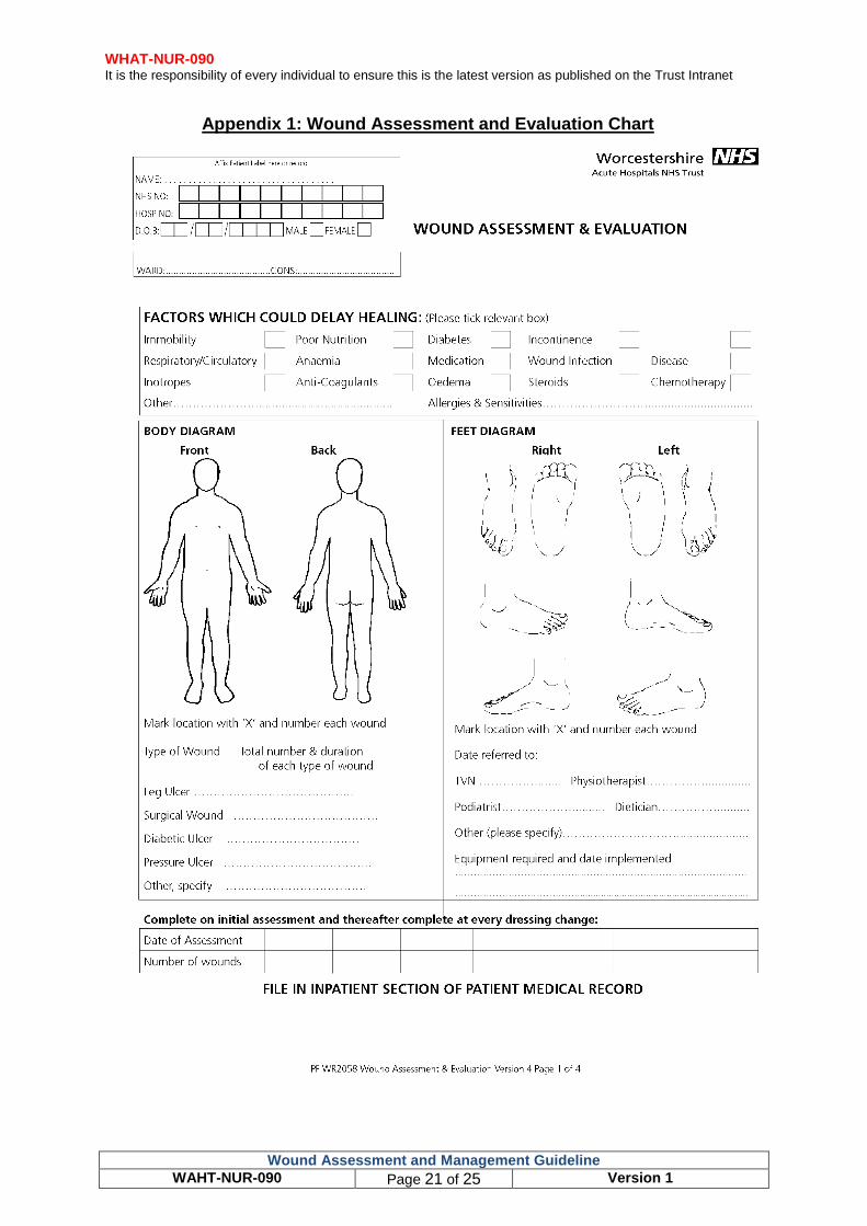
c. A wound assessment must be completed and documented by a registered practitioner using the approved Worcestershire Acute and Hospital trust wound assessment tool (Appendix 1). This should take place during the first contact with the individual and within one week of referral to primary care/ Care home.
d. An individual's ability to heal and the time it takes can vary greatly, and can be influenced by a number of factors. These should be taken into consideration during the assessment (DH 2010a):
i. General physical and psychological health and type and level of concurrent illnesses
ii. Treatment: systemically and locally
iii. Nutritional status
iv. Type of wound, location, depth and extent of damage and type of tissue in wound
v. Wound temperature, moisture level and pH balance
vi. Levels of bacterial colonisation and infection
WAHT-NUR-090 It is the responsibility of every individual to ensure this is the latest version as published on the Trust Intranet
Wound Assessment and Management Guideline
WAHT-NUR-090 Page 4 of 25 Version 1
vii. Blood supply to the wound, peri wound area and oedema of surrounding tissues
viii. Disruption to normal sleep pattern
ix. History of smoking and alcohol consumption
x. Medications that may include; steroids, immune-suppressants, chemotherapy
xi. Environment
e. The Worcestershire Acute and Hospital Trust Wound assessment tool is based upon the TIME framework developed by an International Advisory Board (Falanga 2004). The TIME framework is used to facilitate the assessment of wounds and incorporates assessment and management (Dowsett & Ayello 2004) in relation to the clinical observations and interventions to the cellular level.
T = Tissue non viable or deficient I = Infection and/or inflammation, incorporating the infection continuum (Kingsley et al 2004). M = Moisture imbalance E = Edge of wound, non-advancing or undermined
The following information should be documented at the initial assessment:
4.1 Medical history
a. Individuals past and current medical condition and general health
b. Drug history and current prescribed medications, over the counter medicines and alternative therapies
c. Smoking and alcohol history
d. Allergies: including any previous reactions to dressings, topical applications and natural rubber latex
e. Nutrition and hydration level. Weight, height and Body Mass Index (BMI)
f. Mobility
g. Temperature, Pulse and Blood Pressure, blood glucose level, blood results, urinalysis
h. Previous/planned investigations/procedures. For example: venous/arterial duplex, X-rays, surgery
5. Nutritional assessment and support
a. The prevention and treatment of malnutrition requires assessment and the Malnutrition Universal Screening Tool (MUST) should be utilised taking guidance from the MUST guidelines. (BAPEN 2013)
i. All individuals should have a nutritional screen, which proceeds to full assessment if deficit is suspected (NICE 2006, DH 2010e, Elia 2003, Elia and Russell 2009)

WAHT-NUR-090 It is the responsibility of every individual to ensure this is the latest version as published on the Trust Intranet
Wound Assessment and Management Guideline
WAHT-NUR-090 Page 5 of 25 Version 1
ii. Individuals considered as 'malnourished', or 'at risk' of malnutrition should be managed according to local and national guidance (NICE 2006, DH 2010e, Elia 2003, Elia and Russell 2009)
iii. Nutritionally compromised patients who have wounds may have an increased dietary need and a referral to a Dietician should be considered for further assessment, advice and supplementation (NICE 2006, DH 2010e, Elia 2003, Elia & Russell 2009)
iv. Individuals weight, height and body mass index (BMI) should be recorded at initial assessment and then weekly for community hospital inpatients and a minimum of monthly for individuals in the community
6. Psychological and Social Assessment
a. The following aspects should be considered during the individuals assessment (DH 2010a)
i. Stress level, depression, ability to sleep and where the patient sleeps e.g. bed or chair
ii. Ability to understand cause of wound and ability to participate in care
iii. Factors that may affect concordance with treatment. For example: dementia, cognitive impairment, learning difficulties, behavior, lifestyle choices etc.
iv. Drug/Alcohol dependency
v. Occupation, family structure, carers and their ability to assist with care
vi. Detail attitudes and any avoidance of social activities due to general condition and wound
7 Documentation
a. The following information should be documented on the wound assessment tool;
i. Biographical data, date and time of assessment
ii. Type of wound and any underlying aetiology
iii. Location of the wound on the body
iv. Allergies
V. Wound measurements including depth of damage
vi. Type of tissue in wound bed
vii Colour of tissue in the wound bed
viii. Method and type of debridement, if appropriate
ix. Amount and colour of exudate
x. Presence of any odour.
xi. Condition of surrounding skin and edge of wound
WAHT-NUR-090 It is the responsibility of every individual to ensure this is the latest version as published on the Trust Intranet
Wound Assessment and Management Guideline
WAHT-NUR-090 Page 6 of 25 Version 1
xi. Presence of oedema/lymphoedema.
xiii Wound cleaning fluid (if used)
xiv Presence/signs of infection
xv Dressing selection and regime
xvi Forms should be supported with photographs
xvii Care plans should be individualised and address the factors identified in the holistic assessment
8. Wound Type and Aetiology
a. Wound type and cause should be identified e.g. venous leg ulcer caused by venous hypertension or pressure ulcer caused by pressure/shear/friction etc. Reference should always be made to the appropriate Worcestershire Acute and Hospital Trust guideline. For assessment and management of the diabetic foot please refer to separate trust guidance.
b. Leg ulcers should be assessed using leg ulcer assessment and management guidelines
c. Pressure ulcers should be assessed and treated in accordance with the guidelines for the prevention and management of pressure ulcers
9. Wound Measurements
a. Wound measurements should be recorded on the first assessment and thereafter every four weeks as a minimum. Accurate measurement is an important part of wound assessment and can assist with:
i. Detailing progress or deterioration by comparing dimensions over time
ii. Communication between health professionals
iii. Encourage the individual that healing is progressing
iv. Evidence of skin condition when admitted to, or discharged from the Community Nurse caseload
b. Describe wound depth in terms of the anatomy of the skin and related structures. Use millimeters (mm) or centimeters (cms) to measure undermined tissue with a sterile measure as found in a standard wound dressing pack. The following terms may be useful:
i. Blister: filled with serum or blood or discoloured base
ii. Abscess: filled with pus
iii. Superficial or partial thickness skin loss: skin loss involving epidermis and/or dermis, with or without undermining of adjacent tissue
iv. Full thickness skin loss: damage involving subcutaneous layers, which may expose fat, bone, tendon or joint capsule, with or without undermining of adjacent tissue
v. Sinus: a blind ended tract

WAHT-NUR-090 It is the responsibility of every individual to ensure this is the latest version as published on the Trust Intranet
Wound Assessment and Management Guideline
WAHT-NUR-090 Page 7 of 25 Version 1
vi. Wound Fistulae: an abnormal passage from an internal organ to the body surface
c. A disposable measuring tape can be used to record length and width in cms. Measure the length of the wound along the vertical axis of the body (from head to foot), and the width along the horizontal axis of the body. A sterile probe should be used to measure the depth of the wound or any undermining or sinus. Use an imaginary clock face to increase accuracy of sinus/ undermining position.
10. Wound Photographs
a. Regular photography of the wound provides a useful visual record.
b. Criteria for Wound Photography
i. Obtain written consent (where possible or verbal) from the individual.
ii. Photograph the wound on initial assessment and repeat every four weeks or more frequently if the wound condition changes rapidly.
iii. Photographs should be labeled with the individuals NHS number, name, date of birth, date of photo, wound position and the name of the health care professional who has taken the image. Include a ruled measure to give an indication of scale. Secure/upload in the patient’s records in chronological order or print clearly labeled as above
iv. All photographs should be clear and in focus with an image of the wound and the wound in context
v. Privacy and dignity should be protected and maintained at all times
vi. If photographs are used for training purposes confidentiality must be maintained and appropriate level of consent gained
vii. Images can only be used for publication with specific consent (Individual agreement for photography and release of data for third party use)
viii. Photographs should not be taken using a non NHS mobile phone and images should be deleted from the digital camera once transferred to their secure documentation system
11. Type of Wound Tissue
a. The wound assessment must include a description of the type and amount of tissue present to the wound bed (Ousey &Cook 2012), using the terms epithelialising, granulating, sloughy, necrotic, or non-healing. Different tissue types can exist in the wound at the same time and should be recorded as an estimated percentage of the whole wound e.g. granulation tissue 80% and sloughy tissue 20%. This allows comparison over time. Percentages are used as a guide only and do not need to be precise
b. Necrotic tissue may appear black, hard, dry and leathery or grey in colour and usually indicates devitalised tissue. Assess the patient’s circulation to the affected area before deciding on method of debridement. If digits or heels are necrotic establish the patient’s vascular status using a Doppler or refer for specialist vascular assessment. These wounds should be kept dry until circulation is established

WAHT-NUR-090 It is the responsibility of every individual to ensure this is the latest version as published on the Trust Intranet
Wound Assessment and Management Guideline
WAHT-NUR-090 Page 8 of 25 Version 1
c. Sloughy tissue is also devitalized and includes a build up of dead white cells and may appear yellow or a waxy white in colour. It may appear wet or dry and is usually attached to the wound base (Ousey & Cook 2012)
d. Infected tissue or any associated cellulitis may appear red in colour, which extends beyond the wound margins and periwound edge. If this is associated with any clinical signs and symptoms of infection then systemic antibiotics are indicated with local wound management to control odour, pain and exudate (EWMA 2004, EWMA 2008, DH 2010a). Chronic wounds are often colonised with bacteria therefore diagnosis of infection should not be made solely on the basis of a microbiology swab result. Care should be taken with individuals that have conditions such as diabetes or are immuno-suppressed as they may not exhibit any signs of infection
e. Granulating tissue appears red (strawberry jam in colour and appearance) with small mounds caused by growth of capillary loops and should be protected
f. Epithelialising tissue appears white, pink or blue/mauve in colour. This tissue should be kept warm (body temperature) and moist to facilitate epithelial growth and mitotic cell division (Winter 1962, Hinman and Maibach 1963)
g. Wound colour is related to tissue type and can enhance description of wound status
12. Debridement
a Debridement is the removal of necrotic, devitalised, sloughy, infected tissue, or foreign bodies from a wound (EWMA 2008, Gray et al 2011, Ousey & Cook 2012). The body can debride itself by a natural process called autolysis; however, this may take time if large amounts of slough are present. Slough can provide an environment for bacteria to thrive, increasing the risk of infection. Debridement is recommended as a principle of wound management (EWMA 2008, Gray et al 2011)
b. There are a number of different methods to facilitate wound debridement.
i. Autolytic debridement: where the body naturally removes the devitalised tissue. This process can be enhanced and encouraged by the use of dressings such as hydrogels, hydrocolloids or capillary action dressings, alginates or foam wound dressings
ii. Bio-surgery: sterile larvae (maggots)
iii. Sharp debridement: using a sterile blade, scalpel or scissors. This should only be undertaken by a healthcare professional with specific training and who has achieved the appropriate level of competence.
iv. Surgical debridement: used when there is an urgent clinical need to remove or release devitalised tissue and when fast debridement would speed the individual’s recovery.
c. When deciding whether or not to debride a wound or which method to choose, the clinician should consider the following:
i. The environment
ii. The condition of the individual and their wound
iii. The condition of the surrounding skin

WAHT-NUR-090 It is the responsibility of every individual to ensure this is the latest version as published on the Trust Intranet
Wound Assessment and Management Guideline
WAHT-NUR-090 Page 9 of 25 Version 1
iv. The quality and strength of the underlying blood supply
v. Any risk of an adverse incident
vi. The preference of the individual with a wound
vii. Pain and tolerance level
viii. Equipment and the availability of wound management products
ix. The overall treatment goals (EWMA 2008)
13. Wound Exudate
a. Exudate is the fluid produced as a result of wounding. It will vary in volume, consistency and biochemical composition and may be either beneficial or harmful to underlying tissues and surrounding skin (WUWHS 2007). Some studies indicate that exudate is beneficial to healing acute or superficial wounds while others have found that chronic wound exudate contains enzymes that disrupt healing by degrading the extra-cellular matrix and excoriate surrounding skin. Quantifying the volume of wound exudate can be difficult. This should be recorded as none, low moderate, high. These terms are subjective but can be qualified by using the dressing to record exudates levels, strike through and/or wear time. The colour, viscosity or change in the wound exudate should be noted. The presence of any surrounding oedema/lymphoedema should also be documented
b. Exudate as defined by its colour:
Characteristic Possible cause
Clear, amber Serous exudate, often considered ‘normal’ but may be associated with infection by fibrinolysin-producing bacteria such as staphylococcus aureus may also be due to fluid from a urinary or lymphatic fistula/leak.
Cloudy, milky or creamy
May indicate the presence of fibrin strands (fibrinous exudate – the response to inflammation) or infection (purulent exudate containing white cells and bacteria).
Pink or Red Due to the presence of red blood cells and indicating capillary damage (sanguineous or hemorrhagic exudate).
Green May be indicative of bacterial infection, e.g. pseudomonas aeruginosa.
Yellow or brown
May be due to the presence of wound slough or material from an enteric or urinary fistula.
Grey or blue May be related to the use of silver dressings.
N.B Some medications are known to discolour urine and consideration could be given to drugs as a cause of exudate discolouration when all other causes have been excluded.
c. Exudate as defined by its consistency:
Characteristic Possible cause
High viscosity (thick, often sticky)
High protein content due to: Infection, inflammatory response Necrotic material Enteric fistula Residue from some types of dressings or topical preparations.
Low viscosity (thin, ‘runny’)
Low protein content due to: venous or congestive cardiac disease, malnutrition.

WAHT-NUR-090 It is the responsibility of every individual to ensure this is the latest version as published on the Trust Intranet
Wound Assessment and Management Guideline
WAHT-NUR-090 Page 10 of 25 Version 1
(WUWHS 2007)
d. Chronic wound management techniques and dressings are based on the principle of moisture balance (EWMA 2008, WUWHS 2007, and Cowan 2014). In general this means that:
i. Drier wounds, (except those with poor circulation), should be moistened with dressings that hydrate tissue, e.g. hydrogels, hydrocolloid sheets and pastes
ii. Wounds with excess exudate require dressings that absorb or control fluid, e.g. alginates, gelling fibers, capillary action, foams; negative pressure wound therapy (NPWT), compression bandages and hosiery. Also consider the frequency of dressing changes to minimize any further skin damage.
iii. Surrounding intact skin should be protected from exudate with the use of barrier films and creams to prevent excoriation.
iv Dressings should be selected from the Worcestershire Wound Management Formulary
14. Wound cleansing
a. Routine cleansing of clean granulating wounds with the aim of bacterial removal has been proven to be ineffective (EWMA 2008, WUWHS 2007, Lloyd-Jones 2012, Davies 1999, Lindsey 2007).
b. The rationale to cleanse wounds should include: Removal of debris, (e.g. foreign bodies, dressing residue and devitalised tissue), exudate from the surrounding skin and to refresh the individual. Chronic wounds may be cleansed with tap water, or water which is suitable for drinking (WUWHS 2008, Griffiths et al 2001, and Fernandez et al 2010). Showering/bathing with these wound types is acceptable.
c. Use minimal mechanical force when cleansing or irrigating the wound.
d. Irrigation can be useful for cleaning of a cavity ulcer
e. Appliances used (e.g. bath, shower, bucket or bowl etc.) should be cleaned before and after use in accordance with the infection prevention and control guidance. If a bucket is used it should be lined with a new clinical waste bag or bin liner and the bucket should be kept solely for that specific use and individual, appropriate polythene bags can be obtained from NHS Supply Chain
f. Tap water is not recommended for wounds that can be probed to bone, for those that are immuno- compromised and where the safety of the tap water cannot be assured.
g. Any cleansing solution should be warmed prior to use
15. Infection control principles in relation to wound care
a. When carrying out any wound care intervention the clinician should:
i. Be familiar with Worcestershire Acute and Hospital Trust Infection prevention and control policies and guidelines
WAHT-NUR-090 It is the responsibility of every individual to ensure this is the latest version as published on the Trust Intranet
Wound Assessment and Management Guideline
WAHT-NUR-090 Page 11 of 25 Version 1
ii. Assess the risk of infection and cross infection and plan care accordingly
iii. Maintain hand hygiene and use standard precautions
iv. Use non-woven sterile swabs if cleansing the wound to reduce fiber shedding (cotton wool is not indicated for use)
v. Use a non-touch technique, gloved fingers should not touch the wound surface (Dougherty and Lister 2011)
vi Equipment used should be in accordance with trust and manufacturer guidance eg; forceps, scissors, dressings. Single item products should not be reused (MHRA 2013)
vii Unused part dressings must not be kept for use at the next dressing change or used on other individuals with a wound
16. Topical Antimicrobial/Antiseptic Cleansing Agents
a. Antiseptic solutions (e.g. hypochlorite’s, EUSOL, chlorhexidine and betadine iodine) should not be used for routine wound cleansing as these can cause pain and reduce the proliferation of macrophages and lymphocytes essential to the wound healing process (Naylor et al 2001).
17. Potassium Permanganate
a. The product license for Potassium permanganate is in the treatment of weeping or varicose eczema (Leppard and Ashton 1998, BNF 2014) and is not a wound management product. It should be diluted in lukewarm water to a concentration of 1:10,000 and used as a soak for no more than 20 minutes daily and for a maximum of two weeks (EWMA 2006). The solution stains skin and nails dark brown and can cause skin irritation. It has the potential risk of toxicity if used on large areas over a long period of time (EWMA 2006). This product is not recommended for use across Worcestershire Health and Care and Acute Hospital Trusts. Any individual prescribed for Potassium permanganate should have the treatment rationale confirmed by the prescriber prior to the commencement of treatment and should be regularly reviewed.
18. Wound Infection
a. All chronic wounds will contain bacteria, but not all are infected. Routine swabbing for Microscopy, culture and sensitivity is not required and an unnecessary expense (EWMA 2004). Infection is the result of a complex interaction between the host, organism, wound environment and therapeutic interventions. This is complicated by bacterial virulence (EWMA 2005, EWMA 2006). Identifying wound infection should be viewed as a clinical skill which can be supported by laboratory findings when necessary. A thorough assessment of the individual and their wound is required prior to diagnosis.
b. Observations should be made for:
i. Abscess, cellulitis, an increase in the white blood cell count
ii. Increased wound exudate; serous, seropurulent, haemopurulent, purulent wound fluid
iii. Individual with a wound feels unwell with possible pyrexia

WAHT-NUR-090 It is the responsibility of every individual to ensure this is the latest version as published on the Trust Intranet
Wound Assessment and Management Guideline
WAHT-NUR-090 Page 12 of 25 Version 1
iv. Increased heat production, redness and swelling (cellulitis) to the peri wound
v. Delayed healing or wound breakdown, discoloration of wound and surrounding skin
vi. Granulation tissue that is friable and bleeds easily
vii. Increased, unexpected or a change in the individuals pain experience
viii. Pocketing to the wound bed
ix. Bridging of tissue between cavities
x. Change in wound odour/Malodour
c. NB: The wound aetiology and bacteria will define both the signs and symptoms that are present to the wound and individual. Not all signs or symptoms will be present in all cases. This may be due to the type of bacteria, auto-immune impairment, diabetes mellitus, and quality of vascular supply or the use of medications such as steroids, anti-inflammatory, immune-suppressants and chemotherapy (EWMA 2004). Individuals with wounds that exhibit a spreading cellulitis and/or systemic infection should have a wound swab and blood cultures taken, this is to identify the offending organism and an assessment of the differential diagnosis. The individual should be treated with antibiotics which in some cases may be given intravenously dependent on the severity of the host response. Topical treatment should be considered in accordance with the Wound care formulary.
19. Taking a Wound Swab for microscopy
a. Cleanse the surface of the wound to remove surface bacteria
b. Rub the tip of the swab across the wound in a ‘zig zag’ manner and at the same time rotate the swab handle (Kelly 2003, Levine et al 1976)
c. Complete the microscopy form with sufficient information for laboratory staff and microbiologist to know from where and why the swab was taken and any additional supporting information such as current antimicrobial therapy
d. NB: Systemic treatment for wound infection should not be delayed while waiting for swab results. Topical antibiotics are not generally used in wound care due to the risks of resistance (DH 2010b). Follow microbiology/pharmaceutical guidance on the systemic treatment of wound infection
20. Wound Dressing Selection
a. Choice of dressing, method of debridement and the optimum wound healing environment should be created using modern dressings (NICE 2001). A different treatment or dressing type may be required to meet the needs of the individual with a wound at their different stages of healing. Wound management selection must be made on an individual basis after a thorough assessment and following discussion with the individual with the wound.
b. The following characteristics should be considered in dressing selection (Cowan 2014):
i. The ability to prevent penetration of capillary loops into the dressing material avoiding adherence

WAHT-NUR-090 It is the responsibility of every individual to ensure this is the latest version as published on the Trust Intranet
Wound Assessment and Management Guideline
WAHT-NUR-090 Page 13 of 25 Version 1
ii. Be hypoallergenic, sterile and have a long shelf life
iii. Be cost effective with a supporting evidence base
iv. Maintain high humidity and an optimum pH at the wound / dressing interface
v. Remove excess exudate, toxic components and not release toxic chemicals, particles or fibres into the wound bed
vi. Allow gaseous exchange at the wound interface
vii. Provide thermal insulation to encourage mitotic cell division
viii. Be impermeable to bacteria
ix. Be free from particulate and toxic contaminants
x. Allow removal without causing additional trauma and be comfortable to the individual to wear
xi. Ensure the wound remains moist with exudate but not macerated (except wounds with no underlying circulation, which must be kept dry)
21. Wound re-assessment
a. It is essential for all health care professionals to set a deadline. This should be undertaken according to individual need, and always undertaken if the patient’s condition changes. Maximum period before re-assessment is 1 month but may be as little as daily. Frequency should be based on vulnerability and condition of the patient.
b. Any alterations to the treatment regime will be discussed with the patient, Healthcare Professional and the rationale for this will be documented
i. Rationale:-
ii. Monitor appropriateness of current treatment
iii. Respond to any changes as a result of the re assessment
22. Pain assessment
a. Consideration should be given to pain in relation to wound management and a pain assessment undertaken in patients identified during screening. When a patient has pain the pain assessment tool should be completed. ( Appendix 2). The International Association for the Study of Pain (IASP 2014) defines pain as: “An unpleasant sensory and emotional experience associated with actual or potential tissue damage, or described in terms of such damage”.
b. An individual’s experience of pain is unique, it is complex and influenced by many factors. A systematic and rational approach to the assessment and management of pain is essential. This is a specific role of the clinician and should be documented as with other aspects of the assessment (NMC 2009). A number of publications have been published since 2000 including: Pain at wound dressing change, (EWMA 2002) and, Minimising pain at wound dressing-related procedures (WUWHS 2004) which suggests that we need to consider that all wounds are painful, that over time wounds may become painful, acknowledge that the surrounding skin can become sensitive and painful, the

WAHT-NUR-090 It is the responsibility of every individual to ensure this is the latest version as published on the Trust Intranet
Wound Assessment and Management Guideline
WAHT-NUR-090 Page 14 of 25 Version 1
lightest touch can be intensely painful and that the healthcare professional should know when to refer for specialist assessment.
23. Surgical/plastics referral
a. Referral for surgical/ plastics opinion should be made based on the needs of the individual, their health status, their risk (anaesthetic and surgical intervention), the assessment of psychosocial factors regarding the risk of recurrence, the failure of previous conservative treatment and the positive effect of surgical intervention ( NICE 2008).
24. Individual Centred self care
a. Individuals and carers should be made aware of their wound and the potential risk and/or complications. Treatment and care should take into account the individual needs and preferences, carers and relatives should have the opportunity to be involved in discussions where appropriate.
i. Individuals should be encouraged to maintain their independence and attend appointments at clinics, GP surgeries and wound healing clinics
ii. Individuals and carers who are willing, competent and able to should be taught how to undertake dressing changes based on professional advice
25. Transfer of individuals with a wound between care settings
a. All individuals with wounds who are transferred to any other care setting must have their treatment regime communicated to the appropriate health care professional prior to discharge. Referral and transfer information (Appendix 4)
26. Clinical audit monitoring tool
Standards % Clinical exceptions
Holistic wound assessment will be carried out within the specified time scale
100
All individuals will have their wound size documented within the specified time scale
100
Management will be in accordance with wound assessment and the wound care formulary
100
Regime of care will be communicated on transfer of care setting
100
How will monitoring be carried out?
Ongoing clinical audit, led by Tissue Viability Services within Worcestershire Acute Hospital Trust

WAHT-NUR-090 It is the responsibility of every individual to ensure this is the latest version as published on the Trust Intranet
Wound Assessment and Management Guideline
WAHT-NUR-090 Page 15 of 25 Version 1
27. Monitoring Tool Page/ Section of Key Document
Key control:
Checks to be carried out to confirm compliance with the policy:
How often the check will be carried out:
Responsible for carrying out the check:
Results of check reported to: (Responsible for also ensuring actions are developed to address any areas of non-compliance)
Frequency of reporting:
WHAT? HOW? WHEN? WHO? WHERE? WHEN?
4 A wound assessment must be completed by a registered nurse
Spot checks . requested for every TV referral made
When TV visit wards to assess a referred patient
TV Nurse in charge and if available TV Link nurse to be informed , of requirement and rationale
Ongoing
WAHT-NUR-090 It is the responsibility of every individual to ensure this is the latest version as published on the Trust Intranet
Wound Assessment and Management Guideline
WAHT-NUR-090 Page 16 of 25 Version 1
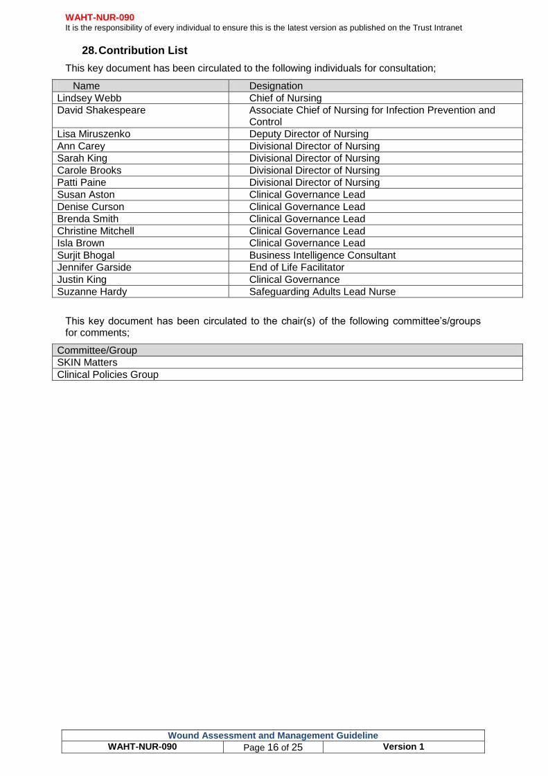
28. Contribution List
This key document has been circulated to the following individuals for consultation;
Name Designation
Lindsey Webb Chief of Nursing
David Shakespeare Associate Chief of Nursing for Infection Prevention and Control
Lisa Miruszenko Deputy Director of Nursing
Ann Carey Divisional Director of Nursing
Sarah King Divisional Director of Nursing
Carole Brooks Divisional Director of Nursing
Patti Paine Divisional Director of Nursing
Susan Aston Clinical Governance Lead
Denise Curson Clinical Governance Lead
Brenda Smith Clinical Governance Lead
Christine Mitchell Clinical Governance Lead
Isla Brown Clinical Governance Lead
Surjit Bhogal Business Intelligence Consultant
Jennifer Garside End of Life Facilitator
Justin King Clinical Governance
Suzanne Hardy Safeguarding Adults Lead Nurse
This key document has been circulated to the chair(s) of the following committee’s/groups for comments;
Committee/Group
SKIN Matters
Clinical Policies Group

WAHT-NUR-090 It is the responsibility of every individual to ensure this is the latest version as published on the Trust Intranet
Wound Assessment and Management Guideline
WAHT-NUR-090 Page 17 of 25 Version 1
29. References
BAPEN (2013) Malnutrition Universal Screening Tool.’http://www.bapen.org.uk/screening-for-malnutrition/must/introducing-must. Accesed November 2014 British National Formulary (2014) British Medical Association and the Royal Pharmaceutical Society of Great Britain. 61. March. www.BNF.org. Accessed Oct 2014. Cowan, T. (2014) Wound Care Handbook 2014-2015 7th edition. MA Healthcare Davies, C. (1999) Cleansing rites and wrongs. Nursing Times. 95, 43, 71-73. DH (2010a) Essence of Care: Benchmarks for prevention and management of pressure ulcers. Department of Health. London. http://www.dh.gov.uk/prod_consum_dh/groups/dh_digitalassets/@dh/@en/@ps/documents/digitalasset/dh_119979.pdf Accessed Jan 2014. DH (2010b) High Impact Actions for Nursing and Midwifery: Your Skin Matters: Pressure ulcer prevention. Institute for innovation and improvement. http://www.institute.nhs.uk/building_capability/hia_supporting_info/your_skin_matters.html Accessed Jan 2014. DH (2010c) National Patient Safety Agency. Patient Safety First: Preventing Pressure Ulcers. Health Foundation. Institute for Innovation and Improvement. http://www.patientsafetyfirst.nhs.uk/Content.aspx?path=/interventions/relatedprogrammes/pressure-ulcers/ Accessed Jan 2014. DH (2010e) Essence of Care Benchmarks for fundamental aspects of care: Benchmark for food and drink: Department of Health. http://www.dh.gov.uk/prod_consum_dh/groups/dh_digitalassets/@dh/@en/@ps/documents/digitalasset/dh_119974.pdf Accessed Jan 2014 Dougherty L. and Lister S. (2011) The Royal Marsden Hospital Manual of Clinical Nursing Procedures. 8th Edition. Wiley-Blackwell Publishing. Oxford. Dowsett, C. & Ayello, E. (2004) TIME principles of chronic wound bed preparation and treatment. British Journal of Nursing. (Tissue Viability supplement) Vol 13, No 15 p s16-21 Elia M (2003), Screening for malnutrition: a multidisciplinary responsibility. Development and use of the Malnutrition Universal Screening Tool (MUST) for adults. MAG, a Standing Committee of BAPEN (ISBN 1 899467 70 X) 2003. Elia M, Russell CA (2009) Combating malnutrition: Recommendations for action: A report from the Advisory Group on Malnutrition led by BAPEN European Pressure Ulcer Advisory Panel Scale EPUAP & NPUAP (2014) Prevention of pressure ulcers: Quick Reference Guide. European Pressure Ulcer Advisory Panel and National Pressure Ulcer Advisory Panel. Washington DC. USA. http://www.epuap.org/guidelines/Final_Quick_Prevention.pdf Accessed Jan 2014. European Wound Management Association (EWMA) (2002)

WAHT-NUR-090 It is the responsibility of every individual to ensure this is the latest version as published on the Trust Intranet
Wound Assessment and Management Guideline
WAHT-NUR-090 Page 18 of 25 Version 1
Position Statement: “Pain at wound dressing changes” London MEP ltd http://ewma.org/fileadmin/user_upload/EWMA/pdf/Position_Documents/2002/Spring_2002__English_.pdf. Accessed November 2014 European Wound Management Association (EWMA) (2004) Position Document: Wound Bed Preparation in Practice. London: MEP Ltd. http://ewma.org/fileadmin/user_upload/EWMA/pdf/Position_Documents/2004/pos_doc_English_final_04.pdf Accessed Nov 2014. European Wound Management Association (EWMA). Position Document: Identifying criteria for wound infection. London: MEP Ltd, 2005. http://ewma.org/fileadmin/user_upload/EWMA/pdf/Position_Documents/2005__Wound_Infection_/English_pos_doc_final.pdf Accessed Nov 2014. European Wound Management Association (EWMA). Position Document: Management of wound infection. London: MEP Ltd, 2006. http://ewma.org/fileadmin/user_upload/EWMA/pdf/Position_Documents/2006/English_pos_doc_2006.pdf Accessed Nov 2014. European Wound Management Association (EWMA) (2008). Position Document: Hard- to-heal-wounds: A holistic approach. London: MEP Ltd. http://www.woundsinternational.com/pdf/content_45.pdf Accessed Nov 2014. Falanga V. (2004). Wound bed preparation: science applied to practice. Introduction in Wound bed preparation. EWMA Position document. MEP Ltd www.ewma.org Fernandez, R., Griffiths, R., Ussia, C. (2010) Water for wound cleansing (review). Cochrane database of systematic reviews CD 003861.Accessed Nov 2014. Gray,D. Chadwick,P. Acton,C et al (2011) Consensus guidance for the use of debridement techniques in the UK. Wounds UK. (7) 1 p77-84 Griffiths, R.Fernandez, R. Ussia, C.Is tap water a safe alternative to normal saline for wound irrigation in the community setting? Journal of Wound Care 2001;10:407–11. Hinman, C. Maibach, H. (1963) Effect of air exposure and occlusion on experimental human skin wounds, Nature. Oct 26. International association for the study of Pain (2014) http://www.iasp-pain.org/ Accessed November 2014 Kelly, F. (2003) Infection control: Validity and Reliability in wound swabbing. British Journal of Nursing, 12, 16, PP 959-964. Kingsley, A. White, R. & Gray, D. (2004) The wound infection continuum: A revised perspective. Applied Wound management Supplement. Wounds UK. Aberdeen: 1 (1): p 13-18 Leppard, B. Ashton, R. (1998) Treatment in Dermatology. Radcliffe Medical Press. Oxford. Levine, N. Lindberg, R. Mason, A. Pruitt, B. (1976). The quantitative swab culture and smear: a quick simple method for determining the number of viable aerobic bacteria on open wounds. J Trauma; 16(2): 89-94. Lindsay, E. (2007) to wash or not to wash: what is the solution for chronic leg ulcers? Wound essentials; 2: 74-83

WAHT-NUR-090 It is the responsibility of every individual to ensure this is the latest version as published on the Trust Intranet
Wound Assessment and Management Guideline
WAHT-NUR-090 Page 19 of 25 Version 1
Lloyd-Jones, M. (2012) Wound cleansing: has it become a ritual or is it a necessity? Wound care S22-26, December MHRA (2013) Single-use Medical Devices: Implications and Consequences of Reuse. The Medicines and Healthcare products Regulatory Agency (MHRA) London. http://www.mhra.gov.uk/Publications/Safetyguidance/DeviceBulletins/CON2024995 Accessed Nov 2014. Naylor, W. Laverty, D.& Mallett, J. (2001) Handbook of Wound Management in Cancer Care. Blackwell Science. Oxford. NICE (2001) Guidance on the use of debriding agents and specialist wound care clinics for difficult to heal surgical wounds. NICE London NICE (2006) Nutrition support in adults: Oral nutrition support, enteral tube feeding and parenteral nutrition. National Collaborating Centre for Acute Care. London. http://guidance.nice.org.uk/CG32/Guidance/pdf/English Accessed Jan 2014. NMC (2009) Nursing and midwifery code of professional conduct. London NICE (2008) Surgical Site Infection: Prevention and Treatment of surgical site infection. National Collaborative Centre for Women’s and Children’s Health. http://www.nice.org.uk/nicemedia/pdf/CG74FullGuideline.pdf Accessed Jan 2014 NICE (2014) Pressure ulcers: the management of pressure ulcers in primary and secondary care. National Institute for Health and Clinical Excellence. London. http://guidance.nice.org.uk/CG29/Guidance/pdf/English Accessed Nov 2014. Ousey, K & Cook, L. (2012) Wound Assessment Made Easy. Wounds UK 8 (2) www.wounds-uk.com/made- easy. Accessed Nov 2014 Palmer C (1999) Patient choice in wound care management: the experience of larval therapy” Presentation given at the Tissue Viability 32nd Conference in Manchester. Posnett, J., Gottrup, F., Lundgren, H. & Saal, G. (2009) The resource impact of wounds on health-care providers in Europe. Journal of Wound Care, 18(4), 154-161. Posnett, J. & Franks, P. (2008) the cost of skin breakdown and ulceration in the UK. In The silent epidemic Smith and Nephew Foundation, Hull. RCN. (2006) Clinical Practice Guidelines: The management of patients with venous leg ulcers. London. Royal College of Nursing. http://www.rcn.org.uk/__data/assets/pdf_file/0003/107940/003020.pdf Accessed Jan 2014. Winter GD. Formation of the scab and the rate of epithelisation of superficial wounds in the skin of the young domestic pig. 1962. Journal of Wound Care 1995. 4:366–7. World Union of Wound Healing Societies (2004) Principles of best practice: Minimising pain at wound dressing-related procedures: A consensus document. London. MEP Ltd. World Union of Wound Healing Societies (WUWHS) (2008). Principles of best practice: Compression in venous leg ulcers. A consensus document. London. MEP Ltd. http://www.wuwhs.org/datas/2_1/9/Compression_VLU_English_WEB.pdf Accessed Jan 2013. World Union of Wound Healing Societies (WUWHS). Principles of best practice: Wound exudate and the role of dressings. A consensus document. London: MEP Ltd.

WAHT-NUR-090 It is the responsibility of every individual to ensure this is the latest version as published on the Trust Intranet
Wound Assessment and Management Guideline
WAHT-NUR-090 Page 20 of 25 Version 1
(2007). http://www.wuwhs.org/datas/2_1/4/consensus_exudate_ENG_FINAL.pdf Accessed Jan 2014.
WHAT-NUR-090 It is the responsibility of every individual to ensure this is the latest version as published on the Trust Intranet
Wound Assessment and Management Guideline
WAHT-NUR-090 Page 21 of 25 Version 1
Appendix 1: Wound Assessment and Evaluation Chart

WHAT-NUR-090 It is the responsibility of every individual to ensure this is the latest version as published on the Trust Intranet
Wound Assessment and Management Guideline
WAHT-NUR-090 Page 22 of 25 Version 1

WHAT-NUR-090 It is the responsibility of every individual to ensure this is the latest version as published on the Trust Intranet
Wound Assessment and Management Guideline
WAHT-NUR-090 Page 23 of 25 Version 1
WHAT-NUR-090 It is the responsibility of every individual to ensure this is the latest version as published on the Trust Intranet
Wound Assessment and Management Guideline
WAHT-NUR-090 Page 24 of 25 Version 1
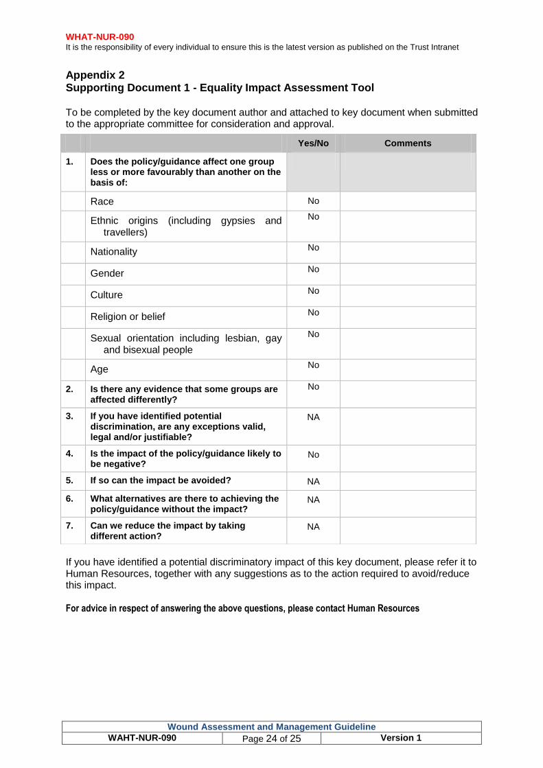
Appendix 2 Supporting Document 1 - Equality Impact Assessment Tool To be completed by the key document author and attached to key document when submitted to the appropriate committee for consideration and approval.
If you have identified a potential discriminatory impact of this key document, please refer it to Human Resources, together with any suggestions as to the action required to avoid/reduce this impact. For advice in respect of answering the above questions, please contact Human Resources
Yes/No Comments
1. Does the policy/guidance affect one group less or more favourably than another on the basis of:
Race No
Ethnic origins (including gypsies and travellers)
No
Nationality No
Gender No
Culture No
Religion or belief No
Sexual orientation including lesbian, gay and bisexual people
No
Age No
2. Is there any evidence that some groups are affected differently?
No
3. If you have identified potential discrimination, are any exceptions valid, legal and/or justifiable?
NA
4. Is the impact of the policy/guidance likely to be negative?
No
5. If so can the impact be avoided? NA
6. What alternatives are there to achieving the policy/guidance without the impact?
NA
7. Can we reduce the impact by taking different action?
NA
WHAT-NUR-090 It is the responsibility of every individual to ensure this is the latest version as published on the Trust Intranet
Wound Assessment and Management Guideline
WAHT-NUR-090 Page 25 of 25 Version 1
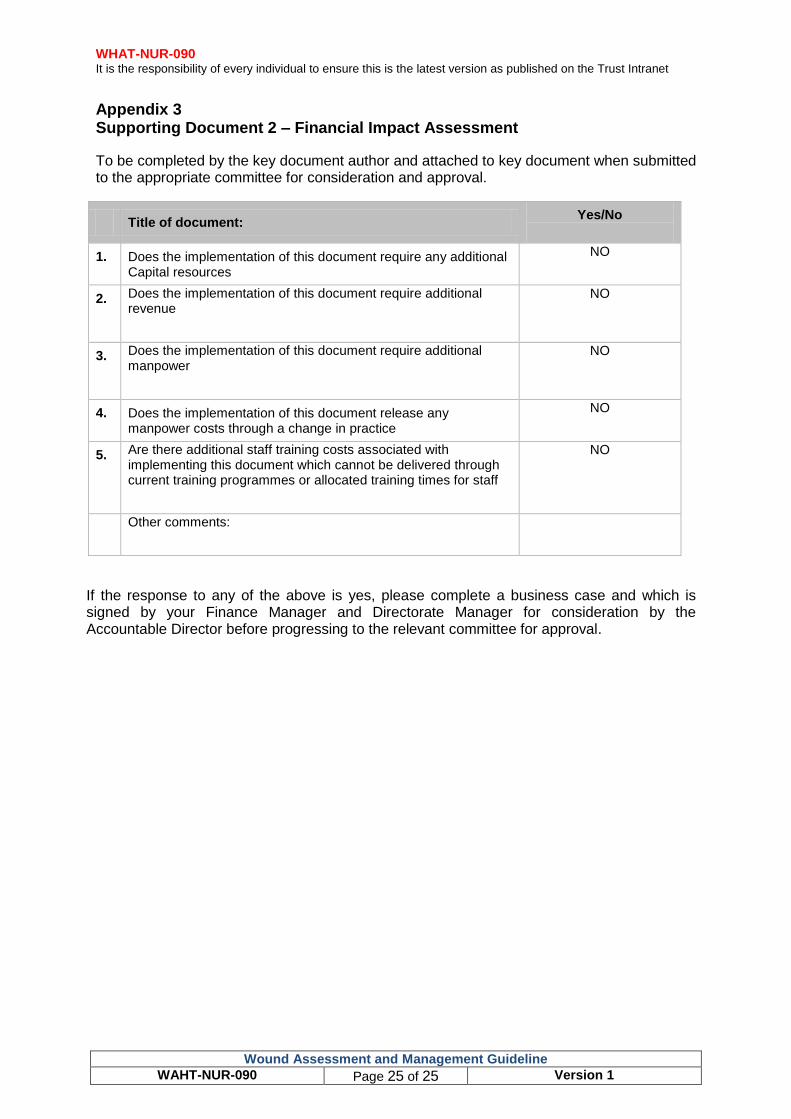
Appendix 3 Supporting Document 2 – Financial Impact Assessment
To be completed by the key document author and attached to key document when submitted to the appropriate committee for consideration and approval.
Title of document: Yes/No
1. Does the implementation of this document require any additional Capital resources
NO
2. Does the implementation of this document require additional revenue
NO
3. Does the implementation of this document require additional manpower
NO
4. Does the implementation of this document release any manpower costs through a change in practice
NO
5. Are there additional staff training costs associated with implementing this document which cannot be delivered through current training programmes or allocated training times for staff
NO
Other comments:
If the response to any of the above is yes, please complete a business case and which is signed by your Finance Manager and Directorate Manager for consideration by the Accountable Director before progressing to the relevant committee for approval.