workshop - dtic
TRANSCRIPT

Institute of Radio Engineering and Electronics Academy of Sciences of the Czech Republic
>th 8m VIENNA OPT(R)ODE WORKSHOP
Book of abstracts
no
<jj
Prague, October 7-8, 1998

8th VIENNA OPT(R)ODE WORKSHOP
Prague, Czech Republic, October 7-8,1998
Organized by:
Institute of Radio Engineering and Electronics, Acad. Sei. CR, Prague Faculty of Electrical Engineering, Czech Technical University, Prague
Sponsored by:
Institute of Radio Engineering and Electronics, Acad. Sei. CR, Prague, Czech Republic Czech Technical University, Prague, Czech Republic
European Research Office of the U. S. Army, United Kingdom Engineering Academy, Prague, Czech Republic
ASAC - Austrian Society for Analytical Chemistry, Austria Devetsil JST s.r.o, Plzeri, Czech Republic
ITC s.r.o., Prague, Czech Republic
Workshop Chairmen:
Miroslav Chomät (Czech Republic) and Otto S. Wolfbeis (Germany)
International Program Committee:
Miroslav Chomät (Czech Republic) Vlastimil Matejec (Czech Republic) Franz L. Dickert (Austria) Boris Edl-Mizaikoff (Austria) Pavel Fiala (Czech Republic) Josef Schröfel (Czech Republic) Jifi Homola (Czech Republic) Otto S. Wolfbeis (Germany)
Local Organizing Committee:
Daniela Berkovä Vlastimil Matejec Miroslav Husäk Radan Slavik Ivan Kasik Petr Tobiska
Workshop Secretary:
Daniela Berkovä

Edited by: V. Matejec, M. Chomät
Published by: Institute of Radio Engineering and Electronics, Academy of Sciences of the Czech Republic, Prague
Printed by: NEOSET, Prague
ISBN 80-901658-9-3

In Memoriam Robert Kellner (1945-1997)
By announcing the 8th Vienna Opt(r)ode Workshop the undersigned have the sad duty to inform the scientific community that our friend and colleague Robert Kellner died at the age of 52, on October 8, 1997. His untimely death has shocked all who knew this excellent scientist with an extraordinary active personality.
Robert Kellner was born in Vienna, Austria, on January 5, 1945. After graduating in Chemistry from the Vienna University of Technology with a Ph.D. in 1971, he decided to pursue an university career, achieved his „Habilitation" in 1975 under the supervision of Prof. Dr. H. Malissa and was appointed Professor at the Institute of Analytical Chemistry of this university in 1978.
Robert Kellner had an extremely successful scientific career which is documented in over 200 publications, numerous, worldwide lectures and in the organization of outstanding international conferences (e.g. ICOFTS 87, EUCMOS 92, AIRS III 98, Vienna Opt(r)ode Workshop Series). His strong commitment to teaching analytical chemistry found its culmination in the conception of the EUROCURRICULUM and the textbook on „Analytical Chemistry" which was published by Wiley-VCH in spring 1998. Robert Kellner has also contributed enormously to the scientific community by his work within the Working Party of Analytical Chemistry (WPAC) of the Federation of European Chemical Societies. The very fruitful development of the WPAC during the last years as well as their transformation into a Division of Analytical Chemistry (DAC) can to a substantial degree be traced back to Robert Kellner's initiatives.
All our further activities in proceeding with the organization of AIRS III will be dedicated to make this symposium a successful scientific event in memory of Robert Kellner.
The initiation of the Vienna Opt(r)ode Workshop Series in 1988 together with Prof. Dr. Otto Wolfbeis followed by the first meeting in Vienna 1989 reflects impressively the spirit and enthusiasm of Robert Kellner. Hence, all our further activities in proceeding with the organization of this 8l symposium will be dedicated to ensure a successful scientific event in memory of Robert Kellner as we will always remember him - a hard working scientist, a reliable colleague and a true friend.
Otto S. Wolfleis Boris Mizaikoff Miroslav Chomät

Foreword
The 8th Vienna Opt(r)ode Workshop is the continuation of a highly successful workshop series started by Prof. Robert Kellner and Prof. Otto S. Wolfbeis in 1989. The 8th Workshop, dedicated to the memory of Prof. Kellner, will be for the first time held in Prague in the Czech Republic. Similarly to the previous Workshops, it will deal with detection systems employing optical sensing principles used in modern Analytical Chemistry. Two directions can be seen in this field, namely, search for new chemical transducers with higher and higher sensitivity as well selectivity and the development of advanced optical techniques capable of fully exploiting the potential of these transducers.
The general aim of the Workshop in Prague is to show the state-of-the-art of the two directions mentioned above. As at the previous Workshops, invited lectures and contributed posters will be presented. We believe that they both will contribute to the main topics of the 8th Workshop by giving both survey and showing recent results and directions.
The invited lectures presented by the invited speakers have been arranged into two sections reflecting basically the main topics of the Workshop. The contributed posters then further develop the subjects of the invited lectures and provide a basis for fruitful discussions among the Workshop participants. The organisers hope that the abstracts in the Proceedings, printed from the received camera-ready texts, may contribute to this discussion.
Finally, the organisers would like to thank you for participating in the Workshop, presenting the contributions and wish you to enjoy the meeting. We also thank the sponsoring organisations for supporting the 8th Vienna Workshop in Prague.
Miroslav Chomät Otto S. Wolßeis Vlastimil Matejec

Contents
Invited Papers - Section I
Frank F. Bier, Frank Kleinjung, and Kerstin Kroger Sensitivity of Fluorescence Based Fibre Optic Hybridization Assays 11
E. Brynda and M. Houska Biosensor J with Surface Immobilized Protein Networks 13
F. L. Dickert, W. Greibl, and M. Tortschanoff Materials for Optrodes by Molecular Imprinting-Solvent Vapour Detection 15
and PAHs in Water
G. Gauglitz Optical Detection Methods and Parallel Screening 17
I. Klimant, G. Neurauter, A. Stangelmeier, and O.S. Wolfbeis A New Way to Design Self Referenced Fluorescence Sensors 18
Gerhard J. Mohr Fluoro- and Chromoreactands - A New Class of Dyes for Optical Sensors 19
O. S. Wolfbeis, I. Klimant, B. König, and A. Klimant Optical Sensors for Microtiterplates 20
Invited Papers - Section II
J. Btirck NIR Evanescent-Wave Sensing of Hydrocarbons Based on Polymer-Clad 22
Optical Waveguides
H. Gagnaire and A. Trouillet SPR Sensing Using Multimode Fibers 24
J. Homola and S.S. Yee Recent Development in SPR Sensing 25
B. Mizaikoff, M. Jakusch, and M. Kraft "EWALD" and "SOFIE" -
Improvements and New Applications of IR Fiberoptic Sensors
Kerstin Usbeck An Optochemical Sensor Basing on Side-Polished Fibre-Optic 29
Bragg Gratings
27
K. Volka Present Status of Utilization of Optical Fibers in Analytical Chemistry
in the Czech Republic

Posters
P01 G. Barkö and J. Hlavay Development of a Fiber Optic Humidity Sensor 32
P02 D. Berkovä, M. Chomät, V. Matejec, I. Kasik, and Z. Berka Detection of Liquid Hydrocarbons by Means of Sensing Modules 34
Built of Bent PCS Optical Fibers
P03 X. Bevenot, C. Veillas, A. Trouillet, M. Clement, and H. Gagnaire Detection of Hydrogen Leakages Using a Fibre Optic Sensor 36
for Aerospace Applications
P04 E. Bonnet, A. Trouillet, C. Veillas, and H. Gagnaire Novel Optical Fibre Refractive Index Sensor Using a Specific Injection 38
of Parallel Light Beam along the Axis
P05 E. Brynda, M. Houska, P. Tobiska, and J. Homola The Suppression of Non-Specific Response to Human Blood Plasma 40 in SPR Sensor for Detection of ß2-Microglobulin
P06 M. Chomät, I. Kasik, V. Matejec, J. Ctyroky, D. Berkovä, and H. Gagnaire Detection of Refractive-Index Changes by Means 4 \
of the Inverted-Graded Index Optical Fibers
P07 J. Doupovec, R. Brunner, J. Zähora, and F. Kvasnik Temperature Induced Light Switching 43
Performed by Capillary Optical Fiber
P08 G. Hennrich, H. Sonnenschein, U. Resch-Genger A Novel Redox Switchable Fluorescence Probe Highly Sensitive 44
for the Copper (H) Cation
P09 P. Hfibek, F. Folttiny, J. Krai, J. Schröfel, and J. Spirkovä-Hradilovä Active Optical Waveguides: 45
Waveguide and Physical Parameters Measurement
P10 Christian Huber, Tobias Werner, Ingo Klimant, Christian Krause, and Otto S. Wolfbeis Luminescence Lifetime Based Fiber Optic Ion Microsensors 47
PI 1 O. Hugon, P. Benech, and H. Gagnaire Molecule Detection with an Integrated Surface Plasmon Transducer 48
P12 M. Huja Micromirror Driven by Electrostatic Actuation 49
PI3 I. Hiittel, J. Gurovic, F. Cerny, and M. Chomät Carbon and Carbon Nitride Planar Waveguides on Silicon Substrates 51
for Optical Sensors
P14 M. Jakusch, M. Kraft, B. Lendl, and B. Mizaikoff Characterization of Polymeric Coating Materials for Infrared Evanescent 53
Wave Sensors
PI5 M. Kraft, M. Jakusch, and B. Mizaikoff Optical Fibres for Environmental Analysis of Seawater Pollutants 54

PI6 L. Jiräsek, Z. Burian, and M. Jiräskovä Light Emitting Diodes as Selective Detectors 55
PI7 L. Kalvoda, R. Lukas, P. Lukäsovä, M. Landl, P. Simon, and F. Kvasnik Distributed Fibre Optic Sensors for Detection and Localisation 56
of Ammonia Leaks
PI8 B. Koväcs, R. Dombi, S. Kunsägi-Mäte, and N. Marek A Development Tool for Sensors Based on the Inner-Filter Effect 58
P19 Bui Thi Thu Lan, K. Töth, and I. Bitter Characterization of Chromogenic Calix[4]arene Derivative Based 59
Sodium-Selective Optical Sensors
P20 W. Lin, N. Jaffrezic-Renault, M. Lacroix, J.-M. Chovelon, H. Gagnaire, C. Veillas, and A. Trouillet The Experimental Studies on Feasibility of the Fibre-Optic Aqueous Sensors 60
Based on Surface Plasmon Resonance on Silver Film
P21 Aleksandra Lobnik, Alenka Majcen Le Marechal, and Otto S. Wolfbeis Characterization of Ormosils 61
P22 J. Maschke, L. Sevcik, C. Vlcek, and Z. Zaorälek Analysis of Measurement Errors for Jones Matrix Elements 63
of Optical Fibers
P23 Vlastimil Matejec, Klaus Rose, Matthias Heinrich, Marie Pospisilovä, Milos Hayer, and Miroslav Chomät Sensitivity of Silica Optical Fibers Coated with ORMOCER®s 65
to Hydrocarbons and to Water
P24 M. Miler and I.Koudela Non-Contact Laser Sensors for Measuring Angular Displacement 67
Based on Light Reflection from a Spiral Filter
P25 P. Nekvindovä, J. Spirkovä-Hradilovä, J. Schröfel, J. Vacik, and V. Pefina Optical Waveguides in Erbium Doped Lithium Niobate: 69
Moderate Temperature Approaches
P26 B. Schelle, P. Dress, H. Franke, G. Kuncovä, J. Pazlarovä, K. Demnerovä, and J. Burkhard Application of a Liquid Core Waveguide for Early Detection of PCB 's 71
P27 P. Simon, M. Landl, and F. Kvasnik NIR Dyes as Sensing Agents for Ammonia 73
P28 R. Slavik E. Brynda, J. Homola, and J. Ctyroky Fiber Optic Surface Plasmon Resonance Biosensor 75
P29 Jifi Sochor, Fethi Abdelmalek, Daniela Berkovä, Miroslav Sedläf, Ivan Kasik, Vlastimil Matejec, Miroslav Chomät, Nicole Jaffrezic-Renault, and Henri Gagnaire Detection of Refractive-Index Changes by Means of Optical Fibers 77
Coated with Dried Gel Layers
P30 J. Spirkovä-Hradilovä, P. Nekvindovä, J. Vacik, and J. Schröfel Tayloring of Lithium vs. Extraordinary Refractive Index Relationship 79
in Proton Exchanged LiNb03 Slab Waveguides

Sensitivity of Fluorescence Based Fibre Optic Hybridization Assays
Frank F. Bier, Frank Kleinjung, Kerstin Kroger
Institute of Biochemistry and Molecular Physiology, University of Potsdam Im Biotechnologiepark, D -14943 Luckenwalde, FRG
The determination of specific nucleic acid sequences within a DNA-sample of unknown composition is of increasing interest in all life sciences and for many applications as well in clinical diagnostics and as in many other analytical concerns. Molecular biologists therefore employ the method of hybridisation: A labelled oligonucleotide of several ten to hundred nucleotides (nt) length, which is complementary to the sequence of interest, is added to the denatured, i.e. single stranded, sample DNA. The first task is to assay a specific sequence within a sample, e.g. a virus, with high sensitivity. The second task concerns the comparison of an unknown sample sequence with a known one, e.g. looking for mutations, this is selectivity. Hybridization is usually performed in combination with labels: One of the single strands, DNA or RNA, is labeled and catches the complementary sequence, the target, out of a sample, which again may be DNA or RNA. This type of hybridization assay ("gene-probe-assay") is one of the most specific assays in the molecular biologists laboratory (1); highest sensitivity is achieved in conjunction with polymerase chain reaction (PCR) (2), or an other DNA amplifying method (3), combined with isotope labeling or with enzyme labels additionaly enhanced by chemiluminescence (4). However, getting the information is still time consuming and laborious. To combine this powerful technique with the sensor approach has been recognized to be very promising since the development of affinity sensors started about 20 years ago, however, it is only very recently that reports are given on successful hybridization on sensor surfaces; for review see e.g. (5). There are two possibilities of making use of fluorescence for DNA determination: Fluorescence labels and fluorscence indicators. While labels do not interfer with the DNA- DNA interaction, indicators specifically bind to DNA preferably with the doublestranded form, influencing the binding strength of the hybrid. This might be used to enhance the formation of the hybrids leading to more sensitive detection schemes. The fiber optic fluorescence sensor is based on the total internal reflection fluorescence (TIRF) technique (6); the device used here is an advancement of a previously described apparatus (7); excitation of the fluorescence is performed from outside, perpendicular to the sensing fibre axis, and only emission light is collected through the fiber via the evanescent field effect. This approach avoids any laborious adjustment and has the potential of being adapted to any other measuring device or processing machine, e.g. a thermo cycler. In an alternative approach we demonstrated the use of planar waveguides for multianalytical measurements. The binding of DNA oligonucleotides to immobilized DNA-targets using a fibre optic fluorescence sensor is demonstrated with the example of 13-mer oligonucleotides. The oligomers were attached to the core of a multimode fibre. The complementary sequence was
11

either end-labelled (with Fluorescein) or the formation of hybrids was detected by use of a fluorescent doublestrand specific DNA ligand (intercalator). The evanescent field was employed to distinguish between bound and not bound species. The template DNA-oligomer was immobilized either by direct coupling to the activated sensor surface or using the avidin- biotin bridge.
By virtue of the labelled oligomers it was possible to determine kinetic constants as well for DNA-DNA (8) hybrids as for the formation of PNA-DNA (Peptide Nucleic Acids, PNA) heterodimers. Parallel measurements with label-free detection means ensures that the influence of the label is neglectable. Single base mismatches in the target sequence were detected. Using a fluorescence indicator, which intercalates to the doublestranded DNA, enhancement of sensitivity is achieved of about 3 orders of magnitude. This efect is due to a shift in binding equilibrium since some intercalators stabilize the double helix. Thus a lower detection limit of the sensor of 70 fM (7.5 attomoles) was obtained (9).
References (1) Nickerson, D.A. (1993), Curr. Opin. Biotechnol. 4, 48. (2) Feddersen, R.M. and Van Ness, B.G. (1989) BioTechniques 7,44. (3) Abramson R.D. and Myers T.W. (1993) Curr. Opin. Biotechnol. 4, 41. (4) DiCesare, I, B. Grossman, E. Katz, E. Picozza, R. Ragusa and T. Woudenberg (1993) BioTechniques 15,
(5) Bier, F.F. and Forste, J.P. (1996), in: F.W. Scheller, F. Schubert and J. Fedrowitz (eds.) Fomtiers in Biosensorics Vol. 1, EXS 80, Birkhäuser, Basel, pp 95-118. (6) Lieberman R.A. (1991) in: O.S. Wolfbeis (ed.) Fiber Optic Chemical Sensors and Biosensors Vol 1 CRC Press, Boca Raton, p. 209. (7) Bier, F.F., W. Stöcklein, M. Böcher, U. Bilitewski und R.D. Schmid (1992) Sens. Act. B 7, 509. (8) Bier. F.F., F. Kleinjung und F.W. Scheller (1997) Sens. Act. B 38-39, 78-82. (9) Kleinjung F., F.F. Bier, A. Warsinke und F.W. Scheller (1997) Anal. Chim. Acta, 350, 51-58
12

Biosensors with Surface Immobilized Protein Networks
E. Brynda and M. Houska
Institute of Macromokcular Chemistry, Academy of Sciences of the Czech Republic 162 06 Prague 6, Czech Republic
Biosensors based on the affinity binding of analytes to specific bioreceptors immobilized on the surface of an optical transducer, such as surface plasmon resonance (SPR) sensor, grating coupler (GC), resonant mirror, and various types of interferometers, are of increasing interest for real-time detection of analytes without the use of labelled reagents. A common principle of these devices consists in modification of the transducer optics by changes of optical parameters of medium adjacent to the transducer surface via interaction with evanescent light wave penetrating into the adjacent medium. The intensity of evanescent light decays exponentially with distance from the transducer surface. From this point of view the most sensitive detection of the analyte binding is right at the transducer surface. A standard approach is to immobilize receptors directly onto the surface by physical adsorption or covalent bonding. Such arrangement of the receptors is not the most convenient for practical applications: (a) In biological media, receptors immobilized by only physical adsorption are replaced by molecules with a higher affinity to the surface, (b) The activity of some receptors is decreased due to conformation changes induced by their interaction with the surface or due to a tight receptor attachment to the surface with an orientation unfavourable for binding the analytes. (c) The number of receptors limited by their two-dimensional arrangement on the surface may not be sufficient to provide a measurable optical response for small analytes because a mass added to the sensor surface by binding the analytes to the receptors is low. (d) A non specific sensor response caused by the adsorption of analytes and other compounds on the bare surface in between immobilized receptors and/or by the interaction of receptors with compounds different from the analyte, e.g. immunoreactions in blood plasma and blood. An immobilization technique based on the consecutive adsorption1'2 of proteins makes possible to prepare assemblies consisting of a controlled number of molecular layers of various proteins arranged according to a predetermined architecture. The multilayers are formed by alternating adsorption of polyanions and positively charged proteins below their isoelectric point and/or adsorption of polycations and negatively charged proteins above their isoelectric point followed with covalent crosslinking of the proteins. Polyelectrolytes are subsequently washed out from the crosslinked protein assembly by rinsing it with a buffer in which the net charge of proteins is inverted. So far the assemblies have been prepared on surfaces of germanium reflection element for ER ATR3, gold SPR sensors4, Ti(Si)02 and Ta205 grating couplers3, Ta205 on gold SPR fibre sensors5, polystyrene ELISA plates, and various polymers1'2. Crosslinked multilayer assemblies are stable, screen completely the sensor surface and, in general, they can provide the closest packing of immobilized bioreceptors in the three-dimensional network of eligible thickness. The capacity of assembly to bind analytes depends on the analyte ability to diffuse into the receptor network and to find enough space for the interaction with receptors. Big compounds like antibody-peroxidase conjugates of m.w. 210000 can interact only with the outmost layer of receptors of a crosslinked assembly, while smaller molecules, such as
13

horseradish peroxidase and human choriogonadotropin (m.w. 44000 and 39000, resp.) can penetrate deeper and interact efficiently with the respective monoclonal antibodies immobilized in two top layers, thus increasing sensitivity of SPR and GC immunosensors. The sensor response to human ß2microglobulin (HB2M) of m.w. 12600 increased with increasing number of immobilized molecular layers of anti-HB2M indicating that the small antigen diffused in the antibody network. It was possible to facilitate the antigen diffusion and binding by increasing the network flexibility. The later was estimated by measuring the extent of the network reversible expansion induced by changes of charge on antibody molecules. The coating of a surface with a multilayer antibody assembly decreased the non specific adsorption of foreign compounds from human blood plasma several times in comparison with a monolayer passivated by consequent adsorption of serum albumin, however, the nonspecific response was still too high probably due to immunoreaction of human plasma with the mouse monoclonal antibodies. The response to HB2M decreased only insignificantly while the non specific response to plasma suppressed many times when a crosslinked antibody assembly was overcoated with a top albumin molecular layer. In this case the crosslinked albumin monolayer is permeable for the antigen but it prevents the interaction of big plasma immunoglobulins with the underlying immobilized antibodies. The fabrication of multilayer assemblies may be a way to the direct detection of small molecules by optical sensors with a bigger number of bioreceptors within the penetration depth of the evanescent wave as well as to medical applications of sensors with suppressed non specific response in blood plasma and blood. This research has been supported by the Grant Agency of the Czech Republic under contracts 303/96/1358, and 102/96/1561 and by Ministry of Education, Youth, and Sports of the Czech Republic under contracts OE 04 (1996) ME 082 (1997).
1. Brynda, E. and Houska, M., Multiple alternating molecular layers of albumin and heparin on solid surfaces, J.Colloid Interface Sei. 183, 18-25 (1996)
2. Brynda, E. and Houska, M. Preparation of organized protein multilayers., Macromol. Rapid Commun. 19, 173-176. (1998)
3. Brynda, E. and Houska, M., Skvor J., and Ramsden J.J., Immobilisation of multilayer bioreceptor assemblies on solid substrates., Biosensors & Bioelectronics, 13 165-172 (1998)
4. Homola J., Pfeifer P., Brynda E., Skvor I, Houska M., Schwotzer G., Latka I., and Wilsch R., Optical biosensing using surface plasmon resonance spectroscopy., Proc SPIE Vol. 3105, 318-324(1997)
5. Slavik R, Brynda E., Homola J., and Ctyroky J., Miniature fiber optic surface plasmon resonance immunosensor Proceeding SPIE, Vol. 3570, 1998
14
Materials for Optrodes by Molecular Iinprinting-Solvent Vapour Detection and PAHs in Water
F. L. Dickert W. Greibl, and M. Tortschanoff
Institute of Analytical Chemistry, Vienna University, Währingerstr. 38, A-1090 Vienna, Austria
Non-covalent interactions are of great importance in many chemical processes and so also in chemical sensors. Compounds such as cyclodextrins, paracyclophanes or calixarenes, have been successfully used as coatings for chemical sensing via molecular hollows. Their synthesis, however, is rather time consuming and therefore other ways to generate selective sensor coatings have been looked for. A very powerful tool was found in the technique of molecular imprinting. Here the analyte is added to solutions of mono- and multifunctional monomers. Especially, in the case of solvent vapour detection the analyte itself can act both as template and reaction medium. After the polymerization has finished, the analyte molecules are removed either by evaporation or by an extraction process. In this way geometrically and functionally adapted cavities in the polymer are formed, generated to bind the analyte molecules by a reinclusion. Furthermore, a major advantage results by the fact, that the sensor material can be directly polymerized on the transducer devices. In addition the highly linked polymers are insoluble in solvents and therefore chemical sensing in liquids is made possible.
Our work shows the application of molecular imprinting techniques for the optical detection of solvent-vapours in air. The enrichment of the analytes by these molecular hollows occurs according to principles of host-guest chemistry. Besides mass-sensitive measurements this process can be followed by optical labels according to absorbance changes. Polycyclic aromatic hydrocarbons are also extracted by imprinted polymers e.g. from drinking water. In this case fluorescence detection is favourably applied.
Two principles of selectivity are realized in optical sensing. In a preequilibrium the analytes are incorporated by concave chemistry due to the molecular cavities of the polymers. This process can be independently followed by mass-sensitive measurements. The optical label then reacts with the analyte preferentially via convex chemistry by a donor-acceptor interaction. For this purpose cholesteric liquid crystals can also be embedded into the molecularly imprinted polymers. In this case selectivity enhancement is possible due to changes of the pitch height of these columnar phases, which is modified by the shape of the intercalated analytes.
Carbenium ions as optical labels were embedded in the polymer. By interaction with an acidic component, e.g. free phenolic groups, these highly coloured ions are stabilized. If solvent molecules are incorporated into the layers the back-reaction occurs and the absorbance is reduced. Molecular imprinting was done with a variety of solvents. In spite of the fact that distinct solvents show the same effect to the pure dye layer imprinting of the polymers makes it possible to tune the sensor response to each analyte of interest.
It was even possible to incorporate the dye and the acidic ingredient into sol-gel layers. This is a major advantage as these layers are highly stable against temperature and corrosive chemicals. As they are insoluble in water they could also be used for the detection of organic solvents in drinking water. Again, these layers can be imprinted with
15

solvents being compatible to the solvolysis necessary for the generation of the glassy layers. By choice of this strategy a tuning to the respective analytes is achieved.
Cholesteric liquid crystals included in polystyrene-films show a similar behaviour. Here the incorporation of organic solvents leads to changes in the pitch-height of the staple and therefore to hypso- or bathochromic effects. Comparing e.g. tetrahydrofuran and ethanol the pure liquid crystals show a quite different response pattern to those included in imprinted polystyrenes. All these results show the great possibilities of molecular imprinting in modifying the sensor responses of optically sensitive materials.
The promising aspects of chemical sensing in liquid phase are shown for the monitoring of polycyclic aromatic hydrocarbons. An on-line detection is desirable as they have a very high carcinogenic potential. These compounds can be selectively characterized by their intensive fluorescence spectra.
This highly sensitive detection even allows to follow traces of PAHs in drinking water down to some ng/1. The PAHs show no pronounced functionality and therefore van der Waals interactions are important, preferentially n-% contributions between the analyte and the molecularly imprinted polymer sensor layer. Therefore, the size of the cavities determines the selectivity and sensitivity of the sensor. The sensor layers were imprinted with a large variety of PAHs. These compounds where then washed out with toluene, leaving hollows of different size. The same procedure can be performed with water if the
respective sulfonates of the PAHs are used as printing molecules. The cavities provide an optimized van der Waals interaction for the anal- yte of interest. Larger molecules are size- excluded and smaller ones are washed out lacking a tight fit. This
Peryiene idea was proved via
fluorescence measure- ments. It showed e.g. that layers imprinted with anthracene have a better selectivity and a stronger response to anthracene than to pyrene and vice versa.
Changing the procedure of layer preparation showed that it is also possible to get optimized results if the print molecules are slightly smaller than the analyte to be detected as shown in the figure above. Non-imprinted layers show nearly no sensor response, which proves the validity of the molecular imprinting strategy. Summarizing, the enrichment of PAHs by molecularly imprinted polymers shows an appreciable enhancement to the direct observation in solution with respect to selectivity and sensitivity. Furthermore, unspecific quenching is diminished since disturbing molecules are excluded from the interior of the layers.
Pyrene ^^
Chiysene
Chiysene Anthracene non Imprinted
4>
03 S3 <
Imprint-molecule
16

Optical Detection Methods and Parallel Screening
Günter Gauglitz
Eberhard-Karls-Universität, Institut für Physikalische und Theoretische Chemie, Auf der Morgenstelle 8, D-72076 Tübingen, Germany
An increasing amount of samples in environmental analysis, new approaches in combinatorial
chemistry-, and an interest in using high throughput screening for pharmaceutical and drugs
require fast and simple detection methods. In all these applications the use of affinity or im-
muno reactions is highly feasible. For this reason, biosensors are used in modern analytics for
the detection of molecular interaction which can seen as additional methods to instrumental
analysis.
This biomolecular interaction process can be detected either in homogeneous or heterogeneous
phase assays partly known from conventional ELISA techniques. For a long time labelled
molecules have been used in this type of analysis. However, since labelling may reduce the bio-
activity of the compound and prevent e.g. antigen/antibody interaction, direct optical detection
is favoured in many application. Quite a few planar optical devices can be used for direct opti-
cal detection, such as surface plasmon resonance, grating couplers, resonant mirrors, and re-
flectometric interference spectroscopy (RIfS). They require a heterogeneous phase test format
to allow the detection of biomolecular interaction at the transducer surface. At present, only
single channel devices or devices with a few additional channels (up to 16) are available. After
a brief review of these optical methods, the speaker will describe in more detail the present
realisation of a high throughput screening system using direct optical detection based on RIfS
technology which allows the simultaneous monitoring of 96 wells on a special microtriterplate.
The application to different interaction measurements such as thrombin-inhibitor and DNA
intercalator measurements.
Labelling is necessary when using homogeneous phase assays. In this case either fluorescence
quenching or alternatively resonance energy transfer can be used. Both approaches allow
measurements in microtiterplates or even miniaturisation down to nanotiterplates. At present
the development is aiming at sample volumes in the nanolitre range. Results in the field of ho-
mogeneous and heterogeneous assays using labelled samples can be demonstrated in the appli-
cation in environmental analysis detecting pesticides in multianalyte or multisample assays.
17

A New Way to Design Self Referenced Fluorescence Sensors
I. Klimant, G. Neurauter, A. Stangelmeier, and 0. S. Wolfbeis
University of Regensburg, Institute of Analytical Chemistry, Chemo- and Biosensors, D-93040 Regensburg, Germany
Fluorescence intensity is the most frequently determined parameter in optical fluorescence sensing and quantitative fluorescence spectroscopy. Its caused by the simplicity of the measuring systems requested for such measurements and the fact that the concentration of a fluorophore correlates with the intensity linearly. On the other hand quantification of intensity signals is frequently complicated due to the effect of fluctuations in the optoelectronical setup, non-defined optical parameters of the sample and loss of light in the optical path. Thus an efficient method to reference this parameter is essential to realise precise measurements. Generally, ratiometric methods were used to solve this problem. Its includes the dual or multiwavelength measurement of a single fluorophore (e.g. for fluorescein) or the addition of standard luminophores showing different spectral properties than the indicator itself. In both cases either at least two light sources or two detectors are requested, making the instrumentation more complicated than systems requested for single wavelength measurements. Disturbing effects which are wavelength dependent (scattering, light absorption) will be not referenced completely with ratiomatric measurements. We present a new and powerful scheme to reference fluorescence intensity signals. The intensity information will be converted either in a phase shift (measurable in the frequency domain) or a time dependent parameter.
An inert reference luminophore with a long luminescence lifetime (at least in the microsecond range) will be added either to the sample with the fluorophore or in the fluorescent sensing layer itself. Both luminophores must have similar excitation and emission spectra to allow the simultaneous excitation with the same light source and the detection with a single photodetector. The lifetime and the quantum yield of the reference luminophore is not effected by any chemical parameter of the sample, since it is embedded in an inert matrix. The phase shift is measured at low modulation frequencies (commonly in the range between 80 Hz up to 80 kHz depending on the lifetime of the reference luminophore used) and depends only from the intensity ratio of the two coimmobilised indicators. The basic principles of the new concept will be presented and examples for self referenced optical sensors will be given.
The potential of the new method in quantitative fluorescence spectroscopy and speciality in optical sensing is discussed.

Fluoro- and Chromoreactands - A New Class of Dyes for Optical Sensors
Gerhard J. Mohr
Centre for Chemical Sensors' Biosensors and bioAnalytical Chemistry ETH Technopark, Technoparkstrasse 1, CH-8005 Zürich, Switzerland
The new concept using fluoro- and chromoreactands as selective sensing elements operates
on the basis of a specific reversible chemical reaction associated with a reversible change of
the reactands spectrum. The change in fluorescence and absorbance is based on the
nucleophilic addition of the analyte to the trifluoroacetyl group of the reactand, thus causing
a change in the degree of electron delocalisation.
C8H-I7
Fluororeactand ETHT 4004 Chromoreactand ETHT 4001
Based on these trifluoroacetyl reactands, optical sensors for alcohols, humidity and amines
can be prepared. The reactands are incorporated into a thin sensor layer and exposed to
sample solutions. The selectivity of the layers to the different species is governed by the
addition of a catalyst, by varying the polymer matrix, by using a perm-selective coating, and
by the pH of the sample solution.
The ethanol sensor membranes exhibit a linear response to aqueous ethanol in the range
of 1 - 50 vol% with a detection limit of 0.5 vol% ethanol. The humidity sensor membranes
show a non-linear response to relative humidity with maximum signal changes in the 1% -
40% relative humidity range. The amine-sensitive membranes exhibit high sensitivity to
lipophilic primary amines, e.g. aqueous 1-butylamine in the range of 1 - 100 mM with a
detection limit of 0.3 mM 1-butylamine.
19
Optical Sensors for Microtiterplates
Otto Wolfbeis, Ingo Klimant, Bettina König, and Angela Klimant
University of Regensburg, Institute of Analytical Chemistry, Chemo- and Biosensors, D-93040 Regensburg, Germany
A variety of materials have been developed in the past years that respond to a (bio)chemical species by a change in their optical properties. Usually, such materials are based on the use of indicator dyes entrapped in a polymer. They may be placed on a planar support to result in so-called planar (spot) sensors, at the tip or the core of a fiber optic light guide, on integrated optical systems or inside capillaries.
Another variation of the use of such materials is its combination with microtiterplate ("microplate") technology. Microplates are widely used in routine analysis and diagnosis because they allow numerous assays to be performed in short time, require small amounts of reagents only, and allow the analytical system to be calibrated in regular intervals. Microplate technology makes efficient use of laboratory roboters.
We have combined the features of microplate technology with those of planar sensors by placing fluorescent chemical sensors in the form of thin layers at the bottom of microplate wells. The sensor "cocktail" (i.e., the solution of the chemically sensitive material in a solvent) was placed on the bottom of the wells, and the solvent left for evaporation (Fig.
sample
bifurcated fiber bundle
micro-plate with integrated sensor spots
excitation 4-
LEDM ' I
f- ^ amplifier PMT i_J
dual phase lock-in
emission
20

Two major kinds of applications are evident: (a) the rapid measurement of blood gases and electrolytes of a large number of samples using the respective chemical sensors; (b) the monitoring of the time course of biochemical/biological reactions (see the Table).
The use of an oxygen-sensitive layer at the bottom of a microplate may serve as an example of the broad applicability of the approach. A 10-um layer was spread on the bottom of the plate along with small quantities of the enzyme glucose oxidase. On addition of a solution of glucose, enzymatic oxidation of glucose leads to depletion of oxygen which is reported by the sensor layer whose fluorescence is monitored from the bottom (Fig. 1). Measurement of luminescence lifetime is the preferred method for sensor interrogation.
Because the analytical information comes from the sensor layer (and not the sample or any components contained therein), the optical properties of the sample do not interfere. This enables the analysis of even strongly colored species such as whole blood. For system calibration, each 10th well is filled with a calibrant solution (containing a known concentration of glucose) which gives a reference signal and can account for temporal and spatial variations.
Aside from enzyme-based assays we have also monitored the course of bacterial activity such as the growth of toxic bacteria in blood and foodstuff. Most bacteria, when growing, consume oxygen and produce carbon dioxide as a result of their metabilic activity. The respective chemical sensors can be applied to monitor such processes. The approach may also be used in tests for sterility, inhibition and activation and hence is considered to be of wide applicability.
Typical optical sensors ("optodes") that have been used in conjunction with microplate technology include those for oxygen, pH, carbon dioxide and ammonia. Typical reactions that may be monitored are summarized in Table 1 which demonstrates the wide scope of the approach. Representative examples will be discussed in some detail.
Table. Chemical species and biological parameters that have been assayed by the combined microplate/optode technology. measurand(s) sensor layer for reaction remarks pH, 02, C02 pH, 02, C02 none measurement in blood K, Na, Ca, Cl K, Na, Ca, Cl none measurement in blood glucose oxygen oxidation enzymatic assay urea ammonia hydrolysis by
urease enzymatic reaction produces ammonia
heavy metals ammonia hydrolysis of urea by urease
inhibition of the activity of urease by heavy metals
toxic bacteria oxygen, C02 metabolism oxygen consumed during growth, C02 produced
sterility oxygen, C02 bacterial growth 02 decreased in presence of bacteria, C02 increased
21

NIR Evanescent-Wave Sensing of Hydrocarbons Based on Polymer-Clad Optical Waveguides
J. Bürck
Forschungszentrum Karlsruhe, Institut für Instrumentelle Analytik (IFIA), P.O. Box 3640, D-76021-Karlsruhe, Germany
Interactions of analyte molecules with the evanescent field of light guided in optical waveguides are among the most promising sensing principles that can be utilized to develop inexpensive and simple, but nevertheless high-performing chemical sensors. Such sensors can have multiple applications, e.g., for the in-situ and continuous monitoring of chemical compounds in the environment and in process analysis. They are usually based on the enrichment of analyte molecules in the polymer coating of a waveguide and a spectroscopic measurement of the species extracted into the polymer. The evanescent wave of light conducted in the waveguide penetrates into the polymer and allows to measure changes in light absorption, emission or reflectance that are induced by the analyte molecules by using one of the conventional spectroscopic techniques. A big advantage of this arrangement is that the optical measurement is scarcely disturbed by matrix effects (e.g. background absorption of water in IR measurements, absorption / fluorescence of interfering compounds, stray light due to turbidity). In the near-infrared (NIR) and Vis spectral range quartz glass optical waveguide technology can be applied, which is far advanced because it has numerous applications in the telecommunications industry. These waveguides compared to waveguides used in the mid- IR range are low-cost materials, have rather low light attenuation (2-6 db/km), good mechanical stability, and are fairly resistant to chemical attack. With these properties in mind research in the field of evanescent wave chemical sensors at the IFIA is focused on the NIR and Vis spectral range and on quartz glass waveguides coated with polysiloxane sensing membranes. With this combination of materials it is possible to design and construct evanescent wave sensors for monitoring of nonpolar hydrocarbons (HCs), e.g., chlorinated hydrocarbons, BTEX (benzene, toluene, ethylbenzene and xylene) aromatic compounds as well as different fuels and mineral oils. The first sensing device developed at our institute is based on a direct evanescent field absorptiometric (EFA) measurement of the CH 1st overtone vibration bands of HC analytes around 1700 nm. It consists of a polysiloxane-clad quartz glass fiber of 210 urn core diameter and 12 -30 m interaction length, which is coiled on a stainless steel / Teflon support and can be adapted by all-silica optical fibers to commercially available NIR spectrophotometers or to a low-cost filter photometer built up at the IFIA.1 In combination with a spectrophotometer multicomponent analysis is possible, while the filter photometer provides a sum signal of the extracted HC compounds. Another sensor that is based on an integrated optical (10) approach uses the same NIR absorptiometric principle. Here, the EFA sensing element is a planar strip waveguide structure produced by Na+/Ag+ ion-exchange in a borosilicate glass substrate, which has interaction lengths with the HC solution of 0.17 up to 0.56 m and 35x20 urn dimensions of the light guiding zone.2 This waveguide element allows easy deposition of the polymer sensing layer and provides higher mechanical stability compared to the fiber-optic sensor. We could prove, that both lengthening of the waveguide and optimization of the refractive index difference between waveguide and polymer leads to an increased sensitivity. For the IO-sensor UV-cured, acrylated polysiloxanes turned out to be mostly suitable as sensing
22

membrane. Normalized to the waveguide interaction length an optimized 10 sensor is by a factor of 27 more sensitive than the fiber-optic version, as could be shown by a comparison of trichloroethene calibration graphs. On the other hand, because of the smaller waveguide dimensions the photometric noise is higher by a factor of 3 up to 10. Due to this and to the limited maximum waveguide length of around 0.6 m of the 10 sensor, at present the fiber- optical sensor yields better limits of detection. Typical LOD values for HCs are in the range of a few mg l"1 for the 10 sensor and a few hundred u.g l"1 for a 30-m fiber-optical sensor. For both sensors the response times for volatile HC species in aqueous solution are in the range of 2-20 minutes and the rate determining step is analyte film diffusion through the aqueous boundary layer. The uptake of volatile HC species in the silicone cladding is completely reversible. The latest aspect in evanescent wave chemical sensing examined at the IFIA is the development of distributed sensing techniques, which could be of great importance for fast detection and location of HC leakage in technical installations of large spatial extension (e.g. pipelines and tanks).3 Here, the principle of Optical Time-Domain Reflectometry (OTDR) is used, which is a well-known technique for characterization of fiber-optic communication systems or for the distributed sensing of temperature using optical fibers. Short light pulses are coupled into a polymer-clad silica fiber. While the light is guided through the fiber a part of it is scattered and transported back to the front end of the fiber. These light signals are focused on a fast detector and registered in the time-domain by a digitizing oscilloscope. Since the light guiding properties of the fiber are affected through the evanescent wave by chemical substances enriched in the silicone cladding the shape of the backscatter signal is changed at the corresponding position where the fiber contacts a chemical. By knowing the time delay between excitation pulse and change in the backscatter signal and the light velocity in the fiber, the position of a chemical along every point of the fiber can be determined. The 'pattern' of backscatter signals (peak or step drop) caused by penetrating HC chemicals, that either change the refractive index, or the absorption and fluorescence properties of the fiber cladding, can be clearly separated from each other and from signals originating from other 'defects' in the fiber. These signals are also quantitatively correlated to the concentration of the analyte. The length of a fiber section that contacts the analyte solution can be obtained from the width of the corresponding backscatter signal. OTDR evanescent wave sensing experiments with a polymer-clad silica fiber performed in our laboratory showed that a length of up to 1.5 kilometer of the sensing fiber is possible. The instrumental setup and various parameters influencing the response of the different evanescent wave sensor systems will be discussed. Furthermore examples for setting up HC calibrations, applications in the in-situ monitoring of HC contaminated waters and the performance of the fiber-optical sensor system in field tests will be presented.
References: 1. Bürck J., Mensch M., 'Portable fiber-optic in-situ monitoring system for hydrocarbons in water' in Gottlieb J et al. [ed.], Proc. of the Conf. 'Field Screening Europe', Karlsruhe, Kluwer Academic Publishers, 1997, pp. 243-46. 2. Mayer J., Bürck J., Ache H.-J., 'Optimization of an integrated optical evanescent wave absorbance sensor for the determination of chlorinated hydrocarbons in water', Fresenius J. Anal. Chem. 354, (1996), 841-847. 3. Bürck J., Sensfelder E., Ache H.-J., 'Distributed sensing of hydrocarbons using evanescent wave interactions in a silicone-clad optical fiber', in Liebermann R. (ed.), Chemical, Biochemical and Environmental Fiber Sensors IX, München, SPIE Proc. Vol. 3105, 1997, pp. 21-30.
23

SPR Sensing Using Multimode Fibers
H. Gagnaire and A. Trouillet
Laboratoire Traittement du Signal et Instrumentation, University of Jean Monnet, 23 rue du Dr P. Michelon, F-42023 Saint-Etienne cedex 2, France
For a bout twenty years, SPR technique has been widely used for chemical sensing and biotechnological applications. Surface plasmons are electromagnetic waves with maximal intensity at the interface between a noble metal (silver or gold) and a dielectric medium. The intensity decays exponentially in both the metal and the dielectric medium with the distance from the interface. Therefore, the excitation of surface plasmon is very dependent of the nature of both the refractive index and the thickness of the dielectric medium near the interface which allows sensing applications. The well known Kretchmann and Otto arrangements have been widely used in order to excite surface plasmons. A TM polarized light and a high refractive index prism in order to produce evanescent waves are needed. In these arrangements, the propagation vector of the evanescent wave parallel to the interface matches the propagation vector of the surface plasmon. So, one can monitor either the angle of incidence of the beam or the wavelength of the light. Nevertheless, these arrangements cannot lead to small sensors and remote sensing is impossible.
The use of a multimode fibre instead of the high refractive index prism allows the realization of more compact devices. The two approaches - modification of the wavelength or variation in the angle of incidence - are also possible. In the Biacore equipment (for biological applications) and at the University of Washington (Seattle - USA) [1], the wavelength of the light is monitored. At the University of Saint-Etienne (France), the « angular » approach has been studied [2]. Neither a white light source nor a spectrometer are necessary and the device is very simple. This lecture will deal with the avantages and the limitations of such an approach. The results obtained from experiments performed in the laboratory and from detection of CFC (using a specific polymer) will be given. Additonal results about the detection of hydrogen leakages will also be given. In this case, the dielectric constant of the metal (palladium) is modified by the presence of the gas. Improvements in the equipment will be shortly introduced (but presented in more details in a poster communication).
[1] R.C. Jorgenson and S.S Yee, A fiber-optic chemical sensor based on surface plasmon resonance, Sensors and Actuators B, 12 (1993) 213-220 [2] A. Trouillet, C. Ronot-Trioli, C. Veillas and H. Gagnaire, Chemical sensing by surface plasmon resonance in a multimode optical fibre, Pure Appl. Opt., 5 ((1996) 227-237
24

Recent Development in SPR Sensing
J. Homola1 and S. S. Yee
Electrical Engineering Department, Box 352500, University of Washington, Seattle, WA 98195, USA on leave from Institute of Radio Engineering and Electronics, Academy of Sciences ofCR,
Chaberskä57, CZ-18251 Prague 8, Czech Republic
Since the first application of surface plasmon resonance (SPR) phenomenon for sensing
almost two decades ago, the SPR sensor technology has made great strides. Numerous SPR
sensing configurations have been developed and SPR optical sensors have been applied for
the measurement of physical, chemical and biological quantities. A great deal of work has
been done in the exploitation of SPR for biosensing where the SPR method has shown a
great potential for affinity biosensors allowing real-time analysis of biospecific interactions
without the use of labeled molecules. The SPR sensor technology has been commercialized
by several companies. In the introduction, we shall attempt to review major development in
SPR sensing focusing primarily on the development of optical systems for SPR sensors.
Analysis of sensitivity and resolution of SPR sensing devices will be presented. The major
body of the presentation will be devoted to discussing emerging trends in SPR sensing
particularly the miniaturization of SPR sensing platforms and development of multichannel
SPR sensing devices.
Major challenges associated with the development of miniaturized SPR sensors will be
outlined. SPR sensing devices based on integrated optical waveguides and optical fibers will
be discussed in more detail. Specifically: (1) We shall discuss an SPR sensor utilizing a
single-mode side-polished optical fiber. Two modes of operation based on wavelength
interrogation of surface plasmons and measurement of optical wave intensity variations due
to excitation of surface plasmons will be described. Application of the fiber optic SPR
sensor for refractometry and biosensing will be discussed and experimental results
demonstrating the sensor's ability to measure bulk refractive index changes and monitor
processes in the closest proximity of the sensing surface such as a growth of a thin layer will
be presented. (2) We shall talk about SPR sensing devices based on planar integrated optical
waveguides including methods for modeling SPR waveguide-based devices, sensor
25

characterization and customization of sensing devices for specific applications.
Experimental results illustrating performance of integrated optical SPR refractometers based
on planar waveguides produced by ion exchange in glass substrates will be presented.
SPR sensors based on wavelength interrogation of SPR in the Kretschmann geometry of the
attenuated total reflection method and their potential for miniaturization and multichannel
sensing will be discussed. Special attention will be given to an SPR sensing configuration
which comprises parallel sensing channels and an optical switch allowing quasi-simultaneous
monitoring of the sensing channels using a single spectrograph. Experimental results
obtained using this sensing configuration for monitoring biospecific interactions will be
reported.
Acknowledgement
Authors would like to acknowledge support of the Grant Agency of the Czech Republic
(contracts 102/96/1561 and 303/96/1358), Defense Advanced Research Projects Agency
(contract DAAL01-96-K-3614), and Center for Process Analytical Chemistry at the
University of Washington (contract 66-9938).
26

"EWALD" and "SOFIE" - Improvements and New Applications of IR Fiberoptic Sensors
B. Mizaikoff, M. Jakusch, and M. Kraft
Institute of Analytical Chemistry, Vienna University of Technology, Getreidemarkt 9/151, A-1060 Vienna, Austria
During the last decades Chemical Sensors have gained considerable acceptance as versatile and
flexible analytical systems for the evaluation of compound- or ion-specific or -selective signals
produced by specific or selective chemical reactions taking place at the interface between the
chemically modified sensor surface and the substrate. The introduction of new fiber optic
materials and enhanced optical sensing schemes has greatly contributed to the fact that sensors
are considered now as the "third supporting pillar of Analytical Chemistry" besides separation
techniques and spectroscopic methods.
The aim of this presentation is to review the state-of-the-art of IR fiber optic sensor systems
using chemically modified mid-infrared (MIR) transparent optical fibers as Chemical IR-
Sensors for the on-line analysis of various analytes such as chlorinated hydrocarbons and
organic compounds in aqueous solution focusing mainly on improvements of the sensor
technology as well as new fields of application.
The development of fiber optical sensors during the last years benefits primarily from the
substantial progress in the field of IR optical fibers, an offspring of the telecommunication
industry. With the investigation of new materials besides the well-known quartz fibers for the
UV/VIS/NIR-range the optical window for fiber optic sensors was enlarged from 0.2 to 20 urn
recently. This spectral range allows to analyze compounds through their characteristic MIR
absorption and is the driving force for modern research in IR optical fiber sensors focusing
predominantly on chalcogenide (As-Se-Te), sapphire (A120?) and silver halide
(AgBr / AgCl) fibers.
Organic contaminants originating from industrial effluents and domestic waste are among the
major contaminants in ground and drinking water posing serious threats to our world water
27

resources. Especially the increasing pollution of seawater demands for robust and accurate
monitoring systems of the marine environment. Hence, the EU-project SOFIE - "Spectroscopy
using optical fibers in the marine environment" (MAS3-CT97-0157) aims at the development of
an exclusively optical measurement system for the continuous perception of selected pollutants
such as chlorinated hydrocarbons, heavy metals, etc. Currently applied analytical techniques for
the determination of organic seawater pollutants are mainly involving discontinuous methods
collecting and analyzing discrete samples. Main focus of our research group within this project
is the development of an underwater IR fiber optic sensor system for in-situ and on-line
monitoring of organic contaminants such as chlorinated hydrocarbons in the marine
environment.
The introduction of a physico-chemical sensor system based upon attenuated total reflection
(ATR) in appropriate mid-infrared (MIR) fiberoptics, known as fiberoptic evanescent field
absorption (FEFA), reveals a new method for in-situ marine monitoring. Silver halide fibers
with an IR-transparent spectral window from 3000 to 500 cm"1 provide entire access to the
fingerprint region of organic components such as chlorinated hydrocarbons. Appropriate
polymer coatings modifying the fiber surface act as enrichment layer for various organic
analytes. Using a compact Fourier transform infrared spectrometer a laboratory sensor system
for multicomponent analysis has already been realized.
Highly sensitive target analysis for wastewater monitoring and analysis requires enhanced
detection capabilities of IR fiberoptic sensors. This can be achieved by introducing MIR tunable
diode lasers, which is realized within the EU-project EWALD - "Evanescent wave absorption
spectroscopy using laser diodes" (ENV4-CT97-0475). Combining these lightsources with
planar monomode MIR waveguides results in highly sensitive monitoring systems enabling the
determination of trace amounts of a wide range of individual organic pollutants by detecting
individual absorption bands in the fingerprint region. Main emphasize of our research team in
this project is focused on the development of new polymer coatings assuring selective
enrichment of the analyte on the sensor surface.
28

An Optochemical Sensor Basing on Side-Polished Fibre-Optic Bragg Gratings
Kerstin Usbeck
Institut für Physikalische Hochtechnologie IPHT, Helmholtzweg 4, D-07743 Jena, Germany
Introduction Single-mode fibre opt(r)odes find increasing applications for characterising chemical pa- rameters. The measurement of transmission spectra presents a very practicable sensor tech- nique. However, the sensor influences a rather broad band of the spectrum, allowing to in- terrogate serial distributions of sensor elements along a fibre-optic transmission line only by time-domain and related demultiplexing techniques. The introduction of Bragg gratings in the waveguide of the evanescent field sensor enables to confine the influence of the chemical to a Bragg wavelength shift of the narrow reflection spectrum of the grating. In single mode fibres, such Bragg gratings are periodic modulations of the fibre core refractive index which can be created by irradiation of excimer laser inter- ference pattern [1]. A multi-point sensor network of many Bragg gratings along one fibre-optic transmission line can be interrogated by measurement of the entire reflected light spectrum, with the in- dividual sensor elements separated by their different Bragg wavelengths. Such sensor net- works are suited to accomplish fast on-line measurements of spatial distributions of chemi- cals with applications, e.g., in chemical and biochemical technology. The increasing market volume of fibre gratings in communication, optical signal processing and Photonics technol- ogy gives reason to expect a low-cost potential for their application in sensor technology, too. In order to describe and to optimise the dependence of the Bragg wavelength on the overlay parameters to be measured, a slab model analysis can be applied for overlay deposited sin- gle-mode fiber half-couplers, with the Bragg grating as a weak perturbation. Beyond this new sensor application, the Bragg wavelength of a fibre grating provides direct determination of the propagation constant of guided light modes. It is possible now, to compare mode parameters in evanescent field coupled multi-layered waveguides by the re- sults from theoretical predictions and from experiments. Extending these investigations will show validity limits for different waveguide models and the calculated guided modes and will lead to the development of new functional elements. Sensor configurations There are several structures with the capability to realise a Bragg wavelength shift by chemicals or thin film overlays near to the single mode fibre core: D-shaped fibres [2], isotropically etched [3] or side-polished fibres [4]. Beside, long-period gratings can gener- ate mode coupling to lossy cladding modes, with the resonance wavelength shifted by chemicals outside of the fibre cladding, too [5]. In order to complete the evanescent field sensor by planar sensitive thin film overlays, the estab- lished technology of preparing side-polished fibres has been used in the investigations at IPHT. The Bragg wavelength shifts of several sensor elements can be measured simultaneously using various spectroscopic techniques: optical filter plates, scanning Fabry Perot interfer- ometer, acousto-optic tuneable filters, matched Bragg grating filters, prism or grating spec- trometers, CCD based compact spectrometers (polychromator). In the optoelectronic interrogation system at IPHT (Fig. 1, [6]), the accuracy of the spectral measurements of Bragg wavelength achieves residual errors of 1pm basing on a polychro- mator and especially developed thermally compensated gratings as wavelength reference.
29

The individual sensor elements in the network of Fig. 1 are addressed by their different Bragg wavelengths, typically
Side-polished fibre Bragg grating sensor network
cnnD) cniD CUED cgmD/ zs •:
SLD 820..850nm
CCD Line Array _
Polvchromator
Holographic grating —
Reference
Fig.l Scheme of the optochemical sensor network
several nm apart from each other. Some of the sensor ele- ments may be constructed with non-exposed Bragg gratings. This allows to measure tem- perature or mechanical strain simultaneously with the chemi- cal properties of the analyte. Cross sensitivity of fibre grat- ings to some of these meas- urands can be considered and numerically eliminated.
Applications The measuring signal of the refractive index of the guided fibre mode is the Bragg wave- length of the grating, with typically 0.1 nm halfwidth and maximum shifts over few nm. In this way, neutrality to intensity losses is guaranteed, even under weak spectral distortions in long transmission lines.
In the experiments, the highest sensitivity of about 2-10"5 occurred as the result of refractive index changes in thick overlays near to iw=l -46. Without additional buffer layers, the most suitable substances to analyse are, e.g., different sorts of petrol or sucrose solutions. Aque- ous salt solutions in the iw*1.33..1.37 region can be detected with <10"3 sensitivity. The concentration of hot solutions of Na2C03, i.e., a typical borehole fluid, can be determined to <+10%, i.e., already this type of fibre-optic sensor is recommended for chemical measure- ments under the hostile environment of very deep boreholes. The theoretical model predicts a substantial increase of sensitivity in the xw=l .0.. 1.3 region by applying a thin high-refractive buffer layer between fibre core and analyte. Beside, such high-refractive layers can be used by themselves as sensitive overlays, if they are, e.g., Si02/Ti02 films of microporous structure with particular sorption properties to humidity or carbon hydrides. In a similar way, electro-optic or magneto-optic thin films are suitable to transduce the influence of the corresponding fields to a Bragg wavelength shift.
References [1] Yun-Jiang Rao, "In-fibre Bragg grating sensors", Meas. Sei. Tech. Vol. 8, pp. 355-375
(1997) [2] G. Meltz, S.J. Hewlett, and J.D. Love, "Fiber grating evanescent-wave sensors", Proc. SPIE
Vol. 2836 Chemical, Biochemical, and Environm. Fiber Sensors VIII, pp. 342-350 (1996) [3] A. Asseh, S. Sandgren, H. Ahlfeldt, B. Sahlgren, R. Stubbe, G. Edwall, "Optical Fiber
Long-Period Gratings for the Refractometry of Aqueous Solutions", Proc. SPIE Vol. 2836 Chemical, Biochemical, and Environmental Fiber Sensors VIII, pp. 57-68 (1996)
[4] K. Usbeck, W. Ecke, A. Andreev, V Hagemann, R. Mueller, R. Willsch, "Distributed Op- tochemical Sensor Network Using Evanescent Field Interaction in Fibre Bragg Gratings", Proceedings of 1st European Workshop on Optical Fibre Sensors, 08-10 July 1998, Peebles Scotland (1998)
[5] V. Bhatia and A.M. Vengsarkar, "Optical fiber long-period grating sensors", Optics Letters Vol. 21, pp. 692-694(1996)
[6] W. Ecke, J. Schauer, K. Usbeck, R. Willsch, and J.P. Dakin, "Improvement of the stability of fiber grating interrogation systems using active and passive polarization scrambling de- vices", Proceedings of 12th Optical Fiber Sensor Conference, pp. 484-487 (1997)
30
Present Status of Utilization of Optical Fibers in Analytical Chemistry in the Czech Republic
K. Volka
Department of Chemical Technology, Institute of Chemical Technology, Technickd 5, Cz-J 6628 Prague 6, Czech Republic
Aim of this contribution is to map an actual state of the appearing of optical fibers
and fiber optic sensors in the focus of interest of university researchers in the analytical and
other departments of the Czech Republic.
Contribution of the Institute of Chemical Technology in Prague to this field in the
last years will be illustrated on the construction of pH fibre optic sensor, based on light
reflection from the pH-sensitive polymer, glucose fibre optic sensor, based on detection of
changes in the light reflected from the layer of immobilized glucose oxidase, and hydrogen
peroxide fibre optic sensor, based on the detection of changes in chemiluminiscence
intensity during the oxidation of luminol in a reaction catalyzed by peroxidase. The
prepared hydrogen peroxide fibre optic sensor was used for the determination of glucose:
during the oxidation of glucose catalysed by glucose oxidase hydrogen peroxide was
produced and its concentration was subsequently determined. Results obtained by the single
fiber optics for determination of glucose in the aqueous solutions by near-infrared
spectroscopy will be also reviewed.
31

Development of a Fiber Optic Humidity Sensor
G. Barko and J. Hlavay
University of Veszprem, Department of Earth and Environmental Sciences Veszprem 8201, P.O.B. 158, Hungary
Several methods are used for the measurement of the air humidity including piezoelectric, conductive and thin-film sensors. The application of these sensors gives the possibility of the on- line measurement. The application fields of the water vapour sensors are extremely wide. The exact determination of the air humidity is necessary to control industrial processes, to avoid the exposure of semiconductor plates - or to perform the proper air condition. In the current project, a fibre optic humidity sensor has been developed.
The sensitive coating was fixed on the end of the light transceiver fibre optic cable. The coating has consisted of homogeneous silica suspension as matrix, and metal-oxide / metal chloride compounds as sensing element. The light receiver cable was set in front of the light sender cable. A PTFE tube was applied to hold the fibres in optical line (Fig. 1.). Porous PTFE membrane with 2 |im average pore size were used to cover the sensing tube. The size of the element was 3 mm in diameter and 10 mm in length.
Fibre optic
20 mW Laser Diode
Data handling
Figure 1.
670 nm 20 mW laser diode was used to drive the light transceiver cable. The amplitude of the laser light was modulated. Silicon light detector was applied to receive the signal from the other cable. The voltage signal was amplified and the result was read by an 12 bit A/D card.
The calibration of the device has been established by vapour space measurements (Figure 2.). The activity of the water vapour can be described using the partial pressure above difference salt solutions.
32

to vacuum pump
Commercial humidity recorder
0
Fibre optic
Salt solution
Humidity sensor
Vacuum tank
Figure 2.
Analytical curve was made using five different salt solutions (see Fig 3.) Good reproducibility has been observed. Different humidity values and sensor response were calculated (see Table 1.) based upon the activity constants of the electrolyte solutions. The lifetime of the sensor was half a year.
Salt NaOH MgCl2
NaBr KBr H20
Activity coefficient 0.070
0.333
0.577
0.807
1.000
R% 7.0 33.3
57.7
80.7
100.0
Signal (m V) 670 803 1197
1902
15586
Table
2,000
1,800
1,600
1,400
1,200
1,000
0,800
0,600
0,400
0,200
0,000 0,
Analytical curve of the humidity sensor
•
y ■* "O.SSWe0-01«'' T== 6,9539
Figure 3.
33

Detection of Liquid Hydrocarbons by Means of Sensing Modules Built of Bent PCS Optical Fibers
Daniela Berkovä, Miroslav Chomät, Vlastimil Matejec, Ivan Kasik, and Zbynek Berka
Institute of Radioengineering and Electronics, Academy of Sciences of the Czech Republic Chaberskd 57, 182 51 Prague 8, Czech Republic
In recent years, a lot of effort has been devoted to the development of simple and
inexpensive optical sensors for the detection of hydrocarbon leakages or hydrocarbon pollution
of water [l]-[3]. One promising approach is based on using bent Polymer Clad Silica (PCS)
optical fibers [2],[3] in visible and NIR region. In these sensors changes of the output optical
power from the fiber are measured which are induced by changes of the refractive index of the
fiber polymeric cladding due to the penetration of hydrocarbons into this polymer. This poster
deals with the detection of hydrocarbons and petrol fuel via bent PCS optical fibers. It
investigates, both theoretically and experimentally, several experimental arrangements of bent
sensing fibers. Results of the investigation of a novel detection structure set-up of the bent fibers
and an output mode filter [3] are shown, too.
Fiber-optic detection modules for sensing experiment were fabricated from PCS fibers
with a core diameter of 0.2 mm. The optical cladding of these fibers was prepared from a
commercially available polydimethylsiloxane polymer (Sylgard 184) and had an outer diameter
of 0.35 mm. These fibers were bent with bending diameters of about 2.5 cm and fixed to
supporting frames which had to ensure a constant bending diameter of the bent fiber during the
experiments. The fibers were bent in an arc shape or wound in a single or multiple turns on the
frame. These sensing modules were immersed in liquid hydrocarbons in an immersing cell and
the output optical power from the fiber was measured by a detector. In some cases the output
mode filter was placed between the fiber output and the detector.
The measured spectral and time responses of the sensing modules to contact with
different amounts of various hydrocarbons and their mixtures will be published in the poster
which show that sensitive modules with rapid and reversible response can be fabricated in this
way (see the time response curve in Fig. 1). This effect can be enhanced by using the mode filter
34

preventing the detection of axial rays.
No sensitivity of the detection modules to water has been observed which reflects a
small value of the partition coefficient of water in this siloxane polymer. A rapid short-term
decrease of the output power after removing the immersing liquid from the cell can be explained
by cooling down the cladding due to evaporation of the immersing liquid remaining on the fiber.
Results of the verification of this explanation in experiments when the sensing module was
immersed in water with different temperature will be given.
A theoretical ray-
optics model has been
developed which takes into
account the refractive index
of the cladding, bending
diameter of the fiber and its
length. This model was used
for the explanation of
experimentally observed
25
3
Ä 15 CD
I Q. ^ 10
Q_
Detection with a mode filter
fiber in air
fiber in petrol
»* •" i^^MJMUUAi «««WW« ^ ».■» »—i
—I— 20
—1— 100
Time [min]
Fig. 1. Response of a PCS fiber bent in an arc of 2.5-cm diameter to petrol fuel
be shown in the poster.
sensitivities of sensing
modules with different
bending diameters and
lengths of fibers. Results of
this theoretical analysis will
This work was financially supported by the Academy of the Czech Republic (project No. 4104)
and by the Grant Agency of the Czech Republic under contract No. 102/96/0939.
[1] J.P. Conzen, J. Bürck and H.J. Ache, Appl. Spectr. 47 (1993) 753-763.
[2] J. Bürck, J.P. Conzen and H.J. Ache, Fresenius J. Anal. Chem. 342 (1992) 394-400.
[3] P. Tobiska, M. Chornat, V. Matejec, D. Berkovä and I. Hüttel, EUROPT(R)ODE IV,
Münster 1998 lecture si 10c.
35

Detection of Hydrogen Leakages Using a Fibre Optic Sensor for Aerospace Applications
X. Bevenot1, C. Veillas1, A. Trouillet1, M. Clement2, and H. Gagnaire1
• Laboratoire Traitement du Signal et Instrumentation, CNRS UMR 5516 ^ 23 rue du Dr Paul Michelon, F-42023 Saint-Etienne Cedex 2, France ' Societe Europeenne de Propulsion (SEP), Division de SNECMA
The SEP builts the engine Vulcain of the european rocket ARIANE V. This is a cryotechnic engine which burns liquid oxygen and liquid hydrogen. The need of hydrogen detection is due to the fact that only 4% of hydrogen in dry air leads to an explosive medium at room temperature. Therefore it becomes necessary to have a protection against hydrogen leakages which can occur along the pipes and near the coupling flanges in the closed area made by the thermic protection of the engine. In order to overcome this drawback we have to develop an hydrogen sensor which can detect 1% of hydrogen in air in a response time lower than 10 seconds. The need of a deported local detection in a severe atmosphere yields to the choice of a fibre optic sensor.
Principle The detection of hydrogen can be made by using palladium as transducing medium. This metal reacts with hydrogen in order to make the hydride PdHx where x is the atomic ratio H/Pd. The absorption of hydrogen is related to a cristallogruphic phase transition represented by the Figure 1 for different temperatures. This phase transition leads to variations of the dielectric constant of palladium which can be used to produce variations of reflectivity of a palladium micro-mirror deposited at the out-put end of an optical fibre. The optical device which uses a standard silica fiber (400 urn diameter core) is presented in the Figure 2. The monochromatic beam of a laser diode is focused in the in-put branch of a Y- junction and the signal reflected by the sensing area (12 nm thick Pd layer) is collected by a photodiode at the out-put of the second branch. Continuous recordings of the power reflected by the palladium micro-mirror have been performed.
laser diode
o T CL Pd
43
fiber optic junction photodiode
Figure 2 Optical device for the detection of H: Figure 1 : Phase diagramm of the hydride PdH. leakages.
Detection at room temperature and at -196°C Figures 3 and 4 present respectively the response and the response time versus the concentration of hydrogen at room temperature. The choice of the presentation with inverted axes has been made here in order to show the similarity of these curves with the phase transition curves of Figure 1. One can observe that the response is greater and quite constant when palladium is in the ß-phase. Nevertheless the response time strongly depends on the concentration with a singular point related to the phase transition. Figures 5 and 6
36
present respectively the response and the response time for the detection of 4% of hydrogen at different temperatures. The singular point of the phase transition is still found but the main observation here is that the response time increases in great proportions when the temperature is lowered, leading to response times inconsistent with the protection against explosions. It follows that the palladium must be maintained in the ß-phase to provide the required lower than 10 seconds response time. Therefore it is necessary to heat locally the palladium layer. This heating can be obtained optically by increasing the laser diode power. Figures 7 and 8 present respectively the response and the response time for the detection of 4% and 100% of hydrogen for different incoming powers on the sensing area at -196°C. The singular point of the phase transition is still observed and these curves are comparable with those of Figures 5 and 6. Furthermore they show that increasing the diode power locally increases the temperature of palladium and lowers the response time. An 80 mW incoming light power is necessary to provide a response time lower than 10 seconds in every case with a response of several percents which is still enough for the detection.
5 10 15 r 6 p o n s e (% )
10 100
temps de montee (s;
Figure 3 : Response at room temperature Figure 4 : Response time
20
S 15 -
«> c o Q.
•O
-5 0
1000
1 00
1 0
0 50 100 temperature du gaz (°C)
-50 0 50 10 0
temperature du gaz (°C)
Figure 5 : Response at different temperatures Figure 6 : Response time
a S c
0.1 -L 30 50 70 90
puissance lumineuse (m W )
Figure 7 : Response
30 50 70 90
puissance lumineuse (m W )
at -196°C Figure 8 : Response time Conclusion
This communication presents results obtained for the detection of hydrogen leakages at room temperature +23°C and at cryogenic temperature -196°C using a fibre optic sensor and palladium as tranducing medium for aerospace applications. These results show that is possible to detect hydrogen leakages with a response time lower than 10 seconds provided that the palladium has been kept in the ß-phase by an optical heating. These results are protected by an industrial patent.
37
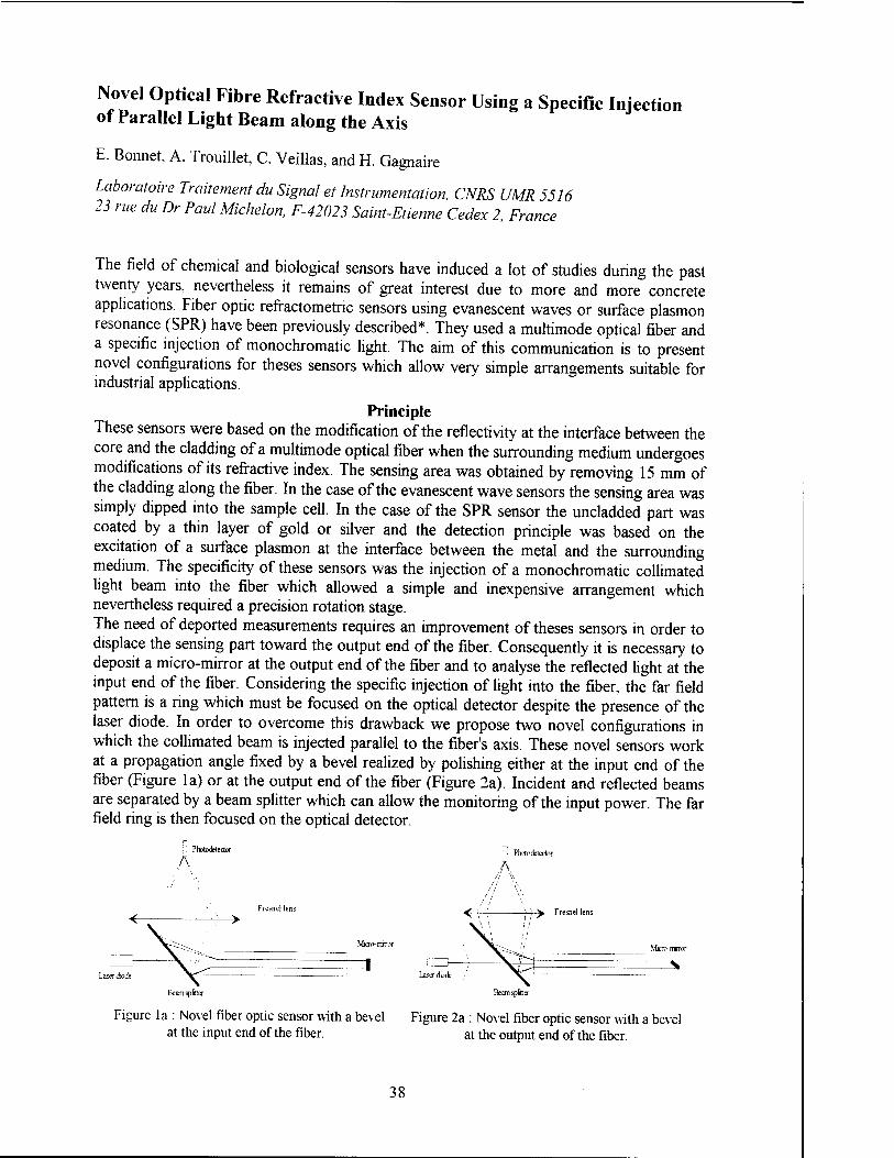
Novel Optical Fibre Refractive Index Sensor Using a Specific Injection of Parallel Light Beam along the Axis
E. Bonnet, A. Trouillet, C. Veillas, and H. Gagnaire
Laboratoire Traitement du Signal et Instrumentation, CNRS UMR 5516 23 rue du Dr Paul Michelon, F-42023 Saint-Etienne Cedex 2, France
The field of chemical and biological sensors have induced a lot of studies during the past twenty years, nevertheless it remains of great interest due to more and more concrete applications. Fiber optic refractometric sensors using evanescent waves or surface plasmon resonance (SPR) have been previously described*. They used a multimode optical fiber and a specific injection of monochromatic light. The aim of this communication is to present novel configurations for theses sensors which allow very simple arrangements suitable for industrial applications.
Principle These sensors were based on the modification of the reflectivity at the interface between the core and the cladding of a multimode optical fiber when the surrounding medium undergoes modifications of its refractive index. The sensing area was obtained by removing 15 mm of the cladding along the fiber. In the case of the evanescent wave sensors the sensing area was simply dipped into the sample cell. In the case of the SPR sensor the uncladded part was coated by a thin layer of gold or silver and the detection principle was based on the excitation of a surface plasmon at the interface between the metal and the surrounding medium. The specificity of these sensors was the injection of a monochromatic collimated light beam into the fiber which allowed a simple and inexpensive arrangement which nevertheless required a precision rotation stage. The need of deported measurements requires an improvement of theses sensors in order to displace the sensing part toward the output end of the fiber. Consequently it is necessary to deposit a micro-mirror at the output end of the fiber and to analyse the reflected light at the input end of the fiber. Considering the specific injection of light into the fiber, the far field pattern is a ring which must be focused on the optical detector despite the presence of the laser diode. In order to overcome this drawback we propose two novel configurations in which the collimated beam is injected parallel to the fiber's axis. These novel sensors work at a propagation angle fixed by a bevel realized by polishing either at the input end of the fiber (Figure la) or at the output end of the fiber (Figure 2a). Incident and reflected beams are separated by a beam splitter which can allow the monitoring of the input power. The far field ring is then focused on the optical detector.
! Photodetector
A jl Phrtodetector
A
7 /' \ '>
<r~
\. ■ ""'-'—.
■>
Fresnel lens
Micro-rrnror
<r- \ \ \!
> Fresnel lens
Micro- mnnr _ 1 Laser diode
; \ Laser diode
Beamsplitter Beamsplitter
Figure la : Novel fiber optic sensor with a bevel Figure 2a : Novel fiber optic sensor with a bevel at the input end of the fiber. at the output end of the fiber.
38

iVß
Figure lb : Schematic propagation of light with a bevel at the in-put end of the fiber.
Figure 2b : Schematic propagation of light with a bevel at the out-put end of the fiber.
Results and performances In the first case (Figure lb) the propagation angle 9 in the fiber is determined by the bevel's angle ß and by the refraction law at the input according to the relation sinß = nc.sin(ß-0) where nc is the refractive index of the fiber core. It follows that with nc =1.457 (silica) ß must be approximately three times Q. In this configuration a sensing area half long as in the previous cases is enough. Experimental results have been obtained with ß = 19° and 37° and an evaporated gold micro-mirror, they show similar response and performances than those obtained with the previous sensors. We measured a maximum sensivity of 3.7 10"5 in the range 1.445-1.455 for the evanescent wave configuration. In the second case (Figure 2b) the propagation angle 6 in the fiber is only determined by the bevel's angle ß and such that 0 = 2.ß which makes the fabrication of the bevel easier. In this configuration only the reflected light presents the right propagation angle. So the sensing length must be the same as that of the sensors without micro-mirror in order to obtain the similar efficiency. Experimental results have been obtained with ß = 6° and an evaporated gold micro-mirror, they show similar response and performances than those obtained with the previous sensors. Figure 3 presents the reflected light power of an evanescent wave sensor for which we measured a maximum sensivity of 10"4 in the range 1.43-1.45. Figure 4 presents the reflected light power of a SPR sensor with an evaporated 50 nm silver layer for which we measured a maximum sensivity of 3.1(H in the range 1.36-1.39.
Refractive index
Figure 3 : Reflected power of an evanescent wave sensor with a 6° bevel at the out-put end.
Indices de refraction
Figure 4 : Reflected power of a SPR sensor with a 50 nm silver layer and a 6° bevel at the out-put end.
Conclusion We have presented novel configurations for evanescent waves and SPR fiber optic sensors which allow very simple arrangements suitable for deported measurements and industrial applications. The dynamic range ( 1.33 to 1.45 ) and sensitivity ( 5.10"5 to 5.10"4 ) of these sensors can be adjusted by the choice of the bevel's angle.
Reference
* C.Ronot-Trioli,A.Trouillet,C.Veillas,H.Gagnaire, Sensors&Actuators A, 54(1996)

The Suppression of Non-Specific Response to Human Blood Plasma in SPR Sensor for Detection of ß2-Microglobulin
E.Brynda1, M.Houska1, P.Tobiska2, and J.Homola2
'Institute of Macromolecular Chemistry, Academy of Sciences of the Czech Republic CZ-162 06 Prague 6, Czech Republic 'Institute of Radio Engineering and Electronics, Academy of Sciences of the Czech Republic Chaberskä 57, CZ-182 51 Prague 8, Czech Republic
A non-specific sensor response caused by the adsorption of analytes and other compounds on bare surface between immobilized receptors or by interaction of receptor molecules with compounds different from the analyte is one of the factors limiting medical applications of sensors for direct analyses in blood plasma and blood. In this paper we demonstrate that the non-specific response of an SPR sensor for detection of ß2-microglobulin may be considerably reduced by coating the sensing antibody layer with a monolayer of bovine serum albumin.
An assembly (I) consisting of two molecular layers of monoclonal antibodies against human ß2-microglobulin (anti-ß2m) and an assembly (II) consisting of two anti-ß2m molecular layers coated with a monolayer of bovine serum albumin (BSA) were immobilized on gold layers of surface plasmon resonance (SPR) chips by consecutive adsorption of proteins and covalent crosslinking with glutaraldehyde1'2. Changes in SPR conditions during the assembly preparation and immunoreaction were monitored by wavelength interrogation technique3. The shifts in the resonant wavelength induced by binding ß2-microglobulin (ß2m) from 1% BSA solution to the assembly I and II were determined to be 2.24 nm and 2.43 nm, respectively. The binding efficiencies of the assemblies I and II were estimated as 0.88 and 0.82 molecule ß2m per 1 molecule anti-ß2m, respectively, by comparing the sensor responses to ß2m binding with those corresponding to the immobilization of anti-ß2m. While the specific response was not significantly reduced by the BSA coating, the non-specific response measured in human blood plasma was efficiently suppressed. The shift in the resonant wavelength of 8.15 nm (assembly I) was reduced to only 0.16 nm (assembly II) by the presence of BSA coating. Obviously, ß2m molecules (m.w. 12600) were able to penetrate through the protective BSA monolayer on the top of the sensitive anti-ß2m assembly while bigger immunoglobulins (m.w. 155000) probably responsible for the response to human plasma due to their immunoreaction with anti-ß2m, were kept apart the anti-ß2m assembly by the crosslinked BSA layer.
1. Brynda, E. and Houska, M. Preparation of organized protein multilayers., Macromol. Rapid Commun. 19, 173-176(1998)
2. Brynda, E. Houska, M, Skvor J., and Ramsden J.J., Immobilisation of multilayer bioreceptor assemblies on solid substrates., Biosensors & Bioelectronics, 13, 165-172 (1998)
3. Homola, J., Pfeifer, P., Brynda, E., Skvor, J., Houska, M., Schwotzer, G, Latka, L., Willsch, R., Optical biosensing using surface plasmon resonance spectroscopy, Chemical, Biochemical and Environmental Fiber Sensors IX, Munich, Germany, June 1997, Proc. SPIE Vol. 3105, 318 - 324 (1997).
40

Detection of Refractive-Index Changes by Means of the Inverted-Graded Index Optical Fibers
Miroslav Chomät1, Ivan Kasfk1, Vlastimil Matejec1, Jiff Ctyroky1, Daniela Berkovä1, and Henri Gagnaire2
Institute of Radio Engineering and Electronics, Academy of Sciences of the Czech Rebublic, Chaberskä 57, CZ-182 51 Prague 8, Czech Republic 'Laboratoire du Traitement du Signal et Instrumentation, Universite Jean Monnet
42023 Saint-Etienne Cedex 2, France
~i ' r
0,000 IGI FIBER 0=410 urn
In recent years, several special fiber-optic sensing structures have been developed with
enhanced evanescent-wave sensitivity for chemical detection. Apart from fibers with special
shapes (e.g. sectorial fibers [1]), multimode fibers with a special refractive-index profile in the
core - inverted-graded index fibers (IGI) [2], have been devised. In preliminary investigations of
the IGI fibers [2] increased sensitivity to changes of the cladding refractive index has been
theoretically predicted and experimentally shown. This poster reports further results on
theoretical and experimental investigations of IGI fibers.
Experimental samples
of IGI fibers were drawn
from preforms with a
refractive-index profile shown
in Fig. 1. Fibers with a core
diameter of 0.41 mm were
drawn and coated with
polydimethylsiloxane polymer
with a refractive index value
of 1.407. For testing the
sensitivity of these fibers to
changes of the cladding
refractive index, the
-0,008 -0,3 -0,2 -0,1 0.0 0,1 0,2
Distance from the center [mm]
0,3
Fig. 1. Refractive-index profile of the drawn IGI fiber
41

25
20
polymeric cladding was removed in lengths from 3 to 10 cm. This bare part of the fiber was
immersed in liquids with known values of the refractive index.
An experimental quasi-point light source has been built set up of a single-mode fiber
with a core diameter of 9 um linked to a semiconductor laser diode by means of an optical
connector. Using precise micropositioners this light source was placed on the axis of a tested
sample of the IG1 fiber and its distance from the input end of the IGI fiber was varied while the
output power from the fiber
was measured. Results of
these measurements similar to
those in Fig. 2 will be shown in
the poster.
Experimental results
have been explained on the
basis of a ray-optics model.
Results of the theoretical
analysis of the fiber sensitivity
taking into account the actual
refractive-index profile of the
drawn IGI fiber and assuming
various values of the cladding
refractive index will be shown
in the poster.
•■••• 850 nm -o— 1300nm
1,32 1,36 1,40 1,44 1,48 1,52
Refractive index of the immersion 1,56
Fig. 2. Sensitivity of experimental IGI fibers to refractive- tindex changes in the optical cladding
This work was financially supported by the Grant Agency of the Czech Republic under contracts
No. 102/95/0939 and No. 102/97/P143.
[1] V. Matejec, M. Chornat, M. Hayer, D. Berkovä, M. Pospisilovä, I. Kasik, Sensors &
Actuators B38-39 (1997) 334-338.
[2] V. Matejec, M. Chornat, I. Kasik, J. Ctyroky, D. Berkovä, M. Hayer, EUROPT(R)ODE
IV. Münster, 1998 poster pI14.
42

Temperature Induced Light Switching Performed by Capillary Optical Fiber
J. Doupovec1, R. Brunner1, J. Zähora1, and F. Kvasnik2
Department of Fiber Optics, Institute of Physics, SAS, Bratislava, Slovak Republic 'UMIST, Dpartment of Instrumentation and Analytical Science, Manchester, UK
Capillary Optical Fiber (COF) serves as a perspective element for liquids refractive index sensing as well as for a gas, fields (electrical and magnetically) and chemical sensing. Its main advantage consist in its use in distributed sensors schemes. The aim of this contribution is to show the new Principle and its use in temperature induced light switching in both "point" and "distributed" arrangement.
The crossectional profile of COF see as follow: the core of this optical fiber has a shape of a ring, with cavity inside. The refractive index of this quartz-based ring is thanks to the Ge - doping slightly higher than the surrounded cladding. By this way is from one side of this core the air (or sensored medium placed in the cavity) and from the another side is quartz-based cladding. This cladding is surrounded by substrate in order to realized the desired geometrical dimension of this profile, and by this way to maintain the multi- or monomode state of this fiber.
The COF refers to so called "evanescent wave (EW) sensor". It means, that a part of the guided optical radiation penetrates both in COF cavity and substrate region, respectively. We have shown how depend the EW penetration depth on the index refraction difference (between core and material placed in the cavity), fiber geometry and applied wavelength, previously [ 1 ], [ 2 ].
The COF designed for temperature switching has a following principle. The COF which maintain the monomode state (with air inside the cavity and wavelength higher than 1000 nm) was filled with CS2 (in the form of thin liquid layer placed on the COF core) and closed in the cavity thanks to the collapsing of both COF ends. By normal conditions is CS2
liquid with index of refraction 1.62, what is much larger than that of COF core. Thank to this properties all light, which originally was guided by COF core, is coupled into this liquid layer (at the normal thermodynamically conditions) and after short distance is optical radiation predominantly guided by it. Thanks to the high attenuation of this supplemental liquid core layer, the radiation is attenuated after short COF length substantially. Now, when this COF is placed in the temperature higher than is the boiling point of CS2, the phase transition of the first order take place. The COF core is surrounded by the CS2 vapor now, with the index of refraction near to 1. By this way the radiation guiding is performed via the COF core with very small attenuation.
Various nuances of this temperature-induced light switch will be shown and discussed. Among them the most important are silanization of the COF inner wall, and maintaining the CS2 pressure inside the cavity and regulation of switching temperature by this manner.
References: [ 1 ] R. Brunner, et al.: Acta Physica Slovaca 45 (1995), 491-498. [ 2 ] J. Doupovec et al.: EUROPT(R)ODE III, Zurich 1996, 150.
Acknowledgment: The authors would like to acknowledge to VEGA grant No. 2/5082/98 and Joint Research Project SAS-BC.
43

A Novel Redox Switchable Fluorescence Probe Highly Sensitive for the Copper (II) Cation
G. Hennrich', H. Sonnenschein2, and U. Resch-Genger1
; Federal Institute for Materials Research and Testing, ^ Rudower Chaussee 5, D-12489 Berlin, Germany ' Humboldt Universität zu Berlin, Institut für Chemie, Hessische Str. 1-2. D-l0115 Berlin, Germany
The fluorescence and cation complexation behaviour of the 1,2,4-thiadiazol derivative (2) and
its corresponding N-imminoyl-thiourea (1) is described. Both forms can be converted into
each other by reversible oxidation or reduction, respectively.
Ox
Red
1 2
Depending on the system's redox state, the heterocyclic (oxidised) and the ring opened
(reduced) form show distinctly different complexation properties towards various heavy metal
cations which is monitored via fluorescence spectrometry. Selective complexation of
physiologically and environmentally relevant cations like Ca2+, Ni2+, Hg2+ and Pb2+ resulting
in fluorescence enhancement is achieved. Especially addition of Cu2+, commonly known as
fluorescence quencher, tremendously increases the quantum yield of 2 in acetonitrile solution
whereas the fluorescence of 1 is quenched completely.
100 |-
p 25
400 450 500 550
wavelength/nm 600 650
fluorescence enhancement of 2 upon addition ofCu2" in acetonitrile solution
44

Active Optical Waveguides: Waveguide and Physical Parameters Measurement
P. Hffbek1. F.Folttiny1, J.Kräl1, J.Schröfel2, and J. Hradilovä3
Czech Technical University in Prague, Faculty of Nuclear Sciences and Physical Engineering, Department of Physical Electronics, Bfehovd 7,115 19 Prague 1, Czech Rebublic
' Czech Technical University in Prague, Faculty of Electrical Engineering, Department of Microelectronics, Technickd 2, 166 27 Prague 6, Czech Rebublic Institute of Chemical Technology, Department of Inorganic Chemistry Technickd 5, 166 28 Prague 6, Czech Republic
Thin-film dielectric waveguides are basic components of integrated optics. The active optical waveguides play an important role in this field. The current fabrication techniques of the thin-film active optical waveguides, such as ion-implantation, ion-exchange, diffusion, epitaxial growth method, and other techniques, result in waveguiding layers with nonuniform refractive index profiles and physical parameters changes. For the design and the fabrication of integrated optical devices, such as amplifiers and lasers, the exact knowledge of waveguide and physical parameters of the active optical waveguides, and their dependence on technological parameters, are required. So, it is important to have a possibility to determine basic waveguide characteristics and waveguide physical parameters accurately and efficiently.
Two groups of the optical methods have been realised for the active optical waveguides characterisation: the methods for the optical waveguides parameters and the refractive index profile determination, and the methods for the optical waveguides physical parameters measurement.
The dark-mode spectroscopy method, together with the reflectivity calculation method (RCM), and the surface reflectivity analysis (SRA), has been selected from the group of published methods [1] for the optical waveguide parameters and the refractive-index profile determination. The active optical waveguides fabricated by ion-implantation, ion-exchange, and laser deposition had been studied by these techniques.
The single isosceles-prism dark-mode spectroscopy has been realised and used to determine the waveguide and substrate modes, and effective-mode indices of the waveguide samples. These data are necessary for refractive index profile determination by the RCM .
The RCM was realised for the refractive index profile determination of multimode waveguides. We made a software package for mathematical simulation of the reflectivity coefficient of the single isosceles-prism technique, and we calculated the reflectivity coefficient as a function of the waveguide mode indices for anticipated (chosen) refractive index profile, characterised by a set of parameters. Theoretically determined effective-mode indices were compared with the effective-mode indices, which where experimentally obtained from the single- isosceles dark-mode spectroscopy. Using numerical optimisation techniques, we determined the optimum refractive index profile parameters, and so the shape of the refractive index profile. The method is applicable for waveguides with more than three modes only. The refractive index
45

profile determination will be demonstrated for the Nd:YAG waveguides fabricated by He* ion implantation.
For single mode waveguides and for optical thin-films that can not work as a guide the SRA technique has been selected and a software package was made. The method is based on the determination of the Fresnel reflectivity of the waveguide surface as a function of the angle of incidence for an anticipated (chosen) refractive index profile, characterised by a set of parameters. From the reflectivity curve measurements we assess initial values of these parameters. By using the numerical optimisation method we have fitted the measured and the calculated curves. We have find the correct parameters, and we determined the refractive index profile. The program has been tested theoretically for a set of refractive index profiles. The results obtained will be presented and limitation of the SRA method will be discussed.
A good knowledge of the fundamental physical parameters, such as waveguide losses, stimulated emission cross section, and fluorescence time decay of fabricated active waveguides, is important for waveguide amplifiers and lasers development.
The Fabry-Perot cavity resonances technique [2] has been prepared for channel optical waveguides loss measurement. The technique is based on the Fabry-Perot resonances of the channel optical waveguide with good polished facets. Losses of such Fabry-Perot resonator are deduced from the optical power resonant and antiresonant transmission measurement. Fully coherent, monochromatic laser light is needed for this measurement, and the cavity resonances must be scanned during the measurement process. An electronically controlled thermoelectrical (Peltier) system was developed for the Fabry-Perot resonator scanning. The first results of this method will be presented.
The modified end-coupling method [3] have been developed for planar active optical waveguides loss measurement. The optical phase conjugation (OPC) was used for automatic and efficient coupling of the measuring beam into the waveguide. The method is a modification of the single-pass transmission-loss measurement technique that is not very accurate because of the unknown laser beam coupling efficiency. The loss coefficient of 0.6cm"1, corrected for Fresnel refraction, was obtained for the ion-implanted Nd:YAG waveguides. The self-pumped phase conjugation in photorefractive BaTi03 single-crystal has been used for OPC of the forward propagated, out-coupled waveguide modes, for the purpose of the loss coefficient measurements. The experimental set-up of such measurement and results obtained will be presented.
The stimulated emission cross section of Ti3+, Er3+, and Nd3+ doped waveguides we determine from comparative fluorescence measurement of the doped waveguide and just the same doped bulk material. The single-photon, time-resolved spectroscopy was used for the fluorescence time decay measurement. Some of the results obtained for Ti:sapphire, Englass, and Nd:YAG waveguides will be presented.
[1] P.D.Townsend, P.J.Chandler, L.Zhang, „Optical effects of ion implantation", Cambridge University Press, 1994.
[2] R.G.Walker, „Simple and accurate loss measurement technique for semiconductor optical waveguides", Electron. Lett., vol.21, p.581, 1985.
[3] D.Fluck, D.H.Jundt, P.Gunter, „Modelling of refractive index profiles of He* ion- implanted KNb03 waveguides based on the irradiation parameters", J.Appl.Phys vol.74, p.6023, 1993.
This research has been supported by grant No. 102/96/1243 GACR.
46

Luminescence Lifetime Based Fiber Optic Ion Microsensors
Christian Huber, Tobias Werner, Ingo Klimant, Christian Krause, and Otto S. Wolfbeis
University of Regensburg, Institute of Analytical Chemistry, Chemo- and Biosensors 93040 Regensburg, Germany
Abstract
Luminescence decay time based fiber optic ion micro-sensors for chloride and potassium are
presented. Fiber tip coatings consisting of the respective ion-selective lipophilic ion carrier,
plasticized PVC, and the ruthenium(IT)tris-4,4'-diphenyl-2,2'-bipyridyl bromothymol blue ion
pair [Ru(dibipy)3BTB2] as a proton donor [1, 2]. The efficacy of radiationless fluorescence
energy transfer from the donor (the ruthenium complex) to the acceptor (BTB) is mediated by
the ion concentration within the samples.
The chloride response is based on the co-extraction of chloride along with protons from
the aqueous sample into a plastiziced PVC membrane, whereas in the presence of potassium
ions in the sample, the neutral BTB becomes deprotonated on potassium ion extraction,
followed by the release of protons. Both processes result in a change of BTB absorbance. The
absorption band of deprotonated BTB overlaps significantly with the emission band of the
ruthenium complex so that radiationless energy transfer can take place.
Multimode quarz/quarz fibers with a core diameter of 100 urn have been used for the
fabrication of the micro-optodes. The sensor chemistry was applied to the prepared fiber tips.
The micro-optode fiber tips were fabricated using a preparation procedure described else where
[3]. After the application of the respective sensor chemistry, multimode fiber sensors were
obtained possessing decay times in the JUS range and allows the use of a relative simple
instrumentation e.g. a solid state light source (LED) for the phase shift measurement.
References:
[1] Ch. Krause, T. Werner, Ch. Huber, I. Klimant, and Otto S. Wolfbeis, Anal. Chem. 1998
(in press).
[2] Ch. Huber, T. Werner, Ch. Krause, I. Klimant and O.S. Wolfbeis, Anal. Chim. Ada 1998,
364,143.
[3] I. Klimant, V. Meyer, M. Kühl, Limnol. Oceanogr., 1995,1159.
47

Molecule Detection with an Integrated Surface Plasmon Transducer
O. Hugon1. P. Benech1, and H. Gagnaire2
; LEMO. INPG-UJF-CNRS UMR 5530, Ecole Nationale Superieure d'Electronique ^ et de Radioelectricite, 23 avenue des Martyrs, BP 257, 38016 Grenoble Cedex, France 2 LTSL CNRS UMR 5516, 23 rue du Docteur Paul Michelon 42023 Saint-Etienne, France
Since the last few years, there is a spectacular growth of interest in the field of the
integrated surface plasmon sensors, certainly because they can offer good performances at a
very low cost with some advantages that are a characteristic of optical components such as
immunity to electrical or electromagnetic interferences or the possibility to work in hostile
environment.
The device described is a classical channel waveguide realised by ion-exchange in a glass
substrate, covered with a thin silver film over a length of about 1mm and the light source is
a 830nm laser diode.
We show that the output light power of the device is a function of the superstrate index. In
order to reach the optimal sensitivity, it is necessary to have a good understanding of the
phenomena that occur inside the component For that purpose, we studied the mode
coupling and propagation in detail to obtain an accurate modellization.
The transfer matrix method with the Newton-Raphson algorithm have been employed to
solve for the propagation modes of the structure. It appears that with the classical scalar
product, those modes are no longer orthogonal. The modal analysis must then be performed
carefully.
We find that the computed response of the component is in good agreement with the
measured values.
Finally, by the use of a sensitive material (squalane: C50H62), we also show an example of
gas detection with the realised device.
48

Micromirror Driven by Electrostatic Actuation
Martin Huja
The Czech Technical University in Prague, Faculty of Electrical Engineering, Department of Microelectronics, Prague, Czech Republic
Abstract. Microengineering principles and techniques are playing an increasing role in the
design and fabrication of components in the fields of micro-optics and optoelectronics. Optical mirrors, gratings, lenses, waveguides air techniques offer a wide range of possibilities to manufacture various components and also batch-produce and integrate them onto a wafer. A new, fast growing discipline called microsystems has evolved from a combination of the three subject areas of mechanics, optics and electronics at micrometer scales.
Introduction IC-compatible microfabrication of micro-opto-electro-mechanical systems
(MOEMS) has recently emerged to serve the increasing demand for low-cost mass- produced optics, compatible with standard silicon microelectronics. This discipline receives more and more attention and very promising results have recently been obtained in the fabrication of mechanical, electrical and optical sensors and actuators.
Micromechanical systems must include large-deflection actuators to create devices, which can interact mechanically with their surroundings. Electrostatic actuators with large non-resonant motion require high voltages and large surface areas to produce useful forces.
Production of microoptical devices is normally conducted in two stages: fabrication of a master copy and subsequent replication. The master can be fabricated and replicated using lithography, which is widely used in microelectronics. Direct laser and e-beam writing are also frequently used when very high precision is demanded. Replication for the fabrication of large arrays of high-quality allows inexpensive optical components with excellent repeatability.
Simulation of Micromirror The micromirror in Figure 1 is driven by electrostatic actuation. The phase of light
reflected off the micromirror can be altered if the micromirror is pulled down towards the substrate. The top electrode is the center region of the micromirror supported by the flexures. The bottom electrode is from PolyO.
■ Metal
HI Poly 1 ■I Poly 0 I | Nitride I I Substrat
Figure 1.: Electrostatic actuated micromirror fabricated MUMPS technology
49

Metal Poly2 Polyl PolvO
Nitride
Figure 2: The stacks of Poly 1, Poly 2 and Metal represent mirror used to drive the actuator electrostatically.
<V =.2463 rv =-.8292 :'.' -.5018
i-ZS—16.29 :-3UFrrp:
Figure3: Input structure for ANS YS simulation
The mirror is a moveable plate, while the access line underneath is the fixed plate. The capacitor electrodes never touch each other, instead the tip of the moving mirror touches the fixed level at a landing pad, which is also conductive and kept at the same potential as the mirror.
The electrostatic micromirror was fabricated using the Multi-User MEMS Processes. The electrostatically actuated micromirror was constructed using thin surface of micromachined polysilicon film on a silicon wafer (Figure2). MUMPs offers three paternal layers of polysilicon and two sacrificial layers of phoshosilicate glass on a base layer of silicon nitride derived from work performed at the Berkeley Sensors and Actuators Center at the university of California.
For mechanical simulation of micromirror was used ANSYS program for finite element analysis and design. The input structure for ANSYS simulation is on the Figure 3.
The micromirror for micromirror array has size 70x70um and distance between micromirror and bottom electrode is 2 (am. The theoretical dilatation simulated by ANSYS program is on the Figure 4.
Conclusions The technology of inexpensive
micromachined adaptive mirrors and tilt correctors has found many applications in modern optics and optoelectronics.
More complex devices are produces at low cost using standard PCB technology to fabricate the mirror carrier, actuator structure, spacer and electrical interconnects. These devices can replace more expensive adaptive optics in applications where high costs prevent further development.
ANSYS 5.3 MAY 26 1998 18:30:13 NODAL SOLUTION 5TEP=! SUB -1 TIME=1
=.:22E-05
.24^E-0S
.493E-&6
.987E-06
.123E-05 •148E-05 .173E-05 .197E-05 .222E-05
Figure 4: Output of ANSYS simulation. Dilatation of micromirror 70x70um. Distance of electrodes 2 urn, V=36V
References
Bums M.D., Bright, V. M. Nonlinear flexures for stabile deflection of an electrostatically actuated micromirror SPIE Vol 3226 p 125-135
Berkeley Sensors and Actuators Center Three-layer polysilicon Surface Micromachining Process, http:/mems.mcnc.org/smumps/Mumps.html
50

Carbon and Carbon Nitride Planar Waveguides on Silicon Substrates for Optical Sensors
I. Hiittel1, J. Gurovic2, F. Cerny2, and M. Chornat3
Institute of Chemical Technology, Prague, Czech Republic 'Faculty of Mechanical Engineering, Czech Technical University, Prague, Czech Republic Institute of Radio Engineering and Electronics, Prague, Czech Republic
1. Introduction With the development of optical communications and lately also of optical sensor
structures [1,2,3], the components of integrated optics used on silicon substrate have been increasingly useful. Their advantages are very good optical parameters, cheap basic material, and, especially, the potential possibility of integrating optical and microelectronic structures.
Silicon substrate is coated with a separate layer which is prepared by thermal oxidation and its thickness is several microns. Presently there are many technological procedures used for the preparation of the upper planar waveguide layer with a higher refraction index than that of a separate layer of silicon oxide. This upper layer is created by dielectrics of anorganic origin and species of organic origin have also been successfully used recently. Anorganic dielectrics are usually layers of silicon oxide and nitride types, so- called oxynitrides, or layers of properly dopped silicon oxide. In our work we have focused on preparation of thin carbon and carbon nitride films for planar optical sensors, which we consider very promising.
2. Experimental Optimization of the preparation of carbon layers for planar optical waveguides
focused on the growth of layers with a reproducible refraction index and thickness. Plasma Enhanced Chemical Vapor Deposition (PECVD) method was used for layer preparation. The source gases were methane, nitrogen, or ammonia. The layers samples for planar optical waveguides were prepared on silicon substrate. The samples were provided with thermically oxidated separating layer of SiCK (thickness 3 urn).
The samples chosen for infrared spectra measurement were prepared on two-sidedly polished silicon wafers. The thickness of the prepared waveguide layers was measured by profilometer Talystep. Optical properties of the waveguides were measured at 633 nm wave length using standard prism spectroscopy. An ellipsometer with rotating analyzer was used for refractive index measuring, the source of radiation being He-Ne laser with 633 nm wave length. The refractive index was measured at twelve points. Infrared (IR) spectroscopy was performed at room temperature using a FTIR Nicolet 740 spectrometer operating in absorption mode at 500 - 400 cm"1 with GOLD Specular Reflectance Equipment manufactured by Spectra Tech UK.
3 Results and discussion The refraction index and its distribution on the substrate is an important parameter of
the layers prepared. The refraction index on carbon and carbon nitride softer layers prepared on the negative electrode was high (2.2 to 2.6) as compared with the harder layers on the positive electrode (1.6 to 1.8).
Infrared absorption spectra were elaborated for chosen samples. The spectra of the softer carbon layers clearly indicate the presence of-(CH2)- groups (characteristic peak in the range 2900 - 3000 cm"1), causing probably low hardness of the layers and lower
51

refraction index (polymeric character of the layers). The spectra of the harder carbon layers with higher refraction index contain fewer of these groups. The absorption bands caused by C-H stretching, peak at 2937 cm"1, respectively at 2863 cm"1, i.e. near the wave number corresponding to asymmetrical and symmetrical stretching in CH, groups These lines indicate the amount of bonded hydrogen in the film. The frequency tail of this band (near and above 3000 cm") can be ascribed to stretching in various = C - H groups High hydrogen content leads to frequent termination of the atomic bond network, which probably results in low hardness of the films and in lower refraction indices. The main feature of t TIR spectrum is a band corresponding to symmetrical stretch - C = C = C - groups at 1070 cm supporting the proposed polymeric structure. In all samples the characteristic carbonyl band (C=0) at 1740 cm'1 can be detected. The origin of this peak is probably in reaction trapped long-lived radicals after deposition with oxygen and water vapor
Optical properties of planar optical waveguides, i.e. samples of the layers deposited on a silicon base with a separation layer of Si02 , were measured. Because of the great difference of values of refraction indices, the „guided,, mode already exists from the layer thickness of approx. 100 nm, and the waveguide is one-mode type with the thickness up to approx. 400 nm, practically independently of the thickness of SiO> layer.
This analysis reflects only the attenuation by radiation and absorption in the silicon substrate. It is possible to expect that the quality of the interface and the structure perfection of waveguide layers is of essential importance for the real attenuation of waveguides due to high concentration of optical radiation in the waveguide layer with the high refraction index. We prepared samples of planar waveguides with great differences between the refraction indices of the core and the casing with the structure of Si/Si02/C From the analysis and from the measured parameters of the layers, the optimum thickness of 320 nm for the waveguide layer for the one-mode waveguide resulted. The attenuation of the prepared planar waveguides was about 1 dB/cm and in the case of the best one it was 0 3 dB/cm.
4 Conclusion Optimization of PECVD technology for preparation of carbon layers suitable for
planar optical waveguides was implemented, and samples of these waveguides prepared Conditions have been set up for channel waveguides implementation for optical sensors and integrated optics elements on silicon substrates.
Acknowledgements The work was realized under financial support of the Grant Agency of the Czech Republic (Nos. 102/96/1610 and 106/97/0498).
References [1] G. Boisde, A. Harmer, Chemical and Biochemical Sensing with Optical Fibres and Waveguides, Boston-London, Artech House; (1996), pp. 177-197. [2] M. Kawachi, Optical and Quantum Electronics, 22 (1990), p.391. [3] S. Goel, J.C. Pincenti, and D.L. Naylor, Applied Optics, 32 (1993), p.318.
52
Characterization of Polymeric Coating Materials for Infrared Evanescent Wave Sensors
M. Jakusch, M. Kraft, B. Lendl, B. Mizaikoff
Vienna University of Technology,Institute of Analytical Chemistry, Getreidemarkt 9/151, A-1060 Wien, Austria
Sensor technology has come to an age, where many of initially very ambitious and risky concepts have proved their applicability and successfully found their way into industrial and commercial systems. However, there are still white spots on the map of potential sensor applications: Especially environmental monitoring and process analysis in harsh environments often suffer from limited endurance of complex (bio-)chemical recognition layers.
In this context, mid infrared spectroscopy is a promising transducer system for chemical sensors, due to its inherent molecular specificity, which allows to choose more simple but stable materials such as common polymers as chemically sensitive materials. Additionally, by means of fiber optic and evanescent wave sensing principles versatile and rugged sensor systems can be easily realized.
In these systems most of the 'recognition task' is performed by the molecular specific transducer. However, even in moderately complex matrices analyte spectra are likely to heavily overlap and prohibit exact speciation. Hence, the nature of the polymer layer, which has to serve as a kind of 'preselecting filter' allowing only enrichment of a certain subclass of all present substances in the active region of the sensor, has still to be carefully chosen.
In order to accurately characterize and compare the sorption and diffusion properties of only a few polymer materials for several analytes a considerable number of single sorption experiments have to be carried out, which can get very time consuming. For this kind of experiments we built up a computer controlled sequential injection calibration system, which enables unattended running of whole calibration sets (e.g. overnight) thus providing a statistically useful amount of data within reasonable time.
Enrichment factors of volatile organic compounds (VOCs) such as BTEX or chlorinated hydrocarbons in thin films of several aliphatic polymers and siloxanes were determined. The resulting selectivity patterns will then be used for further development of coating materials.
This study was part of the EU project ENV4970475, which aims on development of an optimized ER tunable diode laser sensor system for VOCs in waste water.

Optical Fibres for Environmental Analysis of Seawater Pollutants
M. Kraft, M. Jakusch, and B. Mizaikoff
Institute of Analytical Chemistry, TU Wien, Getreidemarkt 9/151, A-1060 Wien, Austria
The maintained health of our surrounding oceans requires frequent monitoring of their numerous physical, chemical and biological parameters. The main obstacles are the diversity of the constituents, the harsh measurement conditions and the enormous size of the water mass. The most widespread approaches for sea water analysis involve sampling. This requires a suitable sampling arrangement and a laboratory with highly specialized equipment and trained personal. Even then these methods usually are too slow, too expensive and far too complicated for measuring time dependant pollution profiles, e.g. near industry sites or tanker cleaning points.
The logical alternative to the standard analysis systems are sensors. Especially optical sensors are well suited for this purpose as it is much easier to build sea water resistant optodes than electrodes. Additionally, varying environmental conditions are less likely to affect the rugged optical sensors than electrochemical devices. For these reasons, the European Union decided to fund an European research project for Spectroscopy using Optical Fibres in the Marine Environment - SOFIE (Contract Nr. MAS3-CT97-0157).
The aim of the SOFIE-project is the development of a modular all-fibre-optic measurement device for the in situ monitoring of pollutants in the marine environment. The overall system consists of different sensors covering a range of optical methods. All sensitive parts are contained in a seawater proof tough body, while only the actual sensing elements are exposed to the seawater. The analytes chosen for the first such device are heavy metal ions, polycyclic aromatic hydrocarbons (PAH's) and chlorinated hydrocarbons (CHCs). However, the modular construction of the system allows the implementation of additional sensors.
The Institute of Analytical Chemistry at the University of Technology Vienna is responsible for the development of a mid-infrared (MIR) fibre optical sensor for the analysis of chlorinated hydrocarbons in water. Chlorinated hydrocarbons, such as trichloroethylene and tetrachloroethylene, are, besides highly toxic chemicals, particularly significant indicators for urban pollution.
The CHC-sensor is based on fibre evanescent wave spectroscopy (FEWS), using silver halide fibres as elongated attenuated reflection elements. Silver halide fibres are highly transparent in the mid-infrared region, highly flexible and can be used for remote sensing distances of up to 5 m. The actual sensor head is a silver halide fibre, coated with a thin, hydrophobic polymer layer. This layer simultaneously enriches the analyte near the attenuation reflection element and protects the fibre from the corrosive seawater.
Two measuring principles are currently under investigation. One approach utilizes tunable diode lasers (TDL's), which are small and rugged. However, only a limited number of compounds could be detected using this system. The alternative introduces a rugged, miniaturized FTIR-spectrometer. This system would have the advantage of allowing multi- component analysis of CHCs as well as of aromatic hydrocarbons. While the use of MCT- detectors cooled by maintenance-free Stirling coolers provides a suitable detector, the crucial point still is the ruggedness of the optical setup, especially when using the FTIR-approach.
54

Light Emitting Diodes as Selective Detectors
L. Jiräsek1, Z. Burian', and M. Jiräskovä2
Department of Microelectronics, Czech Tech. Univ. in Prague, Prague, Czech Republic 'Dep. of Dermatology, 1st Faculty of Medicine, Charles Univ., Prague, Czech Republic
340 370 410 450 490 530 570 610 650 690
lambdafm]
The human organism needs small doses of UV to produce vitamin D3. Too high doses of UV, however, can cause sunburn, degenerative changes, skin cancer, damage the immune system and the eyes. The risk of carcinoma is increased and the detection of three spectral ranges of UV (UV-A, UV-B, UV-C) seem necessary. Therefore we need the high selectivity detector in this region of UV. Common detectors (photoresistors and photodiodes)
Fig. 1 Spectral characteristics (from left) of blue, green, red LED used as photodiode
have relatively wide spectral characteristics (100 nm, e. g. [2]) This selectivity must be increased by special selective optical accessories as optical grating, prism, filter (too complex construction, too large proportions, too expensive). Some Light Emitting Diodes behave as acceptable selective detectors. The characteristics can be influence by type of the dopant. We suppose using these diodes as selective detectors for miniature pocket or „watch" personal dosimeters.
For verification of our idea we tried to check the spectral sensitivity common types of diodes (red-GaP, LQ2134-Bicolour; green-GaP, LQ2134-Bicolour; blue-SiC, L-934BC a L53BC). The measured characteristic especially SiC LED showed a very narrow spectral characteristic (spectral linewidth 10 nm) with maximum sensitivity at 365 nm . In other
parts of the UV and visible light interval we have found no measurable voltage value on these diodes. The resulting spectral characteristics of measured diodes are shown in the Fig. 1. For there comparison we also cite the behaviour of the spectral characteristic of an idealised quantum
detector. In tab. 1. is overview of properties some types of detectors. Perspective materials for LEDs useable for selective detectors are shown in tab. 2. The measurements were performed on the Department of Microelectronics.
In future work we shall endeavour to discover follow-up data(materials) for diodes with narrow spectral characteristics and a potential for adjusting maximum of spectral sensitivity characteristic by the type of dopands enabling a classification of UV spectrum region. Tab. 2 Perspective materials
[1] Jiräskovä, M, Jiräsek, L.: Natural and Artificial Radiation Sources Influence on a Human Body. Invited lecture. Workshop of Illumination Technology, Kosice 1996 [2] Catalogue of LASER COMPONENTS GmbH, Olching 1998
Tab. 1 Source A [nm]
Detector Type A [nm] AA [nm] L-934BC(SiC-blue) LQ2134(GaP-green) LQ2134(GaP-red) SGD-lOO(Si-photodiode)
470 520 650
365 565 660 1000
10 130 45 400
Material Emission A [nm] CdF2 270-700 Cdi_xZnxS 340-540 6H SiC 450-560 GaN 360-570
55

Distributed Fibre Optic Sensors for Detection and Localisation of Ammonia Leaks
L. Kalvoda1, R. Lukas1, P. Lukäsovä', M. Landl2, P. Simon2, and F. Kvasnik3
'Lukas Industrial Research, Doubravinova 1/219, 163 00 Prague 6, Czech Republic -Department of Physical Chemistry, Faculty of Chemical Technology,
3 Slovak Technical University, Radlinskeho 9, 812 37 Bratislava, Slovak Republic Department of Instrumental and Analytical Science, UMIST, PO Box 88, Manchester M60 1QD, United Kingdom
A rapid detection and localisation of ammonia leaks establishes a serious problem for all the industrial facilities utilising large-scale cold-storage rooms, such as breweries, dairies and abattoirs. Every year, several accidents are reported caused by ammonia leakage resulting in severe common distress.
The traditional ammonia detection employs point potentiometric electrode sensors organised in network [1]. These generally suffer from high power consumption, necessity of relatively frequent re-calibration, and restricted coverage of the inspected area.
Numerous optical devices has been proposed in attempt to overcome the former drawbacks (see e.g. [2-7]). One of the most promising concepts is taking advantage of optical time domain reflectometry (OTDR), allowing simultaneous determination of ammonia concentration and leak location over the length of sensing fibre [4,8,9]. An OTDR set-up was employed to testing the distributed ammonia sensing system described in further.
The experimental set-up we used consisted of Photodyne 5500 XFA OTDR unit HP 54615B digital oscilloscope (lGSa/s, 500 MHz bandwidth), 57m of a custom-made sensing optical fibre, and a PC. Light pulses (20 ns width, 1 kHz repeating frequency, ^=850 nm) were launched in the fibre and the reflected light intensity was detected by a Si-photodiode The signal was then amplified by a logarithmic amplifier, analysed by the oscilloscope, and the resulting OTDR curves were sent to PC. Ammonia vapours were developed by evaporation of a 25 % NH4OH/H2O solution at RT. An ammonia/air mixture was pumped into the testing chamber where a short section (4m) of the sensing fibre was placed.
The key part of the system was the sensing fibre transmuting ammonia exposition to variation of the back-reflected light intensity. We employed a silicon-clad silica fibre (200/260 urn core/cladding diameter) with the cladding doped by 5-(4'-diethylaminophenylimino)quinolin- 8-one copper(Il)sulphate complex dye. The method of dye incorporation was similar to those described by Potyrailo and Hiefije [10]. The synthesis and spectral sensitivity of the dye to ammonia was described elsewhere [11,12]. The dye shows a strong absorption band with maximum at 740 nm. The red shoulder extends up to 900 nm. Repeated expositions to ammonia cause reversible drops of the NIR extinction.
Significant variations of the OTDR curves were observed during the ammonia exposition- (i) The intensity of the Fresnel reflection from the remote fibre end increased, as a result of the total light attenuation decrease owing to the local dye absorption decay (ii) The time- dependence of the back-reflected light intensity showed a well-resolved local increase The slope change could be related to the exposed length of the fibre and the ammonia concentration. A simple decision-making algorithm is proposed utilising the former features
56

Acknowledgements
This research was kindly supported by the European Commission (contract number CIPA- CT94-0206).
References
[I] Y. M. Fraticelli, M. E. Meyerhoff, Anal. Chem., 53 (1981) 992. [2] M. R. Shahriary, Q. Zhou, G. H. Siegel, Jr., Opt. Lett., 13 (1988) 407. [3] L.L. Blyler, Jr., R. A. Lieberman, L. G. Cohen, J. A. Ferrara, J. B. MacChesney, Polym.
Eng. Sei., 29.(1989) 1215. [4] R. A. Liebermann, SPIE Vol. 1586 (1991) 80. [5] R. Klein. E. Voges, Sens, and Actuators B, U (1993) 221. [6] C. Preininger, G. J. Mohr, I. Klimant, O. S. Wolfbeis, Anal. Chim. Acta, 334 (1996)
113. [7] T. Grady, T. Buttler, B. D. MacCraith, D. Diamond, M. A. McKervey, Analyst, 122
(1997)803. [8] P. Klocek, R. Beni, J. C. O'Connel, C. P. Van Vloten, SPIE Vol. 618 (1986) 151. [9] J. Bürck, E. Sensfelder, H.-J. Ache, Proc. SPIE Vol. 2836 (1996) 1. [10] R. A. Potyrailo, G. M. Hieftje, ,4«a/. Chim. Acta, 370 (1998) 1. [II] P. Simon, S. Sekretär, B. D. MacCraith, F. Kvasnik, Sens, and Actuators B, 38-39
(1997)252. [12] C. Malins, M. Landl, P. Simon, B. D. MacCraith, Submitted to Sens, and Actuators
(1998).
57

A Development Tool for Sensors Based on the Inner-Filter Effect
B. Koväcs. R. Dombi, S. Kunsägi-Mäte, and N. Marek
Department of General and Physical Chemistry, Janus Pannonius University, Pecs, Hungary
The use of the primary and secondary inner-filter effect (IFE) and/or energy transfer
(ET) in fluorescent optical sensors has two distrinct advantages: /') tunable dinamic range
and //') enhanced sensitivity. The technique was introduced first for pH sensing as cuvet test
[l]and recently was adapted for solid-state optode membrane systems [2,3], A sensor based
on the IFE contains two different dyes: the emission of a fluorescent species is modulated
by a second absorber molecule. In the simplest case the sensor response is influenced by the
spectral overlap and the concentrations of the two dyes and by the binding coefficient(s) of
the analyte. Because of the many parameters, it is not easy to find the best composition of a
sensing membrane and the development requires a lot of experiment.
In this work we describe a novel computing methode based on the theoretical
description of the IFE [4], The results show that two different types of IFE based sensors
exist: a) the passive IFE where the absorber binds the analyte, and b) the active IFE where
both of the dyes are sensitive. By using the experimental absorption and emission spectra of
the dyes the computing results in the following important parameters: the optimal
concentrations of the absorber and fluorescer as a function of the excitation and emission
wavelength, the ex/em wavelength at the highest signal change, and the signal change of the
sensing system as a function of the analyte concentration. Heavy metal sensitive [5] and pH
sensitive dye mixtures were used to test the program.
References (1) Gabor, G., Walt, DR., Anal. Chem., 63 (1991) 793-796. (2) He, H, Li, H, Mohr., G, Koväcs., B., Werner, T., Wolfbeis, O.S., Anal. Chem 65 (1993)123-127. (3) Shortreed. M., Bakker, E., Kopelman, R., Anal. Chem., 68 (1996) 2656-2662. (4) Credi. A., Prodi, L., Spectrochimica Acta Part A, 54 (1998) 159-170. (5) B. Koväcs, R. Dombi, N. Marek, submitted to Microchimica Acta (1998).
This work was supported by the Hungarian Research Foundation (OTKA) F-17324.
58

Characterization of Chromogenic Calix[4]arene Derivative Based Sodium-Selective Optical Sensors
Bui Thi Thu Lan, Klara Töth, and Istvän Bitter
Technical University of Budapest, Institute for General and Analytical Chemistry, Department of Organic Chemical Technology, 1111 Budapest, Szt. Geliert ter 4, Hungary,
Novel chromogenic calix[4]arene derivatives were synthesized and tested in our Laboratory as
ionophores for designing optical sensors. Distinct sodium selectivity was observed with the
chromogenic calix[4]arene derivative with 2,4-dinitro-phenylazo group in the neighbourhood
of the coordination sites. This ligand has been incorporated into plasticized PVC and other
polymers to develop sodium-selective optodes. The analytical characterization of the
plasticized PVC based optical membranes has been investigated in detail. The dynamic
concentration, ion-selectivity behaviour, response time and reproducibility are discussed. The
optical membrane exhibits sodium sensitivity in the 5xl0"2 -10"4 mol/1 concentration range.
Due to the pka value of the ligand the operation pH of the plasticized PVC membrane is
relatively high. The optode membrane components were varied deliberately with the aim of
lowering the operation pH. The effect of the nature of the membrane matrix on the dynamic
range and the operation pH of the relevant optode membrane are explained. The sodium ion
response is interpreted on the basis of the ion-exchange theory, which is underlined by the
results of calixarene mediated ion-transport experiments.
59

The Experimental Studies on Feasibility of the Fibre-Optic Aqueous Sensors Based on Surface Plasmon Resonance on Silver Film
Wenbin Lin1, Nicole Jaffrezic-Renault', Monique Lacroix1, Jean-Marc Chovelon1
Henri Gagnaire", Collete Veillas2, and Alain Trouillet2
'ingenierie et Fonctionnalisation des Surfaces (UMR 5621), Ecole Centrale de Lyon ^ 36 Avenue Guy de Collongue, BP163, 69131 Ecully Cedex, France 'Laboratoire du Traitement du Signal et Instrumentation (UMR 5516), Universite Jean Monnet, 23 Rue du Dr. Paul Michelon, 42023 Saint-Etienne Cedex 2, France
Gold and Silver are ones of most important metals employed to produce the Surface Plasmon Resonance (SPR). Gold has been traditionally used for decades and commercially realized in SPR sensor owing to its physical and chemical stability. Silver can provide the SPR much sharper than Gold that has been theoretically and experimentally verified Unfortunately Silver is very active. It is oxidized as soon as it is exposed to air especially to water that gives rise much difficulty to bring about an aqueous sensor which utilizes Silver as the SPR supporter.
Our optical fibre sensors were elaborated by a multimode silica step-index fibre with the core diameter 600 ^m and the core refractive indice 1.457. A section of the cladding was removed at the middle of the fibre. Then a Silver film about 45nm-55nm was deposited by thermal evaporation. The SPR was excited by a collimated light with 670nm wavelength which illuminated the input end of the fibre from a laser diode. According to our theoretical predictions, if an overlayer of Zr02 (index 1.54 and thickness 20nm approximately) was coated onto the Silver layer, a novel sensor capable to monitor aqueous environment (index varied from 1.33 to 1.35) can be achieved. This kind of optical fibre sensor is very useful to detect most chemical and biochemical systems.
The overlayer of Zr02 was fabricated by Sol-Gel technique. After that, the fiber was cured at 60 C for three hours in order to densify the Zr02 film. Experiments illustrated that the Silver layer was seriously oxidized if the temperature was raised up to 110°C.
We have experimentally realized this kind of fibre-optic aqueous sensor with very good sensitivity (about 2.5x10-). However, its performance deteriorated in a few days It's obvious that the overlayer of Zr02 was not dense enough to protect the Silver from oxidation. Therefore, the acid Thiols with both long chain and short chain were introduced to further experiments. Before the elaboration of the Zr02 layer, the metallized fibres were dipped into the solutions of Thiols for 2 hours in order to construct a monolayer of Thiol on the surface of Silver. The compared experimental results let know that the acid Thiols can prevent, to some degree, the oxidation of the Silver, hence delay the deterioration of the sensor and at the same time, improve the adhesion of the layer of Zr02. Moreover the sensor with the long chain acid Thiol has much more stability and much longer life-time than that with the short chain acid Thiol has. It can be explained as the long chain Thiol can build a monolayer much denser than the short chain Thiol can. This experimental discovery is significant because it makes possible to fabricate a stable fibre-optic aqueous sensor using a long chain acid Thiol for practical applications. Furthermore, an immuno-sensor can be developed.
60

Characterization of Ormosils
Aleksandra Lobnik1, Alenka Majcen Le Marechal1, and Otto S. Wolfbeis2
Faculty of Mechanical Engineering, University ofMaribor Smetanova 17, 2000 Maribor, Slovenia
''University of Regensburg, Institute of Analytical Chemistry, Chemo- and Biosensors D-93040 Regensburg, Germany
The sol-gel technology enables the production of glasses at ambient temperature. In contrast
to conventional glass sintering at high temperature, a great variety of glasses containing
both organic and inorganic additives can be made. Major advantages of sol-gels include
mechanical stability, chemical inertness, optical transparency, low-temperature processing,
and flexibility in shaping configurations. Most sol-gel techniques use water and low
molecular weight alkoxysilanes such as tetramethoxysilane (TMOS) or tetraethoxysilane
(TEOS) as sol-gel precursors. Ormosils (organically modified siloxanes) represent hybrid
systems in which several precursor types such as organotrialkoxysilane or
diorganodialkoxysilane precursors (R'Si(OR)3 or R'2Si(OR)2, respectively) in which the R'
represents a nonhydrolyzable organic substituent, are combined. They have attracted much
attention in recent years because they provide easy introduction of reactive functional
groups which can be subsequently used as covalent anchors for indicators, or they are used
to reduce the functionality (potential number of sites able to form Si-O-Si bonds) of the
alkoxide precursor, impart organic character or derivatize the siloxanes network.
Conventional sol-gels are rather hydrophilic and well suited for sensing ions. A more
hydrophobic material is obtained by preparing ormosils. Ormosils have been attracting
greater interest recently in the area of chemical sensors, mostly to form ormosil-based
optical oxygen or ammonia sensors. A critical parameter in gas sensing is the permeability
and permeation selectivity of the polymer used. Silicones, teflon and plasticized poly(vinyl
chloride) are considered to be a good compromise, owing to their high permeability for
gases and their impermeability to ions. Other materials such as cellulose, poly(vinyl
chloride), poly(vinyl acetate) also have been used for gas sensing but they are less gas-
61

permeable.
In contrast with the fast development of many applications, the exact nature of the
entrapment and the properties of the encapsulating cage are still to be fully understood. The
detailed microstructure of sol-gel glasses depends on a large number of important
processing parameters such as water:sol-gel precursor ratio (R), precursor type, nature of
the catalyst, aging time, aging temperature, drying time and drying temperature.
Investigation of sol-gel systems has proceeded along two fronts: a) examination of the sol-
to-gel-to-glass phase transitions and b) elucidation of the structural, physical, and chemical
properties of the xerogel and glass products. These characterization studies have been
carried out with a variety of techniques including X-ray scattering, infrared, Raman and
NMR spectroscopies, transmission electron microscopy (TEM), and ellipsometry.
The idea of our work was to entrap polarity-sensitive dyes to gain more information
about polarity insight of the ormosil cages. We have used two different solvatochromic
dyes, (a) Nile Red (NR) and (b) ketocyanine (KC) dye, all with unique positive
solvatochromic properties in fluorescence, to study the spectroscopic properties in different
solvents and ormosils. Fluorescence maxima and intensities were used to study NR and KC
doped in ormosils, prepared from various proportions of tetramethoxysilane (TMOS) and
methyltrimethoxysilane (MeTriMOS), octyltrimethoxysilane (OcTriMOS),
phenyltrimethoxysilane (PhTriMOS), dimethyl-dimethoxysilane (DiMeDiMOS) or
diphenyldimethoxysilane (DiPhDiMOS). Layer properties such as temporal and storage
stability were monitored as a function of amount and type of the ormosil precursor.
References:
1. OS. Wolfbeis, R. Reisfeld, I. Oehme, Structure and Bonding, 85 (1996) 52.
2. CJ. Brinker, G.W. Sherer in Sol-Gel Science, Academic Press, San Diego, 1989.
3. A. Lobnik, I. Oehme, I. Murkovic and OS. Wolfbeis, Anal. Chim Acta 367 (1998)
159.
4. A. Lobnik and O.S. Wolfbeis, Sensors and Act., in press.
5. A. Lobnik and O.S. Wolfbeis, Analyst, accepted.
62

Analysis of Measurement Errors for Jones Matrix Elements of Optical Fibers
J. Maschke, L. Sevcik, C. Vlcek, and Z. Zaorälek
Military Academy, Brno, Czech Republic
Polarisation stage of single mode optical fibers for some different physical conditions (rotation, torsion) was measured in accondance with designed methods [1,], [3]. Elements of Jones matrix may be determined by using of computing programme JONES [2,] [4] from 11 optical intesities that are measured for defined arrangement of measuring system. Scattering of measured optical intensity values cause the incorrect determination of Jones matrix elements. One may recognize the effect of error decomposing of Jones matrix on the field of quaternions.
Optical fibre component described by Jones matrix
J = R„.eJ0
&v \Rv.e
AV ~\
Rv.eJ>
RH.e-^J
may be decomposed on the field of quaternions
J = 4 1 tf
0 \)
f r
+ J- 0 1
1 0 +£ 0" o + 4 -h
where is £+<f+£+£ =1.
New part of programme AQUA can examine the effect of optical intensity changes on the Jones matrix and quaternions. Values of intensity entering the programme vary randomelly in reqired range and adequate Jones matrix and quaternions are calculated. Resulting quaternion values are drawn as a points in the coordinate system £3, t,i. Polar coordinates of such point
are p = V? + ?» tg2a = —. Number of calculations in finally 20000. Examples of scatterig
paterns are given in figures 1,2.
According to present knowledge the errors in measurement and calculation have the maximum effect if the phase difference in matrix diagonals has an extreme value (kn, where A: is an integer number). In those cases some of quaternions limit to zero or maximum value.
Method is suitable for analysis of error existing in determination of Jones matrix elements.
References:
[1] VLCEK, C, MASCHKE, J., SEVCIK, L, ZAORÄLEK, Z: Modeloväni prvkü optovläknovych systemü. ZävSreönä vyzkumnä zpräva grantoväio ükolu. VA Brno 1993.
[2] VLCEK, C„ MASCHKE, J„ SEVCIK, L., ZAORÄLEK, Z.: Modeloväni prvkü optovläknovych systemü. Zävöreönä vyzkumnä zpräva ükolu. VA Brno 1995.
[3] SEVCIK, L., ZAORÄLEK, Z., VLCEK, C., MASCHKE, J.,: Stanoveni Jonesovy matice v meznfch stavech mörenf a vypoötu parametrü optovläknovych komponentü. Sbornik konference Radioelektronika 95. ÜREL FEVUTBrno 1995.
63

[4] MASCHKE, J., SEVCIK, L., VLCEK, C, ZAORÄLEK, Z.: Decomposition of Fiber Component Jones Matrix on the Quaternions Field. Proceedings of Conference Radioelektronika 96. ÜREL FE VUT Brno 1996.
13.7.1998 prunet kvaternianu do pudorysny
,ksil+ i T i
Legenda
CR+
Uypoctu celken
Jizni polorouina
Lewa polorouina
ksi3 +
20000
17008
12074
deleni os 0.2 azinut S stp.
ZfiZNflM : jo355.jon **• KOMENTAR : rozptyl intenzit 20
Fig.l Scattering patern for ^0 -0.82, %x «0.007,
13.7.1998 prunet kvaternionu do pudorysny
,ksil+ i T i
Legenda
CR+
Uypoctu celken
Jizni polorouina
La/a polorouina
ksi3+
deleni os 0.2 azinut 5 stp.
ZftZNftM : jo410.jon ** KOMENTAR : rozptyl intenzit 20 V.
Fig.2 Scattering patern for p «0.91, £, « 0, AV « 185°
20000
100S9
17060
64

Sensitivity of Silica Optical Fibers Coated with ORMOCER®s to Hydrocarbons and to Water
Vlastimil Matejec1, Klaus Rose2, Mathias Heinrich2, Marie Pospisilovä1, Milos Hayer1, and Miroslav Chomät1
Institute of Radio Engineering and Electronics, Academy of Sciences of the Czech Republic, Chaberskä 57, 182 51 Prague 8, Czech Republic Fraunhofer-Institut für Silicatforschung ISC, Neunerplatz 2, D-97082 Würzburg, Germany
Materials based on organically modified siloxanes, ORMOCER®s (Trademark of
Fraunhofer-Gesselschaft zur Förderung der angewandten Forschung e.V. in Germany), have
already been used as coatings, bulk or composite materials in electronics, medicine, food
industry etc. They are also applied in optoelectronics as matrices with low optical losses or
protective and active coatings. UV-curable ORMOCER®s have been applied on optical
waveguides as coatings and sensitivity of these materials to gases and organic solvents has been
observed [1],[2]. This paper deals with the experimental and theoretical study of the sensitivity of
silica optical fibers coated with UV-curable ORMOCER®s to water and to hydrocarbons with
the aim of finding ways for employing these materials for the detection of hydrocarbon pollution
in water.
OH 0 H /Me
•JL ° Me °> Me0 J^ O ^- ^
ORMOCER® Dl ORMOCER® B5
Fig. 1. Chemical structures of two ORMOCER samples
Several types of ORMOCER®s with typical chemical structures shown in Fig. 1 have
been investigated. These materials were continuously applied on silica optical fibers during the
fiber drawing process. Fibers with a silica part diameter of 0.4 mm and an ORMOCER® layer
thickness of about 10 urn were drawn. In immersing experiments, fiber segments were brought
into contact with water, aliphatic and aromatic hydrocarbons in a cell and the output power from
the fiber was measured. Both spectral and temporal changes of the output power induced by the
tested chemicals similar to those in Fig. 2 will be shown in the poster.
65

A theoretical model based on ray optics has been developed. In this model only changes
of the refractive indices of the ORMOCER® coatings and immersing chemicals are taken into
account as basic parameters. Calculated relationships between the output optical power and these
basic parameters will be shown. These theoretical curves are used for the explanation of
experimental data and for the 10,
10 „ n 2 0
< -2-
I I I I M I I I I I I I I I I I 1 I I I I I
Sample B5
— Fiber in water -- Fiber in water+petrol ,j',.
— Fiber in oxygen ' '
Active length 0.1 m M111111111111111111111 I | I I I I I M I I | I M I
estimation of the refractive
index of the ORMOCER®
layers .
The interaction of
ORMOCER® materials with
the chemicals is accounted for
by using the partition
coefficients. Approaches for 900 1000 1100 1200 1300 u'oo 1500 16bo i700 Wavelength [nm]
estimating the values of Fig> 2 Spectral changes of ^ ^ optical pQWer from a
partition coefficients of the fiber coated with ORMOCER B5 induced by immersing in chemicals
tested materials from
experiments will be shown as well as the measured values. For explaining these values, the
contact angles or wetting energies of ORMOCER®s and liquid chemicals have been measured
and will be given. All these data show that by changing the chemical structure of ORMOCER®s
their hydrophobicity can be tailored.
This work was supported by the European Community under contract No. CEPA-CT94-0140 and
by the Grant Agency of the Czech Republic under contract No. 102/98/1358.
[1] A. Brandenburg, R. Edelhäuser, F. Hutter, Sensors & Actuators Bll (1993) 438-442.
[2] V. Matejec, K. Rose, M. Hayer, M. Pospisilovä, M. Chornat, Sensors & Actuators
B 38 -39 (1997) 438-442.
66

Non-Contact Laser Sensors for Measuring Angular Displacement Based on Light Reflection from a Spiral Filter
M. Miler, I.Koudela, and I. Aubrecht
institute of Radio Engineering and Electronics, Academy of Sciences of the Czech Republic, 182 51 Prague 8. Czech Republic
AdL
Spiral phase plates are optical spatial filters that are able to form helical wavefronts [1]. This kind of wavefront has one or more singular points (point dislocations) named in optics optical vortices. The optical vortex is a ray around which the phase changes in this case for example linearly. On concentric circles of various radii with centres in the vortex, the same phase function is distributed. This means that a rotational wedge is formed. The beginning and the end of the distribution lie on one straight line starting in the centre. This line ^f ^ represents the jump from the beginning phase to the end phase and it is advantageous if it is integer multiple of 2n. In this contribution the spiral phase filter with only one singular point is considered (Fig.l).
There are several methods how to fabricate a spiral phase plate. In our case the filter was prepared in a photoresist layer illuminated by UV light with the angular gradient and properly developed. The procedure of illumination is based on rotation of the sample masked by a fixed and rotated half-plane opaque metallic screens which is realized in three steps [2]. After development, or strictly speeking etching, the rotational wedge phase filter arises. Using common photoresist (AZ 1350 JSF) as the photosensitive layer, developer (AZ 303) as the etching agent, 200W mercury lamp for illumination, and rotation rate of the sample at 360° per two minutes, one can obtain the wedge phase jump of the order 2% for HeNe laser light.
If the step at the end of the wedge is denoted as At/max, the phase jump after light transmission of light through the end will be Acp'max = (27TAk)(«-l)Aö?max, where n is the
Fig.l
Fig. 2
67

index of refraction of the wedge material. In order to obtain k.2n jump, Mmtx must be equal to Admsx = kXk/(n-\). For the reflection, the phase jump will be A(pr
ma, = (27t^j)2/7A<:/ma„ where generally a different wavelength is supposed. If similarly this expression should be equal to j.2n, and if we wish to calculate which order / of the interference pattern in reflection arises, we obtain / = (2nk)/(n-])(Xk Aj). For k = 1, Xk = A.j, and n « 1.6 the order / of the interference will bey «5.
The samples of the phase filter of this kind with the parameter above have been fabricated. The interference pattern in reflection must have fringes in the radial direction while the number of fringes is five. The interferogram experimentally obtained is in Fig. 2
together with the computer simulated result. Unlike the ideal form of the interference pattern the fabricated sample has slightly curved form because the surface of the photoresist layer is not ideally planar and the thickness of the layer increases slightly with the diameter.
Radial fringes of the reflection inter- ferogram are located in angles according to the direction of incidence of the inspection beam. If a ray impinges on the layer, on which the interference pattern is studied, under the angle of incidence a and the ray propagates in the layer with the index of refraction n under the angle d, the path
Fig- 3 difference 5 between the beams reflected on the upper and lower boundary is given
by the expression S = 2d costf. After differenciation of the last expression and back introduction of the angle of incidence a, one can obtain the dependence of the path difference increment d<*> on the increment of the angle da
, „ . , sina \/l-sin2 a , do = -2d r~r-------■--.-: da
n V«2 -sin2 a The core function on the right side of the last equation has the form sin« cos»/^2 - sin2 a)1'2, and it determines a coefficient for transmission of the angular displacement of the phase plate on the increment of the rotation angle of the radial fringes interferogram. This coefficient is plotted in Fig. 3 from where it can be seen that for small angular displacements around the perpendicular incidence the measurement can be treated as linear. For instance, the accuracy 1.5% can be achieved within limits of ± 10°, and for the accuracy 5.5% the limits are + 20°.
[1] M.W.Beijersbergen, R.P.C.Coerwinkel, M.Kristiensen, J.P.Wocdcrman: Helical- wave front laser beams produced with a spiral phase plate. Opt. Comm. 112/5-6 (1992) 321-7.
[2] M.Milcr, I.Aubrecht, I.Koudela, J.Wagner: Fabrication and properties of light spiral Congress on Modern Optics, 14-17 Sept 1998 Budapest, Hungary. filters. Proc. of the 5,h
68

Optical Waveguides in Erbium Doped Lithium Niobate: Moderate Temperature Approaches
Pavla Nekvindovä,1 Jarmila Spirkovä-Hradilovä1, Josef Schröfel2, Jiff Vacik3, and Vratislav Pefina3
Department of Inorganic Chemistry, Institute of Chemical Technology, 166 28 Prague, Czech Republic
Department of Microelectronics, Faculty of Electrical Engineering, Czech Technical University, 166 27 Prague, Czech Republic
3 Nuclear Physics Institute, Academy of Sciences, 250 68 Rez, Czech Republic
Trivalent erbium doped single crystal lithium niobate has attracted much attention as one of the most promising materials for applications in planar optical amplifiers and waveguide laser. The doping with erbium ions is standardly done during growth of the bulk crystals or by localized doping into the substrate surfaces, i.e. diffusion from erbium metal thin film evaporated onto the substrates surfaces, and by implantation of Er3+ions. Though the both kind of the standard approaches give very good results, they are high-temperature and energy consuming ones.
We have developed a novel technological procedure which allows for fabrication of erbium doped optical waveguides at temperatures which does not exceed 400 °C. The process consists of several steps. The first step is a localized doping of the X-, Y- and Z-cut lithium niobate wafers from a reaction melt of nitrates, which contains 10 wt. % of erbium salt. The doping is performed at 350 °C. Necessary degree of the doping is achieved after 5 hs of the diffusion. Under these conditions the in-diffused erbium content is localised in a very thin layer about 200 nm, however, it can be diffused deeper by subsequent
o.x
ii UB-3-r
Fig. 1. Er3" concentration depth profiles (RBS) for low temperature doped X-, Y- and Z- cuts of lithium niobate (see text) in comparison with high-temperature indiffused erbium (from evaporated thin metal film onto X-cut surface) concentration depth profile
69

annealing at 350 °C in ambient atmosphere. Using the reported approach concentrations of the indiffiised erbium, as measured by RBS, were found to be up to 0.5 wt. % in the Y- and Z-cuts, but they can go easily up to 10 wt. % in the X-cut samples (Fig.l.) Strong anisotropy of the results of the doping can be explained in terms of various wettability as well as surface density of the particular crystallographic orientations of the cuts.
All the erbium doped wafers exhibited waveguiding properties at 633 nm. The "erbium" waveguides were very deep (up to 9 urn) and supported one (Z-cut) and two or three (X- and Y-cuts, resp.) modes with total Ane gomg up to + 0.005. Annealed proton exchange of the erbium doped samples, when performed in Li-containing adipic acid, resulted in fabrication of APE waveguides without any damage of the samples surfaces.
Fig. 2. Comparison of the extraordinary refractive index profiles of "erbium" and Er APE waveguides in the X-cut. (APE: PE 1 hr, 213 °C; A 1 hr, 350 °C, adipic acid cont. 0 5 mol % of Li-adipate)
This research has been carried out with support of both the International Atomic Energy Agency Co-ordinated Research Programme (Project CZE10031) and the Grant Agency of the Czech Republic (Contract 102/96/1243 )
70

Application of a Liquid Core Waveguide for Early Detection of PCB's
B. Schelle1, P. Dress1, H. Franke1, G. Kuncovä2, J. Pazlarovä3, K. Demnerova3, and J. BurkhardJ
'FB Physik, Univ.-GHDuisburg, Lotharstr. 1, 47057 Duisburg, Germany 2 Institute of Chemical Process Fundamentals, Academy of Sciences of the Czech Republic, Prague 6, Czech Republic 3 Institute of Chemical Technology, Technickä 5, Prague 6, Czech Republic
Abstract
A hollow, Teflon coated glass capillary is capable of guiding light in its water filled core representing a liquid core waveguide (LCW). This device represents an efficient tool for the early detecztion of colour changes during the biodegradation of PCB's.
For the detection of low concentrations of pollutants the hollow core or capillary systems appear quite attractive. The test gas or liquid flows through the hollow core and the capillary transmits or guides the optical power. A long interaction length between the light and the measurand is responsible for the low detection limits. In this paper a Teflon coated capillary is used for the analysis of an aqueous solution in the spectral range 360nm < X < 750nm . In this investigation the light is guided in a water core, forming a liquid core waveguide (LCW).
UV glass Tiber (Incoupling)
Tenon AF 2400 coaling (50 urn)
UV glass fiber (ou(coupling) UV glass fiber
Z^-203 mm 660 um
Fig. I: Schematic of the LCW configuration
AH sS» SKF \H Y (^-OOCH K-H K K?°
W MJ «(<V[ ffi>,
Ch Qc Ox Ox
Fig. 2: Formation of the yellow intermediate
am
Qx
71

The analyzed compound is a yellow intermediate which is formed during the biodegradation of PCB's. In an aerobic bioreactor with immobilized cells Pseudomonas species 2 the mixture of PCB's with the commercial name "D 103" is investigated. The assumed reaction is shown in Fig. 2.
The aqueous solution is pumped through the LCW (Fig. 1: "liquid in","- out"). Using a fiber optic spectrometer we have compared the spectroscopic results using a standard 1cm optical cell with a 20.7cm long LCW for series of diluted solutions containing the yellow intermediate. The results are shown in Fig. 3. For the same concentration both absorption spectra are plotted for comparison. In the inset of Fig. 3 the absorption at 397nm is plotted as a function of the concentration of the yellow intermediate for both types of optical cell. The LCW clearly allows the detection of lower concentrations.
1.2-
1.0-
_ *
D 0.8- £ ■
c o 0.6- <*J a. o 0,4- (0
JD (0
0,2-
0,0 000°OCK>00<X>OOOCK>0<XK300^^
400 —T— 500 600 700 800 900
wavelength [nm]
Fig. 3: Comparison of an absorption spectrum of an aqueous solution of the yellow intermediate using sa I cm optical cell (spectrometer) or the LCW cell
72

NIR Dyes as Sensing Agents for Ammonia
P. Simon1, M. Landl', and F. Kvasnik2
'Department of Physical Chemistry, Faculty of Chemical Technology, Slovak Technical University, Radlinskeho 9, 812 37 Bratislava, Slovak Republic
Department of Instrumental and Analytical Science, UMIST, PO Box 88, Manchester M60 1QD, United Kingdom
Photometric devices using a chemical transducer in the form of solid matrix-supported dyes represent a promising class of instruments for gas monitoring over a wide range of concentrations. Dyes absorbing in the region 700-900 nm are of particular importance for the development of distributed sensors since the absorbance of most tested materials is minimal in this region and does not interfere with the absorbance of a NIR chromophore. Furthermore, the absorption wavelength of the dyes matches to the emission wavelength of laser diodes and the optical transmission window of communication fibres.
Recently, several attempts have been made to develop sensors for ammonia sensing in the visible region both in gas and liquid phases [1-6]. A synthesis and testing of the dyes sensitive to ammonia in the NIR region has been published recently [7-9]. This contribution reviews two dyes found to be most suitable for the detection of ammonia in gaseous mixtures: (i) Cu(II) complex of 5-(N'-diethylamino-phenylimino)quinolin-8-one (dye D1),
synthesis carried out according to Kubo et al. [10]; (ii) 1,1,3-methyl-2-[ {1,1,3-methyl-2( lH,3H)-indolylidene- 1-propenyl} ]-5-oxo-2-
furfiirylidene-(3H)-indolium perchlorate (dye D2), synthesised according to [9]. The structures of the dyes are given in the following figure:
(R)2N
N(R)2
H3C CH.
D2
For the measurement of absorbance in the region 400 - 940 nm, the Ocean Optics CCD array spectrometer SI000 has been used.
73

The absorption maximum of the dye Dl before ammonia attack is 738 nm. The reaction of Dl with ammonia obviously results in evolution of the free ligand since its peak at 618 nm appears. Ammonia is obviously a stronger donor of free electron pair than the ligand is and, consequently, ammonia substitutes the ligand in the ligand sphere of the complex. After exposure of the dye to ammonia has been concluded, the peak of the Cu(II) complex recovers which shows that Dl is reversible to ammonia.
D2 exhibits an absorption maximum at 726 nm. The interaction of the dye D2 with ammonia results in the decrease of this absorption peak and a formation of a peak at 498 nm. The decrease in wavelength is apparently brought about by the breakdown of the conjugation of polyenic cation by ammonia. The interaction with ammonia is reversible.
It can be concluded that the most promising MR dyes for ammonia sensing seem to be metal complexes of nitrogen-containing ligands and polymethine dyes. We hope that these types of dyes enable the development of a distributed ammonia sensor.
ACKNOWLEDGEMENTS
The financial support via the grants COPERNICUS CIPA-CT94-0206, INCO COPERNICUS ERBIC15CT0819 and the grant of Slovak Ministry of Education No. 609 is greatly acknowledged.
REFERENCES
[1] J.F.Giuliani, H.Wohltjen and N.L.Jarvis, Optics Letters 8 (1983) 54-56. [2] J.Reichert, W.Sellien and H.J.Ache, Sensors & Actuators A25-27 (1991) 481-482. [3] Y. V.Kostov, Sensors & Actuators B8 (1992) 99-101. [4] Y.Sadaoka, Y.Sakai and Y.Murata, Sensors & Actuators B13-14 (1993) 420-423. [5] R.Klein and E.Voges, Sensors & Actuators Bll (1993) 221-225. [6] RA.Potyrailo, L.A.Mikheenko, P.S.Borsuk, S.P.Golubkov and P.M Talanchuk Sensors & Actuators B21 (1994) 65-70. [7] Simon P., Sekretär S., MacCraith B.D. & Kvasnik F., Sensors and Actuators B38-39 (1997)252-255. [8] Landl M. & Simon P., Chemicke listy 91 (1997) 743-744. [9] Landl M. Simon P. & Kvasnik F., Sensors and Actuators, in press [10] Y.Kubo, K.Sasaki, H.Kataoka and K.Yoshida, J.Chem.Soc.Perkin Trans I 1989, 1469-1472.
74

Fiber Optic Surface Plasmon Resonance Biosensor
R. Slavik1. E. Brynda2. J. Homola1. and J. Ctyroky1
'Institute of Radio Engineering and Electronics, Academy of Sciences of the Czech Republic, Chaberska 57, 182 51 Prague 8, Czech Republic
'Institute of Macromolecular Chemistry, Academy of Sciences of the Czech Republic, Heyrovskeho näm. 2, Prague 6, Czech Republic
Within the last decade miniaturization of surface plasmon resonance (SPR) sensing devices has received a great deal of attention and various SPR sensors using optical waveguides have been reported.
We report a highly miniaturized SPR sensing device based on a single-mode optical fiber. This SPR sensing structure is based on a standard single-mode optical fiber. Its cladding is locally removed to get access to the evanescent field of a guided mode and this area is coated with a thin gold film which may support a surface plasma wave (SPW). The guided mode propagates in the fiber and excites the SPW at the outer interface of the gold layer if the two modes are closely phase-matched [1]. The propagation constant of the SPW depends dramatically on a spatial profile of refractive index of the medium adjacent to the gold film. Therefore changes in the spatial profile of refractive index of medium adjacent to the gold surface cause mismatch of the two modes. As SPW is highly attenuated, the mismatch results in changes in the fiber mode attenuation. Consequently, variations in the spatial profile of refractive index can be sensitively detected by monitoring changes in the output power.
Theoretical analysis and of the SPR sensing structure based on the equivalent planar waveguide approach and the mode expansion and propagation method has been carried out. Design parameters of the sensing structures has been optimized to maximize the sensor sensitivity and resolution.
Laboratory prototypes of fiber optic sensors were fabricated. A part of a single-mode fiber cladding was removed by polishing. An adhesion chromium layer, a thin surface plasmon active gold layer and a tantalum pentoxide tuning overlayer [2] were deposited by vacuum evaporation. In order to characterize the developed sensing device, the sensor was applied to measurement of variations in the refractive index. We measured changes in the output optical power induced by variations in the refractive index of analyte simulated by liquids with known refractive indices. The average sensitivity of the developed SPR fiber sensor was determined to be about 2600 dB/RIU (RIU - refractive index unit). In conjunction with an optoelectronic system which can measure changes in the optical power of 0,5 per cent, the sensor is capable of resolving variations in the refractive index as small as 10"5.
In order to demonstrate the ability of the developed SPR sensing device to perform real-time biosensor analysis a network of monoclonal antibodies against horse radish peroxidase (HRP) was immobilized on the sensor surface by the covalent crosslinking of two molecular layers of antibodies prepared using the consecutive adsorption [3]. Then the interaction between horse
75

radish peroxidase in 1% bovine serum albumin (BSA) solution and immobilized antibodies was monitored.
GO
L_
CD
O Q.
"3 Q.
-18.8
-19.0
-19.2
-19.4
-19.6
-19.8
-20.0
-20.2
-20.4
-20.6
-20.8
-21.0
T . BSA PBS
■
[ \ ■ ■ ^_
f " - I- HRP (0.1 (jg/ml)
" 1 1 1 1 1 1 1 1 ■ ■ ■ ■ 1 ■ ! ( , | . . ,
HRP (0.56 pg/m
1 HRP
1)
(1.67|jg/ml)
HRP (5 |jg/ml)
BSA ■ 1 1 i 1.1 1 1 1 1 . 1 1 1 1 ... 1 1
O
0 tr
\ ■-■-1
■ 1 ■ * 1 1 1 i 1 1 1 1 1
10 15 20 25 30 35 40 45 50
Time [min]
Fig. HRP binding. Response of the sensor to increasing concentrations of HRP molecules.
The realized prototype of the sensor has been demonstrated to be able to detect HPR in concentration of 100 ng/ml. The ultimate sensor detection limit for HRP determined by the sensor system noise (0.5 per cent of optical power) was found to be 30 ng/ml. The presented concept of SPR optical fiber sensing devices may be of interest in numerous areas in which remote, sensitive detection in microvolumes of chemical and biological media is desirable including in vivo applications.
Acknowledgement
Authors would like to acknowledge support of the Grant Agency of the Czech Republic under contracts 102/96/1561 and 303/96/1358.
References
1. J. Homola, and R. Slavik, Fibre-Optic Sensor Based on Surface Plasmon Resonance, Electronics Letters 32 (1996) 480 - 482. R. Slavik, J. Homola, J. Ctyroky, Optical fiber surface plasmon resonance sensor for an aqueous environment, 12th International Conference on Optical Fiber Sensors, Williamsburg. USA, 1997, Technical Digest Series Vol. 16, (1997) 436-439. E. Brynda, M.Houska, J. Skvor, J. J. Ramsden, Immobilisation of multilayer bioreceptor assemblies on solid substrates, Biosensors and Bioelectronics, 13 (1998) 165-172.
2.
3.
76

Detection of Refractive-Index Changes by Means of Optical Fibers Coated with Dried Gel Layers
Ji f i Sochor1, Fethi Abdelmalek2'3, Daniela Berkovä1, Miroslav Sedlä f', Ivan Kasik1, Vlastimil Matejec1, Miroslav Chomät1, Nicole Jaffrezic-Renault2, and Henri Gagnaire3
Institute of Radio Engineering and Electronics, Academy of Sciences of the Czech Republic, Chaberskä 57, 182 51 Praha 8, Czech Republic
'Laboratory of Engineering and Functionallization of Surfaces (UMR 5621), Ecole Centrale de Lyon, BP 163, 69131 Ecully Cedex, France
1* Laboratory of Signal Processing and Instrumentation, Universite Jean-Monnet, 42023 Saint-Etienne, France
Changes of optical properties of dried-gel layers prepared by the sol-gel method have
already been investigated as a basis for chemical detection [l]-[3]. Refractive-index changes
induced by gases and vapours in porous silica layers [2] or in silica layers doped with TiOTi
chains [3] have been detected by means of silica optical fibers used as substrates for these layers.
This poster deals with the detection of refractive-index changes of liquid chemicals containing
OH groups and gaseous hydrocarbons by means of dried-gel silica layers doped with TiOTi
chains and coated on silica 27
m -a
■—'18
ti o
d .,
I I I I I I I I I I rTTTTTTTn
Q.QQ9P Si(0.5)/Ti(0. DD GDD Si(0.8)/Ti(0.
■ **** Si
;> i
,7
>
¥- Wavelength 850 nm
1.44 i Tm-r i i i i i i i | i I i i i i i i i i i
1.47
optical fibers.
Layers composed of
dried gel silica doped with
TiOTi chains were
prepared by the sol-gel
method using alkoxides as
raw materials.
Acetylacetone and/or
methoxyethanol were used 1.45 1.46
Refractive index of immersion Fig. 1. Changes of the output optical power due to immersing gel layers with different content of TiOTi chains as modifiers in the
in mixtures of butanol and benzylalcohol preparation of input sols.
The sols were applied onto silica optical fiber substrates (diameter of 0.4 mm) by the multiple
dip-coating technique. The highest heat-treatment temperature of the prepared gel la ers was
70°C.
The sensitivity of the prepared layers to refractive-index changes was investigated in
immersing experiments when the fibers with the gel layers were brought into contact with liquid
or gaseous chemicals and the output optical power was measured. Changes of the output optical
77

power (see Fig. 1), time response curves and angular distributions of the output power (see Fig. 2)
were measured and will be shown.
A theoretical ray-optic model has been developed which takes into account refractive
indices of the gel layers and of immersing chemicals as basic parameters. The relationship
between the output optical power and these basic parameters will be shown on the basis of
calculated results. These
curves have been used for the
explanation of experimental
results and the estimation of
refractive indices of the
prepared gel layers from
experimental data.
Using these estimated
values of refractive indices of
the layers the partition
5 mol.%, of TiOa
Layer in benzene: 2.8 vol.%, in air
ooooo theory experiment |
20 -20 -15 -10 -5 0 5 10 15 Angle of incidence a. [degree]
. Fig. 2. Angular distributions of the output optical power and coefficients, accounting for their changes due to contact with gaseous benzene
the interaction of gel materials and chemicals, were calculated and will be given. The calculated
parameters show a marked effect of the modifiers.
This work was financially supported by the Grant Agency of the Czech Republic under contract
No. 102/98/1358 and by a joint French-Czech project Barrande 98.
[1] O.S. Wolfbeis, R. Reisfeld, I. Oehme, Structures and Bonding-Optical and Electronic
Phenomena in Sol-Gel Glasses, 85, Springer, Berlin (1996) 51-98.
[2] A. Abdelghani, J.M. Chovelon, N. Jaffrezic-Renault, M. Lacroix, H. Gagnaire, C.Veillas,
D. Berkova, M. Chomat, V. Matejec, Sensors & Actuators B44 (1997) 495-8.
[3] F. Abdelmalek, M. Lacroix, J.M. Chovelon, N. Jaffrezic-Renault, D. Berkova, V. Matejec, I.
Kasik, N. Chomat, EUROPT(R)ODE IV, Münster, 1998 poster pI20
78

Tayloring of Lithium vs. Extraordinary Refractive Index Relationship in Proton Exchanged LiNb03 Slab Waveguides
Jarmila Spirkovä-Hradilovä1, Pavla Nekvindovä1, Jiff Vacik2, and Josef Schröfel3
Deptartment of Inorganic Chemistry, Institute of Chemical Technology, 166 28 Prague, Czech Republic
'Nuclear Physics Institute, Academy of Sciences of the Czech Republic, 250 68 Rez, Czech Republic
Dept. of Microelectronics, Faculty of Electrical Engineering, Czech Technical University, 166 27 Prague 6, Czech Republic
Proton exchange (PE) and annealed proton exchange (APE) in lithium niobate (LiNb03) is an attractive technique for formation of low loss optical waveguides with substantially restored electrooptic coefficient. The waveguides can be used in various devices for harnessing and distribution of optical radiation, sensors and optical amplifiers. The realization of these devices depends largely upon the substantial increase in the extraordinary refractive indices (ne) of the waveguides produced by proton exchange process (up to + 0.12 at X= 633 nm).
Proton exchange fabrication procedure consists of two steps. The first step, proton exchange (PE) , occurs after immersing a lithium niobate wafer into a reaction melt of appropriate proton source and results in replacement of lithium ions from the substrate surface for hydrogen ions from the melt. For obtaining stable waveguides with almost completely restored electrooptic coefficient the second fabrication step, annealing (A) is necessary.
A very important issue for tailoring of APE waveguides parameters is the exact composition within the exchanged region and its behaviour with changing fabrication conditions. In order to resolve this issue a better understanding of lithium atoms distributions in the waveguides is necessary.
We present results of our study of relations between lithium atoms depth profiles and extraordinary refractive index depth profiles of proton exchanged and annealed proton exchanged waveguides fabricated from reaction melts of adipic acid which was used either pure or contained small amounts of lithium adipate (0.5 - 1.5 mol %). Temperature of the melt was kept at 213 °C and the proton exchange times ranged from 30 min. to 6 hr. Annealing occured in ambient atmosphere at 350 °C for, typically, 1 h. The waveguides were optically characterized using prism coupling method at 633 nm and the measured data processing was done by standardly used inverse WKB approximation, which resulted in extraordinary refractive index (m) depth profiles. Lithium concentration (cy) depth profiles were monitored using neutron depth profiling (NDP) method, which is based on a reaction ''Li (n,a) JH. The samples were irradiated with thermal neutron beam from a 6m long neutron guide and the charged reaction products were recorded by means of Si(Au)surface barrier detectors. This non-destructive method enabled us to characterize a large amount of samples.
A typical example of the relations between ne and cy is given in the Fig. 1, where are illustrated ne and cL; depth profiles of an as-exchange and post-exchange annealed X-cut. Both profiles of the as-exchanged sample are of a step-like shape. The character of the both
79

shapes is profoundly changed by annealing, indicating a strong correlation between the optical properties and distribution of lithium in the waveguiding layers.
E i 1 2x10'' E
t 8.0x10°- ffV>*A
f
2 3
depth/urn
a b Fig. 1: Changes of the ne (a) and cLi (b) depth profiles in the as-exchanged (PE) and post- exchanged annealed (APE) waveguides in the X-cut. (PE: 4 hs, adipic acid conto 0 5 mol.% of Li-adipate, A: lh) °'
An influence of structural effects on optical properties of the waveguides is obvious in the Hg. 2, where are presented n* vs. cLi relationships for a series of APE X-cuts. According to the shapes of the dependences the waveguides form two classes: while the a-f waveguides belong mostly to the structural phase a, remaining two waveguides consist of a mixture of the phases.
2.30
2,28 e
■8 2.2ft
0
// -a ^b c
2.20
2.1S
Ü depletion 1%
Fig. 2: Relationships between Ane and depletion of lithium for a series of APE waveguides in the X-cuts, which differed by the proton-exchange times. (PE: (a) 0.5 h, (b) 1 0 h (c) 1 5 h, (d) 2.5 h, (e) 2.5 h and (f) 3.0 h, adipic acid contg 0.5 mol.% of Li- adipate; A: 1 h)
It is known, that APE process can be controled so that the waveguides can reach the single phase condition, now, the information on distribution of lithium ions within the waveguiding region can be used for tailoring of the waveguides properties with apriori given composition of the guiding layers.
This research has been carried out with support of both the International Atomic Energy Agency Co-ordinated Research Programme (Project CZE 10031) and the Grant Agency of the Czech Republic (Contract 102/96/1610)
80

LIST OF CONTRIBUTED POSTERS OF THE 8TH VOW IN PRAGUE
G. Barkö, J. Hiavay Development of a fiber optic humidity sensor University of Veszprem, Department of Earth and Environmental Sciences, Veszprem 8201 P.O.B. 158. HUNGARY
D. Berkovä, M. Chomät, V. Matejec, I, Kasik and Z. Berka Detection of Liquid Hydrocarbons by Means of Sensing Modules Built of Bent PCS Optical Fibers Institute o Radioengineering and Electronics, 182 51 Prague, Czech Republic
X. Bevenot1, C. Veillas1, A. Trouillet1, M. Clement2, H. Gagnaire1
Detection of Hydrogen Leakages Using a Fibre Optic Sensor for Aerospace Applications. ^aboratoire Traitement du Signal et Instrumentation, CNRS UMR 5516 23, rue du Dr Paul Michelon, F-42023 Saint-Etienne Cedex 2, E-mail: [email protected] 2 Societe Europeenne de Propulsion (SEP), Division de SNECMA
E. Bonnet, A. Trouillet, C. Veillas, H. Gagnaire Novel Optical Fibre Refractive Index Sensor Using a Specific Injection of Parallel Light Beam along the Axis. Laboratoire Traitement du Signal et Instrumentation, CNRS UMR 5516 23, rue du Dr Paul Michelon, F-42023 Saint-Etienne Cedex 2, E-mail: [email protected]
E.Brynda1, M.Houska1, P.Tobiska2, J.Homola2
The Suppression of Non-Specific Response to Human Blood Plasma in SPR Sensor for Detection of ß2-Microglobulin.. institute of Macromolecular Chemistry, Academy of Sciences of the Czech Republic, 162 06 Prague 6, Czech Republic 2Institute of Radio Engineering and Electronics, Academy of Sciences of the Czech Republic, Chaberskä 57, 182 51 Prague 8, Czech Republic
M. Chomät1,1. Kasik1, V. Matejec1, J. Cryroky1, D. Berkovä1 and H. Gagnaire2
Detection of Refractive-index Changes by means of the Inverted-graded Index Optical Fibers ^stitute of Radio Engineering and Electronics, 182 51 Praha 8, Czech Republic 2Laboratoire du Traitement du Signal et Instrumentation (UMR 5516),Universite Jean Monnet, Faculte des Sciences et Techniques, 42023 Saint-Etienne Cedex 2, France
J. Doupovec1, R. Brunner1, J. Zähora1 and F. Kvasnik2
Temperature Induced Light Switching Performed by Capillary Optical fiber institute of Physics, SAS, Bratislava, Slovak Republic 2UMIST, Dpt. of Instrumentation and Analytical Science, Manchester, UK
G. Hennrich1, H. Sonnenschein2, U. Resch-Genger1
A Novel Redox Switchable Fluorescence Probe Highly Sensitive for the Copper (H) Cation 1 Federal Institute for Materials Research and Testing, Rudower Chaussee 5, D-12489 Berlin 2 Humboldt Universität zu Berlin, Institut fur Chemie, Hessische Str. 1-2, D-10115 Berlin

P. Hfibek1, F.Folttiny1, J.Kräl1, J.Schröfel2, and J. Hradilovä3
Active optical waveguides: waveguide and physical parameters measurement ^zech Technical University in Prague, Faculty of Nuclear Sciences and Physical Engineering, Department of Physical Electronics, Bfehovä 7, 115 19 Praha 1 2 Czech Technical University in Prague, Faculty of Electrical Engineering, Department of Microelectronics, Technicka 2, 166 27 Praha 6, E-mail: [email protected] 3 Institute of Chemical Technology, Department of Inorganic Chemistry, Technicka 5, 166 28 Praha 6
Ch. Huber, T. Werner, I. Klimant, Ch. Krause and 0. S. Wolfbeis Luminescence Lifetime based Fiber Optic Ion Microsensors University of Regensburg, Institute of Analytical Chemistry, Chemo- and Biosensors, 93040 Regensburg, Germany
0. Hugon1, P. Benech1, H. Gagnaire2
Molecule Detection with an Integrated Surface Plasmon Transducer 1LEMO, INPG-UJF-CNRS UMR 5530, Ecole Nationale Superieure d'Electronique et de Radioelectricite, 23 avenue des Martyrs, BP 257, 38016 Grenoble Cedex, France. E-mail: [email protected] / [email protected] 2 LTSL CNRS UMR 5516, 23 rue du Docteur Paul Michelon, 42023 Saint-Etienne, France. E-mail: [email protected]
M. Huja Micromirror Driven by Electrostatic Actuation The Czech Technical University in Prague, Faculty of Electrical Engineering, Department of Microelectronics, Prague, Czech Republic
1. Hüttel1, J. Gurovic2, F. Cerny and M. Chomat3
Carbon and Carbon Nitride Planar Waveguides on Silicon Substrates for Optical Sensors institute ofChemical Technology, Prague, Czech Republic 2Faculty of Mechanical Engineering, Czech Technical University, Prague, Czech Republic institute of Radio Engineering and Electronics, Prague, Czech Republic
M. Jakusch, M. Kraft, B. Lendl, B. Mizaikoff Characterization of Polymeric Coating Materials for Infrared Evanescent Wave Sensors Vienna University of Technology, Institute of Analytical Chemistry, Wien, Austria
L. Jirasek1, Z. Burian1, M. Jiräskovä2
Light Emitting Diodes as a Selective Detectors department of Microelectronics, Czech Technical University in Prague, Prague, Czech Republic department of Dermatology, 1st Faculty of Medicine, Charles University, Prague, Czech Republic
L. Kalvoda1, R. Lukas1, P. Lukäsovä1, M. Landl2, P. Simon2, F. Kvasnik3
Distributed Fibre Optic Sensors for Detection and Localisation of Ammonia Leaks JLukas Industrial Research, Prague, Czech Republic department of Physical Chemistry, Faculty ofChemical Technology, Slovak Technical University, Bratislava, Slovak Republik department of Instrumental and Analytical Science, UMIST, PO Box88, Manchester, United Kingdom

B. Koväcs, R. Dombi, S. Kunsägi-Mäte, N. Marek A development tool for sensors based on the inner-filter effect Department of General and Physical Chemistry Janus Pannonius University, Pecs, Hungary
M. Kraft, M. Jakusch and B. Mizaikoff Optical Fibres for Environmental Analysis of Seawater Pollutants Institute of Analytical Chemistry, TU Wien, Wien, AUSTRIA
Bui Thi Thu Lan, K. Töth nad I. Bitter Charakterization of Chromogenic Calix[4]Arene Derivative Based Sodium-Selective Optical Sensors Technical University of Budapest, Institute for General and Analytical Chemistry, Dept. Of Oraganic Chemical Technology, 1111 Budapest, Szt. Geliert ter 4, Hungary
W. Lin1, N. Jaftrezic-Renault1, M. Lacroix1, J.M. Chovelon1, H. Gagnaire2, C. Veillas2, A. Trouillet2
The Experimental Studies on Feasibility of the Fibre-Optic Aqueous Sensors Based on Surface Plasmon Resonance on Silver Film
xIngenierie et Fonctionnalisation des Surfaces (UMR 5621), Ecole Centrale de Lyon, 36 Avenue Guy de Collongue, BP163, 69131 Ecully Cedex, France 2Laboratoire du Traitement du Signal et Instrumentation (UMR 5516),Universite Jean Monnet, Faculte des Sciences et Techniques, 23 Rue du Dr. Paul Michelon, 42023 Saint-Etienne Cedex 2, France
A. Lobnik1, A. Majcen Le Marechal1 and O. S. Wolfbeis2
Characterization of Ormosils faculty of Mechanical Engineering, University of Maribor, Smetanova 17, 2000 Maribor, Slovenia 2University of Regensburg, Institute of Analytical Chemistry, Chemo - and Biosensors, D-93040 Regensburg, Germany
J. Maschke, L. Sevcik, C. Vlcek and Z. Zaoralek Analysis of Measurement Errrors for JONES Matrix Elements of Optical Fibers Military Academy, Brno, Czech Republic
V. Matejec1, K. Rose1 , M. Heinrich1, Marie Pospisilova1, Milos Hayer1 and Miroslav Chomät1
Sensitivity of Silica Optical Fibers Coated with Ormocers to Hydrocarbons and to Water xInstitute of Radio Engineering and Electronics, 182 51 Praha 8, Czech Republic 2Fraunhofer-Institut für Silicatforschung ISC, Neunerplatz 2, D-97082 Würzburg, Germany
M. Miler, IKoudela Non.Contact Laser Sensors for Measuring Angular Displacement Based on Light Reflection from a Spiral Filter Institute of Radio Engineering and Electronics, 182 51 Praha 8, Czech Republic
P. Nekvindovä,1 J. Spirkova-Hradilova1, J. Schröfel2, J. Vacik3 and V. Pefina3
Optical Waveguides in Erbium doped Lithium Niobate: Moderate Temperature Approaches lDept. of Inorganic Chemistry, Institute of Chemical Technology, Prague, Czech Republic 2Dept. of Microelectronics, Faculty of Electrical Engineering, Czech Technical University, Prague, Czech Republic

3Nuclear Physics Institute, Academy of Science, Prague, Czech Republic
R. Slavik1 E. Brynda2, J. Homola1, J. Ctyroky1
Fiber Optic Surface Plasmon resonance Biosensor ^stitute of Radio Engineering and Electronics, Chaberskä 57, Prague, Czech Republic 2Institute of Macromolecular Chemistry, Academy of Sciences of the Czech Republic, 162 06 Prague 6, Czech Republic
J. Sochor1, F. Abdelmalek1'2, D. Berkovä1, M. Sedlaf1,1. Kasik1, V. Matejec1, M. Chomat1, N. Jaflrezic-Renault2 and H. Gagnaire3
Detection of Refractive-Index Changes by means of Optical Fibers Coated with Dried Gel Layers 'institute of Radio Engineering and Electronics, Chaberskä 57,182 51 Praha 8, Czech Republic laboratory of Engineering and Functionallization of Surfaces (UMR 5621), Ecole Centrale de Lyon, BP163,69131 Ecully Cedex, France 3Laboratory of Signal Processing and Instrumentation, Universite Jean-Monnet, 42023 Saint-Etienne, France
B. Schelle1, P. Dress1, H. Franke1, G. Kuncovä2, J. Pazlarovä3, K. Demnerova3 and J. Burkhard3
Application of a Liquid Core Waveguide for Early Detection of PCB's XFB Physik, Univ.-GH Duisburg, Lotharstr. 1, 47057 Duisburg, Germany institute of Chemical Process Fundamentals, AS CR, Prague, Czech Republic 3Inst. of Chemical Technology, Prague, Czech Republic
P. Simon1, M.Landl1 and F. Kvasnik2
Nm DYES AS SENSING AGENTS FOR AMMONIA department of Physical Chemistry, Faculty of Chemical Technology, Slovak Technical University, Radlinskeho 9, 812 37 Bratislava, Slovak Republic department of Instrumental and Analytical Science, UMIST, PO Box 88, Manchester M60 1QD, United Kingdom
J. Spirkovä-Hradilovä1, P. Nekvindovä1, J. Vacik2 and J. Schröfel3
Tayloring of Lithium vs. Extraordinary Refractive Index Relationship in Proton Exchanged LiNb03 Slab Waveguides TJept. of Inorganic Chemistry, Institute of Chemical Technology, Prague, Czech Republic Nuclear Physics Institute, Academy of Science, Prague, Czech Republic 3Dept. of Microelectronics, Faculty of Electrical Engineering, Czech Technical University, Prague, Czech Republic

LIST OF INVITED LECTURES OF THE 8TH VOW IN PRAGUE
Otto S. Wolfbeis, I. Klimant, B. König and A. Klimant Optical Sensors for Microtiterplates University of Regensburg, Institute of Analytical Chemistry, Chemo- and Biosensors, D-93040 Regensburg, Germany
Frank Bier, Frank Kleinjung, Kerstin Kroger Sensitivity of Fluorescence Based Fibre Optic Hybridization Assays Institute of Biochemistry and Molecular Physiology, University of Potsdam Im Biotechnologiepark, D -14943 Luckenwalde, FRG
E. Brynda and M. Houska Biosensors with surface immobilized protein networks. Institute of Macromolecular Chemistry, Academy of Sciences of the Czech Republic 162 06 Prague 6, Czech Republic
F. L. Dickert, W. Greibl and M. Tortschanoff Materials for Optrodes by Molecular Imprinting-solvent Vapour Detection and PAHs in Water Institute of Analytical Chemistry, Vienna University, Vienna, Austria
G. Gauglitz Optical detection methods and parallel screening Eberhard-Karls-Universität, Institut fur Physikalische und Theoretische Chemie, Auf der Morgenstelle 8, 72076 Tübingen
I. Klimant, G Neurauter, A. Stangelmeier and O.S. Wolfbeis A new way to design self referenced fluorescence sensors University of Regensburg, Institute of Analytical Chemistry, Chemo-and Biosensors, D-93040 Regensburg, Germany
Gerhard J. Mohr Fluoro- and Chromoreactands. A New Class of Dyes for Optical Sensors Centre for Chemical Sensors/ Biosensors and bioAnalytical Chemistry ETH Technopark, Technoparkstrasse 1, CH-8005 Zürich
J. Bürck NIR Evanescent-wave sensing of hydrocarbons based on polymer-clad optical waveguides Forschungszentrum Karlsruhe, Institut für Instramentelle Analytik (MA), P.O. Box 3640, D- 76021-Karlsruhe, Germany
H. Gagnaire, A. Trouillet SPR Sensing Using Multimode Fibers University of Jean Monnet, France

w J. Homola1, S. S. Yee Recent Development in.SPR Sensing Electrical Engineering Department, Box 352500, University of Washington, Seattle, WA 98195, USA1 on leave from Institute of Radio Engineering and Electronics, Chaberskä 57, CZ-18251 Prague, Czech Republic
B. Mizaikoff, M. Jakusch and M. Kraft "EWALD" and "SOFIE" Improvements and New Applications of IR Fiberoptic Sensors Institute of Analytical Chemistry, Vienna University of Technology Getreidemarkt 9/151, A-1060 Vienna/Austria
Kerstin Usbeck An Optochemical Sensor Basing on Side-polished Fibre-Optic Bragg Gratings Institut fur Physikalische Hochtechnologie IPHT, Jena, Germany
K. Volka Present Status of Utilization of Optical fibers in Analytical Chemistry in the Czech Republic Institute of Chemical Technology, Czech Republic

FINAL LIST OF ATTENDEES OF THE 8TH VOW IN PRAGUE
Fethi Abdelmalek
Gyorgi Barko
Daniela Berkova
Frank F. Bier
Eduard Brynda
Jochen Bürck
Jaroslav Burcik
Zdenek Burian
Frantisek Cerny
Jin Ctyroky
Franz L. Dickert
Jifi Doupovec
Pavel Fiala
Frantisek Folttiny
Henri Gagnaire Günter Gauglitz
Wolfgang Greibl
Milos Hayer
Matthias Heinrich
Günther Hennrich
Jifi Homola
Petr Hfibek
Institute of Radio Engineering and Electronics, Academy of Sciences, Prague, Czech Republic University of Veszprem, Department of Earth and Enviromental Sciences, Veszprem, Hungary Institute of Radio Engineering and Electronics, Academy of Sciences, Prague, Czech Republic Institute of Biochemistry and Molecular Physiology, University of Potsdam, Germany Institute of Macromolecular Chemistry, Academy of Sciences, Prague, Czech Republic Forschungszentrum Karlsruhe, Institut fur Instrumentelle Analytik, Karlsruhe, Germany Czech Technical University in Prague, Faculty of Electrical Engineering, Prague, Czech Republic Czech Technical University in Prague, Faculty of Electrical Engineering, Prague, Czech Republic Czech Technical University in Prague, Faculty of Electrical Engineering, Prague, Czech Republic Institute of Radio Engineering and Electronics, Academy of Sciences, Prague, Czech Republic Institute of Analytical Chemistry, Vienna University, Vienna, Austria Institute of Physics, Slovak Academy of Sciences, Bratislava, Slovak Republic Czech Technical University in Prague, Faculty of Electrical Engineering, Prague, Czech Republic Czech Technical University in Prague, Faculty of Nuclear Sciences and Physical Engineering, Czech Republic University of Jean Monnet, Saint Etienne, France Eberhard-Karls-Universität, Institut fur Physikalische und Theoretische Chemie, Tübingen, Germany Institute of Analytical Chemistry, Vienna University, Vienna, Austria Institute of Radio Engineering and Electronics, Academy of Sciences, Prague, Czech Republic Fraunhofer-Institut Silicatforschung ISC, Würzburg, Germany Federal Institute for Materials Research and Testing, Berlin, Germany Institute of Radio Engineering and Electronics, Academy of Sciences, Prague, Czech Republic Czech Technical University in Prague, Faculty of Nuclear Sciences and Physical Engineering, Czech Republic

Christian Huber
Martin Huja
Miroslav Husäk
Ivan Hüttel
Murthy Chavali
Miroslav Chomät
Nicole Jaffrezic-Renault
Jiri Janta
Jiri Janata
Markus Janotta Jaroslav Jerhot Lubor Jiräsek
Ladislav Kalvoda
Ivan Kasik
Ingo Klimant
Josef Kortän Barna Koväcs
Gabriela Kuncovä
Jens Kürner
Marian Landl
Aleksandra Lobnik
A. Majcen Le Marechal
Vlastimil Matejec
Torsten Mayr
University of Regensburg, Institute of Analytical Chemistry, Regensburg, Germany Czech Technical University in Prague, Faculty of Electrical Engineering, Prague, Czech Republic Czech Technical University in Prague, Faculty of Electrical Engineering, Prague, Czech Republic Institute of Chemical Technology, Prague, Czech Republic Institute of Analytical Chemistry, Vienna University of Technology, Austria Institute of Radio Engineering and Electronics, Academy of Sciences, Prague, Czech Republic Ingenierie et Fonctionnalisation des Surfaces, Ecole Centrale de Lyon, France Institute of Radio Engineering and Electronics, Academy of Sciences, Prague, Czech Republic School of Chem. and Biochem. Georgia Tech Atlanta, USA Vienna University of Technology, Vienna, Austria West Bohemia Universtiy, Plzen, Czech Republic Czech Technical University in Prague, Faculty of Electrical Engineering, Prague, Czech Republic Lukas Industrial Research, Prague, Czech Republic Institute of Radio Engineering and Electronics, Academy of Sciences, Prague, Czech Republic University of Regensburg, Institute of Analytical Chemistry, Regensburg, Germany ITC s.r.o., Prague, Czech Republic Department of General and Physical Chemistry Janus Pannonius University, Pecs, Hungary Institute of Chemiccal Process Fundamentals, Academy of Sciences, Prague, Czech Republic University of Regensburg, Institute of Analytical Chemistry, Chemo- and Biosensors, Regensburg, Germany Department of Physical Chemistry, Faculty of Chemical Technology, Slovak Technical University, Bratislava, Slovak Republic Faculty of Mechanical Engineering,University of Maribor, Maribor, Slovenia Faculty of Mechanical Engineering,University of Maribor, Maribor, Slovenia Institute of Radio Engineering and Electronics, Academy of Sciences, Prague, Czech Republic University of Regensburg, Institute of Analytical Chemistry, Chemo- and Biosensors, Regensburg, Germany

Boris Mizaikoff
Gerhard J. Mohr
Pavla Nekvindovä
Jan Pala
Ondrej Podrazky
Marie Pospisilovä
Jitka Rysavä
Klaus Rose
Burkhard Schelle
Josef Schröfel
Jan Slavik
Radan Slavik
Martin Slunecko
Jifi Sochor
Bohumil Städnik
Peter Simon
Jannila SpirkoväT
Petr Tobiska
Alain Trouillet Kerstin Usbeck
Cestmir Vlcek Karel Volka
Martin Vrnata
Otto S. Wolfbeis
Institute of Analytical Chemistry, Vienna University of Technology, Vienna, Austria Centre for Chemical Sensors/Biosensors and BioAnalytical Chemistry,ETH Teclinopark, Zürich, Switzerland Dept. of Inorganic Chemistryjnstitute of Chemical Technology, Prague, Czech Republic Institute of Radio Engineering and Electronics, Academy of Sciences, Prague, Czech Republic Institute of Chemiccal Process Fundamentals, Academy of Sciences, Prague, Czech Republic Institute of Radio Engineering and Electronics, Academy of Sciences, Prague, Czech Republic Institute of Radio Engineering and Electronics, Academy of Sciences, Prague, Czech Republic Fraunhofer-Institut fur Silicatforschung ISC,Würzburg, Germany FB Physik, Univ.-GH Duisburg, Duisburg, Germany Czech Technical University in Prague, Faculty of Electrical Engineering, Prague, Czech Republic Inst. of Physiology Academy of Sciences, Prague, Czech Republic Institute of Radio Engineering and Electronics, Academy of Sciences, Prague, Czech Republic Czech Technical University in Prague. Faculty of Electrical Engineering, Prague, Czech Republic Institute of Radio Engineering and Electronics, Academy of Sciences, Prague, Czech Republic Institute of Radio Engineering and Electronics, Academy of Sciences, Prague, Czech Republic Department of Physical Chemistry, Faculty of Chemical Technology, Slovak Technical University. Bratislava, Slovak Republic Dept. of Inorganic Chcmistry.Institute of Chemical Technology, Prague, Czech Republic Institute of Radio Engineering and Electronics, Academy of Sciences, Prague, Czech Republic University of-lean Monnet, Saint Etienne. France Institut fur Physikalische Hochtechnologie IPIIT, Jena, Germany Military Academy in Brno, Czech Republic Dept of Inorganic Chemistryjnstitute of Chemical Technology, Prague, Czech Republic Dept. of Inorganic Chcmistryjnstilute of Chemical Technology, Prague, Czech Republic University of Regensburg, Institute of Analytical Chemistry, Chemo- and Biosensors, Regensburg, Germany
giscp ".yrrflöÄ