wood decay fungi of subalpine conifer forests - fs.fed.us · pdf filewood decay fungi of...
TRANSCRIPT

Wood Decay Fungi of Subalpine Conifer Forests
Jessie A. Glaesera and Kevin T. Smithb
U.S. Forest Service, Northern Research Station aOne Gifford Pinchot Drive, Madison, WI 53726 [email protected]
b271 Mast Road, Durham, NH 08324 [email protected]
One of the fundamental skills needed for hazard tree assessment is the evaluation of decay. This
may be a difficult task as we usually only use external symptoms (wounds, basal swellings,
decayed branch stubs), signs (mushrooms, fungal crusts or brackets) or mechanical / indirect
sampling methods (drilling, electrical or sonic resistance) to estimate the amount of sound versus
decayed wood. The ability to identify fruiting bodies of wood decay fungi can give the assessor
additional information on the type and extent of decay in the tree. For example, the presence of a
single fruiting body of Fomitopsis (=Fomes) officinalis usually indicates extensive heartwood
decay while a single fruiting body of Porodaedalea (=Phellinus) pini suggests a limited decay
column above and below the conk (Wallis et al., 1980). In the Midwest, it is usually safe to park
a car underneath a boxelder (Acer negundo) with a fruiting body of Hypsizygus ulmarius
growing from a pruned branch stub but not one with Polyporus squamosus, a fungus that causes
extensive heart rot decay which may lead to failure of major branches (Luley, 2005). In this
paper, we will discuss the most common signs and symptoms of decay fungi associated with
western conifers at higher elevations.
Trees at higher elevations are often under extensive stress and may be wounded more frequently
than those at lower altitudes. The growing season is very short, and the soils are shallow and
poor in organic matter. Eastern slopes of western mountains are often dry with high radiant heat
in the summer. Temperature extremes are common, and freezing damage can be extensive from

late spring and early autumn frosts. One of the most important factors contributing to forest
health and regeneration in subalpine forests is the depth of the snowpack (Peterson, 1998).
Excessive snow or ice loads can break branches and stems, wounding them and allowing fungal
entry. Fire is another important wounding agent, with basal fire scars being a common entry site
for decay fungi. Winter sunscald and frost cracks can also occur when the trunk is exposed to
strong sunlight during the day and sub-zero temperatures at night (Innes, 1998). Filip et al.
(2007) concluded that the rate of conifer mortality is greater in high-elevation forests due to
stress from weather, insects and disease. It is thus particularly important to monitor the health of
trees near campsites and habitations in subalpine regions.
Early studies of wood decay were more concerned with wood products in service rather than
living trees. This resulted in color- and texture-related descriptions of decayed wood (e.g.,
yellow stringy rot, brown crumbly rot, and red ring rot) that are still used in field manuals
(Goheen and Willhite, 2006). Each of these decay types has different physical properties that
affect the amount of strength remaining in the wood. Simple observation of wood in service
showed that for many tree species, the extractive-rich heartwood resisted decay to a far greater
degree than sapwood. In mature forests, however, individual trees may have extensive cavities
surrounded by a more-or-less continuous band of sapwood. The explanation of these patterns is
relevant to evaluating decay as a component of tree hazard.
Wood decay in living trees follows patterns. One pattern of decay is based on the constituents of
the wood cell wall that are degraded. Decay type distinguishes white rot (all cell wall polymers
degraded) from brown rot (cellulose and other sugar polymers degraded while retaining lignin).

Another pattern is the stage of decay from initial infection through incipient to advanced decay
to the potentially complete loss of wood and the formation of a void or cavity. Decay type and
stage have an interactive effect in that significant strength loss occurs for brown rot at much
earlier stages of decay and weight loss than for white rot.
Yet another pattern results from habitat specialization for decay of sapwood or heartwood.
Sapwood is the outer band of wood in the root, stem, or branch that conducts water and contains
living parenchyma cells. Most western conifers produce heartwood from sapwood in the central
core of the tree through aging or maturation. Heartwood does not conduct water nor contain
living tree cells, but depending on species, may contain natural wood-preserving chemical
compounds. When timber yield is the priority, forest management emphasizes heartwood decay.
For tree risk or hazard evaluation, sapwood decay and the loss of strength in the outer, load-
bearing portion of the trunk poses a greater concern.
Early research on infection processes that result in wood decay emphasized the exposure of
heartwood to initiate heart rot. However more recent studies show that decayed wood in the core
of large trees can result from sapwood infection followed by subsequent diameter growth of
sound wood, enclosing the decay (Shortle et al., 2010). Curious patterns such as skeleton decay,
with bands of decay-resistant wood separated by decay pockets or voids, can result from
repeated injuries and periods of infection (Smith and Glaeser, 2013).
One conceptual system to understand the patterns of infection in trees including conifers is
compartmentalization, the boundary-setting process that allows wood to resist the spread of

infection and loss of function after injury. The compartmentalization process involves triggering
preformed constitutive features, such as the aspiration of pits in xylem tracheids, to avoid
desiccation as well as induced shifts in metabolism to form reaction and barrier zones (Shigo,
1984). This happens in both hardwoods and conifers. First promoted in the 1970s in a series of
colorful booklets for a non-technical readership, the Compartmentalization of Decay in Trees
(CODIT) model was presented in an idealized “comic book” format that relied on examples
drawn from northeastern broadleaved species. However, the CODIT informational model is a
simplified representation of compartmentalization and not the process itself (Smith, 2006). For
specialists, more technical treatments have greater relevance for conifers (e.g., Blanchette and
Biggs 1992, Dujesiefken and Liese 2015, Schwarze 2008, Smith et al. 2016). A key feature of
compartmentalization for most conifers is the constitutive and induced production of resin in
specialized groups of cells or canals that resists both desiccation of interior tissues and fungal
and insect attack from outside of the stem.
Wood decay fungi express different levels of pathogenicity - the ability to cause disease. Many
cannot colonize or grow on living trees but can break down and recycle the nutrients from slash,
fallen logs, stumps, snags, dead branches, or wound-killed portions of living trees. These fungi
are termed “saprotrophs.” Others are outright pathogens and can directly colonize living trees. In
nature where living trees may contain dead tissues due to maturation or injury, a clear cut
distinction between saprotroph and facultative pathogen is not always possible. Root and butt
rots are of particular importance in hazard tree assessment since they frequently result in total
tree failure when the damaged root system or tree base can no longer support the weight of the

tree. Failure can occur during windy conditions (wind-throw), after extensive rains or
unexpectedly on a bright, sunny, windless day.
The border between saprotroph and pathogen is not static. Trees stressed by drought, insects,
other diseases or environmental factors may be subject to decay by “opportunistic pathogens”
that would not be able to colonize a tree growing under more ideal conditions. When sapwood is
exposed by mechanical injury, many sap rot fungi can act as primary pathogens and kill and
infect wood cells. These include canker rot and many root rot fungi (Shortle et al., 1996).
Fruiting bodies of sap rot fungi around the outer circumference of the stem can indicate
structural weakness and increased risk for climbing arborists. Yet the mere presence or sign of
infection by a wood decay fungus does not necessarily indicate a high degree of hazard or
warrant removal of the infected tree.
Below are descriptions of wood decay fungi frequently associated with subalpine tree species
including: subalpine fir (Abies lasiocarpa), mountain hemlock (Tsuga mertensiana), whitebark
pine (Pinus albicaulis), western white pine (Pinus monticola), lodgepole pine (Pinus contorta)
and ponderosa pine (Pinus ponderosa). Descriptions are taken from Allen et al. (1996),
Gilbertson and Ryvarden (1986, 1987) and Goheen and Willhite (2006) unless otherwise
specified. Scientific names are according to the most recent nomenclature (as of August, 2016)
according to Zhou et al. (2016a and b). A glossary of mycological terms appears at the end of the
text. Measurements of fruiting bodies follow the standardized notation of Length (from host
surface to outside edge of fruiting body) X Width (across the widest axis of the fruiting body
perpendicular to the host) X Depth (thickness of the fruiting body from top surface to spore-
bearing lower surface).

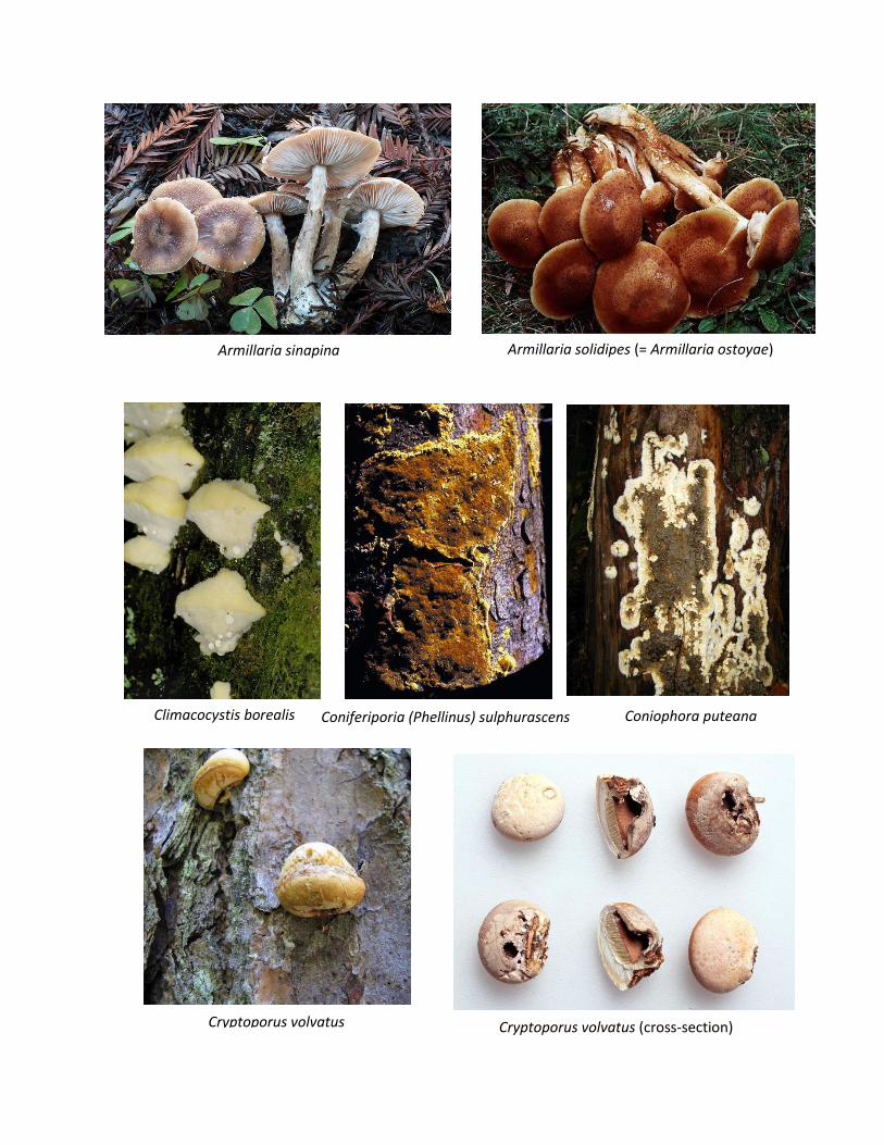
Armillaria solidipes (= Armillaria ostoyae) is the most serious root/butt rot pathogen of conifers
in the western U.S. and Canada (Dettman and van der Kamp, 2001). It is usually associated with
conifers but will also colonize hardwoods. Armillaria solidipes can be differentiated from other
species of Armillaria by its brown cap and stipe, the fairly prominent dark brown scales on the
cap, and the well-developed brown ring (annulus). Mushrooms form in clusters at the base of
affected trees and on the dead wood of fire scars and other wounds. Individual mushrooms can
be quite large – up to 1 foot in diameter in the Pacific Northwest (Burdsall and Volk, 1993).
Although all species of Armillaria are white rot fungi, the wood becomes yellow to brown in
color; advanced decay appears yellow, water-soaked and stringy. Thick mycelial fans form
between wood and bark in infected trees and stumps, and large quantities of resin may flow from
the tree base. Black to reddish brown rhizomorphs may be present. White rot, root/butt rot.
Armillaria sinapina mushrooms are similar to those of A. solidipes except that the dark scales on
the pileus are a bit smaller. A. sinapina is considered only a weak pathogen, usually associated
with hardwoods, but reported on conifers in interior British Columbia and Washington’s
Olympic Peninsula (Banik et al., 1996; Dettman and van der Kamp, 2001). White rot, mild
pathogen.
Climacocystis borealis initially forms a white, mottled rot in the roots and butt of living conifers
and can continue to decay wood of dead trees as a saprotroph. The fruiting bodies can be single
or in overlapping clusters and are found at the base and on roots of living trees, logs and stumps.
They are large (4-15 X 3-15 X 0.5- 4 cm), watery or sappy, and formed annually. The pores are
white (yellow upon drying), large and slightly irregular, 1 – 3 pores/mm. The upper surface is
hairy and white to yellowish with age. It has been found on Abies, Larix, Picea, Pinus,
Pseudotsuga, Tsuga, and occasionally on hardwoods. White root/butt rot.
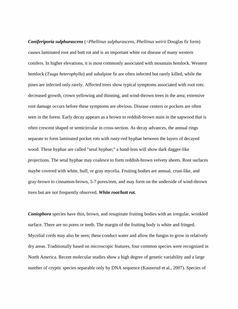
Coniferiporia sulphurascens (=Phellinus sulphurascens, Phellinus weirii Douglas fir form)
causes laminated root and butt rot and is an important white rot disease of many western
conifers. In higher elevations, it is most commonly associated with mountain hemlock. Western
hemlock (Tsuga heterophylla) and subalpine fir are often infected but rarely killed, while the
pines are infected only rarely. Affected trees show typical symptoms associated with root rots:
decreased growth, crown yellowing and thinning, and wind-thrown trees in the area; extensive
root damage occurs before these symptoms are obvious. Disease centers or pockets are often
seen in the forest. Early decay appears as a brown to reddish-brown stain in the sapwood that is
often crescent shaped or semicircular in cross-section. As decay advances, the annual rings
separate to form laminated pocket rots with rusty-red hyphae between the layers of decayed
wood. These hyphae are called "setal hyphae;" a hand-lens will show dark dagger-like
projections. The setal hyphae may coalesce to form reddish-brown velvety sheets. Root surfaces
maybe covered with white, buff, or gray mycelia. Fruiting bodies are annual, crust-like, and
gray-brown to cinnamon-brown, 5-7 pores/mm, and may form on the underside of wind-thrown
trees but are not frequently observed. White root/butt rot.
Coniophora species have thin, brown, and resupinate fruiting bodies with an irregular, wrinkled
surface. There are no pores or teeth. The margin of the fruiting body is white and fringed.
Mycelial cords may also be seen; these conduct water and allow the fungus to grow in relatively
dry areas. Traditionally based on microscopic features, four common species were recognized in
North America. Recent molecular studies show a high degree of genetic variability and a large
number of cryptic species separable only by DNA sequence (Kauserud et al., 2007). Species of

Coniophora form root and butt rots of living conifers but are largely saprotrophic on hardwoods.
They form a brown, cubical rot that sequesters carbon into the soil for long-term storage (USDA
Forest Service, 2011a). Brown root/butt rot.
Cryptoporus volvatus (“Pouch Fungus”) is a saprotroph frequently found on trees killed or
injured by bark beetles, wood borers or fire. This white rot fungus rapidly decays the outer
sapwood of dead trees, forming a grayish brown discoloration in the wood. The fruiting bodies
are initially yellow-brown fading to white, round, small (up to 4 X 5 X 4 cm, but often much
smaller) and are often formed in large quantities over the trunk within a year of tree death. The
characteristic diagnostic feature is the presence of a hard membrane that initially covers the pore
surface. Insects form small holes in this membrane close to the base of the conk, allowing spore
release from the brown pore layer. With time, the membrane erodes away so it may not be
present in older, faded conks. This species is very common on pines and true firs. White sap rot.
Echinodontium tinctorium (“Indian Paint Fungus”) causes significant heart rot in hemlocks and
true firs in western North America; losses in old-growth stands can approach 25 – 50%.
Advanced decay is frequently associated with wind-throw and trunk breakage. The decay is
described as a “brown stringy rot,” but it is actually a form of white rot with significant lignin
decomposition. The brown pigmentation is from the darkly colored mycelium. Ring shake occurs
when the springwood is weakened and the annual rings fall apart, leaving a laminated
appearance to the remaining wood. Decayed wood is soft, light-yellow to brown and may appear
water-soaked, eventually deepening to a dark reddish brown. The fruiting bodies are woody,
perennial ungulate, and can be quite large (up to 40 X 30 X 20 cm thick). The upper surface of

the fruiting body is very dark and may be hairy when young. The pore surface can appear tooth-
like. A diagnostic characteristic is the bright red color within the upper portion of the fruiting
body when it is split open. In the past, Native Americans used this pigment as a body paint, thus
the common name of the fungus. White heart rot.
Fomitiporia tsugina (=Phellinus hartigii) is a saprotroph on western hemlock, Pacific silver fir
(Abies amabilis), subalpine fir, and Douglas fir (Pseudotsuga menziesii), usually developing
from wounds, dead branches, or mistletoe infections, frequently forming on the underside of
branches or in branch crotches. Fruiting bodies are perennial and variable in shape - they can be
ungulate or resupinate, generally 5 - 15 cm wide. The upper surface is initially pale yellowish
brown aging to dark brown or black. The pore surface is grayish brown to purplish brown, pores
circular and small, 5 - 7 pores/mm. Decay is a white rot, often spreading 1 - 2 meters above and
below each fruiting body. The decayed wood frequently has multiple zone lines and appears
bleached, sometimes having light brown areas or streaks. Affected trees often break within 6 m
of the ground. White sap rot.
Fomitopsis officinalis (“Quinine Fungus”) is a brown-rot fungus primarily associated with old
growth conifers, especially those with broken tops, in Oregon and Washington. Fruiting bodies
are not common and usually found at the top of the tree rather than the base. The presence of
even a single conk suggests extensive decay in the trunk. The chalky white to gray fruiting
bodies are hard, perennial, ungulate, and have ridged and cracked surfaces. The conks form on
branch stubs or wounds. They are generally columnar and grow up to 15 X 23 X 45 cm. The
pore surface is white to tan, with circular to angular small pores (4 – 5 pores/mm). The inside of

the conk is soft, white, and has a bitter flavor. Conks contain the compound “agaricin,” which
has medicinal properties. In the past, this fungus was used medicinally against fevers and
wasting diseases, such as tuberculosis. Decayed wood often has thick white to cream-color
mycelial felts. The wood in ponderosa pine is initially red brown or brown; in other hosts the
wood may be light yellow to reddish brown. In advanced decay, the wood is broken down into
reddish-brown cubes, indicative of a brown rot. Brown sap rot.
Fomitopsis pinicola (“Red belt fungus”) is a common brown rotter of conifers in western North
America. Recent taxonomic research (Haight et al., in press) has shown that this fungus is
actually a species complex with two different species present in the Pacific Northwest, northern
California and Alaska. Both species play similar roles in the forest - they are major recyclers of
wood and creators of coarse woody debris. The species cannot be separated macroscopically.
Both fungi produce large (up to 38 X 20 X 15 cm), woody, bracket-shaped perennial conks.
Younger conks are smaller, white and round. As the conks mature, the upper surface becomes
dark gray to black, sometimes with a bright red band at the margin. The pore surface remains
white and has circular pores (5 – 6 pores/mm). Fresh fruiting bodies often have a very sweet
smell. The wood breaks up into the brown cubical decay characteristic of brown rot. Frequently
thick mycelial felts are formed in the shrinkage cracks of the decayed wood. Decay initiates in
the sapwood and progresses rapidly into the heartwood. Brown sap rot.
Ganoderma applanatum (“Artist’s Conk”) has a perennial fruiting body that is 5 – 52 cm wide
or even larger, convex, ungulate to fan-shaped, sessile. The upper surface is hard, concentrically
zonate and furrowed, gray to brown in color. The spore-bearing surface is poroid, white at first,
becoming off-white to dingy yellow with age, staining brown upon bruising (the characteristic
used by artists for etching). Pores are very small, 4 – 6 per mm. This fungus is very common,
and may be solitary or in overlapping clusters on stumps, logs, or wounds of living trees (Binion
et al. 2008). It forms a mottled white rot of roots, root crown and trunks. In the Pacific
Northwest, the fungus is common on conifers. The fungus often enters a tree through wounds in
exposed roots and at the base of the tree. Decay commonly extends 1 – 2 m above and below the
fruiting body. Decline and mortality are more pronounced during periods of environmental
stress. White sap rot.
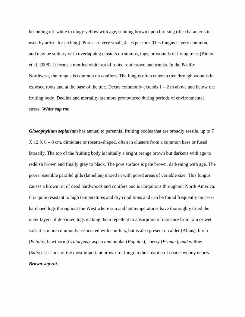
Gloeophyllum sepiarium has annual to perennial fruiting bodies that are broadly sessile, up to 7
X 12 X 6 – 8 cm, dimidiate or rosette-shaped, often in clusters from a common base or fused
laterally. The top of the fruiting body is initially a bright orange brown but darkens with age to
reddish brown and finally gray to black. The pore surface is pale brown, darkening with age. The
pores resemble parallel gills (lamellae) mixed in with pored areas of variable size. This fungus
causes a brown rot of dead hardwoods and conifers and is ubiquitous throughout North America.
It is quite resistant to high temperatures and dry conditions and can be found frequently on case-
hardened logs throughout the West where sun and hot temperatures have thoroughly dried the
outer layers of debarked logs making them repellent to absorption of moisture from rain or wet
soil. It is more commonly associated with conifers, but is also present on alder (Alnus), birch
(Betula), hawthorn (Crataegus), aspen and poplar (Populus), cherry (Prunus), and willow
(Salix). It is one of the most important brown-rot fungi in the creation of coarse woody debris.
Brown sap rot.

Hericium abietis (“Bear’s Head”) fruiting bodies are startlingly white to yellowish, annual and
soft. They are usually found on stumps, old logs, fallen trees or wounds of living trees and have
large numbers of downward pointing spines or teeth that come off the tips of horizontal
branches. Other species of Hericium have longer teeth or are more highly branched, but those
species are primarily associated with hardwoods. Decay is a white pocket heart rot, similar to
that from Porodaedalea pini (see below) but with longer, blunter pockets. In advanced decay, the
wood appears honeycombed. Pits may be filled with yellow to white fibers or may be hollow.
Hericium abietis is most common on true firs, hemlocks and spruces and is frequently found on
the Olympic Peninsula and the mountains of eastern Oregon and Washington. The fruiting
bodies are desired edibles. White sap rot.
Heterobasidion irregulare (= Heterobasidion annosum, p-type) causes a very serious root rot of
most species of pine; ponderosa pine is particularly susceptible, while whitebark pine is
generally not affected (Lockman and Kearns, 2016). The crown of diseased trees are frequently
symptomless until over 50% of the root system has been colonized, although tree growth is
inhibited. Affected trees often attract bark beetles or are more susceptible to wind-throw before
exhibiting crown symptoms. Decay is a white, stringy decay. One diagnostic feature of
Heterobasidion root disease is the production of fruiting bodies at the tree base or from buried
roots. Some of these fruiting bodies may be very small and "popcorn"-like, later expanding into
large (0.5 - 30 cm diam) resupinate (flat) or shelf-like structures. The upper surface of the
fruiting body is reddish brown to almost black. The lower pore surface is cream-colored to
yellow-brown with age. The pore surface is bright white with small pores (7 pores/mm) that are
rather elongated (Otrosina and Garbelotto, 2009). White root rot.

Heterobasidion occidentale (= "Heterobasidion annosum, s-type") is similar to H. irregulare.
Heterobasidion occidentale causes a devastating root rot disease, primarily on species of Abies,
Sequoiadendron, Tsuga, Pseudotsuga and Picea. The fruiting bodies of the two species are
difficult to distinguish from each other. The pore surface of H. irregulare may have more
irregularly shaped pores than those of H. occidentale, but a comparison of multiple fruiting
bodies of each type would be necessary to notice this slight difference (Otrosina and Garbelotto,
2009). White root rot.
Laetiporus conifericola ("Chicken of the Woods," "Sulfur fungus") causes a brown cubical heart
rot in many conifers in the Pacific Northwest. Traditionally this fungus was lumped with other
orange - yellow polypores under the scientific name Laetiporus sulphureus. Modern molecular
techniques were used to demonstrate that L. sulphureus was actually a species complex,
consisting of more than six species with different geographical ranges and host preferences.
Laetiporus conifericola produces annual, large, shelf-like conks that are soft, fleshy, and brilliant
orange to red orange. The pore surface is bright yellow when fresh. These colors fade with time.
Decay usually occurs at the base of trees. Fruiting bodies can be found on dead trees, stumps and
logs or on dead portions of living trees. Initial decay appears as a brown stain that later develops
into brown cubical decay with mycelial mats filling the cracks between the cubes. Brown sap
rot.
Onnia leporina (= Inonotus circinatus) and O. tomentosa (below) cause a white pocket rot in
the roots and butts of conifers throughout the Pacific Northwest and Alaska. Onnina leporina is

generally considered to be less virulent. The fruiting bodies of O. leporina are highly variable
and can be stalked or nonstalked ("sessile"), usually found at the base of trees or stumps. They
are usually not found in large numbers, unlike O. tomentosa, and tend to be larger and thicker
than O. tomentosa, up to 18 cm in diameter and 11 cm thick. The fruiting bodies are annual, the
upper surface can be hairy or smooth, light buff to reddish brown and up to 2 cm thick. The pore
surface is buff to yellowish brown, pores angular, 3 - 4 pores/mm. O. leporina is most commonly
found on pines. White root/butt rot.
Onnia tomentosa (Inonotus tomentosus) has annual fruiting bodies that are generally smaller
and thinner than those of O. leporina, up to 11 cm in diameter, but it is difficult to differentiate
the two species in the field. Those of O. tomentosa are usually stalked (either centrally or
eccentrically). The upper surface of the fruiting body is covered with densely matted woolly
hairs (“tomentose”) and yellowish-brown. The pore surface is initially pale buff and then darker
brown with age. The pores are angular, 2-4 pores/mm. Fruiting bodies often form in large
numbers on roots in old growth spruce stands. This fungus can also colonize pines and other
conifers, including lodgepole pine, western white pine, western hemlock, Douglas fir, western
larch (Larix occindentalis), ponderosa pine, and grand (Abies grandis) and subalpine firs
(Lockman and Kearns, 2016). Like most root and butt rot diseases, colonized trees are prone to
breakage and wind-throw. White root/butt rot.
Perenniporia subacida is usually considered to be a secondary pathogen and is associated with
trees that are suppressed or weakened from biological or abiotic factors, such as drought, insects,
or other pathogens. It forms a butt rot in fir and hemlock and is also a common saprotroph on

both conifers and hardwoods. Decay is usually at the base of the tree or in the roots. The fruiting
bodies are resupinate, crust-like or leathery, and cream-colored to dirty yellow-orange. They are
often on the underside of logs, fallen trees and exposed roots and can be up to 1 m long. The
pores are circular to angular, 5-6 pores/mm. As decay progresses, the decayed springwood forms
masses of stringy fibers with black flecks. The wood becomes delaminated as the annual rings
separate, and yellow to white mycelial mats form between the sheets of laminated decay.
Eventually the wood appears spongy with a yellowish color. The fungus is a white rotter. White
butt rot and sap rot.
Phaeolus schweinitzii ("Velvet top fungus," "Dyer's Polypore") causes a brown cubical heart rot
of the lower bole and roots, usually in older trees, especially those with basal fire scars. The
presence of fruiting bodies on the trunk or woody roots usually indicates a large amount of decay
in the wood; affected trees may have swollen bases and often fall over as wind-throws or break
off at the base of the tree. Fruiting bodies can be mushroom-shaped with a stipe or can grow
directly on the base of affected trees, often emerging from cracks in the bark or wounds. They
can be overlapping or bracket-shaped, up to 25 cm in diameter. The upper surface is velvety in
texture and variable in color - reddish brown, greenish brown, yellow brown, dark brown - often
with a yellow edge. The pore surface is initially orange, becoming greenish-brown, yellowish
brown to rusty brown with age. Pores are angular and large, 1 - 2 pores/mm. Pore walls may
break up and appear tooth-like. Thin, white mycelial felts can often be seen in shrinkage cracks.
The fruiting bodies are highly desired for dying wool and other natural fabrics. Brown heart rot.

Porodaedalea cancriformans (Phellinus cancriformans) forms a white pocket rot that might be
confused with decay from P. pini (below). Unlike most saprotrophs or decay pathogens, P.
cancriformans is a canker rot. The fungus grows into the inner bark, kills the cambium, and then
continues to colonize the wound tissue that is formed in response to the pathogen. The tree is
frequently unable to wall off the pathogen, and a sunken canker develops. As wound wood
continues to form along the edge of the canker, the tree stem appears swollen with the sunken
canker on the center. Trees frequently break at this point. P. cancriformans is a serious canker
disease of firs, including subalpine fir. The fruiting bodies form in proximity to the canker,
usually on the lower portion of the bole and not on branch stubs or whorls. Unlike P. pini, where
a single fruiting body might represent a large decay column, P. cancriformans produces multiple
fruiting bodies, often in large quantities, They are bracket-like with rough, dark, furrowed upper
surfaces, and usually smaller (1 - 7 cm wide) than those of P. pini. The lower surface is dark
cinnamon brown with irregular pores that can be angular or maze-like (“daedaloid”) pores, 2 - 3
pores/mm, as in P. pini. This fungus does not produce “punk knots”: decayed branch stubs that
frequently contain masses of dark fungal hyphae, often swollen by the overgrowth of sound
wood. White canker rot.
Porodaedalea pini (= Phellinus pini) is considered the most common heart rot fungus of living
conifers with a very wide host range that includes Douglas fir, western larch, pines, hemlocks,
spruces, true firs, western red cedar (Thuja plicata) and sometimes incense cedar (Calocedrus
decurrens). The decay is a white pocket rot in which spindle-shaped cavities form in the wood;
the cavities are usually separated by sound wood. In advanced decay, the cavities can combine
into distinct rings that can be seen in cross section. Zone lines are sometimes present. The decay

is usually found in the middle and upper trunk but may extend downward to form a butt rot. The
fungus can also degrade sapwood but the decay process does not continue after tree death.
Fruiting bodies most commonly form at branch stubs or knots but are sometimes found at the
base of the tree. They are perennial, ungulate to bracket-like, or entirely resupinate, and can be
up to 9 X 13 X 8 cm. The upper surface is rough, dark gray to brownish-black or cinnamon
brown. The lower surface is cinnamon brown with irregular pores that can be angular or maze-
like (“daedaloid”) pores, 2 - 3 pores/mm. Infection can result in swollen, resinous, and sterile
"punk knots," unlike P. cancriformans. White heart rot.
Pseudoinonotus dryadeus (= Inonotus dryadeus, “Weeping Conk”) has annual fruiting bodies
that persist, developing at the base of trunk or on roots below the soil surface, are variable in size
but can be very large (up to 75 cm wide), initially soft but becoming dried and cracked with age.
Top surface of fresh fruiting bodies are yellowish to brown, blackening with age, and may have
many droplets of amber-colored exudates. The lower surface is buff with fine circular to angular
pores, 4 – 6 per mm. The fungus causes a slowly developing white rot root disease and butt-rot
with most of decay concentrated in larger roots. Affected trees may have significant amounts of
root decay and an elevated risk of wind-throw (Luley, 2005; Swiecki and Bernhardt, 2006).
White butt/root rot.
Stereum sanguinolentum (“Bleeding Stereum”) can cause extensive heart rot in pine, spruce and
true fir, especially subalpine fir and Englelmann spruce (Picea engelmanii) in the Rocky
Mountain region (USDA Forest Service, 2011b). It is the most important decay fungus
associated with subalpine fir (Worrall and Nakasone, 2009). It is also a strong saprotroph and is

commonly associated with wounds. Even though this fungus produces a white rot decay, the
wood may initially appear to have a reddish stain that later becomes light brown to red-brown. In
advanced decay, the wood is a light brown, fibrous, stringy mass with white mycelial sheets.
Fruiting is more common on stumps and slash than on living trees so the presence of decay may
be difficult to detect. Fruiting bodies are thin, resupinate, and sometimes occur with the upper
margin projecting outward, with a shelf-like appearance. The spore-bearing surface is grey to
light brown, smooth or slightly wrinkled; there are no pores or teeth. If the fruiting body is cut
with a knife, it will bleed a dark red pigment. White heart rot.
Trichaptum abietinum (“Velvet polypore”) is an efficient saprotroph that breaks down the
sapwood of dead trees and slash as a white pocket rot. This fungus is generally restricted to the
sapwood and may form entire rings of fruiting bodies around the circumference of a log or
stump. It can also colonize wound surfaces, sun-scaled trunks and other dead wood in living
trees. The fruiting bodies are small (up to 1.5 X 8 X 0.2 cm), annual, thin, shelf-like, and
produced in large numbers. The upper surface is usually light gray with hairs but will darken
with age. The lower surface can be startlingly purple or violet when fresh but turns a pink to
brown with age. Pores are angular, 4 - 6 pores/mm, and can break apart with age into spines or
ridges. Decayed wood appears honeycombed. The amount of decay is usually extensive by the
time that fruiting bodies are formed. Trichaptum biforme is a closely related species that is found
primarily on hardwoods. Trichaptum fuscoviolaceum is also a saprotroph of conifers. It can be
differentiated from T. abietinum by its toothed ("hydnoid") lower surface. White sap rot.

Veluticeps fimbriata causes a brown pocket rot on stems of spruce and fir. The decay develops
in pockets surrounded by sound wood. In advanced decay, the pockets coalesce to form large
cavities. The fruiting bodies can form on wounds of living trees but are more commonly found
on downed, dead wood. They are usually perennial, resupinate or shelf-like, up to 1.5 X 10 X 4
cm. The upper surface is dark brown to black. The lower surface is gray to light brown, smooth
or warted, often cracked, and without pores. Decayed wood might have the odor of apples.
Veluticeps abietina, a closely related species, is similar but not as common. The two species are
difficult to differentiate without a microscope (Worrall and Nakasone, 2009). Brown sap rot.
References Allen, E., Morrison, D., Wallis, G. 1996. Common Tree Diseases of British Columbia. Victoria, British Columbia, Canada: Canadian Forestry Service, Pacific Forestry Centre.178 pp. Banik, M.T., Volk, T.J., and Burdsall, H.H.Jr. 1996. Armillaria species of the Olympic Peninsula of Washington state, including confirmation of North American biological species XI. Mycologia 88:492-496. Binion, D.E., Stephenson, S.L., Roody, W.C., Burdsall, H.H., Jr., Vasilyeva, L.N., and Miller, O.K., Jr. 2008. Macrofungi of Oak. Morgantown, WV: West Virginia University Press. 467 pp. Blanchette, R.A. and Biggs, A.R. 1992. Defense Mechanisms of Woody Plants Against Fungi. Springer-Verlag. 427 p. Burdsall, H.H. Jr. and Volk, T.J. 1993. The state of taxonomy of the genus Armillaria. McIlvainea 11:4-12. Modified version available at http://tomvolkfungi.net (accessed 08/17/2016). Dettman, J.R. and van der Kamp, B.J. 2001. The population structure of Armllaria ostoyae and Armillaria sinapina in the central interior of British Columbia. Can J. Bot. 79:600-611. Dujesiefken, D. and Liese, W. 2015. The CODIT Principle. Champaign, IL: International Society of Arboriculture. 162 p. Filip, G.M., Schmitt, C.L., Scott, D.W., Fitzgerald, S.A. 2007. Understanding and defining mortality in western conifer forests. West. J. Appl. For. 22:105-115.

Gilbertson, R.L. and Ryvarden,L. 1986: North American Polypores. Vol. 1. Oslo: Fungiflora. 1 – 433. Gilbertson, R.L. and Ryvarden, L. 1987: North American Polypores. Vol. 2. Oslo: Fungiflora. 437-885. Goheen, E.M. and Willhite, E.A. 2006. Field Guide to Common Diseases and Insect Pests of Oregon and Washington conifers. R6-NR-FID-PR-01-06. Portland, OR: USDA Forest Service, Pacific Northwest Region. 327 pp. Haight, J.E., Laursen, G., Glaeser, J., Taylor, L. Phylogeny of Fomitopsis pinicola: a species complex. Mycologia (in press). Innes, J.L. 1998. The impact of climatic extremes on forests: an introduction. . In: The Impacts of Climate Variability on Forests (M. Beniston and J. Innes, eds). New York: Springer. 1-18. Kauserud, H., Shalchian-Tabrizi, K., Decock, C. 2007. Multilocus sequencing reveals multiple geographically structured lineages of Coniophora arida and C. olivacea (Boletales) in North America. Mycologia 99: 705-713. Lockman, I.B., Kearns, H.S.J. (eds). 2016. Forest root diseases across the United States. Gen. Tech. Rep. RMRS-GTR-342. Ogden, UT: USDA Forest Service, Rocky Mountain Research Station. 55 pp. Luley, C. J., 2005: Wood Decay Fungi Common to Urban Living Trees in the Northeast and Central United States. Naples, NY: Urban Forestry LLC. 60 pp. Otrosina, W.J. and Garbelotto, M. 2009. Heterobasidion occidentale sp. nov. and Heterobasidion irreulare nom. Nov.: A disposition of North American Heterobasidion biological species. Mycological Research Peterson, D.L. 1998. Climate and environmental change in high-altitude forests 191-208. Schwarze, F.W.M.R. 2008. Diagnosis and Prognosis of the Development of Wood Decay in Urban Trees. Rowville, Australia: ENSPEC. 336 p. Shigo, A.L. 1984. Compartmentalization: a conceptual framework for understanding how trees grow and defend themselves. Annual Review of Phytopathology 22: 189–214. Shortle, W.C., Smith, K.T., and Dudzik, K.R. 1996: Decay diseases of stemwood: Detection, diagnosis, and management. In: Forest Trees and Palms. Ed. by Raychaudhuri, S.P.; Maramorosch, Karl, New Delhi, India: Oxford & IBH Publishing: 95-109. [Available at http://www.nrs.fs.fed.us/pubs/5514 (accessed 08 16 2016)].

Shortle, W.C., Dudzik, K.R. Smith, K.T. 2010. Development of wood decay in wound-initiated discolored wood of eastern red cedar. Holzforschung 64: 529-536. Smith, K.T. 2006. Compartmentalization today. Arboricultural Journal 29: 173-184. Smith, K.T., Arbellay, E., Falk, D.A., and Sutherland, E.K. 2016. Macroanatomy and compartmentalization of recent fire scars in three North American conifers. Canadian Journal of Forest Research 46: 535-542. Smith, K.T. and Glaeser, J.A. 2013. Skeleton decay. Arborist News 22 (3): 32-34. Swiecki, T.J. and Bernhardt, E.A. 2006. A Field Guide to Insects and Diseases of California Oaks. PSW-GTR-197. Washington D.C.: U.S. Forest Service, Pacific Southwest Research Station. 152 pp. U.S. Department of Agriculture, Forest Service, Forest Health Protection, Rocky Mountain Region. 2011a. Coniphora root and butt rot. http://www.fs.usda.gov/Internet/FSE_DOCUMENTS/stelprdb5353724.pdf (08/17/2016). U.S. Department of Agriculture, Forest Service, Forest Health Protection, Rocky Mountain Region. 2011b. Rust-red stringy rot and red heart rot. Available at http://www.fs.usda.gov/Internet/FSE_DOCUMENTS/stelprdb5347115.pdf (accessed 08/16/2016). Wallis, G., Morrison, D., Ross, D. 1980. Tree hazards in recreation sites in British Columbia. B.C. Ministry of Lands, Parks and Housing. Canadian Forestry Service. Joint Report No. 13. 52 pp. Reprinted in 1992. Worrall, J.J. and Nakasone, K.K. 2009. Decays of Engelmann spruce and subalpine fir in the Rocky Mountains. FS-R6-RO-FIDL#150/006-2009. Portland, OR: USDA Forest Service, Pacific Northwest Region.12 pp. Zhou, L.W., Nakasone, K.K., Burdsall, H.H.Jr., Ginns, J., Vlasák, Miettinen, O., Spirin, V., Niemelä,T., Yuan, H.S., He, S.H., Cui, B.K., Xing, J.H., Dai, Y.C. 2016. Polypore diversity in North America with an annotated checklist. Mycol. Progress 15:771-790. Zhou, L.W., Vlasák, J., Dai, Y.C. 2016. Taxonomy and phylogeny of Phellinidium (Hymenochaetales, Basidiomycotina): A redefinition and the segregation of Coniferiporia gen. nov. for forest pathogens. Fungal Biology 120:988-1001. Glossary - Definitions of Mycological Terms (Gilberston and Ryvarden, 1987) Annulus – a ring found on the stipe of certain mushrooms. Daedaloid – maze-like Dimidiate – semi-circular in outline when viewed from above.
Hydnoid – resembles teeth. Mycelium – the vegetative stage of the fungus, usually observable as a mass of individual threads, termed “hyphae.” Ochraceous – a yellowish buff color. Pileus – the portion of a fruiting body with a sterile upper surface and a fertile lower surface. In gilled mushrooms, the pileus is the mushroom cap or button. Resupinate – flat and appressed to the stem or root surface. Rhizomorph – a macroscopic strand, often resistant to drying, that spreads throughout the soil.
Black in Armillaria species. Sessile – without a stipe. Stipe – stalk-like or stem-like structure that supports the pileus. Stipitate – with a stipe. The stipe can be central or attached laterally (eccentric). Tomentose – with densely matted hyphae that give a wool-like texture. Ungulate – hoof-shaped. Photo Credits: Our thanks go to the following photographers for allowing us to use their stunning photos: Andrew Khitsun (www.wisconsinmushrooms.com); Michael Woods (www.mykoweb.com); Thomas Volk (http://botit.botany.wisc.edu/toms_fungi/); Eileen Goheen (U.S. Forest Service); Jim Worrall (U.S. Forest Service); Andreas Kunze (commons.wikimedia.org); and USDA Forest Service, Northern and Intermountain Region , USDA Forest Service, Bugwood.org; Larry Grand (deceased), North Carolina State University.
Armillaria sinapina Armillaria solidipes (= Armillaria ostoyae)
Climacocystis borealis Coniferiporia (Phellinus) sulphurascens Coniophora puteana
Cryptoporus volvatus Cryptoporus volvatus (cross-section)

Echinodontium tinctorum
Echinodontium tinctorum
Echinodontium tinctorum (cross section)
Fomitiporia tsugina = Phellinus hartigii
Fomitopsis pinicola
Fomitopsis officinalis
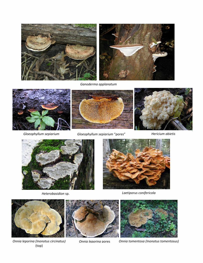
Ganoderma applanatum
Gloeophyllum sepiarium Gloeophyllum sepiarium “pores” Hericium abietis
Heterobasidion sp. Laetiporus conifericola
Onnia leporina (Inonotus circinatus) (top)
Onnia leporina pores Onnia tomentosa (Inonotus tomentosus)

Perenniporia subacida
Phaeolus schweinitzii Porodaedalea cancriformans (Phellinus cancriformans)
Porodaedalea pini (Phellinus pini)

Pseudoinonotus (Inonotus) dryadeus
Stereum sanguinolentum Trichaptum abietinum
Veluticeps fimbriata
