wood anatomical studies of important mangrove species from
TRANSCRIPT

Project code: IWST/WPEW/EXT/MHFD/111
Wood Anatomical Studies of Important Mangrove Species
from Maharashtra Sea Coast for the Identification.
Project Completion Report
Submitted to
Mangrove Foundation, Maharashtra
By
S. Shashikala
Institute of Wood Science and Technology
(Indian Council of Forestry Research and Education)
(An Autonomous Body of Ministry of Environment, Forest and Climate Change,
Govt. of India)
P.O. Malleswaram, Bengaluru – 560 003
2020

Project code: IWST/WPEW/EXT/MHFD/111
Wood Anatomical Studies of Important Mangrove Species
from Maharashtra Sea Coast for the Identification.
Project Completion Report
Submitted to
Mangrove Foundation, Maharashtra
By
S. Shashikala
Wood Properties and Uses Division Institute of Wood Science and Technology
(Indian Council of Forestry Research and Education)
)An Autonomous Body of Ministry of Environment, Forest and Climate Change,
Govt. of India)
P.O. Malleswaram, Bengaluru – 560 003
2020

Project Profile
Project Title Wood Anatomical Studies of
Important Mangrove Species from
Maharashtra Sea Coast for the
Identification
Project Investigator
Ms. S. Shashikala, ACTO
Wood Properties and Uses (WPU)
Division
Co-Investigators
Dr. M. Sujatha, CTO, WPU Division
Dr. S.R. Shukla, Sci-G, WPU
Division
Mr. N. Mohan Karnat, IFS,
Former I/C Director
Research Team
Ms. Krupa. M, Project Assistant
Mr. Jean Simon, STA
Mr. H.C. Shiva Prasad, Technician
Ms. Vanathi Varatha Raj, STA
Date of Commencement of
the project
April 2017
Date of completion of the
Project
30th June 2020
Total budget of the project Rs. 24,35,000/-
Expenditure Rs. 24,32,625/-
Equipment Purchased
under the project
Nikon microscope model Ci-POL
with software NI Elements-BR-
(Rs. 11,48,017)
External hard Disk- (Rs. 3999/-)

Contents
Sl. No. Topic Pg. No.
1 Introduction 1
2 Objectives 4
3 Review of Literature 4
4 Materials and methodology 6
5 Results and discussion 16
6 Conclusion 57
7. Summary 59
8 Acknowledgements 61
9 References 62
10 Photomicrographic plates (Plate 1 to 6) 67-72
11 Selected photos taken during field visits. 73-74
12 Appendix I - IAWA list of card key microscopic features
for hardwood identification
75
13 Appendix II - List of microscopic card key features for
identification of mangrove species
80
14 Appendix III - Key for separation of Mangrove species
based on anatomical and physical features
82

List of Illustrations
Species Plate
Aegiceras corniculatum (L.) Blanco
1
Avicennia marina Vierh.
1
Avicennia officinalis Linn.
1
Bruguiera cylindrica (L.) Blume.
2
Bruguiera gymnorrhiza (Linn.) Lamk.
2
Ceriops tagal (Perr.) C.B. Robinson
2
Cynometra iripa Kostel.
3
Excoecaria agallocha Linn.
3
Heritiera littoralis Dryand.
3
Kandelia candel Linn.
4
Lumnitzera racemosa Willd.
4
Rhizophora apiculata Bl.
4
Rhizophora mucronata Lam.
5
Sonneratia alba J.Smith.
5
Sonneratia apetala Buch.-Ham.
5
Sonneratia caseolaris (Linn.) Engler
6
Xylocarpus granatum Koen.
6

List of Tables
Table
No.
Title Page
No.
1 Details of sampling sites
8
2 Moisture content, specific gravity (sp.gr) and volumetric
shrinkage (VS%) of different mangrove species
16
3 Qualitative features of mangrove woods
25 -30
4 Quantitative features of mangrove woods
31 - 35
5 Vulnerability (VI) and Mesomorphy (MI) of different
mangrove species
56

List of Figures
Figure
No.
Title Page
No.
1 Set up for determining the physical properties 9
2 Wood chips before and after the maceration and individual
wood elements as seen under microscope after the maceration
12
3 Microtomy of wood sections using sliding microtome. 13
4 Determination of vessel frequency in 1mm x1mm area grid.
Vessels in green colour are considered for the measurement.
14
5 Image analysis system (Nikon microscope model Ci- POL with
software NI Elements-BR)
14
6 Specific gravity (Sp.gr) (a) and volumetric shrinkage (VS%)
(b) of different mangrove species
19

1
1. Introduction
Mangroves are salt tolerant, halophytic woody plants distributed along the
tropical and subtropical tidal margins (Duke, 2006). Mangroves are significantly
different from other vegetation as they survive in an environment, an interface
between land and sea marked with fluctuations in salinity, exposed to winds and tides
and to extreme events such as cyclones, hurricanes and tsunamis (Alongi, 2002).
Mangroves provide a range of ecosystem services, including coastal protection from
waves, wind and weather events (Ewel et al. 1998; Koch et al. 2009; Mazda et al.
2006) and are increasingly recognized for their role in carbon sequestration (Mcleod
et al. 2011)
Mangroves are crucial for coastal areas as they help in preventing soil erosion
and also act as a catalyst in reclaiming land from seas. They exhibit a variety of
adaptations viz. morphology, anatomy, physiology, seed and seedlings development
and succession mechanisms. They are subjected to strong tidal flows and waves as
well as high wind speeds that may cause structural damage to trees. Therefore,
survival and establishment of mangrove trees under different environments partly
depend on their anatomical structure, wood density and mechanical strength (Van
Gelder et al. 2006; Curran et al. 2008). In addition to the wind and wave forces,
mangroves grow in soils that vary in salinity. When rainfall run-off is limited, soil
pore-water high in inter-tidal zone becomes highly saline due to evaporation of
seawater (Robert et al. 2009). High salinity simulates to drought condition and results
in an increase in tension in water column within xylem, which can impose mechanical
stresses on xylem vessels (Hacke et al. 2001; Jacobsen et al. 2005).
Mangroves occupy less than 1% of the world's surface and are mainly found
between the Tropic of Cancer and the Tropic of Capricorn on all continents covering
an estimated 75 percent of the tropical coastline worldwide. There are more than 18
million ha. of global mangroves inhabiting in 112 countries and territories in the
tropical and subtropical region. Around 34 major and 20 minor mangrove species
belonging to about 20 genera in over 11 families have been recorded globally
(Tomlinson, 1986). India with a long coastline of about 7516.6 km. including the
island territories, has a mangrove cover of about 6,749 sq. km. the fourth largest
mangrove area in the world (Naskar & Mandal, 1999). These mangrove habitats

2
comprise three distinct zones: East coast habitats having a coast line of about 2700 km
facing Bay of Bengal, West coast habitats with a coast line of about 3000 km facing
Arabian sea, and Island Territories with about 1816.6 km coastline.
As per India State of Forest Report (ISFR, 2019), mangroves constitute around
4975 sq.km accounting 3% of the world‟s mangrove vegetation, 0.15 per cent of India‟s
land area, along the East Coast (80%) and West Coast (20%) with Sundarbans in West
Bengal accounting for almost half of it. The Indian mangroves comprise approximately
59 species in 41 genera and 29 families. Of these, 34 species belonging to 25 genera
and 21 families are present along west coast. There are about 25 mangrove species
which have restricted distribution along the East coast and are not found on the West
coast. Similarly, there are eight species of mangroves which have been reported only
from the West coast. There are approximately 16 mangrove species reported from the
Gujarat coast, while Maharashtra has about 20 species, Goa 14 species and Karnataka
10. Mangroves are being utilized for construction, fuel, fodder, barks for tannin
extraction, fruits and young shoots are used as vegetable, medicinal use, protection
from natural calamities such as during Tsunami. Because of the high specific gravity
of Rhizophoraceous wood, they are preferred for firewood. They are also used for
boat building, brick-burning and to make poles. Avicennia marina and Sonneratia
alba are commonly found plant species in mangrove ecosystems.
Six of Maharashtra‟s districts have mangrove cover: Mumbai city, Mumbai
suburbs, Raigarh, Ratnagiri, Sindhudurg and Thane. Around 320 km2 of the state of
Maharashtra is under mangrove cover (ISFR, 2019). Because of the high salinity of
the soil, around 60 -70 per cent of Mumbai mangroves comprise A. marina. S. alba is
a front mangrove species and prefers non swampy intertidal zones. In the low
intertidal zone, it can be the dominant species along with A. marina, forming a tree
line along the seaward margin of its range.
Though legal protection is afforded to this ecosystem by way of legislation in
the form of Coastal Regulation Zone Notification and mangrove areas being notified
as protected forests, many a times the legal provisions are not sufficient to curb illegal
activities. Felling of trees for fuel wood and wood products is a threat to the mangrove
ecosystems. Among the current threats to mangrove ecosystems, the ever-increasing
human pressure on coastal areas is one of the most serious one. In addition to human-

3
induced threats, natural hazards such as cyclones, storms and floods frequently occur
in the region, threatening several coastal ecosystems, including mangroves. Land
reclamation for construction activity, aquaculture, agriculture and tourism is a major
threat to mangrove ecosystems in Maharashtra. Illegal logging and encroachment of
mangrove forests are the cause of many economic as well as ecological problems.
Wood density is an important characteristic defining the mechanical properties
and its performance such as resistance to breakage during high winds (Curran et al.
2008; Niklas and Spatz, 2010) and to boring insects and pathogens (Bell et al. 2006).
Wood density and other properties are dependent on anatomical characteristics such
as presence of vessels and fibres and their arrangements in wood (Jacobsen et al.
2005; Preston et al. 2006, Santini et al. 2012).
Knowledge of presence and distribution of various anatomical wood elements
as well as physical properties of mangrove species is essential to identify different
species and understand their woody material for suggesting value-added utilization.
Although numerous works carried out on field identification of mangroves based on
characters of bark, leaf, fruiting and branching pattern etc., not much work has been
carried out on wood anatomical characters for identification of Mangroves in general
and mangroves of Maharashtra in particular. The wood structure is different in
different species depending on the proportions, size and distribution of various cell
elements like vessels, fibres, parenchyma and rays. No two woods have exactly the
same structure and structural patterns seen on the end surface as it were the finger
prints of wood by which the identity of any timber can be established. The macro
(gross) and microstructure of wood material of a timber species is being used as
fingerprint for its accurate and reliable identification and inexpensive compared to
other forms of identification like chemical methods (mass spectrometry, near infrared
spectroscopy, stable isotopes, radio-carbon), and genetic methods (DNA barcoding,
population genetics/phylogeography, DNA fingerprinting) each with potential
application to forensic timber identification. Timber identification has traditionally
been provided by wood anatomists through the examination of the internal structure
of wood. As anatomical characters can be influenced by both genetic and
environmental factors, combinations of characters can be used to differentiate taxa.
Standard anatomical characters are described according to the terminology of the

4
International Association of Wood Anatomists (IAWA) (Wheeler et al. 1989) and
identification of unknown sample is obtained through comparison to reference
materials.
Due to lack of information on wood anatomy of mangrove species of
Maharashtra state, from the point of wood identification, it becomes difficult to
identify and differentiate them from non-mangrove species in case of legal issues
arise. Keeping this in view, the present study was undertaken to evaluate the
anatomical characters and a few important physical properties of mangrove species
distributed in different locations of Maharashtra. This study provides valuable data on
wood identification and related physical properties of wood of some important
mangrove species.
2. Objectives
1. To study microstructure and important physical properties of important
mangrove species selected from two locations.
2. To create database of anatomical properties of mangrove species from
Maharashtra seacoast for their identification.
3. Review of Literature
Although numerous works carried out on field identification of mangroves
based on characters of bark, leaf, flowering, fruiting and branching pattern etc., not
much work has been carried out on wood anatomical characters for identification of
mangroves in general and mangroves of Maharashtra in particular. The studies
conducted by researchers on few aspects on Mangroves are as follows: Macroscopic
characters or gross structure on mangrove species are available in Indian woods - Vol
I-VI (Choudhury and Ghosh, 1958, Anon 1963, Rao and Purkayastha 1972,
Purkayashta, 1982 & 1985, Rathuri et al. 2001), but not on minute anatomy. Wood
anatomy of some species of mangroves were described by Pearson and Brown (1932).

5
Detailed description on leaf, floral and seed morphology of each family along with
utilization was discussed by Tomlinson (1986). Rao et al. (1987 and 1989) have
studied the systematic position of Sonneratia samples in comparison with Duabanga
samples of Burma and Andaman collected from the xylarium, FRI, Dehradun,
wherein they have reported the presence of the reticulate perforation type in wood and
aerial root of S. caseolaris. Krishnamurthy and Sigamani (1987) have studied wood
anatomy of stem, root and pneumatophores of Avicennia marina and A. officinalis
from mangrove forests of Pichavaraum, Tamil Nadu. They have noticed the presence
of successive cambial activity, lack of ring porous wood and septate fibres, presence
of small vessels when compared with the other members of the family. Banerjee and
Rao (1990) have listed out the uses and studied ecology of mangroves of Mahanadi
Delta and Sundarbans in their work on mangroves of Orissa coast. Lindorf (1994)
have studied eco-anatomical wood features of species from the point of adaptation of
19 species of very dry forest in Venezuela to the xeromorphic conditions. Naskar and
Mandal (1999) have studied the morpho-anatomical structures of few Indian
Mangroves. Rao et al. (2005) have studied anatomy of selected species of mangroves
from Coondapura, Karnataka. Shashikala and Rao (2013) have reported the status of
work carried out on wood anatomy of Indian mangroves; special wood anatomical
features along with properties and utilization of selected mangrove species. Robert et
al. (2011) have examined the occurrence of growth rings in six mangrove species
grown in Gazi Bay, Kenya and opined that the presence of growth rings is dependent
on climatic conditions that results in a variation of soil water salinity over the year
and suggested to study more than a year and include different sizes to assess the
distinctness of growth rings. Report on the existence of Heritirera littoralis Dryand in
Sindhudurg district was made and morpho-taxonomy, phenology, seedling
morphology was studied (Shaik et al. 2011). Vidyasagaran et al. (2014), Anoop et al.
(2013), have worked on eco-anatomical aspects on mangroves from Kerala. Naskar
and Prathip Kumar (2015) have studied eco-anatomical and physiological adaptations
of selected mangrove species.
There are varied opinions regarding the species belonging to true mangrove
and mangrove associates specially Acanthus spp., Pemphis acidula, Acrostichum spp.
Clerodendrum inerme, Heritiera littoralis, Hernandia nymphaeifolia, Xylocarpus
granatum and Excoecaria agallocha and are considered as controversial species. True

6
mangroves differ from mangrove associates physiologically and ecologically in their
ability to survive in mangrove environment. Wang et al. (2010), have differentiated
between true mangroves and mangrove associates based on leaf traits and salt
contents and classified Pemphis acidula and Xylocarpus granatum as true mangroves.
However, in the present study, Excoecaria agallocha, Heritiera littoralis and
Xylocarpus granatum are also included along with other mangrove species and are
described anatomically. Surya and Hari (2017) have studied the Rhizophoraceae
species of Kerala with respect to their stem anatomy. Mariam et al. (2018) have
studied fibre length, vessel length and ray height of tree species occurring in
Bangladesh mangrove forests. Cynometra iripa which is considered as mangrove
associate by Tomlinson (1986) is also included in the present study.
4. Materials and methodology
4.1 Materials
Seventeen mangrove species from nine families representing twelve genera were
studied for the purpose of identification based on their wood anatomy (macro and
microstructure) and also certain physical properties. Two feet billet from three tree of
each species from stem wood or branch wood were collected for the study.
Name of the species studied:
1. Aegiceras corniculatum (L.) Blanco (Myrsinaceae)
2. Avicennia marina Vierh. (Avicenniaceae)
3. Avicennia officinalis Linn. (Avicenniaceae)
4. Bruguiera cylindrica (L.) Blume. (Rhizophoraceae)
5. Bruguiera gymnorrhiza (Linn.) Lamk. (Rhizophoraceae)
6. Ceriops tagal (Perr.) C.B. Robinson (Rhizophoraceae)
7. Cynometra iripa Kostel (Fabaceae)
8. Excoecaria agallocha Linn. (Euphorbiaceae)
9. Heritiera littoralis Dryand (Sterculaiaceae)
10. Kandelia candel Linn. (Rhizophoraceae)
11. Lumnitzera racemosa Willd. (Combretaceae)
12. Rhizophora apiculata Bl. (Rhizophoraceae)
7
13. Rhizophora mucronata Lam. (Rhizophoraceae)
14. Sonneratia alba J.Smith. (Sonneratiaceae)
15. Sonneratia apetala Buch.-Ham.(Sonneratiaceae)
16. Sonneratia caseolaris (Linn.) Engler (Sonneratiaceae)
17. Xylocarpus granatum Koen. (Meliaceae)
4.2 Study area and sampling sites
Maharashtra state comprises an area of 3,07,690 sq. km with a coast line about
720 km having 54 creeks and their tributaries with an area of about 65,500 ha under
coastal saline soils. The pH (at 25°C) of the water sample ranged between 6-6.9 in
Airoli and 7-7.5 in Sindhudurg. The Electrical conductivity ranged between 33600-
34800 µs/ cm at 25°c in Airoli and 27900 - 45000 µs/cm at 25°C in Sindhudurg.
The water samples were collected during January, April and December.
Samples from stem wood and branch wood of seventeen species of
mangroves were collected from Thane (Airoli mangrove area), Panvel, West
Mumbai (Daravali) and Sindhudurg (Tarkarli, Thakur wadi, Trikuta, Khavane wadi,
Nivthi, Hadi, Dongrawadi of Achra and Jamdulwadi) in Maharashtra state. Table 1
reflects the actual place of sample collection against each species.
4.3 Methodology
Immediately after the samples were collected, they were given prophylactic
treatment with FAA (A mixture of 5 ml of formaldehyde (40%), 5 ml of glacial
acetic acid and 90 ml ethyl alcohol) to prevent them getting affected by fungus. Later
the collected samples were brought to Institute of Wood Science and Technology
(IWST) laboratory for further procedures and study.
The wood samples were converted to study/ determine the physical properties
(moisture content, specific gravity, shrinkage) and anatomical properties. Due to the
small girth of the sample only volumetric shrinkage was studied. Wherever the girth
of the samples is less than 12 cm, shrinkage samples could not be prepared. In case of
L. racemosa the girth of the sample was less and also was not in sound condition (the
sample was decayed partially), hence shrinkage studies could not be conducted.

8
Table 1: Details of sampling sites
Sl.
No.
Species Place of collection
1 Aegiceras corniculatum (L.)
Blanco
Panvel
Khavane wadi,( Sindhudurg)
2 Avicennia marina Vierh. Thane and Dongrawadi (Sindhudurg)
3 Avicennia officinalis Linn. Thane and Dongrawadi (Sindhudurg)
4 Bruguiera cylindrica (L.) Blume. Panvel and Daravali (Mumbai West)
5 Bruguiera gymnorrhiza (Linn.)
Lamk.
Tirkuta (Sindhudurg)
Achra (Sindhudurg)
Nivthi (Sindhudurg)
6 Ceriops tagal (Perr.)
C.B.Robinson
Daravali ( Mumbai West)
7 Cynometra iripa Kostel.
Achra ( Sindhudurg)
8 Excoecaria agallocha Linn.
Thane
Achra ( Sindhudurg)
Khavane wadi (Sindhudurg)
9 Heritiera littoralis Dryand.
Khavane wadi and Nivti (Sindhudurg)
10 Kandelia candel Linn. Hadi, Tarkarli, Jamdulwadi
(Sindhudurg)
11 Lumnitzera racemosa Willd.
Thakurwadi ( Sindhudurg)
12 Rhizophora apiculata Bl.
Achra (Sindhudurg)
13 Rhizophora mucronata Lam.
Daravali ( Mumbai West)
14 Sonneratia alba J. Smith.
Thane
15 Sonneratia apetala Buch.-Ham.
Thane
16 Sonneratia caseolaris (Linn.)
Engler
Khavane wadi (Sindhudurg)
17 Xylocarpus granatum Koen.
Jamdulwadi (Sindhudurg)
4.3.1. PHYSICAL PROPERTIES

9
The moisture content of wood means the relationship between the mass of
water in it and the mass of the timber without the water. Almost all physical
properties of wood vary with moisture content and it is therefore necessary that the
moisture content and its basic density/specific gravity of wood be determined. Fig. 1
shows the setup for determining the physical properties. Following physical properties
were evaluated.
4.3.1.1. Moisture content: The specimens, after conversion (sample size 2 x 2 x
2.5cm), were weighed (Wi) and kept in hot air oven for 48 hours at 103± 20 C until
constant weight was attained. Weight of the over-dried specimens were noted down
(Wo). Percentage of moisture content was determined using the formulae:
MC(%) = (Wi-Wo/Wo) *100
Fig. 1: Set up for determining the physical properties
4.3.1.2. Specific gravity: The samples were converted to 2 x 2 x 2.5 cm. The
specimens were saturated with water. After saturated, excess water was removed by
wiping with a cloth, were weighed for their initial weight and their volume was

10
measured using either water displacement method or Mercury displacement method.
Specimens were kept in hot-air oven for 48 hours at 103± 2° C until constant weight
was attained. The specimens were taken out from the hot-air oven, and weighed
accurately to 0.001g. Specific gravity (Sp.gr.) was determined using following
formula:
Sp.gr. = Wo / Vg.
where Wo is the weight of the oven-dry wood sample and Vg is the volume of the
same wood sample saturated in water.
4.3.1.3. Volumetric shrinkage: For the study of shrinkage, samples were converted
to 2 x 2 x 6 cm3 dimensions and saturated in water. The specimens were weighed
initially to 0.001 g and the volume was determined by immersion method correctly to
0.01cc. A suitable vessel (beaker), half filled with water or mercury kept on the pan of
a weighing balance and weighed accurately to 0.001g. The specimen was then
completely dipped in water/mercury using a needle as shown in Fig. 1 and weighed
again. The difference of the two readings is the volume of the specimen. The
specimen was taken out of water, wiped with dry cloth and allowed to air season
under room conditions and weighed periodically until moisture content of about 12
per cent was attained. The volume was determined once again for the air dried
specimen. The specimen was kept in hot air oven at 103± 2°C until constant weight is
attained. After oven drying, the specimen was again weighed and the volume of the
specimen was determined by immersion as before. Volumetric shrinkage (%) from
initial condition to required dry condition is determined using the formula
VS (%) = ((Vi-Vr)/ Vi) × 100
Where Vi = volume in cc at initial condition (usually green)
Vr = volume in cc at the required dry condition at r percent moisture content (usually
12 per cent moisture content or oven dry condition).
4.3.2. ANATOMICAL PROPERTIES

11
Data on anatomical properties were collected from:
(1). Macerated material and
(2). Using the microslides.
4.3.2.1. Macerations
Wood slivers/chips from radial plane of the specimen is taken in a test tube and
cooked in water till all slivers are settled down/saturated. A pinch of potassium
chlorate and 30% Nitric acid is added and cooked further till the slivers get bleached.
The excess acid is removed by washing with water repeatedly and the colourless
macerated material is shaken till all the wood elements get separated (Jane, 1970).
Fig. 2 shows the wood slivers before and after maceration where separated wood
elements after the maceration process can be seen. This macerated material is put on
microscopic slides and observed under the polarised microscope and quantification of
fibre length, fibre diameter, vessel element length were carried out with the help of
image analysis connected to microscope. Septa of fibre are observed under
microscope. 30 individual un-broken fibres and vessels were measured for their
length and width per tree sample. Fibre wall thickness was calculated by deducting
fibre lumen diameter from fibre diameter.
4.3.2.2. Preparation of microslides
Transverse section or Cross section (TS ), Tangential Longitudinal section (TLS) and
Radial longitudinal section (RLS) of 15-20 µm were cut using Thermo Scientific
Microm HM 430 Sliding microtome (Fig. 3). Permanent microslides were prepared
as per standard laboratory procedure using safranin- aniline blue stain. DPX mountant
was used as resin to cover the slide with cover glass. During the observation of slides
of few species, presence of tension wood fibres were observed. Hence to study the
tension wood structure, permanent micro-slides were also prepared by following
standard procedure using Azure II stain that stains lignified tissues lights blue and
highly cellulosic tissues dark blue. Sections were observed with a light microscope.
Data on vessels, fibres and rays, inter vessel pitting, ray vessel pitting were also
collected from permanent microslides for anatomical characterisation. Ten to twenty
five fields were randomly chosen from these microslides for collecting data on vessel
frequency. Vessels viewed under a grid of 1mm × 1mm were considered for counting
for determining their frequency (Fig. 4).

12
Wood chips (slivers) before maceration
Macerated wood as a suspension
Separated fibres of macerated wood Separated vessels of macerated wood
Fig. 2: Wood chips before and after maceration and individual wood elements as seen
under microscope after the maceration

13
Vessel diameter, their arrangement, percentage of solitary and radial multiples of
vessels, and vessel frequency were measured from the transverse (TS) section.
Parenchyma distribution and its pattern were noted down. Growth rings, if any were
noted down from transverse section. Data on ray frequency collected based on 10
counts over a tangential distance of 1mm. Ray height, ray width, both in cells and
distance were measured from end to end of a ray. Thirty measurements were recorded
for each tree of each species. Cells per parenchyma strand was noted down from
Tangential longitudinal section (TLS). Nature of perforation plates, pitting were
examined in TLS as well as RLS. Measurement of inter vessel pits were recorded
using TLS. Mean values for a species were arrived based on data recorded for three
samples/ specimens of that species for a particular character. Cellular composition of
the rays were noted down using RLS. Presence of crystals, their location (fibres, ray,
and parenchyma cells), and distribution were recorded from TLS and RLS. Laborlux
12 S Pol connected to image analysis system through Leica DFC digital camera HD
290 and Nikon microscope model Ci- POL with software NI Elements-BR (Fig. 5)
were used for the quantification of anatomical properties and for capturing the
photomicrographs of permanent micro-slides.
Fig. 3: Microtomy of wood sections using sliding microtome.

14
Fig. 4: Determination of vessel frequency in 1mm x1mm area grid. Vessels in green
colour are considered for the counting.
Fig. 5: Image analysis system (Nikon microscope model Ci- POL with software NI
Elements-BR)
4.3.2.3. Description of woods
Species have been described based on macroscopic and microscopic observations
along with range of specific gravity. Gross structure has been described using hand
lens (10x) and microscopic features were described under 40x magnification. The
different classification for general features (hardness, weight, vessel size, vessel
frequency, ray size, frequency) were based on Ramesh Rao and Juneja. (1992). The
absence of characters are not recorded unless it is important for identification purpose.
The terminology followed in describing the microscopic features of the wood was
based on “Multilingual glossary of terms used in Wood Anatomy” (Anon, 1964) and

15
IAWA list of microscopic features for hardwood identification” (Wheeler et al.1989).
The microscopic card key characters for identification is given in Appendix I.
Distribution of the species given under description was based on Tomlinson (1986),
Naskar and Mandal (1999).
For each quantitative character mean (average), minimum and maximum values along
with standard deviation is given for physical properties and mean, minimum and
maximum values are given for anatomical properties.

16
5. Results and Discussion
Objective 1 : To study microstructure and important physical properties of important
mangrove species selected from two locations.
5.1. PHYSICAL PROPERTIES
Table 2 shows the average values of moisture content, specific gravity and volumetric
shrinkage along with standard deviation and range in parenthesis for different species.
Table 2: Moisture content, specific gravity (sp.gr) and volumetric shrinkage
(VS%) of different mangrove species along with standard deviation Range of
values are given in the parenthesis.
Species
(abbreviations)
Moisture
content (%)
Sp.gr VS (%)
Aegiceras corniculatum
(T) (AC(T))
97 ± 19
(77- 117)
0.486 ± 0.026
(0.468- 0.523)
9.33 ± 0.42
(8.85- 9.87)
Aegiceras corniculatum
(S) (AC(S))
60 ± 8
(58- 72)
0.645 ± 0.014
(0.628- 0.660)
9.52 ± 1.03
(8.26- 10.63)
Avicennia marina (T)
Am (T))
99 ± 5.2
(92 - 106)
0.606 ± 0.018
(0.584- 0.627)
15.24 ± 0.60
(14.49- 16.05)
Avicennia marina (S)
(Am(S))
44 ± 9.5
(31 - 58)
0.836 ± 0.098
(0.703- 0.980)
14.18 ± 1.09
(12.46- 5.28)
Avicennia officinalis
(T) (AO (T))
126 ± 3
(122 - 129)
0.514 ± 0.008
(0.504- 0.523)
12.54 ± 1.43
(11.49- 5.36)
Avicennia officinalis
(S) (AO (S))
65 ± 2
(62- 68)
0.706 ± 0.009
(0.692- 0.717)
10.90 ± 0.23
(10.44- 1.03)
Bruguiera cylindrica
(BC)
75 ± 4
(69- 80)
0.658 ± 0.013
(0.635- 0.673)
10.28 ± 0.48
(9.79- 11.10)
Bruguiera gymnorrhiza
(BG)
57 ± 1
(55-58)
0.749 ± 0.025
(0.711-0.787)
10.61 ± 0.42
(9.85-11.50)
Ceriops tagal (CT) 64 ± 3
(62-69)
0.755 ± 0.016
(0.727-0.778)
11.91 ± 0.50
(11.13- 2.62)

17
Cynometra iripa (CI) 55 ± 2
(51-57)
0.762 ± 0.025
(0.735- 0.808)
12.40 ± 2.40
(11.08- 7.26)
Excoecaria agallocha
(EA)
174 ± 17
(150- 191)
0.351 ± 0.023
(0.305-0.369)
8.62 ± 1.61
(5.56- 9.54)
Heritiera littoralis (HL) 93 ± 2
(91-96)
0.614 ± 0.009
(0.604- 0.628)
9.18 ± 0.92
(8.32- 10.57)
Kandelia candel (KC)
103 ± 2
(100- 105)
0.517 ± 0.018
(0.495- 0.527)
12.10 ± 2.66
(9.03- 13.77)
Rhizophora apiculata
(RA)
47 ± 1
(46- 49)
0.849 ± 0.008
(0.838- 0.855)
12.33 ± 0.64
(11.67- 3.21)
Rhizophora mucronata
(RM)
51 ± 3
(48- 56)
0.799 ± 0.041
(0.745- 0.836)
14.27 ± 2.08
(12.75- 7.54)
Sonneratia alba (SA) 175 ± 9
(161-187)
0.391 ± 0.016
(0.368- 0.409)
12.60 ± 2.24
(10.15- 6.10)
Sonneratia apetala
(SAP)
136 ± 7
(123-146)
0.461 ± 0.018
(0.426- 0.495)
10.58 ± 1.77
(8.80 - 13.60)
Sonneratia caseolaris
(SC)
107 ± 5
(98- 112)
0.467 ± 0.015
(0.445- 0.489)
8.56 ± 0.90
(7.89- 9.83)
Xylocarpus granatum
(XG)
41 ± 8
(27- 52)
0.679 ± 0.014
(0.663- 0.694)
8.11 ± 0.84
(6.99- 9.58)
Lumnitzera racemosa
(LR)
82 ± 7
(73- 98)
0.667 ± 0.028
(0.613- 0.712)
-*
*Shrinkage not studied as the sample size was small and partially decayed.
(S) samples from Sindhudurg District and (T) samples from Thane creek
5.1.1. Moisture content : It was observed that members belonging to Rhizophoraceae
family showed moisture content in the range of 47-103%, Sonneratiaceae family
members showed a range of 107-175% and in Avicennia species the samples collected
from Thane showed a higher moisture content (99-126%) than from samples of
Sindhudurg district (44-65%). In general, the moisture content ranged from 41 % (X.
granatum) to 175% (S. alba).

18
5.1.2. Specific gravity : In case of A. marina, A. officinalis and A. corniculatum,
samples were collected from both Thane creek and Achra (Sindhudurg). Interestingly
all three species showed higher specific gravity values in samples collected from
Sindhudurg district compared to Thane creek (Fig. 6 a). In case of different species of
Rhizophoraceae, specific gravity was found to be higher in Rhizophora apiculata
followed by R. mucronata, C. tagal, B. gymnorrhiza, B. cylindrica and least was in K.
candel. In case of Sonneratiaceae, higher specific gravity was found in S. caseolaris
(0.467) followed by S. apetala and then by S. alba (0.391). However, the values
obtained for S. alba and A. corniculatum in previous studies (Rao et al. 2005 and
Shashikala and Rao, 2013) showed a higher specific gravity (0.556, 0.476 and 0.589).
Excepting these, the range of specific gravity values in the present study is in
concurrence with the range of values obtained in previous studies. In remaining
species, C. iripa showed highest (0.762), to a least in E. agallocha (0.351). Santini et
al. (2012) have studied the variation in density in A. marina and correlated it with
wood anatomy which says higher wood density was associated with large xylem
vessel diameter and low proportion of phloem in wood. Wood density is highly
dependent on anatomical characters associated with conductive tissue of trees and the
fibre matrix.
5.1.3. Volumetric shrinkage : A. marina and A. officinalis from Thane creek showed
higher values of volumetric shrinkage (15.24% and 12.54%) compared to samples
collected from Sindhudurg (14.28% and 10.90) respectively, whereas a very small
decrease in the values was seen in A. corniculatum samples collected from Thane
creek. Rhizophoraceae family members have shown a range of 10.28% (B. cylindrica)
to 14.27% (K. candel). Sonneratiaceae family members showed 8.11% (S. caseolaris)
to 10.58% (S. apetala) (Fig. 6b). The data in the present study could not be compared
with any other reports made on mangrove species, as no published reports were
available. As the density of wood increases, the shrinkage and expansion caused by
moisture usually increase. Density or specific gravity is determined by the percentage
of cell wall material. The proportion of tissue is a key anatomical factor in the
volumetric shrinkage of wood (Zhang & Zhong, 1992).

19
Fig 6: (a) Specific gravity (Sp.gr) and (b) volumetric shrinkage (VS%) of different
mangrove species. Error bar indicates standard deviation. Species abbreviations are
given in Table 2.
5.2. ANATOMICAL PROPERTIES
Descriptions of each species were made based on macroscopic and microscopic
observation. The qualitative features of each property are depicted in Table 3 (a-c).
For easy comparison, the quantitative features are shown in Table 4 (a-c) comprising
six species from the family Rhizophoraceae in first group (4 a), three species from the
family Sonneratiaceae in second group (4 b) and remaining species from different
family are placed in third group (4 c).
0.000
0.200
0.400
0.600
0.800
1.000
AC
(T)
Ac
(S)
Am
(T)
Am
(S)
Ao
(T)
Ao
(S) BC
BG CT CI
EA HL
KC
RA
RM SA
SAP SC XG
Sp. g
r.
a
0.00
4.00
8.00
12.00
16.00
20.00
AC
(T)
Ac
(S)
Am
(T)
Am
(S)
Ao
(T)
Ao
(S) BC
BG CT CI
EA HL
KC
RA
RM SA
SAP SC XG
VS
(%)
Axis Title
b
20
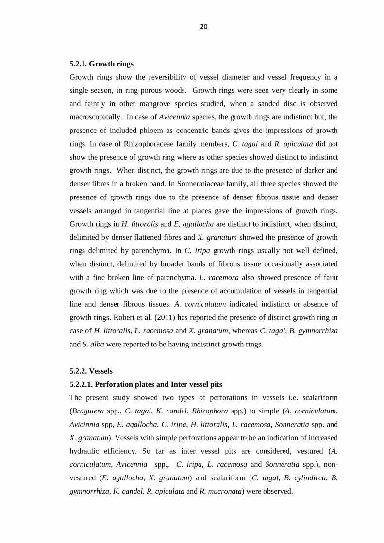
5.2.1. Growth rings
Growth rings show the reversibility of vessel diameter and vessel frequency in a
single season, in ring porous woods. Growth rings were seen very clearly in some
and faintly in other mangrove species studied, when a sanded disc is observed
macroscopically. In case of Avicennia species, the growth rings are indistinct but, the
presence of included phloem as concentric bands gives the impressions of growth
rings. In case of Rhizophoraceae family members, C. tagal and R. apiculata did not
show the presence of growth ring where as other species showed distinct to indistinct
growth rings. When distinct, the growth rings are due to the presence of darker and
denser fibres in a broken band. In Sonneratiaceae family, all three species showed the
presence of growth rings due to the presence of denser fibrous tissue and denser
vessels arranged in tangential line at places gave the impressions of growth rings.
Growth rings in H. littoralis and E. agallocha are distinct to indistinct, when distinct,
delimited by denser flattened fibres and X. granatum showed the presence of growth
rings delimited by parenchyma. In C. iripa growth rings usually not well defined,
when distinct, delimited by broader bands of fibrous tissue occasionally associated
with a fine broken line of parenchyma. L. racemosa also showed presence of faint
growth ring which was due to the presence of accumulation of vessels in tangential
line and denser fibrous tissues. A. corniculatum indicated indistinct or absence of
growth rings. Robert et al. (2011) has reported the presence of distinct growth ring in
case of H. littoralis, L. racemosa and X. granatum, whereas C. tagal, B. gymnorrhiza
and S. alba were reported to be having indistinct growth rings.
5.2.2. Vessels
5.2.2.1. Perforation plates and Inter vessel pits
The present study showed two types of perforations in vessels i.e. scalariform
(Bruguiera spp., C. tagal, K. candel, Rhizophora spp.) to simple (A. corniculatum,
Avicinnia spp, E. agallocha. C. iripa, H. littoralis, L. racemosa, Sonneratia spp. and
X. granatum). Vessels with simple perforations appear to be an indication of increased
hydraulic efficiency. So far as inter vessel pits are considered, vestured (A.
corniculatum, Avicennia spp., C. iripa, L. racemosa and Sonneratia spp.), non-
vestured (E. agallocha, X. granatum) and scalariform (C. tagal, B. cylindirca, B.
gymnorrhiza, K. candel, R. apiculata and R. mucronata) were observed.

21
There is a little comparative information exists on these aspects from the point of
view of eco-anatomical study (Carlquist, 1988). Scalariform perforation plates also
distinguishes the mangrove Rhizophoraceae from their terrestrial relatives (Vliet,
1976). Vijayan et al. (2017) reported the presence of vestured pits in A. corniculatum,
and Rao et al. (2005) in Avicennia spp., whereas vestured pits were not observed in
these two species in the present study which is in supportive of studies made by
Metcalfe and chalk ( 1950).
5.2.2.2. Vessel frequency
Mean vessel frequency among the species varied from 7/mm2 (C. iripa and H.
littoralis) to as high as 296/mm2 (A. corniculatum). In Rhizophoraceae family, lowest
vessel frequency was found in R. mucronata (23/mm2) and highest was found in C.
tagal (53/mm2). In Sonneraciaceae family, lowest vessel frequency was found in S.
alba (31/mm2) and S. caseolaris (31/mm
2) whereas, S. alba showed 46/mm
2. In the
species belonging to third group, lowest was found in H. littoralis and C. iripa
(7/mm2) and highest was found in A. corniculatum (296/mm
2).
Greater the vessel frequency shows the potential advantage of greater redundancy
(Carlquist, 1988). Anoop et al. (2013) and Vijayan et al. (2017) have reported very
low vessel frequency in case of A. corniculatum whereas Shashikala and Rao (2013)
have reported the vessel frequency as 355/ mm2 which are on the higher side
compared to the present study. Sun and Lin (1997) have reported the frequency of A.
corniculatum grown under different soil salinities are in the range of 273/ mm2 to
488/mm2. According to Santini et al. (2013), the vessel morphology varies with the
soil salinity values. Jansonnius (1950) was of the opinion that the vessel density
(vessel frequency) was in proportion to the frequency of inundation. According to
him, the vessel density in A. corniculatum was up to 150 per sq.mm and was
narrower than 50 µm in diameter and vessel densities in mangrove woods are 2 - 10
times higher than the wide range of other woods. The high densities of narrow
vessels could be related to overcome the frequent high tensions in xylem vessels.
5.2.2.3. Tangential vessel diameter
Mean tangential vessel diameter among the species varied from 35 µm (A.
corniculatum) to 111 µm (H. littoralis). In Rhizophoraceae family, mean tangential

22
vessel diameter was highest in B. gymnorrhiza (70 µm) and lowest vessel diameter of
42 µm was found in C. tagal. In Sonneratiaceae family, S. caseolaris (93 µm)
showed highest vessel diameter and S. alba (74 µm) showed lowest vessel diameter.
In the third group, H. littoralis found to have highest vessel diameter and A.
corniculatum has lowest vessel diameter.
Panshin (1932) found that the tangential diameter of the vessels were found to be in
the range of small to very small i.e. less than 100 µm in the woods of the Philippine
mangroves and the same was also confirmed by Jansonnius (1950). The present study
also confirms the results of the previous studies for all the species studied except H.
littoralis. It is also observed that in most of mangrove species, the vessels have
thicker walls compared to other terrestrial wood species. In the present study, the
vessel frequency of A. corniculatum is higher and the vessel diameter is in the range
as mentioned by Anoop et al. (2013) and Vijayan et al. (2017).
5.2.2.4. Vessel element length
Among all the species studied, mean vessel element length varied from 212 µm (A.
corniculatum) to 759 µm (B. gymnorrhiza). In Rhizophoraceae family, B.
gymnorrhiza had longer vessel element length (759 µm) and C. tagal had the shorter
vessel length (mean 506 µm). In Sonneratiaceae family, S. alba had the longest
vessels (mean 580 µm) and S. apetala had the shortest vessel (450 µm). While in third
group, E. agallocha (587 µm) and A. corniculatum (212 µm) had longest and
shortest vessels. Vessel elements were storied in A. corniculatum, H. littoralis and X.
granatum.
Ghose and Das (2001) have found that vessel members of the plants growing on
slopes (frequent tidal influence) have smaller dimensions, but more or less similar
length / width ratios and specialization indices in comparison to those of the ridge
(occasional tidal influence) regions. The vessel element length of B. gymnorrhiza and
S. apetala were shorter in the present study as compared to what has been reported by
Mariam et al. (2018). Zimmermann (1983) concluded that longer vessels confer
greater conducting efficiency, shorter vessels confer greater safety.
23
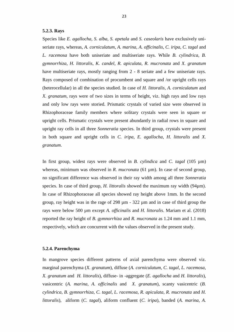
5.2.3. Rays
Species like E. agallocha, S. alba, S. apetala and S. caseolaris have exclusively uni-
seriate rays, whereas, A. corniculatum, A. marina, A. officinalis, C. iripa, C. tagal and
L. racemosa have both uniseriate and multiseriate rays. While B. cylindrica, B.
gymnorrhiza, H. littoralis, K. candel, R. apiculata, R. mucronata and X. granatum
have multiseriate rays, mostly ranging from 2 - 8 seriate and a few uniseriate rays.
Rays composed of combination of procumbent and square and /or upright cells rays
(heterocellular) in all the species studied. In case of H. littoralis, A. corniculatum and
X. granatum, rays were of two sizes in terms of height, viz. high rays and low rays
and only low rays were storied. Prismatic crystals of varied size were observed in
Rhizophoraceae family members where solitary crystals were seen in square or
upright cells. Prismatic crystals were present abundantly in radial rows in square and
upright ray cells in all three Sonneratia species. In third group, crystals were present
in both square and upright cells in C. iripa, E. agallocha, H. littoralis and X.
granatum.
In first group, widest rays were observed in B. cylindica and C. tagal (105 µm)
whereas, minimum was observed in R. mucronata (61 µm). In case of second group,
no significant difference was observed in their ray width among all three Sonneratia
species. In case of third group, H. littoralis showed the maximum ray width (94µm).
In case of Rhizophoraceae all species showed ray height above 1mm. In the second
group, ray height was in the rage of 298 µm - 322 µm and in case of third group the
rays were below 500 µm except A. officinalis and H. littoralis. Mariam et al. (2018)
reported the ray height of B. gymnorrhiza and R. mucronata as 1.24 mm and 1.1 mm,
respectively, which are concurrent with the values observed in the present study.
5.2.4. Parenchyma
In mangrove species different patterns of axial parenchyma were observed viz.
marginal parenchyma (X. granatum), diffuse (A. corniculatum, C. tagal, L. racemosa,
X. granatum and H. littoralis), diffuse- in -aggregate (E. agallocha and H. littoralis),
vasicentric (A. marina, A. officinalis and X. granatum), scanty vasicentric (B.
cylindrica, B. gymnorrhiza, C. tagal, L. racemosa, R. apiculata, R. mucronata and H.
littoralis), aliform (C. tagal), aliform confluent (C. iripa), banded (A. marina, A.

24
officinalis, C. iripa, C. tagal, E. agallocha, K. candel and X. granatum). Parenchyma
was totally absent in S. alba, S. apetala and S. caseolaris. Storied parenchyma was
observed in A. corniculatum and H. littoralis. Prismatic crystals were observed in
parenchyma cells in C. iripa, H. littoralis and X. granatum.
5.2.5. Fibres
Septate fibres were observed in Bruguiera spp. K. candel, S. alba, S. apetala, S.
caseolaris and X. granatum and rest of the species were having non septate fibres.
Fibres were very short ranging from 395 µm (A. corniculatum) to very long (more
than 1 mm) in all Rhizophoraceae family members. In general, mean fibre length was
more than 1 mm in all Rhizophoraceae members and in Avicennia spp, C. iripa, H.
littoralis and L. racemosa. The fibre length values reported by Mariam et al. (2018)
for B. gymnorrhiza, Heritiera spp. and S. apetala were lower as compared to the
values obtained in the present study. Fibre diameter varied from 15.14 µm (C. iripa)
to 25.74 µm (E. agallocha). Vijayan et al. (2017) have reported the fibre length in A.
corniculatum as 482 µm and fibre diameter as 27 µm. Fibres were thin walled (E.
agallocha) to thick to very thick walled (Rhizophoraceae family). Storied fibres were
observed in case of A. corniculatum, H. littoralis and X. granatum. Mariam et al.
(2018) reported shorter fibres (below 800 µm) in case of S. apetala, whereas in the
present study the average fibre length is 960 µm for the same species.

25
Table 3 (a) : Qualitative features of mangrove woods
Vessels
Species Colour Growth ring Diffuse porous Perforation type Inter vessel pitting Storied
A. corniculatum White to yellow to
red
- + Simple Alternate,
minute
+
A. marina Shades of brown - + Simple Alternate, minute to
small
-
A. officinalis Sap wood whitish
yellow Heartwood
Greyish brown
- + Simple Alternate, minute to
small
-
B. cylindrica
Shades of brown ± + Scalariform Scalariform -
B. gymnorrhiza
Reddish brown ± + Scalariform Scalariform -
C. tagal
Orange brown to
reddish brown
- + Scalariform Scalariform -
C. iripa
Pinkish brown to
reddish brown
+ + Simple Alternate, small,
vestured
-
E. agallocha
Yellow to pale yellow ± + Simple Alternate, small to
medium
-
H. littoralis Pale pink to pale
brown
± + Simple Alternate, minute to
small
+
K. candel
Light brown ± + Scalariform Scalariform -
L. racemosa
Brown to greyish
brown
+ + Simple Alternate, small,
vestured
-
R. apiculata
Reddish brown - + Scalariform Scalariform -

26
R. mucronata
Reddish brown + + Scalariform Scalariform -
S. alba Light brown to
reddish brown
± + Simple
Alternate, medium to
large, vestured
-
S. apetala
Light brown to
chocolate brown
± + Simple Alternate, medium to
large, vestured
-
S. caseolaris Light brown to
chocolate brown
± + Simple
Alternate, medium to
large, vestured
-
X. granatum Reddish brown to
pinkish brown
+ + Simple Alternate, minute
+

27
Table 3 (b) : Qualitative features of mangrove woods
Parenchyma
Species Marginal Diffuse Diffuse-in-
aggregate
Vasicentric Aliform Confluent Banded Storied Crystals
A. corniculatum - + - - - - - + -
A. marina - - - + - - + - -
A. officinalis - - - + - - + - -
B. cylindrica
- - - +s - - - - -
B. gymnorrhiza
- - - +s - - - - -
C. tagal
- + - ± + ± + - -
C. iripa
± - - - - + + - +
E. agallocha
- - + - - - + - -
H. littoralis ± + + +s - - - + +
K. candel
- - - - - - + - -
L. racemosa
± + - +s - - - - -
R. apiculata
- - - ±s ± - - - -

28
R. mucronata
- - - ±s ± - - - -
S. alba - - - - - - - - -
S. apetala
- - - - - - - - -
S. caseolaris - - - - - - - - -
X. granatum + + - + - - + + +
s- Scanty

29
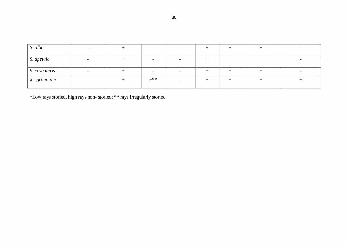
Table 3 (c) : Qualitative features of mangrove woods
Rays Fibres
Species Homocellular Heterocellular Storied Sheath cells Crystals Septate Fibre pit simple Fibre pit narrow
bordered
A. corniculatum + ± +* - - - + -
A. marina - + - - + - + -
A. officinalis - + - - + - + -
B. cylindrica
- + - + + ± + ±
B. gymnorrhiza
- + - + + ± + ±
C. tagal
- ± - ± + - + -
C. iripa
- + - - + - + -
E. agallocha
- + - - + - + -
H. littoralis - + +* + + - + ±
K. candel
- + - ± + + + ±
L. racemosa
- + - - - - + -
R. apiculata
- + - - + - + ±
R.mucronata
- + - - + - + ±
30
S. alba - + - - + + + -
S. apetala
- + - - + + + -
S. caseolaris - + - - + + + -
X. granatum - + ±** - + + + ±
*Low rays storied, high rays non- storied; ** rays irregularly storied

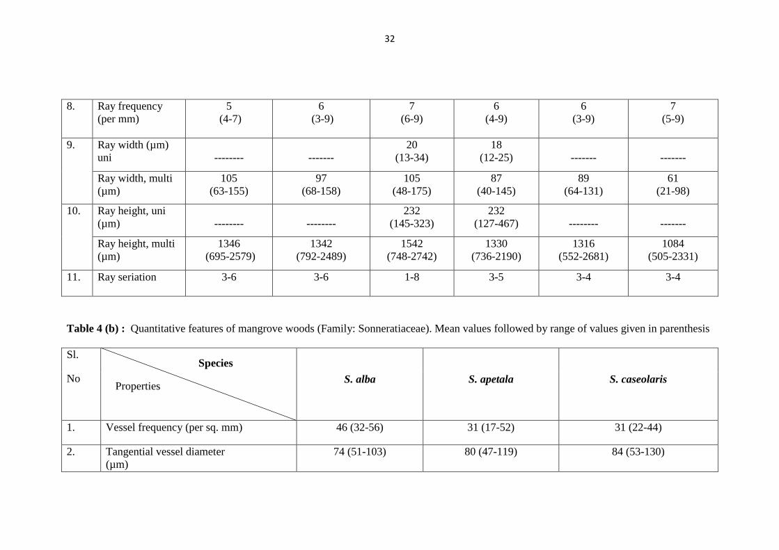
31
Table 4 (a) : Quantitative features of mangrove woods (Family: Rhizophoraceae). Mean values followed by range of values given in
parenthesis
Sl.
No
Bruguiera
cylindrica
Bruguiera
gymnorrhiza
Ceriops tagal Kandelia candel Rhizophora
apiculata
Rhizophora
mucronate
1. Vessel frequency
(per sq. mm)
29
(20-40)
31
(19-42)
53
(33-88)
41
(22-72)
25
(10-39)
23
(13-35)
2. Tangential vessel
diameter
(µm)
61
(40-88)
70
(45-94)
42
(29-59)
59
(39-89)
68
(42-93)
61
(32-89)
3. Vessel element
length (µm)
720
(379-880)
759
(311-1121)
506
(301-710)
582
(374-880)
693
(334-1013)
633
(352 -860)
4. Fibre length (µm) 1132
(879-1361)
1344
(919-1783)
1072
(845-1290)
1148
(876-1568)
1416
(1025-2077)
1428
(1020-1884)
5. Fibre diameter
(µm)
25.13
(15.76-35.95)
24.02
(14.23-30.67)
23.21
(13.81-30.82)
22.69
(15.11-31.75)
23.27
(13.88-28.36)
25.04
(18.28-32.10)
6. Fibre lumen
diameter (µm)
10.80
(5.46-18.70)
8.55
(4.74-17.39)
7.14
(2.65-14.16)
9.87
(5.84-16.08)
6.40
(3.33-12.07)
5.92
(1.89-11.23)
7. Fibre double wall
thickness (µm)
14.33
(8.14-20.52)
15.47
(9.17-22.33)
16.07
(9.32-22.85)
12.83
(7.02-17.95)
16.87
(7.54-24.43)
19.13
(11.88-26.77)
Species
Properties
32
8. Ray frequency
(per mm)
5
(4-7)
6
(3-9)
7
(6-9)
6
(4-9)
6
(3-9)
7
(5-9)
9. Ray width (µm)
uni
--------
-------
20
(13-34)
18
(12-25)
-------
-------
Ray width, multi
(µm)
105
(63-155)
97
(68-158)
105
(48-175)
87
(40-145)
89
(64-131)
61
(21-98)
10.
Ray height, uni
(µm)
--------
--------
232
(145-323)
232
(127-467)
--------
-------
Ray height, multi
(µm)
1346
(695-2579)
1342
(792-2489)
1542
(748-2742)
1330
(736-2190)
1316
(552-2681)
1084
(505-2331)
11. Ray seriation 3-6 3-6 1-8 3-5 3-4 3-4
Table 4 (b) : Quantitative features of mangrove woods (Family: Sonneratiaceae). Mean values followed by range of values given in parenthesis
Sl.
No
S. alba S. apetala S. caseolaris
1. Vessel frequency (per sq. mm) 46 (32-56) 31 (17-52) 31 (22-44)
2. Tangential vessel diameter
(µm)
74 (51-103) 80 (47-119) 84 (53-130)
Properties
Species

33
3. Vessel element length (µm) 580 (348-848) 450 (225-712) 459 (253-698)
4. Inter -vessel pitting (µm) 8.80 (6.34-11.47) 6.45 (3.65-10.59) 6.60 (4.4-9.94)
5. Ray- vessel pitting (µm) 6.81 (3.37-11.66) 7.92 (4.13-14.5) 11.14 (6.06-22.07)
6. Fibre length (µm) 1040 (734-1404) 960 (730-1182) 868 (609-1160)
7. Fibre diameter (µm) 22.66 (12.61-31.46) 20.23 (10.4-27.93) 22.25 (14.69-28.74)
8. Fibre lumen diameter (µm) 15.08 (8.02-24.39) 13.32 (6-20.72) 15.75 (8.13-22.87)
9. Fibre double wall thickness (µm) 7.57 (2.89-12.07) 6.91 (2.07-12.52) 6.51 (4.4-11.44)
10. Ray frequency (per mm) 11 (8-15) 15 (11-19) 12 (9-17)
11. Ray width (µm) -uni 21 (12-29) 18 (13-27) 20 (11-27)
Ray width (µm) -multi ------ ------ ------
12. Ray height (µm) -uni 298 (117-595) 322 (162-713) 275 (96-519)
Ray height (µm)- multi ------ ------ ------
13. Ray seriation 1 1 1

34
Table 4 ( c) : Quantitative features of mangrove woods (Family: Myrsinaceae, Avicenniaceae, Fabaceae, Euphorbiaceae, Sterculiaceae,
Combretaceae, Meliaceae). Mean values followed by range of values given in parenthesis
Sl.
No
A.
corniculatum
A.
marina
A.
officinalis
C. iripa E. agallocha H. littoralis L. racemosa X. granatum
1. Vessel
frequency
(per sq. mm)
296
(220-354)
34
(14-67)
22
(12-38)
7
(5-11)
13
(7-28)
7
(5-12)
49
(28-92)
32
(26-44)
2. Tangential
vessel
diameter
(µm)
35
(23-47)
62
(34-109)
72
(45-103)
98
(66-132)
62
(33-94)
111
(81-156)
48
(31-68)
57
(38-78)
3. Vessel
element
length (µm)
212
(151-245)
245
(106-383)
235
(97-395)
310
(155-424)
587
(266-928)
288
(148-389)
492
(300-703)
245
(155-305)
4. Inter -vessel
pitting (µm)
3.74
(3.07-4.51)
4.14
(3.21-
5.46)
3.59
(2.34-4.36)
5.03
(4.15-6.31)
5.87
(3.64-8.35)
3.57
(2.33-4.9)
4.73
(3.47-7.47)
3.18
(2.64-3.96)
5. Ray- Vessel
Pitting (µm)
3.09
( 2.38-4.13)
2.62
(2.27-
3.01)
2.64
(2.08-3.13)
4.09
(3.25-4.77)
6.13
(5.46-8.35)
4.04
(3.64-4.67)
3.73 (2.39-
4.55)
3.17
(2.60-3.79)
6. Fibre length
(µm)
395
(276-503)
1006
(768-
1272)
1030
(714-1555)
1394
(1062-1661)
982
(721-1278)
1586
(1108-2235)
1013
(769-1351)
750
(581-903)
7. Fibre
diameter
(µm)
24.18
(16.29-33.07)
19.97
(12.92-
29.85)
19.71
(12.07-
26.85)
15.14
(11.56-
20.09)
25.74
(14.44-
36.59)
18.68
(12.72-
23.94)
16.73 (12.31-
21.72)
21.06 (15.82-
24.96)
Species
Properties

35
8. Fibre lumen
diameter
(µm)
14.97
(9.95-22.15)
10.57
(5.81-
16.37)
9.59
(4.22-18.08)
6.98
(4.57-10.96)
19.46
(9.31-29.57)
8.77
(3.74-14.22)
8.89
(5.28-12.67)
14.73 (11-
19.21)
9. Fibre double
wall thickness
(µm)
9.21
(5.14-15.38)
9.39
(5.17-
15.41)
10.12
(5.49-17.1)
8.15
(4.25-14.32)
6.38
(3.29-10.92)
9.91
(4.86-14.69)
7.84
(4.23-12.08)
6.33
(4.26-8.83)
10. Ray
frequency
(per mm)
3 (1-4) 9 (7-13) 10 (7-14) 8 (6-9) 10 (8-16) 5 (4-7) 13 (9-16) 9 (7-11)
11. Ray width uni
(µm)
17 (11-23) 14.58
(7.63-
25.43)
15
(9-27)
-------
21.3
(13.03-
28.18)
22
(12-33)
27
(18-39)
24
(15-30)
Ray width
multi (µm)
31
(22-43)
31.84
(18.3-
60.33)
30
(17-47 )
34
(23-45)
-------
94
(49-146)
-------
42
(29-54)
12. Ray height,
uni (µm)
156
(94-224)
200
(108-429)
201
(121-382)
-------
338
(159-596)
213
(157-290)
379
(155-585)
116
(88-187)
Ray height,
multi (µm)
236
(141-387)
361
(168-
737)
838
(284-1674)
332
(232-457)
-------
713
(223- 1369)
-------
297
(157-506)
13. Ray seriation 1-4 3-5 3-5 1-3 1 (rarely 2) 4-7 1 1-4

36
Objective 2: To create database of anatomical properties of mangrove species from
Maharashtra seacoast for their identification.
For any wood sample to be identified, a reference material is needed. This reference
material can be in the form of either or combination of published literature, data base
in electronic media, xylarium samples, and photo-micrographs. The authentic samples
collected for the project study are preserved in IWST xylarium. Data base in the form
of description is generated based on physical and anatomical properties studied.
Based on the physical and anatomical properties, a list of card key features for
identification of seventeen mangrove species studied is compiled as Appendix-II. An
attempt has been made to prepare an artificial key for separation of all seventeen
species and listed in appendix III.
Anatomical description of wood along with their distribution of all seventeen
mangrove species studied under this project is given below in alphabetical order of
species.
Aegiceras Gaertn.
(Family- Myrsinaceae)
A genus comprising about 200 species of shrubs and small trees occur in the tropics
of Asia and Africa. Twenty five species are reported to be found in India of which one
species found in Maharashtra is studied and described as below.
A. corniculatum (L.) Blanco
The tree - A shrub or small tree up to a height of 6 m. It occurs in the mangrove
swamps and tidal creeks. Bark - Light to dark grey, sometimes pale brown with
reddish tinge, smooth, thin, about 0.4 - 4.0 mm thick.
Description of Wood
(PLATE: 1)
General Features - Wood whitish to red tinge, turning yellow on aging, soft to
moderately hard, light to moderately heavy (sp.gr. 0.468-0.660; oven dry), straight
grained, very fine textured.

37
Gross Structure - A diffuse porous wood. Growth rings indistinct. Vessels very
small to minute, indistinct even under hand lens, very numerous (220 - 354 /mm2),
solitary and in radial multiples of 2 - 6, infrequently in clusters, round to angular,
evenly distributed, sometimes with a tangential pattern, open, vessel lines indistinct
on longitudinal surfaces. Parenchyma sparse, scanty vasicentric (incomplete),
indistinct even under hand lens. Rays moderately broad to fine, broader ones are
widely spaced and are visible to eye. Ripple marks present, distinct under the hand
lens.
Minute Structure - Vessels are in radial multiples of 2 - 6, commonly 4 (solitary 34
%), round to oval in outline, open, 23 µm - 47 µm in tangential diameter (mean 35
µm), perforation plate simple, intervessel pits minute, 3.07 µm - 4.51 µm (mean 3.74
µm) in diameter, oval, alternate, vessel ray pits similar to intervessel pits with distinct
borders, 2.38 µm - 4.13 µm (mean 3.09 µm) in diameter, vessel elements are 151 µm
- 245 µm (mean 212 µm) in length, storied. Axial parenchyma is diffuse, consisting of
a few cells round the vessels, 2 - 4 cells / parenchyma strand with some fusiform cells,
storied. Rays are 1 - 4 /mm (t), uni-seriate and up to 4 seriate, uni-seriate rays are 94
µm - 224 µm (mean 156 µm) high or up to 15 cells in height and 11 µm - 23 µm
(mean 17 µm) in width, multi seriate rays are 141 µm - 387 µm (mean 236 µm ) high
or up to 19 cells in height, 22 µm- 43 µm (mean 31 µm) in width, low rays storied,
high rays non-storied, homocellular to weakly heterocellular, composed of entirely
procumbent cells or with one row of upright and/or square marginal cells. In ray cells,
large thin walled idioblast to form non crystalliferous cysts were observed. Sheath
cells not observed. Fibres are with simple pits, seen on both radial and tangential
longitudinal walls, non- septate, storied, fibres are 276 µm - 503 µm (mean 395 µm)
long, 16.3 µm - 33.1 µm (mean 24.2 µm) in diameter, thin walled, double wall
thickness is 5.14 µm - 15.38 µm (mean 9.2 µm).
Avicennia Linn.
(Family- Avicenniaceae)
A genus of about 14 species of shrubs to trees occurring in the warmer parts of the
world in the coastal regions along with other species. Around 6 species belong to the
Indo-West Pacific Islands. The genus is uniform in its gross morphology and anatomy

38
but for some useful diagnostic features in the field like bark colour and texture. Three
species are distributed in Indian coast, of which two species found in Maharashtra are
studied. These two species are indistinguishable from each other and the description is
given below.
1. A. marina Vierh.
The tree - A small bushy evergreen tree, up to 14 m high. Pencil like pneumatophores
emerge above ground level. Bark - Smooth, whitish-brown to greyish brown with
faint red tinge, lenticellate, thin (about 0.5 - 1.6 mm ), stiff, brittle flakes.
2. A. officinalis Linn.
The tree - A tree up to 20 m high and up to 2 m girth. Pencil like pneumatophores
emerge above ground level and larger compared to other species. Bark - Greyish-
brown to greenish- brown, thin (about 1.3 - 1.8 mm), smooth to coarse, with fine
vertical fissures.
Description of wood
(A. marina and A. officinalis)
(PLATE- 1)
General features - Sapwood and heartwood distinct to indistinct. Heartwood reddish
- brown darkening on exposure. Sapwood lighter than heartwood, light brown in A.
marina and wood whitish -yellow to brownish yellow or light greyish -brown without
distinction into sapwood and heartwood in the samples of A. officinalis examined.
Wood moderately hard to very hard, moderately heavy to very heavy (sp.gr. 0.584 -
0.980; oven dry) in A. marina and moderately hard to hard, moderately heavy to
heavy (sp.gr. 0.504 - 0.717; oven dry) in A. officinalis, dull, straight-grained to
interlocked grained, no distinct odor, fine to coarse -textured with a pleasant figure on
the longitudinal surfaces due to presence of included phloem.
Gross structure - A diffuse porous wood. Growth ring boundaries indistinct or
absent, but due to the presence of concentric bands of included phloem, which often
gives the impression as growth rings / marks. Layers of xylem separated by layers of
phloem and conjunctive parenchyma tissue containing a row of stone cells and
isolated strands of phloem tissue. Vessels moderately large to very small, distinct

39
under the hand-lens, moderately numerous to numerous, 12 - 67 /mm2 (14 - 67 /mm
2
in A. marina and 12 - 38 /mm2
in A. officinalis), evenly distributed, solitary, in short
radial multiples of 2 - 5 rarely in clusters, round to oval, occasionally filled with
yellow deposits, vessel lines indistinct. Parenchyma distinct, vasicentric around the
pores, other than conjunctive tissue. Rays fine to very fine, inconspicuous but visible
with hand lens, closely spaced. Included phloem in small islands, along with thick
bands of conjunctive tissues alternating with layers of fibrous tissues in tangential
band.
Minute structure - Vessels are solitary and in radial multiples of 2 - 7 (solitary 48
%), round to oval in outline, open, 34 µm -109 µm in tangential diameter (mean 62
µm -72 µm), perforation plate simple, intervessel pits are minute to small, 2.34 µm -
5.46 µm (mean 3.6 µm - 4.14 µm) in diameter, angular, alternate, pits leading to rays
and parenchyma are similar to inter vessel pits, vessel elements are 97 µm - 395 µm
(mean 240 µm). Axial parenchyma is vasicentric and as bands. Conjunctive
parenchyma containing a band of stone cells, 2 - 6 cells/ parenchyma strand. Rays are
3-5 seriate, few are uniseriate and few are partially bi-seriate, 7 - 14/mm (t).
Uniseriate rays are 108 µm - 429 µm (mean 200 µm) high or up to 16 cells in height
and 7.63 µm - 27 µm (mean 15 µm) in width composed of either only procumbent
cells or mix of procumbent and upright cells, multiseriate rays are very variable in
height, 168 µm - 1674 µm (mean 361µm - 838 µm) high or up to 55 cells in height
and 17 µm - 60.33 µm (mean 30 µm - 32 µm ) in width. Rays are heterocellular,
composed of procumbent cells with upright and square cells mixed throughout the
ray. Prismatic crystals observed in upright/square and procumbent ray cells, styloids
or elongated and small crystals observed in A. officinalis in upright/square and
procumbent ray cells. Ray cells containing one or more prismatic crystals. Fibres are
with minute pits, restricted to radial walls, non-septate, 714 µm -1555 µm (mean
1006 µm -1030 µm) long, 12.07 µm - 29.85 µm (mean 19.97 µm) in diameter, thin to
thick walled, double wall thickness is 5.17 µm - 17.1µm (mean 9.39 µm -10.12µm).
Included phloem in small islands, along with thick bands of conjunctive parenchyma
tissues with a thin layer of stone cells alternating with layers of fibrous tissues.

40
Bruguiera Lam.
(Family- Rhizophoraceae)
A small genus of about six species of trees occurring in mangroves from East Coast of
Africa to the Pacific Islands, South East Asia and North Australia. Out of three
species found in Maharashtra, two species were studied. The woods of these two
species are very similar in structure and are indistinguishable and described below.
1. Bruguiera cylindrica (Linn.) Blume.
The tree - A small tree up to 20 m in height and up to 60 cm in girth. Knee like
pneumatophores are present. Bark - Dark brown to greyish brown, with few lenticels,
about 1.3 - 3.5 mm thick.
2. Bruguiera gymnorrhiza (Linn.) Lamk
The tree - A tree of 9 - 25 m in height and up to 1 - 2 m in girth. Knee like
pneumatophores are present. Bark - Brownish black to dark grey with large corky
lenticular patches, coarse, about 1.6 - 3.6 mm thick, valuable tanning material.
Description of wood
(B. cylindrica and B. gymnorrhiza)
(PLATE: 2)
General features- Sapwood and heartwood indistinguishable in the samples
examined. Wood light brown to yellowish-brown to reddish brown; moderately hard
to very hard, moderately heavy to very heavy (sp.gr. 0.635 - 0.787; oven dry); straight
to shallowly interlocked-grained, fine textured, often exhibiting distinct silver grain
on radial surface.
Gross structure- A diffuse porous wood. Growth rings indistinct to distinct, when
distinct delimited by dark colored fibres. Vessels small to very small, distinct only
under the hand lens, moderately numerous to numerous (19 - 42/ mm2), evenly
distributed, solitary and in radial multiples of 2 - 4, occasionally in clusters of 3 - 4,
round, open, vessel lines inconspicuous. Parenchyma, scanty paratracheal, indistinct
to just visible under the hand lens. Rays moderately broad, visible to the eye, lighter
in color, somewhat widely spaced.

41
Minute structure- Vessels are in radial multiples of 2 - 4 and solitary, occasionally in
clusters of 3 - 4, solitary vessels 48 – 55 %, round in outline, open, 45 µm - 94 µm in
tangential diameter (mean 61 µm - 70 µm), perforation plate scalariform (6 - 16
bars), oblique, inter-vessel pits abundant, scalariform, vessel ray pits with much
reduced borders to simple, pits round to horizontal- gash like, scalariform, unilaterally
compound and coarse. Vessel elements are 311 µm - 1121 µm in length (mean 720
µm - 759 µm). Solid granular contents infrequently present in vessels. Axial
parenchyma is scanty vasicentric, average number of cells per parenchyma strand is 4
- 8. Prismatic crystals are absent. Rays are 1 - 7 seriate, (commonly 3 - 6 seriate,
rarely uni- seriate), 3 - 9 /mm (t). Rays are commonly more than 1 mm, 695 µm-
2579 µm (mean 1346 µm) high or up to 55 cells in height and 63 µm - 158 µm (mean
97 µm - 105 µm) in width, heterocellular, composed of procumbent cells with 2 - 4
rows of square and upright cells. Prismatic crystals observed in upright/square and
procumbent ray cells. Sheath cells present. Fibres are with minute pits on tangential
and radial walls, occasionally septate, 879 µm - 1783 (mean 1132 µm- 1344 µm)
long, 14.23 µm - 35.95 µm (mean 24.02 µm - 25.13 µm) in diameter, thick walled,
double wall thickness is 8.14 µm - 22.33 µm (mean 14.33 µm -15.47 µm ). Silica not
observed. Granular and amorphous content frequently present in ray and axial
parenchyma cells.
Ceriops Arnott.
(Family- Rhizophoraceae)
A genus comprising two species of shrubs or small trees found on the muddy shores
of Indian and West Pacific oceans, Australia. One species found in Maharashtra was
studied and described below.
Ceriops tagal (Perr.) C. B. Robinson
The tree - A small evergreen tree or shrub up to 9 m in height and 30-60 cm in girth
with many short buttresses. Bark - Pale reddish to orange, smooth and slightly
fissured, lenticellate, peeling off in thick flakes or strips, 2 - 6 mm thick, giving a
valuable tanning material.

42
Description of wood
(PLATE: 2)
General features - Sapwood and heartwood indistinct in the samples studied. Wood
orange brown to reddish orange turning dark reddish brown with age. Wood hard to
very hard, heavy to very heavy (sp.gr. 0.727 - 0.778; oven dry), straight grained, fine
to very fine textured.
Gross structure - A diffuse porous wood. Growth rings indistinct. Vessels small to
very small, visible only under lens, vessels arranged in no specific pattern, numerous
to very numerous (33 - 88 / mm2), solitary and in radial multiples of 2 - 6, round in
outline, vessel lines inconspicuous. Parenchyma visible only under the hand lens,
vasicentric, aliform and sometimes connecting a few adjacent vessels. Rays fine to
moderately broad, just visible to naked eye, evenly and closely spaced.
Minute structure - Vessels are in radial multiples of 2 - 6, four are more common in
radial rows (solitary 38%), round in outline, open, 29 µm - 59 µm in tangential
diameter (mean 42 µm), perforation plate scalariform (6 - 12 bars), thick, inter-vessel
pits scalariform, oblique; vessel ray pits are vertically uni-laterally compound, with
oval to rounded ray pit, shield shaped vessel ray pitting, scalariform, vessel elements
are 301 µm - 710 µm in length (mean 506 µm). Axial parenchyma is diffuse,
vasicentric at places, aliform confluent, sometimes in bands (3 to 5 cells); average
number of cells per parenchyma strand is 5 - 8. Rays are 2 - 8 seriate, commonly 4
seriate, occasional uniseriate, 6 - 9 /mm (t), uniseriate rays are 145 µm - 323 µm
(mean 232 µm) high or up to 8 cells in height and 20 µm (13 µm -34 µm) in width,
multiseriate rays are commonly more than 1mm, 748 µm - 2742 µm (mean 1542 µm)
in height and 48 µm - 175 µm (mean 105 µm) in width. Rays are homocellular to
heterocellular, composed of procumbent ray cells with square and upright cells
restricted to marginal cells with infrequent sheath cells. Prismatic crystals observed in
upright/square and procumbent ray cells. Fibres are with minute pits, confined to
radial wall, non-septate, fibres are 845 µm - 1290 µm (mean 1072 µm) long, 13.81
µm - 30.82 µm (mean 23.21 µm) in diameter, medium thick to thick walled, double
wall thickness is 9.32 µm - 22.85 µm (mean 16.07 µm). Granular and amorphous
content present in both ray and axial parenchyma.
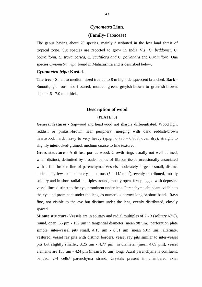
43
Cynometra Linn.
(Family- Fabaceae)
The genus having about 70 species, mainly distributed in the low land forest of
tropical zone. Six species are reported to grow in India Viz. C. beddomei, C.
bourdillonii, C. travancorica, C. cauliflora and C. polyandra and C.ramiflora. One
species Cynometra iripa found in Maharashtra and is described below.
Cynometra iripa Kostel.
The tree - Small to medium sized tree up to 8 m high, deliquescent branched. Bark -
Smooth, glabrous, not fissured, mottled green, greyish-brown to greenish-brown,
about 4.6 - 7.0 mm thick.
Description of wood
(PLATE: 3)
General features - Sapwood and heartwood not sharply differentiated. Wood light
reddish or pinkish-brown near periphery, merging with dark reddish-brown
heartwood, hard, heavy to very heavy (sp.gr. 0.735 - 0.808; oven dry), straight to
slightly interlocked-grained, medium coarse to fine textured.
Gross structure - A diffuse porous wood. Growth rings usually not well defined,
when distinct, delimited by broader bands of fibrous tissue occasionally associated
with a fine broken line of parenchyma. Vessels moderately large to small, distinct
under lens, few to moderately numerous (5 - 11/ mm2), evenly distributed, mostly
solitary and in short radial multiples, round, mostly open, few plugged with deposits;
vessel lines distinct to the eye, prominent under lens. Parenchyma abundant, visible to
the eye and prominent under the lens, as numerous narrow long or short bands. Rays
fine, not visible to the eye but distinct under the lens, evenly distributed, closely
spaced.
Minute structure- Vessels are in solitary and radial multiples of 2 - 3 (solitary 67%),
round, open, 66 µm - 132 µm in tangential diameter (mean 98 µm), perforation plate
simple, inter-vessel pits small, 4.15 µm - 6.31 µm (mean 5.03 µm), alternate,
vestured, vessel ray pits with distinct borders, vessel ray pits similar to inter-vessel
pits but slightly smaller, 3.25 µm - 4.77 µm in diameter (mean 4.09 µm), vessel
elements are 155 µm - 424 µm (mean 310 µm) long. Axial parenchyma is confluent,
banded, 2-4 cells/ parenchyma strand. Crystals present in chambered axial

44
parenchyma cells. Rays are 6 - 9/ mm (t), 1-3 seriate, 232 µm - 457 µm (mean 332
µm) in height or up to 24 cells high, 23 µm - 45 µm (mean 34 µm) in width,
heterocellular, composed of procumbent cells with one marginal row of upright and
square cells. Sheath cells not observed. Prismatic crystals observed in upright/square
and procumbent ray cells. Fibres are with simple minute pits, more numerous on
radial walls than on the tangential walls, non-septate, fibres are 1062 µm - 1661µm
(mean 1394 µm) long, 11.56 µm - 20.09 µm (mean 15.14 µm) in diameter, thick to
very thick walled, double wall thickness is 4.25 µm - 14.32 µm (mean 8.15 µm).
Excoecaria Linn.
(Family- Euphorbiaceae)
A genus of about 40 species of trees and shrubs distributed in Old world Tropical
mangals, Africa and Asia. About 8 species occur in India of which one species, E.
agallocha found in Maharashtra is studied and described below. This species occurs
in mangroves but also occasionally in inland stations.
Excoecaria agallocha Linn.
The tree - A large shrub or small evergreen tree but occasionally attains a height of
12 m and 1.5 m girth. Bark - Grey, smooth and shining when green, becoming
fissured lenticels prominent on younger twigs exudes a poisonous milky juice which
is injurious to human eye and irritable to skin.
Description of wood
(PLATE: 3)
General features - Sapwood and heartwood is indistinct, wood yellow to pale yellow,
turning white or greyish- yellow on ageing, soft, light (sp.gr. 0.305 - 0.369; oven dry);
straight grained, fine textured.
Gross structure - A diffuse porous wood. Growth rings indistinct to distinct, when
distinct delimited by flattened fibres. Vessels small to very small, moderately few to
numerous (7 - 29 /mm2), solitary and in radial multiples of 2 - 6, unevenly distributed,
round to oval in outline, open, vessel lines distinct to inconspicuous on longitudinal
surfaces. Parenchyma visible under hand lens, diffuse to diffuse-in-aggregates,

45
tending to form net like structure with rays at places. Rays fine to very fine, distinct
under the hand lens, closely spaced.
Minute structure- Vessels are solitary and in radial multiples of 2 - 6, (solitary 56%),
round to oval in outline, open, 33 µm - 94 µm in tangential diameter (mean 62 µm),
perforation plate simple, intervessel pits small to medium, 3.64 µm - 8.35 µm (mean
5.87 µm), polygonal, alternate, vessel ray pits with distinct borders similar to
intervessel pits but slightly larger 5.46 µm - 8.35 µm in diameter (mean 6.13 µm),
vessel elements are 266 µm - 928 µm (mean 587 µm) long. Axial parenchyma is
diffuse to diffuse-in-aggregate, sometimes in narrow bands forming mesh like
structure with rays, 2 - 4 cells/ parenchyma strand. Rays are 8 - 16/ mm (t), 1 - 2
seriate, 159 µm - 596 µm (mean 338 µm) in height or up to 21 cells high and 13 µm -
28 µm (mean 21 µm) in width, heterocellular, composed of procumbent cells with
upright and square cells in marginal rows. Prismatic crystals observed in
upright/square and procumbent ray cells. Ray cells containing one or more prismatic
crystals. Laticifers tubes observed in radial longitudinal section. Fibres are with
minute pits, more numerous on radial walls than on tangential walls, non-septate,
fibres are 721 µm -1278 µm (mean 982 µm) long, 14.44 µm - 36.59 µm (mean 25.74
µm) in diameter, thin walled, double wall thickness is 3.3 µm - 10.92 µm (mean 6.38
µm).
Heritiera Alton
(Family- Sterculiaceae)
A small genus of 31 species of trees confined to Indo- West Pacific Islands, tropical
Asia, Africa and Australia. At least 5 species are known to occur in India of which H.
fomes and H. littoralis are littoral species and rest are from inland. One species H.
littoralis found in Maharashtra, which is said to be a rare species in Maharashtra and
is described as below.
Heritiera littoralis Dryand.
The tree - A medium to large sized tree up to 25 m in height and 1-2 m girth.
Prominent buttresses, plank or ribbon roots are seen. Bark - Greenish grey or
discoloured with longitudinal fissures, about 1.0-2.0 mm thick.

46
Description of wood
(PLATE: 3)
General features - Sapwood and heartwood is distinct, sapwood pale pink to pale
brown gradually merging into reddish brown heartwood, darkening on ageing, dull,
hard, moderately heavy (sp.gr. 0.604 - 0.628; oven dry); straight to somewhat
interlocked grained, medium to fine textured, slightly oily with smooth feel.
Gross structure - A diffuse porous wood. Growth rings distinct to indistinct, when
distinct delimited by flattened, fibrous bands and discontinued fine axial parenchyma
line. Vessels moderately large to small, visible to the eye, moderately few (5 - 12 /
mm2), solitary and in radial multiples of 2 - 3, also as cluster of 3 - 4, evenly
distributed, round in outline, often filled with reddish gummy deposits, vessel lines
distinct on longitudinal surfaces. Parenchyma visible under the hand lens, as fine,
closely spaced, diffuse and diffuse-in-aggregates forming a net like structure with
rays, sometimes as continuous lines delimiting growth lines, paratracheal parenchyma
scanty or vasicentric. Rays moderately broad, distinct to eye prominent under the
hand lens, lighter in colour, closely spaced, ripple marks present feebly visible.
Minute structure - Vessels are solitary and in radial multiples of 2 - 3, rarely up to 4
(solitary 80%), round in outline, open, 81 µm - 156 µm in tangential diameter (mean
111 µm), perforation plate simple, intervessel pits minute to small, 2.33 µm - 4.9 µm
(mean 3.57 µm) in diameter, oval, alternate, vessel ray pits similar to intervessel pits,
distinctly bordered, vessel elements are 148 µm-389 µm (mean 288 µm) long, storied.
Axial parenchyma is diffuse to diffuse-in-aggregate, sometimes in narrow bands
forming mesh like structure with rays, scanty paratracheal, 4 cells/ parenchyma
strand, storied. Crystals present in chambered axial parenchyma cells. Rays of two
distinct sizes. 4 - 7/ mm (t), 1 - 7 seriate (4 - 5 seriate are commonly present),
uniseriate rays are 157 µm - 290 µm (mean 213 µm) high or up to 11 cells in height,
12 µm -33 µm (mean 22 µm) in width, multiseriate rays are 223 µm - 1369 µm (mean
713 µm) in height or up to 64 cells high, 49 µm -146 µm (mean 94 µm) in width, low
rays are storied, high rays are non-storied, heterocellular, body of rays cells composed
of procumbent with mostly two rows of upright and/ square marginal cells. Prismatic
crystals present in upright and/or square cells. Sheath cells absent. Silica present.
Fibres are with simple to minutely bordered pits, non-septate, fibres are 1108 µm -

47
2235 µm (mean 1586 µm) long, 12.72 µm - 23.94 µm (mean 18.68 µm) in width, thin
to thick walled, double wall thickness is 4.86 µm - 14.69 µm (mean 9.91 µm), storied.
Gum ducts of traumatic type absent in the samples studied.
Kandelia W. & A.
(Family- Rhizophoraceae)
The only species of the genus of the Tropics ranging from the Ganges Delta, Burma,
South East Asia to South China. Description of the species found in Maharashtra is
given below.
Kandelia candel Linn.
The tree - A small tree or shrub up to 9 m high and 75 cm in girth. Aerial roots
generally absent like other members of Rhizophoraceae. Bark - Greyish or reddish
brown, smooth, peeling off in flakes, thin about 2.1 - 4.7 mm.
Description of wood
(PLATE: 4)
General features - Wood is light brown in colour, soft to moderately hard, light to
moderately heavy (sp.gr. 0.495 - 0.527; oven dry), straight grained, coarse textured
and lustrous.
Gross structure - A diffuse porous wood. Growth rings are indistinct or distinct due
to the zones of fibrous tissue devoid of parenchyma tissue. Vessels small to very
small, distinct under hand lens, vessel frequency highly variable, numerous to very
numerous (22 - 72 /mm2), distinct only under the hand lens, evenly distributed,
solitary and in radial multiples of 2 to 5, sometimes in clusters, round to oval, open,
vessel lines are inconspicuous. Parenchyma abundant, visible to the eye, distinct
under the hand lens as short wavy bands enclosing the vessels alternating with fibrous
tissue. Rays are moderately broad, closely spaced, distinct under the hand lens, radial
flecks are common.
Minute structure - Vessels are solitary, in radial multiples of 2 - 5, sometimes in
clusters of 2 - 6 (solitary 36%), round in outline, open, 39 µm - 89 µm in tangential
diameter (mean 59 µm), perforation plate scalariform (5 - 12 bars), inter-vessel pits
scalariform extending over the entire inter-vessel walls, vessel ray pits with much

48
reduced borders to apparently simple, pits round to scalariform, unilaterally
compound and coarse, mean vessel elements are 374 µm - 880 µm (mean 582 µm) in
length. Parenchyma is paratracheal, banded, 4 - 7 cells wide with strands of 6 - 7 cells.
Prismatic crystals are absent. Globular and amorphous content present in axial
parenchyma. Rays are 3 - 5 seriate and very few uniseriate, 4 - 9 /mm (t), uni-
seriate rays are 127 µm - 467 µm (mean 232 µm) high or up to 12 cells in height and
12 µm - 25 µm (mean 18 µm) in width, multiseriate rays are commonly more than
1mm, 736 µm - 2190 µm (mean 1330 µm) in height and 40 µm - 145 µm (mean 87
µm) in width, heterocellular, body of the ray cells are procumbent with square and
upright cells restricted to marginal rows. Prismatic crystals observed in
upright/square ray cells. Sheath cells infrequent. Silica observed in ray cells. Fibres
are angular with simple to minutely bordered pits, restricted to radial walls, septate,
fibres are 876 µm - 1568 µm (mean 1148 µm) long, 15.11 µm - 31.75 µm (mean
22.69 µm) in diameter, medium thin walled to thick walled, double wall thickness is
7.02 µm - 17.95 µm (mean 12.83 µm).
Lumnitzera Willd.
(Family - Combretaceae)
A genus comprising of two species viz. L. littorea and L.racemosa of small trees or
shrubs inhabiting the mangrove swamps from East Africa to Western Pacific Islands,
Malaysia, North Australia and Indo- China. Both the species are found in India and L.
racemosa found in Maharashtra and described as below.
Lumnitzera racemosa Willd.
The tree - A small, straight-stemmed tree, up to 10 m high but mostly as a shrub.
Bark - Brown to greyish brown, rough, thin, about 1.1 - 3.0 mm thick. It is rich in
tannin and may be used for tanning leather.
Description of wood
(PLATE: 4)
General features - Sapwood and heartwood distinct. Sapwood greyish-brown to
yellowish brown and heartwood is reddish brown, somewhat lustrous, moderately

49
hard, moderately heavy (sp.gr. 0.613 - 0.712; oven dry); straight grained, fine to very
fine textured.
Gross structure - A diffuse porous wood. Growth rings indistinct to distinct, when
distinct delimited by dark latewood fibres sometimes may be due to a tangential
arrangement of vessels. Vessels small to very small, distinct only under the hand lens,
numerous to very numerous (28 - 92 /mm2), usually in radial multiples of 2 - 4
(sometimes up to 6), occasionally in clusters, round to oval in outline, occasionally
filled with white chalky deposits, vessel lines indistinct. Parenchyma scanty
paratracheal, indistinct under the hand lens. Rays fine to very fine, closely and evenly
spaced.
Minute structure - Vessels are solitary and in radial multiples of 2 - 4, occasionally
in clusters (solitary 32%), round to oval in outline, 31 µm - 68 µm in tangential
diameter (mean 48 µm), perforation plate simple, intervessel pits small, 3.47 µm -7.47
µm (mean 4.73 µm) in diameter, polygonal, alternate, vestured, vessel ray pits with
distinct borders, similar to intervessel pits, 2.39 µm - 4.55 µm (mean 3.73 µm) in
diameter, vessel elements are 300 µm - 703 µm (mean 492 µm) in length. Axial
parenchyma is diffuse, scanty vasicentric, 3 - 4 cells/ parenchyma strand without
crystals. Rays are exclusively uni-seriate, 9 - 16/ mm (t), 155 µm - 585 µm (mean
379 µm ) high or up to 22 cells in height and 18 µm - 39 µm (mean 27 µm) in width,
heterocellular, composed of weak procumbent cells and upright and /or square cells
without prismatic crystals. Fibres are with minute simple pits numerous on radial than
on tangential wall, non-septate, fibres are 769 µm - 1351 µm (mean 1013 µm) long,
12.31 µm - 21.72 µm (mean 16.73 µm) in diameter, moderately thin to thick walled,
double wall thickness is 4.23 µm - 12.08 µm (mean 7.84 µm). Solid amorphous
content present in vessels, rays and parenchyma.
Rhizophora Linn.
(Family- Rhizophoraceae)
A pantropical genus of about seven species of trees occurring on muddy tropical
shores. Two species R. apiculata and R. mucronata occur mixed in the same habitats
in India and both these species found in Maharashtra are studied. These two species
are similar in anatomical structure and the description is given below.

50
1. Rhizophora apiculata Bl.
The Tree - A medium sized to tall tree up to 20 m and 60 m in girth. Stilt roots
looping from lower branches and trunk base. Bark - Brownish to whitish grey,
longitudinally fissured, about 1.5 - 4.7 mm thick, rich in tannin.
2. Rhizophora mucronata Lam.
The Tree - A medium sized to tall tree up to 20 m and 60 m in girth. Stilt roots
emerging in arches from the lower trunk and prop roots grow downward from
branches. Bark- Greyish black, reddish brown, scaly, about 2.1-5 mm thick, rich in
tannin.
Description of wood
(R. apiculata and R. mucronata)
(PLATE: 4 and 5)
General features - Wood dark reddish brown darkening on exposure, sapwood and
heartwood indistinct in the material studied, dull. Wood very hard, very heavy (sp.gr.
0.745 - 0.855; oven dry), straight grained, fine textured.
Gross structure - A diffuse-porous wood. Growth rings indistinct, but in R.
mucronata distinct due to the distribution of vessels more at the end of the ring than
in the beginning of the ring and darker fibrous tissue giving an impression of growth
mark. Vessels small to very small, moderately numerous to numerous (10-39 / mm2),
solitary and in radial multiples of 2 - 4, rounded in outline, vessel lines inconspicuous
on longitudinal surface. Parenchyma indistinct under the hand lens, scanty
paratracheal. Rays are distinct under the hand lens, fine to moderately broad, fairly
closely spaced, but not easily visible due to the colour which is almost same as
background.
Minute structure - Vessels are solitary and in radial multiples of 2 - 4 vessels
(solitary 74%), round in outline, open, 32 µm - 93 µm in tangential diameter (mean 61
µm - 68 µm), perforation plate scalariform (7 - 12 bars), inter-vessel pits scalariform,
vessel ray pits with much reduced borders to apparently simple, pits horizontal,
scalariform, unilaterally compound and coarse, vessel elements are 334 µm -1013 µm
long (mean 633 µm - 693 µm). Solid granular content infrequently present in vessels.
Axial parenchyma is scanty to vasicentric, sometimes aliform, average number of

51
cells per parenchyma strand is 5 - 8. Prismatic crystals are absent. Rays 1 - 5 seriate
(commonly 3 - 4 seriate, occasionally uni-seriate), 3 - 9 / mm (t), commonly more
than 1mm, 505 µm - 2681 µm (mean 1084 µm - 1316 µm) in height and 21 µm -131
µm (mean 61 µm - 89 µm) in width, heterocellular, body of the ray cells are
procumbent with square and upright cells. Prismatic crystals observed in
upright/square and procumbent ray cells. Globular, granular and amorphous content
present in both rays and axial parenchyma. Sheath cells absent. Fibres are with
simple to minutely bordered pits, restricted to radial walls, non-septate, 1020 µm -
2077 µm (mean 1420 µm) long, 13.88 µm - 32.10 µm (mean 23.27 µm - 25.04 µm)
in diameter, thick walled, double wall thickness is 7.54 µm - 26.77 µm (mean 16.87
µm - 19.13 µm). Silica not observed.
Sonneratia Linn.
(Family - Sonneratiaceae)
A small genus of 4 - 5 littoral species of trees, growing in mangrove swamps, found in
the mangrove habitats of the Old World Tropics, East Coast of Africa to South East
Asia, Pacific Islands, North Tropical Australia. It is a typical constituent of
mangroves recognised by its tall conical pneumatophores arising from horizontal
roots. The genus was treated previously under the family Lythraceae (Naskar and
Mandal, 1999). Three species found in Maharashtra are studied and all the three
species are similar in their anatomical structure and the description is given below.
1. Sonneratia alba J. Smith
The tree - A tree up to15 m in height and 1 m in girth. Aerial root woods are very
common and widely spread varying in their length and width. Bark - Greyish brown
and with lenticels, smooth, about 2.0 - 4.1 mm thick.
2. Sonneratia apetala Buch.-Ham.
The tree - A moderate-sized tree up to the height of about 15 m and girth of 2 m.
Aerial root woods are very common and widely spread varying in their length and
width. Bark - Dark brown to greyish black, with horizontal lenticels and fissures,
about 2.0 - 5.0 mm thick.

52
3. Sonneratia caseolaris (Linn.) Engler
The tree - A small evergreen tree 6-15 m in height and about 1 m in girth. Aerial root
woods are very common and widely spread varying in their length and width. Bark -
Dark brown and rough, about 1.0 - 3.8 mm thick.
Description of wood
(S. alba, S. apetala and S. caseolaris)
(PLATE - 5 and 6)
General features - Sapwood is light yellowish brown, (heartwood was not observed
in the samples studied but earlier studies reports heartwood as light reddish brown to
chocolate brown in colour) but in S. caseolaris sapwood is light grey, sharply
demarcated from light brown to chocolate brown heartwood. Wood is soft and light
(sp.gr. 0.368 - 0.409; oven dry) in S. alba, wood moderately hard, moderately heavy
(sp.gr. 0.426 - 0.495; oven dry) in S. apetala and S. caseolaris exhibiting wide
variation due to varying proportion of sapwood, lustrous, straight to interlocked
grained, even and fine-textured.
Gross structure - A diffuse-porous wood. Growth rings indistinct to distinct, when
distinct impressions of growth rings are due to denser flattened fibres. Vessels are
moderately large to very small, distinct under hand lens, moderately numerous to very
numerous (17 - 56/mm2), both solitary and in radial multiples of 2 - 3, rarely in
clusters, oval, open; vessel lines inconspicuous. Parenchyma indistinct or absent.
Rays are very fine, just visible under the hand lens, closely spaced and evenly
distributed.
Minute structure - Growth rings are distinct delimited by radially flattened fibres.
Vessels are in radial multiples of 2 - 3, solitary vessels 22% (S. apetala) to 59% (S.
alba), oval in outline, open, 47 µm - 130 µm in tangential diameter (mean 74 µm - 84
µm), perforation plate simple, inter-vessel pits medium to large, 4 µm - 11.47 µm
(mean 6.34 µm - 8.80 µm), alternate, round, polygonal, vestured; vessel ray pits more
or less gash like, and palisade up to 22 µm; vessel elements 225 µm - 848 µm long
(450 µm - 580 µm) with tails at one or both ends. Parenchyma is absent. Rays 8 - 19
/mm (t), predominantly uni-seriate and occasionally to rarely partly bi- seriate, 96 µm
- 713 µm (mean 298 µm - 322 µm) in height or up to 30 cells high, 11 µm - 29 µm
(mean 18 µm -21 µm) in width, heterocellular, composed of procumbent and

53
square/upright cells. Solitary rhomboidal crystals are common in ray cells. Crystals
present in radial alignment in procumbent and upright ray cells in S. alba and only in
upright and/or square ray cells in S. apetala and S. caseolaris. Fibres are with minute
pits, non septate to septate, 609 µm - 1404 µm (mean 868 µm - 1040 µm ) long,
diameter is very variable, 10.40 µm -31.46 (mean 20.23 µm - 22.66 µm), thin to
thick walled, double wall thickness is 2.07 - 12.52 µm ( mean 6.51 µm - 7.58µm).
Note: Scalariform, scalariform branched, reticulate perforation plates were reported in
earlier studies (Rao et al. 1987, 1989) where as in the present study only simple
perforation plates were observed.
Xylocarpus Koen
(Family- Meliaceae)
The littoral species occurring in Asia, Australia and Philippines. According to Bor,
three species of Xylocarpus occur in India and Burma viz (1) X. molluccensis- found
in Andamans; (2) X. granatum found in Andamans and in the coastal forests of India,
Pakistan and Burma; (3) X. gangeticus found in the Andamans only and can be
distinguished from X. granatum by the presence of pneumatophores. One species X.
granatum found in Maharashtra studied and described below.
Xylocarpus granatum Koen.
The tree - A small to moderate-sized tree, stem often crooked, 12 - 20 m. in height,
about 1 - 1.8 m. in girth, generally lacking pneumatophores. Trunk base with well-
developed buttresses. Bark - Greenish-grey, smooth, peels off in flakes, 1.5 - 2.9 mm
thick.
Description of wood
(PLATE: 6)
General features- Sapwood and heartwood distinct. Sapwood narrow, pale pink to
brownish grey, heartwood reddish brown to dark pinkish brown turning darker with
age. Wood moderately hard, moderately heavy (sp.gr 0.663 - 0.694; oven dry),
straight grained, fine and even textured.

54
Gross structure- A diffuse-porous wood. Growth rings distinct, delimited by
concentric bands of parenchyma, closely spaced, distinct to eye in sapwood. Vessels
small to very small, distinct only under the hand lens, numerous (26 - 44 / mm2),
occasionally crowded at the beginning and end of the growth ring. Solitary or in radial
multiples of 2 - 3, occasionally in clusters, round to oval in outline, occluded with
reddish brown gummy deposits in heartwood, tyloses absent, vessel lines indistinct.
Parenchyma as thin concentric bands delimiting growth rings, also as paratracheal,
distinct under the hand lens. Rays fine to very fine, brownish in colour merging with
colour of the heartwood, ripple marks not very distinct. Traumatic gum canals not
observed in the specimens studied.
Minute structure- Vessels are in solitary and in radial multiples of 2 - 3, occasionally
in clusters (solitary 47%), round to oval in outline, occluded with reddish brown
gummy deposits in heartwood, 38 µm - 78 µm in tangential diameter (mean 57 µm),
perforation plate simple, intervessel pits minute, 2.64 µm - 3.96 µm in diameter (mean
3.18 µm), alternate, vessel ray pits with distinct borders, similar to intervessel pits in
size and shape, vessel elements are 155 µm - 305 µm (mean 245 µm) in length,
storied. Axial parenchyma is as thin concentric bands, delimiting growth rings, also as
diffuse and vasicentric, 4 - 6 cells/ parenchyma strand with rhomboidal crystals,
storied. Rays are 1 - 4 seriate, commonly 2 - 3 seriate, 7 - 11/ mm (t), infrequent
uniseriate rays present, 88 µm - 187 µm (mean 116 µm) high or up to 5 cells in height
and 15 µm - 30 µm ( mean 24 µm) in width. Multiseriate rays are 157 µm - 506 µm
(mean 297 µm) high or up to 21 cells in height and 29 µm - 54 µm (mean 42 µm) in
width and irregularly storied. Rays are heterocellular, composed of procumbent cells
and one row of upright and /or square cells restricted to marginal rows. Prismatic
crystals present in both procumbent and upright cells. Fibres are with simple to
minutely bordered pits confined to radial walls, septate, 581 µm - 903 µm (mean 750
µm) long, 15.82 µm - 24.96 µm (mean 21.06 µm) in diameter, thin to moderately
thick walled, double wall thickness is 4.26 µm - 8.83 µm (mean 6.33 µm).

55
5.3. Ecological considerations
The effect of ecological parameter on wood properties viz. vessel diameter, vessel
frequency and vessel member length is defined as eco-anatomy of wood (Lindorf,
1994) which affect the hydraulic efficiency and safety of a tree in its habitat, in
interaction with the environment. It is also mentioned that vessels of mangroves have
thick walls. According to Zimmermann (1983) longer vessel elements are known to
confer greater conductive efficiency while shorter ones play greater safety role.
According to Carlquist (1977) short, narrow vessel elements resist high tension in
water columns. To some extent there is an inverse correlation between diameter of
vessels and their number per sq.mm. By dividing mean vessel diameter by its
frequency of transections one gets a value which may vary. A low value for this ratio
indicates a great redundancy of vessels. According to him, the more numerous the
vessels per sq.mm., the less the chance that disabling of a given number of vessels by
air embolisms formed under water stress would seriously impair water conduction in a
plant. A low value for this ratio would therefore indicate a capacity for withstanding
water stress or freezing. Carlquist termed this ratio as „vulnerability‟ and this ratio
when multiplied by mean vessel member length gives values which he termed
„mesomorphy‟. Species showing higher values indicate the mesomorphic nature of
the plant. Plants have evolved depending upon the above factors of water conducting
safety and water conducting efficiency. The former have narrow vessels more in
number, while the later has larger vessel diameter and fewer in number. Due to the
saline environment factors of mangrove ecosystem, the xylem sap is at negative
absolute pressure which results in formation of embolism; this in turn reduces
hydraulic transport efficiency and leads to death of plant. Studies made by Tomlinson,
(1986), Shashikala and Rao (2013), Anoop et al. (2013) shown that mangrove woods
are known to have specialized eco-anatomical features to overcome this stressful
environment. A comparative table showing mesomorphy and vulnerability of some
common species obtained in current and previous studies are presented in Table 5.

56
Table 5: Vulnerability and Mesomorphy of different mangrove species
Rao. et.al. (2009) Anoop et.al.(2013) Present Study
Species VI MI VI MI VI MI
Aegiceras corniculatum 0.08 19 0.26 118.85 0.07 15
Avicennia marina
1.44 379 0.91 347 1.65 404
Avicennia officinalis
2.79 773 2.98 936 2.77 652
Bruguiera cylindrica 1.84 1064 2.33 3158 1.66 1192
Bruguiera gymnorrhiza - - 2.25 1888 1.68 1273
Ceriops tagal - - - - 0.68 344
Cynometra iripa - - - - 11.29 3499
Excoecaria agallocha 4.17 2430 6.95 5660 4.63 2715
Heritiera littoralis - - - - 11.57 3333
Kandelia candel
- - 3.14 1587 1.00 582
Lumnitzera racemosa 0.56 205 - - 0.73 361
Rhizophora apiculata 2.25 1330 3.39 5126 1.92 1331
Rhizophora mucronata 4.86 3360 3.67 3512 2.22 1404
Sonneratia alba 0.72 271 1.22 526 1.26 731
Sonneratia apetala 1.02 440 - - 2.13 958
Sonneratia caseolaris - - 1.99 814 2.77 1273
Xylocarpus granatum - - - - 1.41 345
VI- Vulnerability Index, MI- Mesomorphy Index
From the above Table 5, it can be seen that the lowest vulnerability (VI) and
mesomorphy (MI) values is for Aegiceras corniculatum (0.07 and 15 respectively)
and highest vulnerability is for H. littoralis (11.57) and C. iripa for mesomorphy
(3499). According to the studies made by Shashikala and Rao (2013), Anoop et al.
(2013), Vijayan et al. (2017), A. corniculatum showed the least value for vulnerability
and mesomorphy, though the values were differing. The values obtained for
vulnerability and mesomorphy in the current studies are comparable to the results
obtained by Shashikala and Rao (2013), excepting R. mucronata, S. alba and
S.apetala.

57
Researchers have varied opinions on how water and its salinity affect the quantitative
features of vessels. It is generally believed that the diameter and length of the vessels
in the xylem of xerophytes tend to be smaller while their frequency is greater
(Carlquist 1975). Mangrove plants actually grow in drought habitats (salinized soil)
which have a physiological influence on the plants. When soil salinity increases,
available water become less leading to smaller diameter and higher frequency of
vessels and decreased length in vessels.
6. Conclusion
Seventeen species from 12 genera belonging to 9 families collected from three
districts of Maharashtra state coastline have been studied for various anatomical and
physical properties. The species included are A. corniculatum, A. marina, A.
officinalis, B. cylindrica, B. gymnorrhiza, C. tagal, C. iripa, E. agallocha, H.littoralis,
K. candel, L. racemosa, R. apiculata, R. mucronata, S. alba, S. apetala, S. caseolaris
and X. granatum. The study involved physical properties (moisture content, specific
gravity/ basic density and volumetric shrinkage) and anatomical properties from the
point of view of identification. The results indicated the moisture content varied
between 41% (X. granatum) to 175% (S. alba). Specific gravity varied between 0.351
(E. agallocha) to 0.849 (R. apiculata) and volumetric shrinkage in the range of 8.11%
(X. granatum) to 15.24% (A. marina). The volumetric shrinkage indicated that
species are similar to physical properties of Tectona grandis (teak) suggesting that
they may be utilised in a better way as a timber subjected to their availability in large
quantity. Samples collected from Sindhudurg showed higher density and lower
shrinkage compared to wood samples collected from Thane district.
Anatomical properties were studied based on macroscopic and microscopic features
and combination of many of these features result in the genus/species level
identification. The primary data so obtained are used for describing each species, and
codification of the data was done in accordance with card key features, International
Association of Wood Anatomists (IAWA) list of microscopic features for hardwood
identification (Wheeler et al. 1989). An attempt was made to prepare the key for
separation of mangrove species. Of the seventeen species studied, Rhizophoraceae
members could be separated from other mangrove species based on the presence of

58
character scalariform perforations in vessel elements. Among six species of
Rhizophoraceae family studied, only K. candel and C. tagal could be identified up to
species level whereas, Bruguiera and Rhizophora species could be identified only up
their genus level due to overlapping of anatomical characters which made species
inseparable from other species based on anatomical characters, but using their
morphological characters, the species level identification could be made in field.
Presence of occasional septate fibres in Bruguiera spp. could be used as
distinguishing feature in separating it from Rhizophora spp. Wood of Avicennia
species can be distinguished from other mangrove species due to the presence of
concentric included phloem, whereas, again wood of both the species of Avicennia
was anatomically inseparable from other, except the higher ray height which made A.
officinalis distinct from A. marina. This distinct feature to be used very carefully
while separating A. marina and A. officinalis, as only six samples were studied for
each species. In case of three Sonneratia species studied, anatomically not much
difference was found, as most of the anatomical characters were overlapped, hence
only up to genus level identification could be made. Wood of S. alba with lower
specific gravity may be separated from S. apetala and S. caseolaris but environmental
effect like locality also to be considered as some times this will play a vital role in
determining the specific gravity. A. corniculatum could be easily identified from
other mangrove species due their small to minute vessel diameter and higher vessel
frequency. Due to the presence of vestured pits in vessels of L. racemosa, this species
could also be separated from other mangrove species. In general, wood of mangrove
species have smaller and numerous vessels compared to the other terrestrial woody
species. Hence, when identification of wood of mangroves is made for legal purpose,
it is appropriate to use wood anatomy, along with additional tools of identification
like genetic identification database, molecular markers/DNA markers/ chemical
markers for more accurate identification up to the species level.

59
7. Summary
Mangroves are group of trees and shrubs that line the coastal intertidal zone which
play a vital role in stabilizing the coastline soil erosion, mitigating the negative impact
of climate change. Despite their importance, they are vanishing due to natural as well
as anthropological reasons. Though mangroves are being protected under various forest
laws, they are destroyed, illegally transported to various locations and encroachment of
land is happening. Identification of cut size woods and its source population of wood
pose a real challenge to the enforcement agencies due to lack or scarce of information of
mangrove wood anatomy. The macro (gross) and microstructure of wood material of a
timber species is being used as fingerprint for its accurate and reliable identification.
Hence, a research project to study the anatomical structure and few physical properties of
mangroves distributed in the coastal areas of Maharashtra state was under taken and was
funded by Mangrove Foundation, Maharashtra. Seventeen mangrove species (A.
corniculatum, A. marina, A. officinalis, B. cylindrica, B. gymnorrhiza, C. tagal, C.
iripa, E. agallocha, H.littoralis, K. candel, L. racemosa, R. apiculata, R. mucronata,
S. alba, S. apetala, S. caseolaris and X. granatum) from twelve genera belonging to
nine families, distributed along coastal Maharashtra were studied for their anatomical
and physical properties.
Physical properties included determination of moisture content, specific gravity and
volumetric shrinkage. The species belonging to Rhizophoraceae family showed
moisture content in the range of 47 - 103%, Sonneratiaceae family members showed a
range of 107 - 175 % and in Avicennia species the samples collected from Thane
showed higher moisture content (99 - 126%) than from samples collected from
Sindhudurg district (44 - 65 %). In general, specific gravity varied from 0.351 (E.
agallocha) to 0.849 (R. apiculata) in the species studied, in A. corniculatum and two
species of Avicennia collected from Sindhudurg and Thane, it was found that the
specific gravity was higher in the samples collected from Sindhudurg district as
compared to the samples collected from Thane. Of the three species studied from
Sonneratiaceae, S. alba found to have lower specific gravity compared to S. apetala
and S. caseolaris. Volumetric shrinkage was found to vary from 8.11% to 15.24 %
which was quite high when compared with the standard teak (6.8%). Anatomical

60
parameters viz. fibre morphology (fibre length, fibre diameter and fibre wall
thickness), vessel morphology (vessel diameter, vessel element length, vessel
frequency), ray morphology (ray height, ray width), inter vessel pitting and ray vessel
pitting was quantified for each species. Qualitative features like presence of growth
rings, nature of perforation plates and pitting, distribution and pattern of parenchyma,
presence of septations in fibres, presence and location of prismatic crystals were noted
down. The primary data so obtained are used for describing each species, and
codification of the data was done in accordance with card key features, International
Association of Wood Anatomists (IAWA) list of microscopic features for hardwood
identification (Wheeler et al. 1989). An artificial key for separation of species was
prepared. Occurrence of tension wood was observed in two species S. caseolaris and
E agallocha. Vulnarability and mesomorpy values were also calculated based on the
data generated, for finding their ecological considerations.
The outcome of the project was summarised in the form of a book “ Morphology and
wood anatomy of Mangrove species” (under publication) where in, anatomical
descriptions along with morphological description of these seventeen species have
been given. The book will be of benefit to researchers and educational institutes,
especially to the mangrove related researchers. This can serve as an identification
guide in the field as well as in laboratories. Reference material in the form of wood
discs, wood samples and permanent slides of these mangrove species have been
deposited in xylarium and wood anatomy laboratory of IWST for future
reference/research.

61
8. Acknowledgements
The investigating team is extremely grateful to the Director, Group Coordinator
(Research) and Head, Wood Properties and Uses Division, Institute of Wood Science
and Technology, Bengaluru for their constant support, and encouragement in
completing this project.
The team is also very grateful to the APCCF and Executive Director, Mangrove
Foundation, Mumbai, Maharashtra for providing the financial assistance for the
project. Sincere thanks are also due to Mrs. Seema Adgaonkar, ACF, MMCU, Dr.
Mayur Bothe, ACF, Ms. Prateeksha Karade, Forest Guard and Mr. Shilwanth, Forest
Guard, Maharashtra Forest Department; Dr. Sheetal Pachpande, Assistant Director,
Projects, Mangrove Foundation, Maharashtra and Prof. (Dr.) N. S. Chavan, Shivaji
University, Kolhapur, Maharashtra for their commendable help and guidance in the
field while identifying the mangrove tree species. The team takes this opportunity to
thank Mr. P Vaidya, Finance Department, Dr. Manas Manjrekar, Deputy Director,
Research & Capacity Building, Mangrove Foundation and Dr. Siddesh Surve,
Capacity Building Officer, Mangrove Foundation.
Special thanks are due to Dr. R. Vijendra Rao, Scientist- G, (Retd.), IWST for his
valuable and constructive suggestions and guidance during the research work. The
team also wishes to express sincere thanks to Dr. S. K. Sharma, Scientist- G, Dr. Anil
K. Sethy, Scientist-E, WPU division and acknowledge other staff of the WPU
Division, and Accounts section, IWST for their support.

62
9. References
Alongi, D. M. 2002. Present state and future of the world´s mangrove forests.
Environmental Conservation. 29: 331-349.
Anon. 1964. Multilingual glossary of terms used in Wood Anatomy, Zurich
Committee on Nomenclature, IAWA.
Anon. 1963. Indian Woods. Their Identification, Properties and Uses. II Manager of
Publications, Delhi, 386 pp.
Anoop, E. V., Job, J. N. and Fasil, M. M. 2013. Eco-anatomical characteristics of the
wood of certain selected mangrove species of Kerala. In: Bhatt, J. R., Ramakrishna,
M., Sanjappa, M., Ramadevi, O. K., Nilaratna, B. P. and Venkataraman, K. (eds.),
Mangroves in India: their Biology and Uses. Zoological Survey of India, Kolkata, pp.
445-456.
Banerjee, L. K. and Rao, T. A. 1990. Mangroves of Odisha Coast and their ecology.
Bishen Singh Mahendra Pal Singh, Dehra Dun, India, 118 pp.
Bell, T., Freckleton, R. P., and Lewis, O. T. 2006. Plant pathogens drive density-
dependent seedling mortality in a tropical tree. Ecology Letters. 9: 569-574.
Carlquist, S. 1975. Ecological strategies of xylem evolution. Univ. California Press,
Berkeley, 259 pp.
Carlquist, S. 1977. Ecological factors in wood evolution: a floristic approach. Amer. J.
Bot., 646 (7): 887-896.
Carlquist, S. 1988. Comparative wood anatomy: Systematic ecological and
evolutionary aspects of dicotyledon wood. Springer -Verlag Publications, Berlin, 434
pp.
Chowdhury, K. A. and Ghosh, S. S. 1958. Indian Woods. Their Identification,
Properties and Uses. I. Manager of Publications, Delhi, 234 pp.
Curran, T. J., Gersbach, L. N., Edwards, W. and Krockenberger, A. K. 2008. Wood
density predicts plant damage and vegetative recovery rates caused by cyclone
disturbance in tropical rainforest tree species of North Queensland, Australia. Austral.
Ecology. 33: 442-450.

63
Duke, N. C. 2006. Australia´s Mangroves. The authoritative guide to Australia´s
mangrove plants. University of Queensland, Brisbane.
Ewel, K.C., Twilley, R. R. and Ong, J. E. 1998. Different kinds of mangrove forests
provide different goods and services. Global Ecology Biogeography Letters, 7: 83-94.
Ghose, M., and Das, S. 2001. Structural characteristics of vessel elements in stems of
some mangroves. http://duduf 2. free. Fr/ Discussion/ Sauren_vessel.htm. 1-10.
Hacke, U., Sperry, J. S., Dockman, W. T., Davis. S. D. and McCulloh, K. A. 2001.
Trends in wood density and structure are linked to prevention of xylem implosion by
negative pressure. Oecologia. 126: 457-461.
ISFR. 2019. http://fsi.nic.in/forest-report-2019
Jacobsen, A. L., Ewers, F.W., Pratt, R. B., Paddock, W. A. and Davis, S. D. 2005. Do
Xylem Fibres Affect Vessel Cavitation Resistance? Plant Physiology, 139: 546-556.
Jane, F. W. 1970. The structure of wood. A and C Black Ltd., London, 478 pp.
Jansonnius, H. H. 1950. The vessels in the wood of Javan mangrove trees. Blumea,
6: 465-9.
Koch, E. W., Barbier, E. B., Silliman, B.R., Reed, D. J., Perillo, G. M. E., Hacker, S.
D., Granek, E. F., Primavera, J. H., Muthiga, N., Polasky, S., Halpern, B. S.,
Kennedy, C. J., Kappel, C. V. and Wolanski, E. 2009. Nonlinearity in ecosystem
services: Temporal and spatial variability in coastal protection. Frontiers in Ecology
and the Environment, 7: 29-37.
Krishnamurthy, K. V. and Sigamani, K. 1987. Wood anatomy of two South Indian
species of Avicennia. Feddes Repertorium, 98 : 537-542.
Lindorf, H. 1994. Eco-Anatomical wood features of species from a very dry tropical
forest. IAWA Journal, Vol. 15 (4). 361-371.
Mariam, H., Paul, A. K. and Jahangir Alam, A. H. M. 2018. Variation in fibre
length, vessel length and ray height of sixteen tree species occurring in mangrove
forests of Bangladesh. Indian Forester, 144 (2): 191-193.
Mazda, Y., Magi, M., Ikeda, Y., Kurokawa, T. and Asano, T. 2006. Wave reduction
in a mangrove forest dominated by Sonneratia sp. Wetlands Ecology and
Management, 14: 365-378.
Mcleod, E., Chmura, G. L., Bouillon, S., Salm, R., Björk, M., Duarte, C. M.,
Lovelock, C. E., Schlesinger, W. H. and Silliman, B.R. 2011. A blueprint for blue

64
carbon: toward an improved understanding of the role of vegetated coastal habitats in
sequestering CO2. Front Ecol Environ., 9: 552-560.
Metcalfe, C.R., and Chalk, L. 1950. Anatomy of the Dicotyledons: leaves, stem and
wood in relation to Taxonomy with notes on economic uses (Vol. 1 & 2). Oxford
University Press, London, 1500 pp.
Naskar, K. R. and Mandal, R. 1999. Ecology and biodiversity of Indian mangroves. I
& II. Global status & Morpho-anatomy of mangroves. Daya Publishing House, Delhi,
754 pp.
Naskar, S. and Pratip Kumar. 2015. Anatomical and physiological adaptations of
mangroves. Wetlands Ecology and Management, 23, Issue 3, pp 357–370.
Niklas, K. and Spatz, H 2010. Worldwide correlations of mechanical properties and
green wood density. American Journal of Botany, 97: 1-8.
Panshin, A. J. 1932. An anatomical study of the woods of the Philippine mangrove
swamps. Philippi. J.Sci., 48(2): 143-205.
Pearson, C. R. and Brown, H. P. 1932. Commercial timbers of India, Vol-I & II.
Govt. of India, Central Publication Branch, Calcutta, 1150 pp.
Preston, A. K., Cornwell, K. W. and Denoyer, J. L. 2006. Wood density and vessels
traits as distinct correlates of ecological strategy in 51 California coast range
angiosperms. New Phytologist,170: 807-818.
Purkayastha, S. K. 1982. Indian Woods. Their Identification, Properties and Uses. IV.
Controller of Publications, Delhi, 172 pp.
Purkayastha, S. K. 1985. Indian Woods. Their Identification, Properties and Uses. V.
Controller of Publications, Delhi, 164 pp.
Ramesh Rao, K. and Juneja, K. B. S. 1992. A handbook for field identification of fifty
important timbers of India. 123 pp.
Rao R.V., Shashikala, S. and Sawanth, S. G. 2005. Anatomy of Branch wood and
aerial root wood of certain mangroves. J.Timb.Dev.Assoc., 51(3&4): 54-69.
Rao, K. R. and Purkayastha, S. K. 1972. Indian woods: Their identification, properties
and Uses. III. The Manager of Publications, Delhi, 262 pp.

65
Rao, R. V., Babulal Sharma, Chauhan, L. and Dayal, R. 1987. Reinvestigations of the
wood anatomy of Duabanga and Sonneratia with particular reference to their
systematic position. IAWA Bulletin, n.s. 8 (4): 337-345.
Rao, R. V., Babulal Sharma, and Dayal, R. 1989. Anatomy of aerial root wood of
Sonneratia caseolaris (l.) Engler (Sonneratioideae). IAWA Bulletin n.s., Vol. 10 (4),
1989: 374-378.
Rao, R. V., Rathuri, R. D., Luxmi Chauhan. and Dayal, R. 1992. Anatomy of little
known Andaman Timbers. ICFRE, P.O. New forest, F.R.I. Dehra Dun, 102 pp.
Rathuri, R. D., Luxmi Chauhan, Sangeeta Gupta. and Vijendra Rao. R. 2001. Indian
woods: Their identification, properties and Uses. Vol. VI. Forest Research Institute,
P.O. New Forest, Dehra Dun, 197 pp.
Reddy, C. S. 2008. Field Identification Guide for Indian Mangroves. Bishen Singh
Mahendra Pal Singh, Dehra Dun, 100 pp.
Robert, E. M. R., Schmitz, N., Kirauni, H. A. and Koedam, N. 2009. Salinity
fluctuations in the mangrove forest of Gazi bay, Kenya: lessons to take for future
research. Nature and Faune, 24: 89-95.
Robert, E. M. R., Schmitz, N., Okello, J. A., Boeren, I., Beckman, H. and Koedam,
N. 2011. Mangrove growth rings: fact or fiction? Trees, 25: 49-58.
Santini, N. S., Schmitz, N and Lovelock, C. E. 2012. Variation in wood density and
anatomy in a widespread mangrove species. Trees, 26 (5), 1555-1563.
Santini, N. S., Schmitz, N, Bennion, V. and Lovelock, C.E. 2013. Anatomical basis of
the link between wood density and mechanical strength. Functional Plant Biology, 40
(4), 400-408.
Shaikh, S. S., Gokhale, M. V. and Chavan, N. S. 2011. A report on the existence of
Heritiera littoralis Dryand on the coast of Maharashtra. The Bioscan, 6(2): 293-295.
Shashikala, S. and Rao, R. V. 2013. Indian Mangroves-Wood Anatomical Perspective
and Utilization. In Mangroves of India: Their Biology and Uses. In: Bhatt, J. R.,
Ramakrishna, M., Sanjappa, M., Ramadevi, O. K., Nilaratna, B. P. and
Venkataraman, K. (eds.), Mangroves in India: their Biology and Uses. Zoological
Survey of India, Kolkata, 423-436.
Sun. Q. K. and Lin, P. 1997. Wood structure of Aegiceras corniculatum and its
ecological adaptations to salinities. Hydrobiologia, 352: 61–66.

66
Surya, S. and Hari., N. 2017. Stem anatomical characteristic of Rhizophoraceae
species in Kerala. International Journal of Biology Research, 2 (2): 100-103.
Tomlinson, P. B. 1986. The Botany of Mangroves. Cambridge University Press,
Massachusetts. 419 pp.
Van Gelder, H., Poorter, L. and Sterck, F. 2006. Wood mechanics, allometry, and life-
history variation in a tropical rain forest tree community. New Phytologist, 171: 367-
378.
Vidyasagaran, K., Nibu, K. and Anoop, E.V. 2014. Anatomy of selected woody
mangroves in the West coast of Kerala. Indian Journal of Plant Sciences, 3 (1) 70-74.
Vijayan, A. S., Anoop, E. V. and Vidyasagaran, K. 2017. Eco-anatomical
characterisation of a true mangrove, Aegiceras corniculatum Blanco, found in the
West Coast of India. Journal of Trop. Agri., 54: 115-119.
Vliet, G. J. Van, C. M. 1976. Wood anatomy of Rhizophoraceae. Leiden Bot. Series,
3: 20-75.
Wang, L., Mu, M., Li, X., Lin, P. and Wang, W. 2010. Differentiation between true
mangroves and mangrove associates based on leaf traits and salt contents. Journal of
Plant Ecol., 4(4): 1–10.
Wheeler, E. A., Baas, P. and Gasson, P. E. 1989. IAWA List of Microscopic Features
for Hardwood Identification, IAWA journal, IAWA Bulletin n. s. 10 (3): 219–332.
Zhang, S. Y. and Zhong, Y. 1992. Structure ‐ property relationship of wood in East
Liaoning oak. Wood Science and Technology, 26 (2), 139 ‐ 149.
Zimmermann, M. H. 1983. Xylem structure and ascent of sap. Springer-Verlag,
Berlin, 143 pp.

67
PLATE-1
T.S. of Aegiceras corniculatum T.L.S of Aegiceras corniculatum
T.S. of Avicennia marina T.L.S of Avicennia marina
T.S of Avicennia officinalis T.L.S of Avicennia officinalis

68
PLATE-2
T.S. of Bruguiera cylindrica T.L.S of Bruguiera cylindrica
T.S. of Bruguiera gymnorrhiza T.L.S of Bruguiera gymnorrhiza
T.S. of Ceriops tagal T.L.S of Ceriops tagal

69
PLATE-3
T.S. of Cynometra iripa T.L.S of Cynometra iripa
T.S. of Excoecaria agallocha T.L.S of Excoecaria agallocha
T.S. of Heritiera littoralis T.L.S of Heritiera littoralis

70
PLATE-4
T.S. of Kandelia candel T.L.S of Kandelia candel
T.S. of Lumnitzera racemosa T.L.S of Lumnitzera racemosa
T.S. of Rhizophora apiculata T.L.S. of Rhizophora apiculata

71
PLATE-5
T.S. of Rhizophora mucronata T.L.S of Rhizophora mucronata
T.S. of Sonneratia alba T.L.S. of Sonneratia alba
T.S. of Sonneratia apetala T.L.S. of Sonneratia apetala

72
PLATE-6
T.S. of Sonneratia caseolaris T.L.S. of Sonneratia caseolaris
T.S. of Xylocarpus granatum T.L.S. of Xylocarpus granatum

73
Selected photos taken during field visits
Thane creek during high
tides
Thane creek during low
tides
Sample collection at Thane
creek (A. marina)

74
Sample collection at Panvel
( B.cylindrica)
Sample collection ( C.tagal,
R. mucronata ) at Dharawali,
West Mumbai
Sample collection ( H.
littoralis) at Khavne wadi,
Sindhudurg

75
Appendix-I
IAWA list of card key microscopic features for hardwood identification
(Wheeler et al. 1989)
ANATOMICAL FEATURES
1. Growth ring boundaries distinct
2. Growth ring boundaries indistinct or absent
3. Wood ring-porous
4. Wood semi-ring-porous
5. Wood diffuse-porous
6. Vessels in tangential bands
7. Vessels in diagonal and / or radial pattern
8. Vessels in dendritic pattern
9. Vessels exclusively solitary (90% or more)
10. Vessels in radial multiples of 4 or more common
11. Vessel clusters common
12. Solitary vessel outline angular
13. Simple perforation plates
14. Scalariform perforation plates
15. Scalariform perforation plates with ≤ 10 bars
16. Scalariform perforation plates with 10 - 20 bars
17. Scalariform perforation plates with 20 - 40 bars
18. Scalariform perforation plates with ≥ 40 bars
19. Reticulate, foraminate, and / or other types of multiple perforation plates
20. Intervessel pits scalariform
21. Intervessel pits opposite
22. Intervessel pits alternate
23. Shape of alternate pits polygonal
24. Minute - ≤ 4 µm
25. Small - 4 - 7 µm
26. Medium - 7 - 10 µm
27. Large - ≥ 10 µm
28. Range of intervessel pits
29. Vestured pits
30. Vessel-ray pits with distinct borders; similar to intervessel pits in size and
shape throughout the ray cell
31. Vessel-ray pits with much reduced borders to apparently simple: pits rounded
or angular
32. Vessel-ray pits with much reduced borders to apparently simple: pits
horizontal (scalariform, gash-like) to vertical (palisade)
33. Vessel-ray pits of two distinct sizes or types in the same ray cell
34. Vessel-ray pits unilaterally compound and coarse (over 10 µm)
35. Vessel-ray pits restricted to marginal rows
36. Helical thickenings in vessel elements present
37. Helical thickenings throughout body of vessel element
38. Helical thickenings only in vessel element tails
39. Helical thickenings only in narrower vessel elements
40. Mean tangential diameter of vessel ≤ 50 µm

76
41. Mean tangential diameter of vessel 50 - 100 µm
42. Mean tangential diameter of vessel 100 - 200 µm
43. Mean tangential diameter of vessel ≥ 200 µm
44. Mean, Standard deviation, range n=x
45. Vessels of two distinct diameter classes, wood not ring-porous
46. ≤ 5 vessels per square millimetre
47. 5 - 20 vessels per square millimetre
48. 20 - 40 vessels per square millimetre
49. 40 - 100 vessels per square millimetre
50. ≥100 vessels per square millimetre
51. Mean, Standard deviation, range n=x
52. Mean vessel element length ≤ 350 µm
53. Mean vessel element length 350 - 800 µm
54. Mean vessel element length ≥ 800 µm
55. Mean, Standard deviation, range n=x
56. Tyloses common in vessels
57. Tyloses sclerotic
58. Gums and other deposits in heartwood vessels
59. Wood vesselless
60. Vascular / vasicentric tracheids present
61. Fibres with simple to minutely bordered pits
62. Fibres with distinctly bordered pits
63. Fibre pits common in both radial and tangential walls
64. Helical thickenings in ground tissue fibres
65. Septate fibres present
66. Non-septate fibres present
67. Parenchyma-like fibre bands alternating with ordinary fibres
68. Fibres very thin-walled
69. Fibres thin- to thick-walled
70. Fibres very thick-walled
71. ≤ 900 µm Mean fibre lengths
72. 900-1600 µm
73. ≥ 1600 µm
74. Mean, Standard deviation, range n=x
75. Axial parenchyma absent or extremely rare
76. Axial parenchyma diffuse
77. Axial parenchyma diffuse-in-aggregates
78. Axial parenchyma scanty paratracheal
79. Axial parenchyma vasicentric
80. Axial parenchyma aliform
81. Axial parenchyma lozenge-aliform
82. Axial parenchyma winged-aliform
83. Axial parenchyma confluent
84. Axial parenchyma unilateral paratracheal
85. Axial parenchyma bands more than three cells wide
86. Axial parenchyma in narrow bands or lines up to three cells wide
87. Axial parenchyma reticulate
88. Axial parenchyma scalariform
89. Axial parenchyma in marginal or in seemingly marginal bands
90. Fusiform parenchyma cells

77
91. Two cells per parenchyma strand
92. Four (3-4) cells per parenchyma strand
93. Eight (5-8) cells per parenchyma strand
94. Over eight cells per parenchyma strand
95. Unlignified parenchyma
96. Rays exclusively uniseriate
97. Ray width 1 to 3 cells
98. Larger rays commonly 4 - to 10 seriate
99. Larger rays commonly > 10-seriate
100. Rays with multiseriate portion(s) as wide as uniseriate portions
101. Aggregate rays
102. Ray height > 1 mm
103. Rays of two distinct sizes
104. All ray cells procumbent
105. All ray cells upright and / or square
106. Body ray cells procumbent with one row of upright and / or square marginal
cells
107. Body ray cells procumbent with mostly 2-4 rows of upright and / or square
marginal cells
108. Body ray cells procumbent with over 4 rows of upright and / or square
marginal cells
109. Rays with procumbent, square and upright cells mixed throughout the ray
110. Sheath cells
111. Tile cells
112. Perforated ray cells
113. Disjunctive ray parenchyma cell walls
114. ≤ 4 / mm, rays per millimetre
115. 4-12 / mm
116. ≥12 /mm
117. Wood rayless
118. All rays storied
119. Low rays storied, high rays non-storied.
120. Axial parenchyma and / or vessel elements storied
121. Fibres storied
122. Rays and / or axial elements irregularly storied
123. Number of ray tiers per axial mm
124. Oil and / or mucilage cells associated with ray parenchyma
125. Oil and / or mucilage cells associated with axial parenchyma
126. Oil and / or mucilage cells present among fibres
127. Axial canals in long tangential lines
128. Axial canals in short tangential lines
129. Axial canals diffuse
130. Radial canals
131. Intercellular canals of traumatic origin
132. Laticifers or tanniniferous tubes
133. Included phloem, concentric
134. Included phloem, diffuse
135. Other cambial variants
136. Prismatic crystals present
137. Prismatic crystals in upright and / or square ray cells

78
138. Prismatic crystals in procumbent ray cells
139. Prismatic crystals in radial alignment in procumbent ray cells
140. Prismatic crystals in chambered upright and / or square ray cells
141. Prismatic crystals in non-chambered axial parenchyma cells
142. Prismatic crystals in chambered axial parenchyma cells
143. Prismatic crystals in fibres
144. Druses present
145. Druses in ray parenchyma cells
146. Druses in axial parenchyma cells
147. Druses in fibres
148. Druses in chambered cells
149. Raphides
150. Acicular crystals
151. Styloids and / or elongate crystals
152. Crystals of other shapes (mostly small)
153. Crystal sand
154. More than one crystal of about the same size per cell or chamber
155. Two distinct sizes of crystals per cell or chamber
156. Crystals in enlarged cells
157. Crystals in tyloses
158. Cystoliths
159. Silica bodies present
160. Silica bodies in ray cells
161. Silica bodies in axial parenchyma cells
162. Silica bodies in fibres
163. Vitreous silica
NON-ANATOMICAL INFORMATION
164. Europe and temperate Asia (Brazier and Franklin region 75)
165. Europe, excluding Mediterranean
166. Mediterranean including Northern Africa and Middle East
167. Temperate Asia (China), Japan, Russia
168. Central South Asia (Brazier and Franklin region 75)
169. India, Pakistan, Sri Lanka
170. Burma
171. Southeast Asia and Pacific (Brazier and Franklin region 76)
172. Thailand, Laos, Vietnam, Cambodia (Indochina)
173. Indo-Malaysia: Indonesia, Philippines, Malaysia, Brunei, Papua, New
Guinea and Solomon Islands
174. Pacific Islands (including New Caledonia, Samoa, Hawaii, and Fiji)
175. Australia and New Zealand (Brazier and Franklin region 77)
176. Australia
177. New Zealand
178. Tropical mainland Africa and adjacent islands (Brazier and Franklin region
78)
179. Tropical Africa
180. Madagascar & Mauritius, Réunion & Comores
181. Southern Africa (south of the Tropic of Capricorn) (Brazier and Franklin
region 79)
182. North America, north of Mexico (Brazier and Franklin region 80)

79
183. Neotropics and temperate Brazil (Brazier and Franklin region 81)
184. Mexico and Central America
185. Carribean
186. Tropical South America
187. Southern Brazil
188. Temperate South America including Argentina, Chile, Uruguay, and S.
Paraguay (Brazier and Franklin region 82)
189. Tree
190. Shrub
191. Vine / liana
192. Wood of commercial importance
193. Basic specific gravity low, ≤ 0.40
194. Basic specific gravity medium, 0.40-0.75
195. Basic specific gravity high, ≥ 0.75
196. Heartwood colour darker than sapwood colour
197. Heartwood basically brown or shades of brown
198. Heartwood basically red or shades of red
199. Heartwood basically yellow or shades of yellow
200. Heartwood basically white to grey
201. Heartwood with streaks
202. Heartwood not as above
203. Distinct odour
204. Heartwood fluorescent
205. Water extract fluorescent
206. Water extract basically colourless to brown or shades of brown
207. Water extract basically red or shades of red
208. Water extract basically yellow or shades of yellow
209. Water extract not as above
210. Ethanol extract fluorescent
211. Ethanol extract basically colourless to brown or shades of brown
212. Ethanol extract basically red or shades of red
213. Ethanol extract basically yellow or shades of yellow
214. Ethanol extract not as above
215. Froth test positive
216. Chrome Azurol-S test positive
217. Splinter burns to charcoal
218. Splinter burns to a full ash: Colour of ash bright white
219. Splinter burns to a full ash: Colour of ash yellow-brown
220. Splinter burns to a full ash: Colour of ash other than above
221. Splinter burns to a partial ash

80
Appendix-II
List of microscopic card key features for identification of mangrove
species
Species Card Key Features
Aegiceras
corniculatum (L.)
Blanco
2, 5, 6, 10v,13, 22, 24, 25, 30, 40, 50, 52, 61, 63, 66, 68, 71, 76,
78, 91, 92, 97, 98, 104, 106, 114, 115v, 119, 120, 121, 168,
169, 171, 172, 173, 175, 189, 190,194.
Avicennia marina
Vierh.
2, 5, 10v, 13, 22, 25, 30, 41, 48, 49, 52, 61, 66, 69, 72, 78v,
79, 85, 91, 92, 93,97, 98v, 105v, 109, 115, 116, 133, 136, 137,
138, 152, 154, 169, 178, 180, 189, 194, 195, 196, 197, 200.
Avicennia
officinalis Linn.
2, 5, 10v, 13, 22, 24v, 25, 30, 41, 47, 48, 52, 61, 66, 69, 72,
79, 85, 91, 92, 93, 97, 98v, 104v, 105v, 109, 115, 116, 133,
137, 138, 151, 152, 154, 169, 178, 180, 189, 194.
Bruguiera
cylindrica (Linn.)
Bl.
2, 5, 14, 15, 16, 20, 32, 34, 41, 48, 53, 56v, 61, 63, 65v, 66,
69, 70v, 72, 73, 75, 78, 92, 93, 97, 98, 102, 104, 106v, 107,
110, 115, 136, 137, 138, 169, 171, 173, 189, 192, 194, 197,
199v.
Bruguiera
gymnorrhiza (Linn.)
Lamk.
1, 2v , 5, 14, 15, 16, 20, 32, 34, 41, 49v, 53, 56v, 61, 63, 65v,
69v, 70, 72, 75, 78, 92v, 93, 98, 102, 107, 110v, 114v, 115,
136, 137, 138, 159v, 160v, 169, 170, 171, 172, 178, 180, 189,
192, 194, 195, 197.
Ceriops tagal
(Perr.)
C.B.Robinson
2, 5, 14, 15, 16, 20, 31, 32, 34, 41, 48, 49, 53, 61, 66, 69v, 70,
72, 76, 80v, 83, 85v, 86, 93, 98, 102, 107, 108v, 110v, 115,
136, 137, 138, 169, 171, 173, 178, 179, 189, 192, 195, 197,
198.
Cynometra iripa
Linn.
1v, 2, 5, 13, 22, 24, 25, 29, 30, 41, 46, 47, 52, 53, 58, 61, 66,
69, 72, 83, 85, 91, 92, 97, 106, 115, 136, 137, 138,142, 169,
171, 172, 173, 189, 192, 194v, 195, 196, 197, 201.
Excoecaria
agallocha Linn.
1v, 2, 5, 13, 22, 23, 25, 26, 30, 41, 47, 48, 52, 53, 54, 61, 66,
68, 69v, 72, 76, 77, 86v, 91, 92, 96, 104v, 109, 115, 116, 132,
136, 137, 138, 168, 169, 170, 171, 173, 189, 193, 199.
Heritiera littoralis
Dryand.
1v, 2, 5, 13, 22, 24, 25, 30, 42, 47, 52, 53, 61, 66, 69, 72, 73,
76, 77, 78, 89v, 91v, 92, 98, 106, 107, 115, 119, 120, 136, 137,

81
142, 159, 160, 169, 170, 173, 175, 179, 178, 180, 189, 194,
196, 197, 198.
Kandelia candel
Linn.
1v, 2, 5, 14, 15, 16, 20, 31v, 32, 34, 40, 41, 48, 49, 53, 54, 61,
65, 69, 71v, 72, 85, 93, 98, 102, 107, 108, 110v, 115, 159, 160,
169, 171, 173, 189, 190.
Lumnitzera
racemosa Willd.
1v, 2, 5, 10v, 13, 22, 23, 24v, 25, 29, 30, 40, 41, 48, 49, 52v,
53, 61, 66, 69, 71, 72, 76, 78, 92, 96, 105, 115, 116, 168, 169,
171, 172, 173, 174, 175, 176, 178, 179, 180, 189v, 190.
Rhizophora
apiculata Bl.
2, 5, 14, 15, 16v, 20, 31, 32, 40, 41, 47, 48, 53, 54, 61, 69v, 66,
70, 72, 73, 75v, 78, 93, 97, 98v, 102, 109, 115, 136, 137, 138,
169, 170, 171, 172, 173, 174, 175, 176,178,180, 189, 195, 197,
198.
Rhizophora
mucronata Lam.
1, 2v, 5, 14, 15, 16v, 20, 31, 32, 40, 41, 47, 48, 53, 54, 61, 66,
70, 72, 73, 75v, 78, 93, 97, 98v, 102, 109, 115, 136, 137, 138,
169, 171, 172, 173, 174, 175, 176,178,180, 189, 195, 197, 198.
Sonneratia alba
J.Smith.
2, 5, 13, 22, 25v, 26, 29, 30, 32, 40v, 41, 48, 49, 53, 61, 65,
66, 69, 72, 75, 96, 104v, 106, 107, 115, 116, 136, 137, 138,
139, 168, 169, 170, 178, 180, 189, 193, 194, 197.
Sonneratia apetala
Buch.-Ham.
1v, 2, 5, 13, 22, 23, 25, 26, 29, 31v, 32, 41, 42, 47, 48, 49, 53,
56, 61, 65, 66v, 69, 72, 75, 96, 107, 116, 136, 137, 139, 168,
169, 170, 171, 173, 189, 192, 194, 196, 197.
Sonneratia
caseolaris (Linn.)
Engler
1v, 2, 5, 13, 22, 23, 25, 26, 29, 31v, 32, 41, 42v, 48, 49v, 53,
56, 61, 65, 68, 69, 72, 75, 96, 107, 115, 116, 136, 137, 139,
140v, 168, 169, 170, 171, 173, 189, 192, 193, 194, 197.
Xylocarpus
granatum Koen.
1, 5, 13, 22, 23v, 24, 30, 41, 48, 49, 52, 58, 61, 65, 69, 71, 76v,
78, 79, 85, 86, 89, 92, 93, 97, 98v, 104v, 106, 115, 120, 122,
136, 137, 138, 141, 168, 169, 170, 171, 172, 173, 189, 192,
194, 196, 197, 198.
Information like chemical characters from 204 - 221 were not included in the key.

82
Appendix-III
Key for separation of Mangrove species based on anatomical and
physical features 1. Included phloem present 2
1. Included phloem absent 3
2. Maximum height of the ray >1000µm Avicennia officinalis
2. Maximum height of the ray <1000µm Avicennia marina
3.Vessel frequency per mm2 > 100, all wood elements
storied, high rays non- storied
Aegiceras corniculatum
3. Vessel frequency per mm2 < 100 4
4. Perforation plate simple 5
4. Perforation plate scalariform 10
5. Inter vessel pits vestured 6
5. Inter vessel pits non vestured 8
6. Rays exclusively uniseriate 7
6. Rays multiseriate Cynometra iripa
7. Parenchyma absent, rays with crystal, ray vessel pitting
horizontal, gash like to vertical Sonneratia alba#
Sonneratia apetala
Sonneratia caseolaris
7. Scanty vasicentric parenchyma, rays without crystals,
ray vessel pitting similar to inter vessel pitting
Lumnitzera racemosa
8. Exclusively uniseriate rays Excoecaria agallocha
8. Multiseriate rays 9
9. Parenchyma as diffuse-in-aggregate, fibres non septate,
maximum ray height and fibre length >1000 µm Heritiera littoralis
9. Parenchyma as bands, crystals in parenchyma and rays
fibres septate, length of fibres <1000µm Xylocarpus granatum
10. Parenchyma banded, fibres frequently septate Kandelia candel
10. Parenchyma scanty to vasicentric 11
11. Fibres occasionally septate, scalariform perforation
plates upto 16 bars. Wood color yellowish brown,
moderately hard, moderately heavy, specific gravity <
0.800
Bruguiera cylindrica#
Bruguiera gymnorrhiza
11. Fibres non-septate 12
12. Wood color orange, mean vessel frequency >40/mm2,
shield shaped vessel ray pitting Ceriops tagal
12. Scalariform perforation plates up to 12 bars. Wood
color dark brown, heavy to very heavy, hard to very hard Rhizophora apiculata#
Rhizophora mucronata
#- Differentiation among the species could not be made on the basis of wood
anatomy.