wide-field multiphoton imaging through scattering media ...wavelength at 800 nm delivers 140-fs...
TRANSCRIPT

SC I ENCE ADVANCES | R E S EARCH ART I C L E
OPT ICS
1SUPA, School of Physics and Astronomy, University of St. Andrews, North Haugh,St. Andrews KY16 9SS, UK. 2School of Medicine, University of St. Andrews, NorthHaugh, St. Andrews KY16 9FT, UK. 3Biological Sciences, University of Southampton,University Road, Southampton SO17 1BJ, UK. 4CRUK Edinburgh Centre, Institute ofGenetics and Molecular Medicine, The University of Edinburgh, Crewe Road South,Edinburgh EH4 2XR, UK.*Present address: SUPA, Department of Physics, University of Strathclyde, RottenrowEast, Glasgow G4 0NG, UK.†Corresponding author. Email: [email protected]
Escobet-Montalbán et al., Sci. Adv. 2018;4 : eaau1338 12 October 2018
Copyright © 2018
The Authors, some
rights reserved;
exclusive licensee
American Association
for the Advancement
of Science. No claim to
originalU.S. Government
Works. Distributed
under a Creative
Commons Attribution
License 4.0 (CC BY).
Dow
n
Wide-field multiphoton imaging throughscattering media without correctionAdrià Escobet-Montalbán1, Roman Spesyvtsev1*, Mingzhou Chen1, Wardiya Afshar Saber2,Melissa Andrews3, C. Simon Herrington4, Michael Mazilu1, Kishan Dholakia1†
Optical approaches to fluorescent, spectroscopic, and morphological imaging have made exceptional advancesin the last decade. Super-resolution imaging and wide-field multiphoton imaging are now underpinning majoradvances across the biomedical sciences. While the advances have been startling, the key unmet challenge todate in all forms of optical imaging is to penetrate deeper. A number of schemes implement aberration cor-rection or the use of complex photonics to address this need. In contrast, we approach this challenge by im-plementing a scheme that requires no a priori information about the medium nor its properties. Exploitingtemporal focusing and single-pixel detection in our innovative scheme, we obtain wide-field two-photonimages through various turbid media including a scattering phantom and tissue reaching a depth of up toseven scattering mean free path lengths. Our results show that it competes favorably with standard point-scanning two-photon imaging, with up to a fivefold improvement in signal-to-background ratio while showingsignificantly lower photobleaching.
loa
on July 12, 2020http://advances.sciencemag.org/
ded from
INTRODUCTIONA suite of powerful, disruptive optical imaging approaches across thephysical and biomedical sciences has recently emerged. Super-resolutionimaging led to new studies looking at nanometric features within cellsthat have revealed intricate aspects of subcellular processes (1–5). At thelarger scale, methods such as optical coherence tomography (6) andlight-sheet imaging (7) are taking hold in fields such as opthalmology,neuroscience, and developmental biology. In tandem with the require-ment for a fast, wide-field visualization and super-resolved imagingacross biomedicine, a grand challenge is to perform such imagingthrough highly scattering (turbid) media, namely, tissue. In particular,this is essential to move from superficial surface imaging to functionalimaging at depth (8–11), which is crucial for biomedical areas includingneuroscience. To address this area, aberration correction can be imple-mented (12). However, this does not readily take into account the prop-erties of the medium, and actual retrieval of the emitted signal fromdepth in the medium can still be challenging. Key advances haveemerged by a consideration of the propagation of light within a complexmedium. In this field, a number of approaches use dynamic wavefrontshaping for illumination of the sample with a calculated input complexwavefront (13–16), which can focus light upon an embedded guide star.In essence, one determines the transmissionmatrix of the sample in thisprocess (11, 17, 18). While this is powerful, the requirement of a guidestar restricts the approach. Furthermore, it requires determination of theproperties of the medium at one or more individual points, making itvery challenging to implement for wide-field imaging.
An important advance would be the realization of a fast, wide-fieldimaging approach that would deliver and retrieve light from any givenplane within a sample, even in the presence of turbidity. This would be
without the requirement to characterize or even actively correct theaberrating effect of the turbid medium. Our approach to achieving thisgoal exploits temporal focusing (TF) microscopy (19, 20). By using thetemporal rather than spatial degree of freedom, scanning of the opticalaxis for image reconstruction is avoided. Consequently, TF may recordwide-field multiphoton images (19, 21). In addition, a little-recognizedfacet of TF is its ability to deliver light through scattering media. Thisability has been used to project optical patterns for applications suchas optogenetics, providing photostimulation at remarkable depths(9, 22, 23). Although TF can deliver light through a scattering mediumvery efficiently, collecting the emitted fluorescent light back throughthe same medium (i.e., truly achieving imaging) has not been accom-plished to date. Separately, there has been the emergence of single-pixel detection, sometimes termed computational ghost imaging (24).In this form of imaging, known patterns illuminate an object, a single-element photodetector records the light intensity that is either transmittedor backscattered by the object, and images are reconstructed with theappropriate algorithm (25, 26).
However, while these studies in TF microscopy and in single-pixeldetection have shown promise, none of them has addressed the chal-lenge of correction-free wide-field imaging through turbid media. Thescheme that we present here, which we call TempoRAl Focusing mi-croscopy with single-pIXel detection (TRAFIX), uses a judiciouscombination of TF illumination with single-pixel imaging to obtainwide-field images of fluorescent microscopic samples within or evenbeyond biological tissues, in the presence ofmultiple scattering, withoutaberration correction or characterization of the turbid medium.
RESULTSPrinciple of the techniqueTF is based on decomposing an incident ultrashort pulsed light fieldinto its constituent wavelengths with a diffraction grating. Each wave-length propagates along an individual path in the optical system, andthese wavelengths constructively recombine to regain the original pulseduration only at the plane conjugate to the grating, generating axiallyconfined mutiphoton excitation. In TRAFIX, orthonormal light pat-terns (in a Hadamard basis) are temporally focused through a turbid
1 of 9

SC I ENCE ADVANCES | R E S EARCH ART I C L E
on July 12, 2020http://advances.sciencem
ag.org/D
ownloaded from
medium to illuminate a fluorescent microscopic sample of interest. Theuse of TF for this projection ensures the retention of the integrity ofthese patterns at any given plane within the turbid media (Fig. 1A).This can be regarded as due to the fact that ballistic photons remainunperturbed all the way to the object plane and arrive at the same time,contributing to the reconstitution of the pulse. In addition, the super-position of wavelets of slightly different wavelengths at the focal planeresults in nearly speckle-free propagation through long distances inscattering media, as recognized by Papagiakoumou et al. (9). We con-firm these aspects here with a numerical simulation. The same princi-ple has been previously used for reducing out-of-focus excitation inline-scanning multiphoton microscopy (20). A scattering mediummay affect the spatial and temporal degrees of freedom of an input fielddifferently. In the time domain, the temporal profile of femtosecondpulses is not significantly distorted at substantial imaging depths suchas 1-mm-thick brain tissue (27). As a consequence, TF may inducemuch more efficient multiphoton excitation when compared to stan-dard point-focusing where spatial speckle greatly reduces the photondensity at the focal spot. Consequently, TF is more robust than con-
Escobet-Montalbán et al., Sci. Adv. 2018;4 : eaau1338 12 October 2018
ventional focusing, resulting in a more intense fluorescence signal gen-erated at large depths (28), which is a major attribute for our approach.
The total intensity emitted by the fluorescent sample under eachillumination pattern is collected by the same objective after passinga second time through the scattering material, in a configuration rem-iniscent of a single-pixel imaging. In this way, we remove the require-ment for any spatial resolution on the imaging path, which, in turn,means that we can readily tolerate the scrambling of the emitted flu-orescence through the scattering medium (Fig. 1B). We retain exactspatial information of where the sample is illuminated by virtue ofusing patterned illumination. This allows an original form of TF mi-croscopy to be realized, enabling the use of the full penetration cap-abilities of TF beams for imaging at depth (29).
In the present experiments, an illumination laser with a centralwavelength at 800 nm delivers 140-fs pulses (80-MHz repetition rate,average output power up to 4 W) onto the sample, and the emittedfluorescent photons are detected by an electron-multiplying charge-coupled device (EMCCD) camera, which is used as a single-pixel de-tector. The epifluorescence configuration of TRAFIX makes it readily
B
A
Fig. 1. Working principle of TRAFIX. (A) A femtosecond laser beam is expanded onto a spatial light modulator (SLM) that generates Hadamard patterns. Subse-quently, the beam is diffracted from a grating, and the Hadamard patterns are projected onto a fluorescent sample after propagating through a scattering medium.Fluorescent light emitted by the sample is collected by the same objective after passing through the scattering medium a second time (epifluorescence geometry), andthe total intensity is measured by a single-pixel detector. (B) A TF beam propagates through a turbid medium with minimal distortion, retaining the integrity ofillumination patterns in the sample plane. Emitted fluorescent photons scatter as they propagate back through the tissue. In contrast to standard TF microscopy,TRAFIX tolerates scrambling of back-propagating light since only an intensity measurement is performed. In a single-pixel measurement, the fluorescent target issequentially illuminated with Hadamard patterns (yn), and the total intensity detected is stored as a coefficient (wn). Gray background in the second column denotesregions of zero intensity. By adding up the Hadamard patterns weighted by their respective coefficients, an image of the fluorescent sample is reconstructed.
2 of 9
SC I ENCE ADVANCES | R E S EARCH ART I C L E
on July 12, 2020http://advances.sciencem
ag.org/D
ownloaded from
suitable for a suite of biomedical applications. An additional micro-scope takes reference images of the fluorescent sample in a transmis-sion geometry analogous to previous reports (9, 22). Reference imagesare taken with a CCD camera under uniform TF illumination acrossthe field of view (FOV) (see Materials and Methods). We stress thatthis additional reference system is not required for imaging. Once allpatterns have been sequentially projected and their intensity coefficientshave been measured, images are reconstructed using an orthogonalmatching pursuit (OMP) algorithm (30). The OMP algorithm de-termines which patterns contribute most effectively to the image recon-struction and sums them up to create an image (see the supplementarymaterials). The number of pixels in the retrieved image is determinedby the size of the Hadamard basis used in the measurement. An n × npixel image requires a Hadamard basis containing N = n2 patterns.Therefore, depending on the pixel resolution required, a differentnumber of Hadamard patterns (typically 4096 or 1024) are encodedon an SLM. The acquisition time of the microscope is given by T =2n2(texp + tSLM), where texp is the exposure time of the camera usedas a single-pixel detector and tSLM is the time required to refreshthe Hadamard patterns on the SLM (including data transmission).
As TRAFIX is based on patterned illumination, it lends itself tocompressive sensing measurements (26, 31). One of the main advan-tages of compressive sensing is that sparse signals can be reconstructedwith fewer samples than required by Nyquist sampling theory. Interms of microscopy, it means that one does not need to measure withthe full set of Hadamard patterns to obtain a good-quality image. Thecompression ratio (CR) = N/M denotes how many patterns are usedto reconstruct the image in relation to the total number of patterns inthe Hadamard basis (26). Here, M is the number of patterns used inthe reconstruction algorithm. For example, a 64-pixel by 64-pixelimage requires a measurement with a Hadamard basis containing4096 patterns. Consequently, a CR of 2 corresponds to using only halfof the total patterns to reconstruct the image (i.e., 2048 patterns), a CRof 4 uses only 1024 patterns, and so on.
As we describe below, to demonstrate the performance of TRAFIX,we imaged various microscopic fluorescent samples making use of fullHadamard bases to obtain a high-quality image. Additional compressedimages were obtained a posteriori to demonstrate that compressivesensingmeasurements are possible in this configuration (see the supple-mentary materials).
Imaging through scattering mediaTo begin with, 400-nm-diameter green fluorescent beads and fixedhuman embryonic kidney (HEK) 293T/17 cells labeled with greenfluorescent protein (HEK293T/17-GFP) were imaged through scat-tering phantoms, designed to mimic the scattering properties of biologicaltissue, and through unfixed human colon tissue. A custom-made flu-orescent microstructure was then imaged through fixed rat brain tissue.As scattering dominates over absorption in the range of wavelengthsconsidered in our investigation (32, 33), we use scattering mean freepath (ls) as a reference value to quantify imaging depth. The ls valuesfor the scattering media used in the experiments presented here areapproximately 140, 85, and 55 mm for the scattering phantom, colontissue, and brain tissue, respectively (see Materials and Methods). FullHadamard bases of either 32-pixel by 32-pixel or 64-pixel by 64-pixelresolution were projected onto the samples, and the resulting imageswere reconstructed with different CRs. The lateral resolution of themicroscope is defined as twice the pixel size in the reconstructedimages, and thus, it depends on the FOV. Using a Hadamard basis
Escobet-Montalbán et al., Sci. Adv. 2018;4 : eaau1338 12 October 2018
containing 32 pixels by 32 pixels and an FOV of 90 mm by 90 mm,the resolution is 5.6 mm, and for a larger basis of 64 pixels by 64pixels, it is 2.8 mm (fig. S7). We measured the depth resolution inthe absence of any scattering layer for a 40× numerical aperture(NA) = 0.8 objective to be 4.7 ± 0.5 mm (fig. S4) and initially testedthe microscope’s performance in imaging beads and HEK cellswithout scattering (see the Supplementary Materials).
To quantify image quality, we measured the signal-to-backgroundratio (SBR) for all images presented in this work (see Materials andMethods). All values are summarized in table S1. To assess the effective-ness of this technique in photolocalization, we estimated the spacingbetween fluorescent beads and the size of cells in the reference imageand in the reconstructed images and calculated their deviation fromthe reference value (see the Supplementary Materials).
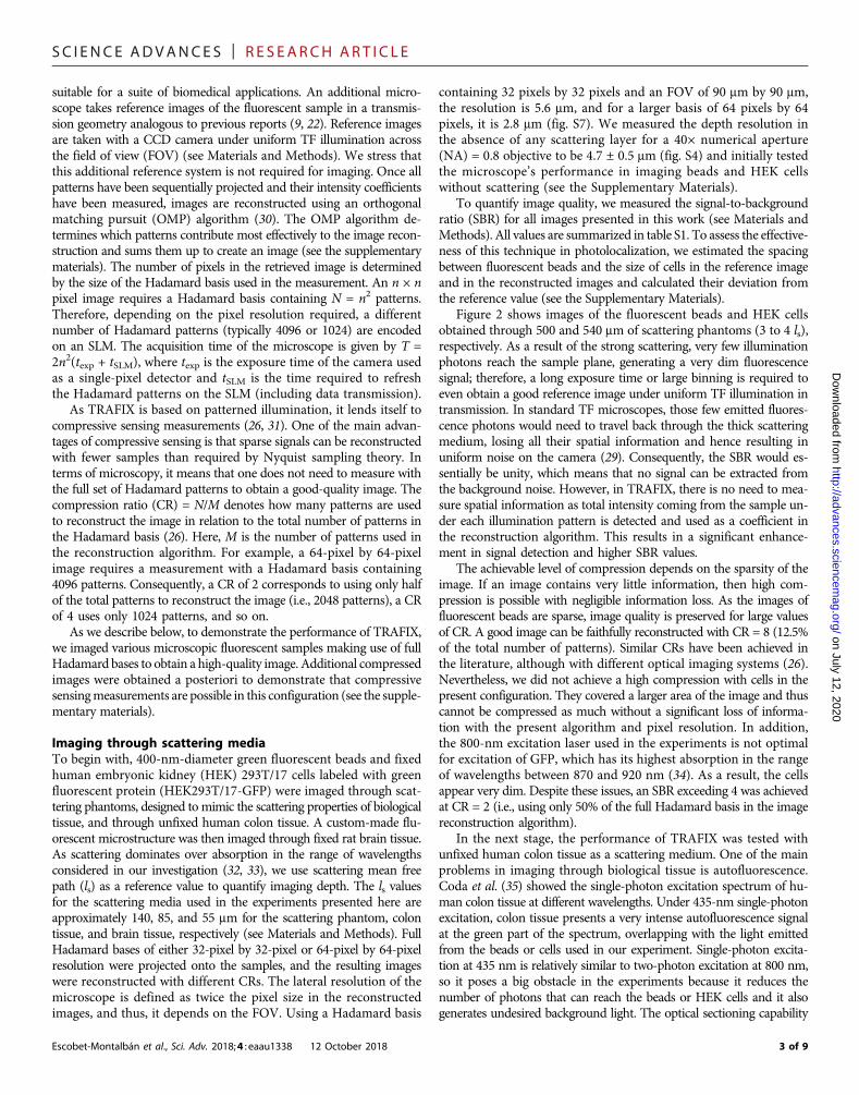
Figure 2 shows images of the fluorescent beads and HEK cellsobtained through 500 and 540 mm of scattering phantoms (3 to 4 ls),respectively. As a result of the strong scattering, very few illuminationphotons reach the sample plane, generating a very dim fluorescencesignal; therefore, a long exposure time or large binning is required toeven obtain a good reference image under uniform TF illumination intransmission. In standard TF microscopes, those few emitted fluores-cence photons would need to travel back through the thick scatteringmedium, losing all their spatial information and hence resulting inuniform noise on the camera (29). Consequently, the SBR would es-sentially be unity, which means that no signal can be extracted fromthe background noise. However, in TRAFIX, there is no need to mea-sure spatial information as total intensity coming from the sample un-der each illumination pattern is detected and used as a coefficient inthe reconstruction algorithm. This results in a significant enhance-ment in signal detection and higher SBR values.
The achievable level of compression depends on the sparsity of theimage. If an image contains very little information, then high com-pression is possible with negligible information loss. As the images offluorescent beads are sparse, image quality is preserved for large valuesof CR. A good image can be faithfully reconstructed with CR = 8 (12.5%of the total number of patterns). Similar CRs have been achieved inthe literature, although with different optical imaging systems (26).Nevertheless, we did not achieve a high compression with cells in thepresent configuration. They covered a larger area of the image and thuscannot be compressed as much without a significant loss of informa-tion with the present algorithm and pixel resolution. In addition,the 800-nm excitation laser used in the experiments is not optimalfor excitation of GFP, which has its highest absorption in the rangeof wavelengths between 870 and 920 nm (34). As a result, the cellsappear very dim. Despite these issues, an SBR exceeding 4 was achievedat CR = 2 (i.e., using only 50% of the full Hadamard basis in the imagereconstruction algorithm).
In the next stage, the performance of TRAFIX was tested withunfixed human colon tissue as a scattering medium. One of the mainproblems in imaging through biological tissue is autofluorescence.Coda et al. (35) showed the single-photon excitation spectrum of hu-man colon tissue at different wavelengths. Under 435-nm single-photonexcitation, colon tissue presents a very intense autofluorescence signalat the green part of the spectrum, overlapping with the light emittedfrom the beads or cells used in our experiment. Single-photon excita-tion at 435 nm is relatively similar to two-photon excitation at 800 nm,so it poses a big obstacle in the experiments because it reduces thenumber of photons that can reach the beads or HEK cells and it alsogenerates undesired background light. The optical sectioning capability
3 of 9
SC I ENCE ADVANCES | R E S EARCH ART I C L E
on July 12, 2020http://advances.sciencem
ag.org/D
ownloaded from
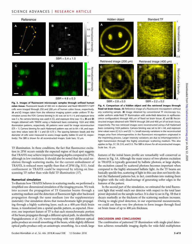
of TF would seem to circumvent this impediment; however, astemporally focused laser pulses propagate longer distances throughscattering tissue, the resulting excitation plane becomes thicker (fig. S4)(36), and consequently, some autofluorescence is excited in the colontissue, resulting in high noise levels even in the reference images takenin transmission. Despite the intense autofluorescence light emitted bythe colon tissue, we succeeded in imaging both 400-nm fluorescentbeads and HEK cells through 250 and 200 mm (~3 ls), respectively,obtaining high SBR values (Fig. 3). An additional image taken in ascattering phantom with fluorophores extending its entire volumeconfirms that TRAFIX can be used in presence of out-of-focus back-ground fluorescence (fig. S8).
The photolocalization analysis looked at spacing between adjacentbeads and cell size in a given image. It was satisfactory in the case ofimages through the scattering phantom obtaining deviations smallerthan 3% for beads in any CR (table S3). We also obtained acceptableresults in the case of cells for the image with no compression (table S2).In general, larger deviations were observed for the images through co-lon tissue presumably because of very low photon count reaching thedetector and distortions caused by inhomogeneities in the tissue.
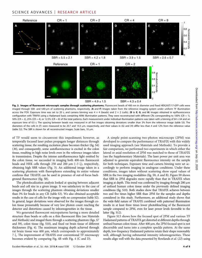
We generated fluorescent micropatterns having a more detailedstructure than beads or cells on a thin fluorescent film (see Materialsand Methods) and imaged them through a scattering phantom (figs. S7and S9), colon tissue (fig. S10), and fixed rat brain tissue of differentthicknesses (Fig. 4). The maximum imaging depth achieved throughrat brain tissue was 400 mm, which corresponds to approximately7 ls. The improvement of TRAFIX over conventional TF microscopybecomes evident by comparing Fig. 4B with Fig. 4 (C and D).
Escobet-Montalbán et al., Sci. Adv. 2018;4 : eaau1338 12 October 2018
A simple point-scanning two-photon microscope (2PM) wasdeveloped to compare the performance of TRAFIX with this widelyused imaging approach (see Materials and Methods). To provide afair comparison, we performed two experiments in which either thelateral or axial resolution of 2PM was matched to those of TRAFIX(see the Supplementary Materials). The laser power per unit area wasadjusted to generate equivalent fluorescence intensity on the samplefor both techniques. Exposure time and camera binning were set ac-cordingly to perform imaging in analogous conditions. Under theseconditions, images taken without scattering show equal values ofSBR in the two imaging modalities (fig. S9, A and B). Figure S9 showsthat SBR in 2PM degrades more rapidly than that in TRAFIX whenimaging at depth. This trend was confirmed by imaging through 200 mmof unfixed human colon tissue under the previously defined imagingconditions (fig. S10). Both studies show that TRAFIX achieves betweentwo and five times higher SBR than 2PM for the samples and depthsconsidered in this study. Furthermore, an additional study shows thatthe wide-field nature of TRAFIX combined with patterned illuminationresults in at least three times lower photobleaching of the fluorescentsample compared to 2PM, even for laser power levels favorable to thelatter (fig. S12).
Figure S13 shows how the focused spot of 2PM and various TFHadamardpatterns of TRAFIX get distorted at different depths throughunfixed human colon tissue. After 400 mm, the 2PM focused spot is notdiscernible and turns into a complete speckle pattern. At the samedepth, low-frequency Hadamard patterns retain their shape reasonablywell, although having substantial intensity inhomogeneities. Theseresults align well with the data presented by Rowlands et al. (22) using
A B EDCCS (CR = 1)
Reference CR = 1 CR = 2 CR = 8CR = 4
1
2
3
F G
Reference CR = 1 CR = 2
1
2
SBR = 5.5 ± 2.1 SBR = 4.2 ± 1.8 SBR = 3.9 ± 1.5 SBR = 2.6 ± 0.7
SBR = 4.9 ± 1.5 SBR = 4.3 ± 0.4
H
Fig. 2. Images of fluorescent microscopic samples through scattering phantoms. Fluorescent beads of 400 nm in diameter and fixed HEK293T/17-GFP cells wereimaged through 500- and 540-mm of scattering phantoms, respectively. (A and F) Images taken from the reference imaging system under uniform TF illuminationacross the FOV. Exposure time was set to 20 s, and camera binning was 4 × 4 (beads) and 2 × 2 (cells). (B to E, G, and H) Images obtained in epifluorescenceconfiguration with TRAFIX using a Hadamard basis containing 4096 illumination patterns. They were reconstructed with different CRs corresponding to 100% (CR = 1),50% (CR = 2), 25% (CR = 4), or 12.5% (CR = 8) of the total patterns. Each measurement under individual illumination patterns was taken with a binning of 64 × 64 and anexposure time of 0.5 s. The spacing between beads was measured in all five images obtaining deviations smaller than 3% from the reference image (table S3). Thediameters of the cells in (F) were measured to be 20.7 and 14.3 mm, respectively, and their values in (G) and (H) differ less than 4 and 12% from the reference value(table S2). The SBR is shown for all reconstructed images. Scale bars, 10 mm.
4 of 9
SC I ENCE ADVANCES | R E S EARCH ART I C L E
on July 12, 2020http://advances.sciencem
ag.org/D
ownloaded from
TF illumination. In these conditions, the fact that fluorescence excita-tion in 2PM occurs outside the expected region of focal spot suggeststhat TRAFIXmay achieve improved imaging depths compared to 2PM,although in low resolution. It should also be noted that the axial res-olution through scattering media, for the current embodiment ofTRAFIX, is reduced more rapidly than that of 2PM (fig. S11). Axialconfinement in TRAFIX could be improved by relying on line-scanning TF rather than wide-field TF illumination (37).
Numerical simulationTo illustrate how TRAFIX behaves in scatteringmedia, we performed asimplified one-dimensional simulation of the imaging process.We tookinto account the propagation of TF Gaussian beams through ascatteringmedium and the detection of fluorescent light after backwardpropagation through the same medium (see the supplementarymaterials). Our simulation shows that monochromatic light propagat-ing through a highly scattering layer, such as a 400-mm-thick braintissue, is transformed into a spatial speckle pattern, as expected. How-ever, one important feature of TF is that each monochromatic portionof the beampropagates through a different optical path. As identified byPapagiakoumou et al. (9), waves traveling with very different opticalpaths produce an overall smoothing of the beam, while slightly differentoptical paths produce only an anisotropic smoothing. As a result, large
Escobet-Montalbán et al., Sci. Adv. 2018;4 : eaau1338 12 October 2018
features of the initial beam profile are remarkably well conserved asshown in Fig. 5A. Although the main source of two-photon excitationin TRAFIX is typically generated by ballistic photons, at large depths,the excitation caused by scattered photons becomes important whencompared to the highly attenuated ballistic light. As the TF beams arebasically speckle free, scattering of light in this case does not heavily dis-tort the Hadamard patterns but, in fact, contributes into making thembrighter with the only disadvantage of generating softer edges in thefeatures of the pattern.
In the second part of the simulation, we estimated the total fluores-cent light that would reach our detector with respect to the total laserpower deposited on the sample surface. It is clear to see in Fig. 5B that itdecays markedly as the thickness of the scattering medium increases.Owing to single-pixel detection, in our experimental measurements,we could use those very few photons to form images through fixedrat brain tissue of up to 400 mm thickness.
DISCUSSION AND CONCLUSIONSThe combination of patterned TF illumination with single-pixel detec-tion achieves remarkable imaging depths for wide-field multiphoton
C D
1 2
3
4 5
A
Reference
1
2
3
SBR = 5.4 ± 1.0
SBR = 4.6 ± 0.3N
ormalized intensity
Norm
alized intensity Fig. 3. Images of fluorescent microscopic samples through unfixed humancolon tissue. Fluorescent beads of 400 nm in diameter and fixed HEK293T/17-GFPcells were imaged through 250 and 200 mm of human colon tissue, respectively.(A and C) Images taken from the reference imaging system under uniform TF illu-mination across the FOV. Camera binning in (A) was set to 4 × 4, and exposure timewas 5 s. No camera binning was used in (C), and exposure time was 15 s. (B and D)Images obtained with TRAFIX using a Hadamard basis containing 1024 and 4096illumination patterns, respectively. All patterns were used for image reconstruction(CR = 1). Camera binning for each Hadamard pattern was set to 64 × 64, and expo-sure time values were (B) 1 s and (D) 0.75 s. The spacing between beads and thediameter of cells were measured to assess image quality (tables S3 and S2, respec-tively). The SBR is shown for all reconstructed images. Scale bars, 10 mm.
A
Retrieved image (200
C
SBR = 2.8 ± 0.3
Standard TF
Retrieved image (400 m)
D
B
SBR = 2.2 ± 0.2
)m
Fig. 4. Comparison of a hidden object and the retrieved images throughfixed rat brain tissue. (A) Reference image of a fluorescent micropattern withoutany scattering sample. (B) Image obtained by conventional TF microscopy (i.e.,under uniform wide-field TF illumination with wide-field detection in epifluores-cence configuration) through 400 mm of fixed rat brain tissue. (C and D) Recon-structed images obtained with TRAFIX through 200 and 400 mm of rat brain tissue,respectively. The two retrieved images were reconstructed using a full Hadamardbasis containing 1024 patterns. Camera binning was set to 64 × 64, and exposuretime values were (C) 0.2 s and (D) 1 s. Small intensity variations in the reconstructedimages arise from inhomogeneities in the fluorescent micropattern originated inthe imprinting process. Larger intensity variations are due to inhomogeneities inlight transmission through the highly anisotropic scattering medium. This alsoapplies to figs. S7, S9, S10, and S12. The SBR is shown for all reconstructed images.Scale bar, 10 mm.
5 of 9

SC I ENCE ADVANCES | R E S EARCH ART I C L E
on July 12, 2020http://advances.sciencem
ag.org/D
ownloaded from
microscopy as it provides a way of exciting fluorescent structuresdeeper inside turbid media than existing imaging techniques and canefficiently collect the emitted light in an epifluorescence configuration.We have demonstrated the effectiveness and potential of TRAFIX byimaging fluorescent beads of 400 nm in diameter and fixed HEK293T/17-GFP cells through a layer of a scattering phantom with a thicknessof more than 500 mm (~4 ls), without any aberration correction, guidestar, or detector placed in/behind the turbid media. We then imaged abright custom fluorescent microstructure through rat brain tissue ofhundreds of micrometers in thickness, reaching a maximum imagingdepth of ~7 ls. In addition, we showed that TRAFIX works well undertypical biological research conditions by imaging both fluorescentbeads and HEK cells through depths of more than 3 ls of unfixedhuman colon tissue and even in the presence of intense backgroundfluorescence.
The main factor that limits imaging depth of TRAFIX is the pen-etration depth of the TF Hadamard patterns. Propagation of TF beamsthrough very large distances in scattering media results in distortionson the illumination patterns mainly caused by refractive index changesin the sample. These distortions generate a basis mismatch that mayresult in deformities in the reconstructed images (38). In addition, theuse of a one-dimensional diffraction grating generates horizontal shiftsin the illumination Hadamard patterns that could potentially be mini-mized by dispersing the patterns isotropically. A future embodimentusing dispersion in two dimensions with two perpendicular diffractiongratings would be an improvement. Despite these present issues,TRAFIX is capable of imaging at remarkable depths with low powerper unit area over a large FOV. Its performance may be further im-proved by combining it with wavefront correction, making it possibleto maintain spatial integrity of the illumination patterns even beyondthe current limits. Furthermore, the imaging depth could be signifi-cantly improved by relying on longer wavelengths and higher-ordermultiphoton processes such as three-photon excitation (see the Sup-plementary Materials) (22, 39, 40).
The results presented here align well with previous work publishedin the literature such as the study by Rowlands et al. (29). They com-pared the penetration depth of standard TF microscopy with 2PM. A
Escobet-Montalbán et al., Sci. Adv. 2018;4 : eaau1338 12 October 2018
careful look at their measured modulation transfer functions (MTFs)shows a remarkably higher contrast under TF illumination at depthsup to 100 to 150 mm with respect to 2PM. At larger depths, the MTFof the standard TF microscope drops markedly because of the im-possibility of retaining any spatial information in the detection system.In contrast, our novel TRAFIX approach uses single-pixel detection toefficiently collect fluorescent light, extending the high performance ofTF to deeper regions. Our comparison with 2PM shows that TRAFIXachieves an enhancement of up to five times in SBR when imagingthrough a scattering phantom and unfixed human colon tissue. More-over, photobleaching of the sample is substantially reduced as a resultof using wide-field TF patterned illumination rather than a focusedhigh-intensity beam (see the Supplementary Materials). Image resolu-tion and SBR can be further improved by using new approaches suchas digital microscanning (41).
Here, we also demonstrate that compressive sensing measurementsare possible in TRAFIX by showing reconstructed images a posteriori.However, in an actual compressive sensing measurement with no apriori knowledge of the sample, choosing the most appropriate illumi-nation patterns is critical. Since the amount of information carried byeach pattern is uneven, it is important to wisely choose the order in whichthey are projected to optimize image quality and acquisition speed (42).
In the present embodiment of TRAFIX, the acquisition speed ismainly limited by the exposure time of the EMCCD camera and theslow refresh rate of the SLM (see the Supplementary Materials). A typ-ical image obtained using a full basis of 1024 patterns (32 pixels by32 pixels) through a moderate thickness of a scattering sample currentlytakes ~5 min. This time is increased to up to 1 hour when imaging inhigh resolution (64 pixels by 64 pixels) with a full basis through themost challenging conditions shown in this article. To speed imagingup, the EMCCD camera would be replaced with a fast, sensitive pho-todetector such as a photomultiplier tube (PMT) and the imaging speedwould no longer be limited by the exposure time of the detector. Inaddition, as TRAFIX currently uses binary Hadamard patterns, theSLM may be replaced with a significantly faster digital micromirror de-vice, which may run at tens of kilohertz. These changes, combined withthe new advances in compressive imaging, suggests that frame rates for
100 200 300 400 500 600Tissue thickness (µm)
0
0.1
0.2
0.3
0.4
0.5
Tot
al fl
uore
scen
ce p
ower
(%
)
250 300 350 400 450 500 550Lateral beam profile (µm)
0
0.5
1
1.5
2E
xcita
tion
inte
nsity
(a.
u.)
×10–4
TF beamA B
Fig. 5. Numerical simulation of TRAFIX in scattering media. (A) Simulated TF laser beams at the focal plane through a 400-mm-thick brain tissue. The solid red curveindicates the smoothed-out lateral beam profile, taking all monochromatic components of the laser pulse into account. (B) Total fluorescence power collected with anNA = 0.8 microscope objective for different thicknesses of brain tissue. Incident laser power at sample surface is set to 100 arbitrary units (a.u.).
6 of 9

SC I ENCE ADVANCES | R E S EARCH ART I C L E
Dow
128-pixel by 128-pixel images can be increased to more than ~30 Hz(43, 44), enabling studies in time-varying turbulence (45).
Since our scheme can be easily implemented in a standard multi-photon microscope, we believe that one of its main applications willbe in the field of optogenetics where it would lend itself to achievelong-term simultaneous imaging and photoactivation of neuronalnetworks with minimal photodamage deep inside the brain. Last, asthe polarization state of the illumination light does not change overpropagation in scattering media through the range of distances nor-mally considered for imaging (see the Supplementary Materials) (46),TRAFIX could also be combined with polarization-resolved imagingtechniques (47).
In summary, TRAFIX is a novel approach for deep multiphotonimaging that presents an increased SBR compared to the ubiquitous2PM while also reducing photobleaching of the sample. In addition,the almost speckle-free propagation of TF illumination patterns sug-gests that TRAFIX may surpass the maximum imaging depth limit of2PM and may be very beneficial for long-term biological studies, par-ticularly in neuroscience.
on July 12, 2020http://advances.sciencem
ag.org/nloaded from
MATERIALS AND METHODSExperimental setupsTempoRAl Focusing microscopy with single-pIXel detectionAn illumination laser (Coherent Chameleon Ultra II) delivers 140-fspulses with an 80-MHz repetition rate, up to 4-W average outputpower at a variable central wavelength between 680 and 1080 nm.The central wavelength of the laser was set to 800 nm for all the ex-periments performed. The illumination beam was expanded fourtimes to cover the active area of a phase-only SLM (LCOS-SLM,Hamamatsu Photonics). The SLM was then imaged onto a blazedreflective grating (1200 g/mm) with a 4f (f = 400 mm) telescope tocreate wide-field TF illumination. The first diffraction order fromthe SLM was transmitted through an iris in the telescope, while allother orders were blocked. The beam was diffracted from the grat-ing, and all wavelengths were collimated with an f = 400 mm lens re-layed onto the back focal aperture of the illumination objective. Twodifferent illumination objectives were used in this work. A water dip-ping objective (40× NA = 0.8; Nikon), which is enclosed in a custom-made chamber filled with water, generates a TF illumination plane witha size of 90 mm by 90 mm. The highest average laser power per unitarea used in this configuration is 64 ± 5 mW/mm2. An air objective (20×NA = 0.75; Nikon) was used for additional studies presented in theSupplementary Materials, as accordingly specified. Backscattered fluo-rescent light propagated through the turbid media and was collected byan EMCCD camera run without amplification (iXonEM+ 885, AndorTechnology) via the same illumination objective in epifluorescenceconfiguration. To provide reference images for this paper, forwardlyemitted photons from the sample were collected by a CCD camera(Clara, Andor Technology) in transmission via a long working distanceair objective (100× NA = 0.7; Mitutoyo). Appropriate short-pass filterswere used to reject the illumination laser at 800 nm and transmit flu-orescence below 700 nm. In contrast to other single-pixel imagingapproaches (25, 26), the EMCCD camera with 64 × 64 binning wasused as a bucket detector instead of using a single-element detectorsuch as a PMT or an avalanche photodiode. Using high binning helpsreducing the effect of readout noise. All objectives, samples, and cam-eras were attached on the body of an inverted microscope (Eclipse Ti,Nikon) accordingly.
Escobet-Montalbán et al., Sci. Adv. 2018;4 : eaau1338 12 October 2018
Point-scanning two-photon microscopeThe 2PM shares the same setup as TRAFIX, except for the diffractiongrating and a lens that are replaced with a mirror to obtain a focusedbeam on the focal plane of the illumination objective. A Nikon 20×NA = 0.75 air objective was used for all experiments. A variable iriswas used to adjust the size of the focused spot. An X-Y-Z motorizedstage (Nano-LP200, Mad City Labs) scanned the sample in a stepwisemotion across the fixed focused beam covering the entire FOV. Thesame binned EMCCD camera run with no amplification (iXonEM+885, Andor Technology) collected the fluorescent light emitted bythe sample.
Fluorescent and scattering samplesFluorescent layer and fluorescent micropatternA 200-nm-thin layer of super-yellow polymer spin-coated on aglass coverslip was used to characterize the profile and depth resolutionof the TF beam (see the Supplementary Materials). It was also used togenerate a fluorescent micropattern that was then imaged throughscattering media. The negative of a pattern of interest was encodedon the SLM, and the thin fluorescent layer was placed at the focal planeof the microscope without any scattering layer. The laser power was setto the maximum, and the negative pattern was photobleached on thefluorescent film. Therefore, the only portion of the FOV that remainedfluorescent was exactly the desired micropattern.Fluorescent beadsGreen fluorescent polymer microspheres (G400, Duke Scientific)with a diameter of 0.39 mm were used to test the performance of theimaging system. A very small amount of beads was deposited on a glasscoverslip and placed on top of the scattering samples to image themthrough the turbid media.HEK cellsHEK293T/17-GFP cells were used to demonstrate the capability of themicroscope in imaging real biological samples through scattering.HEK293T/17 cell line obtained fromAmericanTypeCultureCollectionwas cultured inDulbecco’smodified Eagle’smediumGlutaMAX-I sup-plementedwith 10% fetal bovine serum and 1%penicillin/streptomycinand was transfected using TransIT-LT1 transfection reagent with theVesicular Stomatitis Virus glycoprotein (VSV-G) pseudotyped lenti-virus vector and the packaging plasmid psPAX2 to deliver the plasmidpLenti-GFP-Puro. Regarding the sample preparation, the HEK293T/17-GFP cells were replated onWorld Precision Instruments FluoroDishpoly-D-Lysine–coated cell culture dishes at a low density to achieveideal imaging conditions. The day after plating, before imaging, theHEK293T/17-GFP cells were fixed in a phosphate-buffered saline 4%paraformaldehyde solution. The scattering samples were attached di-rectly on the bottom of the dishes containing the cells.Scattering phantomPolystyrene beads of 1 mm in diameter were used as scatterers tosimulate a turbid sample. The beads were purchased in a 1% concen-tration solution in water (Microbead NIST Traceable Particle SizeStandard, 1.00 mm; Polysciences). The solution was thoroughly stirredin a vortex mixer and then mixed with a 1% solution of agarose inwater (preheated above melting point). Agarose and beads were mixedin the vortex mixer again and placed into sample wells of variable height.The wells consisted of a 100-mm glass slide with multiple 90-mm vinylspacers stacked on top of each other. An additional coverslip was placedon top to seal the well. The concentration of polystyrene beads in thesample was chosen to roughly match the scattering coefficient of realbiological tissue (48). Using an on-line Mie scattering calculator (49),
7 of 9
SC I ENCE ADVANCES | R E S EARCH ART I C L E
on July 12, 2020http://advances.sciencem
ag.org/D
ownloaded from
we determined the reduced scattering coefficient of our phantom toapproximately be m′s≈ 7.5 cm−1, corresponding to a mean free pathof about ls ¼ ð1� gÞ=m′s≈ 140 mm, where g is the anisotropy factor(g ~ 0.9 for most biological tissue at the wavelength considered inthis investigation). Different scattering phantoms were used in someexperiments in the Supplementary Materials as appropriately specified.Rat brain tissueSimilar to our previous work (50), rat brain tissue was obtained fromadult Sprague Dawley rats, in accordance with the UK Animals (Sci-entific Procedures) Act 1986. It was fixed and then sectioned into slicesat thicknesses of 200 and 400 mm. The mean free path of the rat braintissue was estimated by measuring the ratio of the incident laser in-tensity I0 with the intensity of the ballistic photons IB and by applyingan exponential law IB = I0 exp(− L/ls), where L is the thickness of thebrain tissue (18). The obtained value of ls = 55 ± 9 mm is consistentwith other measurements reported in the literature for rat (51) andmouse brains (52).Colon tissueThin fragments of unfixed normal human colon tissue were used asscattering samples. They were stored in a freezer at −80°C and mountedbetween two coverslips. Their thickness ranged from 200 to 250 mm,and their reduced scattering coefficient was approximately 12 cm−1
(53, 54), giving a mean free path of ls ≈ 85 mm. The colon tissue sam-ple used in this study was obtained from the Tayside Tissue Bank,Ninewells Hospital and Medical School, Dundee (tissue request no.TR000289) with appropriate ethical permission.
Image quality quantificationSignal-to-background ratioSBR is defined here as SBR = msig/mbg, where msig and mbg are the averagevalues of the signal and the background, respectively. In the case of cellsand fluorescentmicropatterns, two small regionsof interestweredefined—one containing fluorescent structures and the other corresponding tothe background. In the images of beads, the highest-intensity pixels inthe beads were used as signal value, and the maximum intensity pixelsin the rest of the image were used as background noise. Uncertainty isgiven by the SD of all averaged measurements. All values of SBRpresented in this work are summarized in table S1.Cell sizeAs the cells used here appear approximately spherical, their diameterwas estimated by taking a measurement of their size in two orthogonaldirections and averaging the obtained values. Their diameter was deter-mined by fitting a Gaussian function to the intensity values and measur-ing its full width at half maximum. Table S2 shows the diameters of thecells that appear in the different images presented in this work. Devia-tions in cell size between reconstructed and reference images are ex-pressed as a percentage error.Bead spacingThe central pixel for each individual bead was located, and the dis-tance between beads was measured in the reference image and inall retrieved images with different CRs (table S3). Deviations in beadspacing between reconstructed and reference images are expressed as apercentage error.
SUPPLEMENTARY MATERIALSSupplementary material for this article is available at http://advances.sciencemag.org/cgi/content/full/4/10/eaau1338/DC1Note S1. Numerical simulationsNote S2. Single-pixel detection and compressive sensing
Escobet-Montalbán et al., Sci. Adv. 2018;4 : eaau1338 12 October 2018
Note S3. Microscope characterizationNote S4. Comparison between TRAFIX and point-scanning two-photon microscopy (2PM)Note S5. Polarization state evaluationFig. S1. Numerically simulated TF laser beam propagating through 400 mm of brain tissue.Fig. S2. Properties of a numerically simulated TF laser beam through brain tissue.Fig. S3. Effect of scattering on the beam profile with and without TF.Fig. S4. Depth profile of a TF beam through a scattering phantom.Fig. S5. Characterization of a TF beam through a scattering phantom.Fig. S6. Images of fluorescent microscopic samples without scattering.Fig. S7. Comparison of a hidden object and retrieved images through a scattering phantomwith different resolution.Fig. S8. Image of 4.8-mm fluorescent beads in a volumetric scattering phantom.Fig. S9. Comparison of SBR of TRAFIX and point-scanning two-photon microscopy (2PM) at depth.Fig. S10. Comparison of TRAFIX and point-scanning two-photon microscopy (2PM) throughhuman colon tissue.Fig. S11. Axial confinement in TRAFIX and point-scanning two-photon microscopy (2PM).Fig. S12. Photobleaching comparison of TRAFIX and point-scanning two-photon microscopy(2PM).Fig. S13. Effect of scattering on illumination beams in point-scanning two-photon microscopy(2PM) and TRAFIX.Fig. S14. Effect of turbid media on light polarization.Table S1. SBR measured for all the images shown in this work.Table S2. Cell diameters of all images shown in this work.Table S3. Beads spacing corresponding to all images shown in this work.References (55–61)
REFERENCES AND NOTES1. E. Betzig, G. H. Patterson, R. Sougrat, O. W. Lindwasser, S. Olenych, J. S. Bonifacino,
M. W. Davidson, J. Lippincott-Schwartz, H. F. Hess, Imaging intracellular fluorescentproteins at nanometer resolution. Science 313, 1642–1645 (2006).
2. S. W. Hell, J. Wichmann, Breaking the diffraction resolution limit by stimulated emission:Stimulated-emission-depletion fluorescence microscopy. Opt. Lett. 19, 780–782 (1994).
3. B.-C. Chen, W. R. Legant, K. Wang, L. Shao, D. E. Milkie, M. W. Davidson, C. Janetopoulos,X. S. Wu, J. A. Hammer III, Z. Liu, B. P. English, Y. Mimori-Kiyosue, D. P. Romero, A. T. Ritter,J. Lippincott-Schwartz, L. Fritz-Laylin, R. D. Mullins, D. M. Mitchell, J. N. Bembenek,A.-C. Reymann, R. Böhme, S. W. Grill, J. T. Wang, G. Seydoux, U. S. Tulu, D. P. Kiehart,E. Betzig, Lattice light-sheet microscopy: Imaging molecules to embryos at highspatiotemporal resolution. Science 346, 1257998 (2014).
4. J. M. Pullman, J. Nylk, E. C. Campbell, F. J. Gunn-Moore, M. B. Prystowsky, K. Dholakia,Visualization of podocyte substructure with structured illumination microscopy (SIM):A new approach to nephrotic disease. Biomed. Opt. Express 7, 302–311 (2016).
5. D. Westmoreland, M. Shaw, W. Grimes, D. J. Metcalf, J. J. Burden, K. Gomez, A. E. Knight,D. F. Cutler, Super-resolution microscopy as a potential approach to diagnosis of plateletgranule disorders. J. Thromb. Haemost. 14, 839–849 (2016).
6. D. Huang, E. A. Swanson, C. P. Lin, J. S. Schuman, W. G. Stinson, W. Chang, M. R. Hee,T. Flotte, K. Gregory, C. A. Puliafito, J. G. Fujimoto, Optical coherence tomography.Science 254, 1178–1181 (1991).
7. R. M. Power, J. Huisken, A guide to light-sheet fluorescence microscopy for multiscaleimaging. Nat. Methods 14, 360–373 (2017).
8. J. Bertolotti, E. G. van Putten, C. Blum, A. Lagendijk, W. L. Vos, A. P. Mosk, Non-invasiveimaging through opaque scattering layers. Nature 491, 232–234 (2012).
9. E. Papagiakoumou, A. Bègue, B. Leshem, O. Schwartz, B. M. Stell, J. Bradley, D. Oron,V. Emiliani, Functional patterned multiphoton excitation deep inside scattering tissue.Nat. Photonics 7, 274–278 (2013).
10. O. Katz, P. Heidmann, M. Fink, S. Gigan, Non-invasive single-shot imaging throughscattering layers and around corners via speckle correlations. Nat. Photonics 8, 784–790(2014).
11. S. Kang, S. Jeong, W. Choi, H. Ko, T. D. Yang, J. H. Joo, J.-S. Lee, Y.-S. Lim, Q.-H. Park,W. Choi, Imaging deep within a scattering medium using collective accumulation ofsingle-scattered waves. Nat. Photonics 9, 253–258 (2015).
12. M. J. Booth, Adaptive optical microscopy: The ongoing quest for a perfect image.Light Sci. Appl. 3, e165 (2014).
13. D. Débarre, E. J. Botcherby, T. Watanabe, S. Srinivas, M. J. Booth, T. Wilson, Image-basedadaptive optics for two-photon microscopy. Opt. Lett. 34, 2495–2497 (2009).
14. M. Rueckel, J. A. Mack-Bucher, W. Denk, Adaptive wavefront correction in two-photonmicroscopy using coherence-gated wavefront sensing. Proc. Natl. Acad. Sci. U.S.A. 103,17137–17142 (2006).
15. T. Čižmár, M. Mazilu, K. Dholakia, In situ wavefront correction and its application tomicromanipulation. Nat. Photonics 4, 388–394 (2010).
16. A. P. Mosk, A. Lagendijk, G. Lerosey, M. Fink, Controlling waves in space and time forimaging and focusing in complex media. Nat. Photonics 6, 283–292 (2012).
8 of 9

SC I ENCE ADVANCES | R E S EARCH ART I C L E
on July 12, 2020http://advances.sciencem
ag.org/D
ownloaded from
17. S. M. Popoff, G. Lerosey, R. Carminati, M. Fink, A. C. Boccara, S. Gigan, Measuring thetransmission matrix in optics: An approach to the study and control of light propagationin disordered media. Phys. Rev. Lett. 104, 100601 (2010).
18. A. Badon, D. Li, G. Lerosey, A. C. Boccara, M. Fink, A. Aubry, Smart optical coherencetomography for ultra-deep imaging through highly scattering media. Sci. Adv. 2,e1600370 (2016).
19. D. Oron, E. Tal, Y. Silberberg, Scanningless depth-resolved microscopy. Opt. Express 13,1468–1476 (2005).
20. G. Zhu, J. van Howe, M. Durst, W. Zipfel, C. Xu, Simultaneous spatial and temporalfocusing of femtosecond pulses. Opt. Express 13, 2153–2159 (2005).
21. T. Schrödel, R. Prevedel, K. Aumayr, M. Zimmer, A. Vaziri, Brain-wide 3D imaging ofneuronal activity in Caenorhabditis elegans with sculpted light. Nat. Methods 10,1013–1020 (2013).
22. C. J. Rowlands, D. Park, O. T. Bruns, K. D. Piatkevich, D. Fukumura, R. K. Jain, M. G. Bawendi,E. S. Boyden, P. T. So, Wide-field three-photon excitation in biological samples.Light Sci. Appl. 6, e16255 (2016).
23. N. C. Pégard, A. R. Mardinly, I. A. Oldenburg, S. Sridharan, L. Waller, H. Adesnik,Three-dimensional scanless holographic optogenetics with temporal focusing (3D-SHOT).Nat. Commun. 8, 1228 (2017).
24. M. J. Padgett, R. W. Boyd, An introduction to ghost imaging: Quantum and classical.Philos. Trans. A Math. Phys. Eng. Sci. 375, 20160233 (2017).
25. E. Tajahuerce, V. Durán, P. Clemente, E. Irles, F. Soldevila, P. Andrés, J. Lancis, Imagetransmission through dynamic scattering media by single-pixel photodetection.Opt. Express 22, 16945–16955 (2014).
26. V. Durán, F. Soldevila, E. Irles, P. Clemente, E. Tajahuerce, P. Andrés, J. Lancis, Compressiveimaging in scattering media. Opt. Express 23, 14424–14433 (2015).
27. O. Katz, E. Small, Y. Bromberg, Y. Silberberg, Focusing and compression of ultrashortpulses through scattering media. Nat. Photonics 5, 372–377 (2011).
28. B. Sun, P. S. Salter, M. J. Booth, Effects of sample dispersion on ultrafast laser focusing.J. Opt. Soc. Am. B 32, 1272–1280 (2015).
29. C. J. Rowlands, O. T. Bruns, M. G. Bawendi, P. T. C. So, Objective, comparative assessmentof the penetration depth of temporal-focusing microscopy for imaging various organs.J. Biomed. Opt. 20, 061107 (2015).
30. J. A. Tropp, A. C. Gilbert, Signal recovery from random measurements via orthogonalmatching pursuit. IEEE Trans. Inf. Theory 53, 4655–4666 (2007).
31. E. J. Candès, M. B. Wakin, An introduction to compressive sampling. IEEE Signal Process.Mag. 25, 21–30 (2008).
32. W. F. Cheong, S. A. Prahl, A. J. Welch, A review of the optical properties of biologicaltissues. IEEE J. Quantum Electron. 26, 2166–2185 (1990).
33. A. N. Yaroslavsky, P. C. Schulze, I. V. Yaroslavsky, R. Schober, F. Ulrich, H.-J. Schwarzmaier,Optical properties of selected native and coagulated human brain tissues in vitro in thevisible and near infrared spectral range. Phys. Med. Biol. 47, 2059–2073 (2002).
34. M. Drobizhev, N. S. Makarov, S. E. Tillo, T. E. Hughes, A. Rebane, Two-photon absorptionproperties of fluorescent proteins. Nat. Methods 8, 393–399 (2011).
35. S. Coda, A. J. Thompson, G. T. Kennedy, K. L. Roche, L. Ayaru, D. S. Bansi, G. W. Stamp,A. V. Thillainayagam, P. M. W. French, C. Dunsby, Fluorescence lifetime spectroscopyof tissue autofluorescence in normal and diseased colon measured ex vivo using afiber-optic probe. Biomed. Opt. Express 5, 515–538 (2014).
36. H. Dana, S. Shoham, Numerical evaluation of temporal focusing characteristics intransparent and scattering media. Opt. Express 19, 4937–4948 (2011).
37. M. E. Durst, G. Zhu, C. Xu, Simultaneous spatial and temporal focusing in nonlinearmicroscopy. Opt. Commun. 281, 1796–1805 (2008).
38. Y. Chi, L. L. Scharf, A. Pezeshki, A. R. Calderbank, Sensitivity to basis mismatch incompressed sensing. IEEE Trans. Signal Processing 59, 2182–2195 (2011).
39. N. G. Horton, K. Wang, D. Kobat, C. G. Clark, F. W. Wise, C. B. Schaffer, C. Xu, In vivothree-photon microscopy of subcortical structures within an intact mouse brain.Nat. Photonics 7, 205–209 (2013).
40. K. Toda, K. Isobe, K. Namiki, H. Kawano, A. Miyawaki, K. Midorikawa, Temporal focusingmicroscopy using three-photon excitation fluorescence with a 92-fs Yb-fiber chirpedpulse amplifier. Biomed. Opt. Express 8, 2796–2806 (2017).
41. M.-J. Sun, M. P. Edgar, D. B. Phillips, G. M. Gibson, M. J. Padgett, Improving the signal-to-noise ratio of single-pixel imaging using digital microscanning. Opt. Express 24,10476–10485 (2016).
42. M.-J. Sun, L.-T. Meng, M. P. Edgar, M. J. Padgett, N. Radwell, A Russian dolls ordering of theHadamard basis for compressive single-pixel imaging. Sci. Rep. 7, 3464 (2017).
43. C. F. Higham, R. Murray-Smith, M. J. Padgett, M. P. Edgar, Deep learning for real-timesingle-pixel video. Sci. Rep. 8, 2369 (2018).
44. Z.-H. Xu, W. Chen, J. Penuelas, M. Padgett, M.-J. Sun, 1000 fps computational ghostimaging using LED-based structured illumination. Opt. Express 26, 2427–2434 (2018).
45. M. P. Edgar, G. M. Gibson, R. W. Bowman, B. Sun, N. Radwell, K. J. Mitchell, S. S. Welsh,M. J. Padgett, Simultaneous real-time visible and infrared video with single-pixeldetectors. Sci. Rep. 5, 10669 (2015).
Escobet-Montalbán et al., Sci. Adv. 2018;4 : eaau1338 12 October 2018
46. H. B. de Aguiar, S. Gigan, S. Brasselet, Polarization recovery through scattering media.Sci. Adv. 3, e1600743 (2017).
47. X. Chen, O. Nadiarynkh, S. Plotnikov, P. J. Campagnola, Second harmonic generationmicroscopy for quantitative analysis of collagen fibrillar structure. Nat. Protoc. 7, 654–669(2012).
48. S. L. Jacques, Optical properties of biological tissues: A review. Phys. Med. Biol. 58,R37–R61 (2013).
49. S. Prahl, Mie scattering calculator (2007); http://omlc.org/calc/mie_calc.html.50. M. Chen, J. Mas, L. H. Forbes, M. R. Andrews, K. Dholakia, Depth-resolved multimodal
imaging: Wavelength modulated spatially offset Raman spectroscopy with opticalcoherence tomography. J. Biophotonics 11, e201700129 (2018).
51. M. Oheim, E. Beaurepaire, E. Chaigneau, J. Mertz, S. Charpak, Two-photon microscopy inbrain tissue: Parameters influencing the imaging depth. J. Neurosci. Methods 111, 29–37(2001).
52. E. Chaigneau, A. J. Wright, S. P. Poland, J. M. Girkin, R. A. Silver, Impact of wavefrontdistortion and scattering on 2-photon microscopy in mammalian brain tissue. Opt.Express 19, 22755–22774 (2011).
53. H. J. Wei, D. Xing, G. Y. Wu, H. M. Gu, J. J. Lu, Y. Jin, X.-Y. Li, Differences in opticalproperties between healthy and pathological human colon tissues using a Ti:sapphirelaser: An in vitro study using the Monte Carlo inversion technique. J. Biomed. Opt. 10,44022 (2005).
54. A. N. Bashkatov, E. A. Genina, V. I. Kochubey, V. S. Rubtsov, E. A. Kolesnikova, V. V. Tuchin,Optical properties of human colon tissues in the 350–2500 nm spectral range.Quant. Electron. 44, 779–784 (2014).
55. M. Chen, C. Dainty, F. S. Roux, Speckle evolution with multiple steps of least-squaresphase removal. Phys. Rev. A 84, 023846 (2011).
56. M. Chen, K. Dholakia, M. Mazilu, Is there an optimal basis to maximise optical informationtransfer? Sci. Rep. 6, 22821 (2016).
57. S. T. Flock, B. C. Wilson, M. S. Patterson, Total attenuation coefficients and scatteringphase functions of tissues and phantom materials at 633 nm. Med. Phys. 14, 835–841(1987).
58. S. J. Olivas, Y. Rachlin, L. Gu, B. Gardiner, R. Dawson, J.-P. Laine, J. E. Ford, Characterizationof a compressive imaging system using laboratory and natural light scenes. Appl. Opt. 52,4515–4526 (2013).
59. J. M. Phillips, Compressed sensing and orthogonal matching pursuit, in CS 6140 DataMining (University of Utah, 2017).
60. E. Papagiakoumou, V. de Sars, D. Oron, V. Emiliani, Patterned two-photon illumination byspatiotemporal shaping of ultrashort pulses. Opt. Express 16, 22039–22047 (2008).
61. C. Boudreau, T.-L. Wee, Y.-R. Duh, M. P. Couto, K. H. Ardakani, C. M. Brown, Excitation lightdose engineering to reduce photo-bleaching and photo-toxicity. Sci. Rep. 6, 30892 (2016).
Acknowledgments:We thank J. Glackin and P. Manousiadis from University of St. Andrews forpreparing the uniform thin fluorescent layers of super-yellow polymer and J. Nylk, Z. Yang,G. Bruce, and F. Gasparoli for useful discussions and support. Funding: This work wassupported by the U.K. Engineering and Physical Sciences Research Council for fundingthrough grants EP/P030017/1 and EP/M000869/1 and has received funding from the EuropeanUnion’s Horizon 2020 Programme through the project Advanced BiomEdical OPTICALImaging and Data Analysis (BE-OPTICAL) under grant agreement no. 675512, The CunninghamTrust, and The RS MacDonald Charitable Trust. K.D. acknowledges the financial support ofE. Killick and S. Gurney. Author contributions: A.E.-M. and R.S. developed the experimentalsetup with guidance from K.D. M.M. and R.S. developed the initial code for single-pixelmicroscopy, which A.E.-M. adapted to the presented experiments. A.E.-M. performed theexperiments, recorded the presented data, and performed the analysis with input andguidance from K.D. M.C. developed the numerical model with input from K.D. and A.E.-M. W.A.S.,M.A., and C.S.H. provided the cell and tissue samples. A.E.-M., M.C., and K.D. wrote the paperwith input from the other authors. K.D. conceived and supervised the study. Competinginterests: K.D. is an inventor on a patent related to this work filed by the University ofSt. Andrews (U.K. patent application no. 1715385.9, filed 22 September 2017). All other authorsdeclare that they have no competing interests. Data and materials availability: All dataneeded to evaluate the conclusions in the paper are present in the paper and/or theSupplementary Materials. The data that support the findings of this study are available uponrequest from the corresponding author K.D. The data are not publicly available because ofthe intellectual property filed with regard to the work presented.
Submitted 10 May 2018Accepted 7 September 2018Published 12 October 201810.1126/sciadv.aau1338
Citation: A. Escobet-Montalbán, R. Spesyvtsev, M. Chen,W. A. Saber, M. Andrews, C. Simon Herrington,M.Mazilu, K. Dholakia,Wide-fieldmultiphoton imaging through scatteringmediawithout correction. Sci.Adv. 4, eaau1338 (2018).
9 of 9

Wide-field multiphoton imaging through scattering media without correction
Herrington, Michael Mazilu and Kishan DholakiaAdrià Escobet-Montalbán, Roman Spesyvtsev, Mingzhou Chen, Wardiya Afshar Saber, Melissa Andrews, C. Simon
DOI: 10.1126/sciadv.aau1338 (10), eaau1338.4Sci Adv
ARTICLE TOOLS http://advances.sciencemag.org/content/4/10/eaau1338
MATERIALSSUPPLEMENTARY http://advances.sciencemag.org/content/suppl/2018/10/05/4.10.eaau1338.DC1
REFERENCES
http://advances.sciencemag.org/content/4/10/eaau1338#BIBLThis article cites 59 articles, 6 of which you can access for free
PERMISSIONS http://www.sciencemag.org/help/reprints-and-permissions
Terms of ServiceUse of this article is subject to the
is a registered trademark of AAAS.Science AdvancesYork Avenue NW, Washington, DC 20005. The title (ISSN 2375-2548) is published by the American Association for the Advancement of Science, 1200 NewScience Advances
BY).Science. No claim to original U.S. Government Works. Distributed under a Creative Commons Attribution License 4.0 (CC Copyright © 2018 The Authors, some rights reserved; exclusive licensee American Association for the Advancement of
on July 12, 2020http://advances.sciencem
ag.org/D
ownloaded from