why minimally invasive skin sampling techniques? a bright scientific future
TRANSCRIPT
Introduction
Minimally invasive skin sampling techniques expose the subject to less discomfort and potential risk of complications. More invasive methods such as suction blistering require more time for the blistering process and may alter biomarker structure during that proce-dure. Shave biopsies and full-thickness punch biopsies require a physician to perform the technique, may remove larger portions of dermal tissue, and can expose the subject to higher levels of discomfort, trauma, and postbiopsy infection or scarring (1). Original minimally invasive technique, such as Pachtman’s techniques (described below), were used to collect skin bacterial
flora for colony counts. However, today, such collec-tion techniques can also obtain cells containing genetic material and biochemical markers of internal bodily functions and reactions. For example, the collected biomarkers can be used for patient monitoring, such as glucose and amino acid levels, therapeutic drug lev-els, or biochemical responses to exogenous toxins or other exposures. For instance, exposure to a particular toxin or irritant can lead to the body’s production of a protein in response to the exogenous stressor. Stratum corneum harvested through noninvasive means can yield messenger ribonucleic acid (mRNA) containing sequence coding for that particular protein, present in exposed vs. nonexposed populations.
Cutaneous and Ocular Toxicology, 2011; 30(1): 1–6
C R I T I C A L R E V I E W
Why minimally invasive skin sampling techniques? A bright scientific future
Christina Y. Wang1, and Howard I. Maibach2
1Department of Occupational and Environmental Medicine, Kaiser Permanente, San Francisco, California, USA, and 2Department of Dermatology, University of California, San Francisco, San Francisco, California, USA
AbstractBackground: There is increasing interest in minimally invasive skin sampling techniques to assay markers of molecular biology and biochemical processes. Objective: This overview examines methodology strengths and limitations, and exciting developments pending in the scientific community.
Materials and methods: Publications were searched via PubMed, the U.S. Patent and Trademark Office Website, the DermTech Website and the CuDerm Website. The keywords used were noninvasive skin sampling, skin stripping, skin taping, detergent method, ring method, mechanical scrub, reverse iontophoresis, glucose monitor-ing, buccal smear, hair root sampling, mRNA, DNA, RNA, and amino acid.
Results and conclusions: There is strong interest in finding methods to access internal biochemical, molecu-lar, and genetic processes through noninvasive and minimally invasive external means. Minimally invasive techniques include the widely used skin tape stripping, the abrasion method that includes scraping and detergent, and reverse iontophoresis. The first 2 methods harvest largely the stratum corneum. Hair root sampling (material deeper than the epidermis), buccal smear, shave biopsy, punch biopsy, and suction blis-tering are also methods used to obtain cellular material for analysis, but involve some degree of increased invasiveness and thus are only briefly mentioned. Existing and new sampling methods are being refined and validated, offering exciting, different noninvasive means of quickly and efficiently obtaining molecular material with which to monitor bodily functions and responses, assess drug levels, and follow disease proc-esses without subjecting patients to unnecessary discomfort and risk.
Keywords: Noninvasive; minimally invasive skin sampling; skin stripping; detergent method; ring method; mechanical scrub; reverse iontophoresis; glucose monitoring; DNA; RNA; amino acid
Address for Correspondence: Howard I. Maibach, M.D., University of California, San Francisco, Department of Dermatology, 90 Medical Center Way, Surge 110, Box 0989, University of California, San Francisco, CA 94143-0989, USA. Tel.: (415) 476-2468. Fax: (415) 753-5304. E-mail: [email protected]
(Received 15 July 2010; revised 11 August 2010; accepted 13 August 2010)
ISSN 1556-9527 print/ISSN 1556-9535 online © 2011 Informa Healthcare USA, Inc.DOI: 10.3109/15569527.2010.517230 http://www.informahealthcare.com/cot
Cutaneous and Ocular Toxicology
1
2011
1
30
6
1556-95271556-9535© 2011 Informa Healthcare USA, Inc.10.3109/15569527.2010.517230
15 July 201013 August 201011 August 2010
COT
517230
LCOT
Cut
aneo
us a
nd O
cula
r T
oxic
olog
y D
ownl
oade
d fr
om in
form
ahea
lthca
re.c
om b
y N
orth
Car
olin
a St
ate
Uni
vers
ity o
n 05
/10/
13Fo
r pe
rson
al u
se o
nly.

2 C. Y. Wang and H. I. Maibach
We briefly touch on buccal smears and hair root sampling. These methods (as opposed to semen sampling) have been used to obtain genetic material for chromosome analyses in a patient with mosaic Klinefelter’s syndrome (2). Hair root sampling has yielded proteins and mRNA coding for proteins of interest (3,4) that have helped investigators facilitate genetic testing for X-linked Alport syndrome by direct sequencing of hair root COL4A5 mRNA (5). Hair root genetic material can also be used to evaluate expres-sion of specific genes in normal skin and inflamma-tory and neoplastic skin lesions (6). Hair root sampling can access subepidermal cells and is somewhat more invasive and can be more uncomfortable than the noninvasive methods we are concentrating on, which largely gather the stratum corneum. Interestingly, elec-trothermal atomic absorption spectrophotometry of scalp hair samples has been used to investigate asso-ciations between toxic elements, such as arsenic and lead, and myocardial infarctions (7). Of note, infrared spectroscopy is also used to analyze biological mate-rial in a minimally invasive manner. Facial stratum corneum components have been assessed over time by Fourier-transformed infrared spectroscopy with attenuated total reflection (FTIR-ATR), in order to fol-low molecular dynamic changes over time in relation to skin diseases such as atopic dermatitis (8).
Techniques reviewed
Abrasion techniques
Pachtman’s glass cylinder technique uses a sterile wood scraper to scrape the skin 20 times within the ringed area of a 23-mm diameter glass cylinder pressed firmly against the skin site, with 2 mL of brain–heart infusion broth placed within it. The broth is then aspi-rated 3 times in a 5-mL pipette, and 1 mL is withdrawn and diluted in 9 mL of water, to give a 1:10 concentra-tion. Further 10-fold dilutions are prepared from the original tube, and 1 mL of each dilution is plated in brain–heart infusion agar in sets of 4. Two plates are incubated aerobically at 37°C for 5 days, the other 2 plates are incubated anaerobically at 37°C for 7 days, and then plate colony counts are made. A variation for the scalp is made by using cotton swabs soaked in brain–heart infusion broth instead of wood scrapers within the glass ring (9). This method has been uti-lized extensively at the University of California, San Francisco, by Aly et al., to collect skin surface bac-teria (10), and by Lo et al., to sample potassium and chloride ion loss across delipidized and tape-stripped skin (11).
Other investigators often cross-reference and/or modify Pachtman’s glass cylinder technique. Shaw et al. noted that scrubbing methods using detergent or surfactant can increase dispersion, and they used 0.1% Triton X-100 in a normal phosphate-buffered saline at pH 7.4. Calcium alginate swabs and moistened rayon swabs were used to scrub a 16-cm2 area of skin circumscribed by a sterile plastic template. After the swabbing was completed, the swab head was broken off into 4 mL of collecting media in a sterile capped tube. Shaw et al. also used Teflon spatulas (instead of Pachtman’s wood scraper) to scrape a 5-cm2 area of skin demarcated by sterile Teflon rings containing 2 mL of collecting media; the fluid was then withdrawn with a syringe and added to 2 mL of collecting media in a sterile, capped tube. The samples were agitated, 1-mL aliquots were plated and incubated, and the colonies were counted (12). Keswick and Frank also used a modified technique using a glass cylinder and a rubber policeman to scrub the skin instead of swabs or wooden scrapers (13). Williamson and Kligman and McBride et al. both used a modified Pachtman cup scrub technique using glass cylinders and a blunted Teflon policeman to scrub the skin (14,15). Different authors using the terms Teflon spatula, rubber police-man, and blunted Teflon policeman appear to describe similar tools for skin scrubbing (Figure 1).
Holt used a modified Pachtman scrub technique by using the pipette tip, used to introduce Triton X-100 surfactant into the glass cylinder, as the actual scrub-bing tool instead of a wooden or Teflon spatula. He also described a unique method called velvet pad imprint technique. Sterilized velvet pads moistened with ster-ile infusion broth, backed with heavy aluminum foil, were firmly applied to the skin surface without lateral motion. After about 10 seconds of contact with the skin surface, the pad was removed and evenly pressed onto 5% horse blood agar and MacConkey agar media. Care was taken not to smear the pad across the agar surface, as this led to “tailed” or duplicate bacterial colonies. The agar plates were incubated aerobically for 48 hours at 37°C, and the colonies were counted. It was also noted that the surfactant scrub procedure seemed to wash more squamous cells from the skin and freed more bacteria from each squame than did the velvet pad imprint method, which sampled only
Figure 1. Blunted Teflon policeman. http://www.silcom.com/~css/Image139.jpg.
Cut
aneo
us a
nd O
cula
r T
oxic
olog
y D
ownl
oade
d fr
om in
form
ahea
lthca
re.c
om b
y N
orth
Car
olin
a St
ate
Uni
vers
ity o
n 05
/10/
13Fo
r pe
rson
al u
se o
nly.

Minimally invasive skin sampling techniques 3
the bacteria lying on the skin surface (16,17). Thus, it may follow that scrub techniques obtain more cellu-lar material from which to obtain genetic material as markers of biological processes, such as mRNA, which code for proteins serving as inflammatory markers or indicators of exposure to a certain chemical or compound.
More recently, Lee et al. described an abrasion tool (minimally invasive epidermal abrasion sam-pling device, Unilever Research and Development, Trumbull, CT) (U.S. patent US7087063 B2) used to collect epidermal skin samples with resultant 20 µg of protein and/or 300 ng of total RNA with each single abrasion sample. The device consists of a central shaft that holds the laser-cut hollow tube probe in a split chuck. The conical chuck is connected by a threaded rod to a knurled disk at the top of the central shaft. One turns the disk to tighten the chuck on the probe, and rotates a textured, abrasive-surfaced epidermal probe against the skin surface with moderate pressure. The trained operator inserts a probe into the hand-held device using forceps, making sure the probe extends 2 mm from the end of the positioning collar. The operator places the tool on the skin, with the probe against the area of interest on the skin surface with firm consistent pressure, and rotates the handle for 9 full turns (~1 turn/ second). The threaded rod is then unscrewed and the probe is ejected, by pushing on the knurled disk, into a small tube filled with buffer solu-tions and transported on dry ice for laboratory analysis (1) (Figure 2).
Harper et al. described a technique that involved a saline skin wash without the abrasion step, to obtain biomolecules called matrix metalloproteinases, in order to assess atopic dermatitic inflammatory proc-esses (18). Normal saline is drawn into a plastic pipette, expelled into a polypropylene cylinder placed on the skin, and aspirated 3 times to wash the skin surface. The wash saline is then drawn back up into the pipette for analysis.
Skin stripping
Skin stripping involves adhesive tape applied to the skin surface and peeled off numerous times to strip stratum corneum material for analysis. Tape stripping can be used for drug level quantification and moni-toring (19). Cellular material obtained in this manner also contains mRNA, which can be used as a biomar-ker for various immunologic and other molecular and biochemical reactions. For instance, specific mRNA codes for certain proteins in subjects with allergic versus irritant contact dermatitis (20–22). Kezic et al. showed that genotypic biomarker levels of natural
moisturizing factor in the stratum corneum can be more consistently obtained via tape stripping than via an extraction technique using skin patches con-taining potassium hydroxide (23). Biologic material thus obtained noninvasively can help shed light on internal disease processes. There are various makes and brands of skin stripping tape. One example is the D-Squame skin stripping method using disks of synthetic polyacrylate ester adhesive material (24, 25; CuDerm Corporation, Dallas, TX, USA). DermTech International (26; La Jolla, CA, USA) has several patents pending (27; Vogt, Thomas: 20070281314;
B
A
C D
Figure 2. “Abrasion device and probes developed in this study. All abrasion probes are made of stainless steel. (A) Hand-held abra-sion device. (B) Hand-made prototype SR (solid rod) probe. (C) Manufactured prototype SR probe. (D) Enlarged view of the head of the manufactured SR probe. (E) Enlarged view of the head of the laser-cut HT (hollow tube) probe.” Source: Lee JM, Carson R, Arce C, Mahajan M, Lobst S. Development of a minimally invasive epider-mal abrasion device for clinical skin sampling and its applications in molecular biology. Int J Cosmet Sci 2009;31(1):27-39; Figure 1A, B, C, D, E. © 2009 Unilever. Journal compilation. © 2009 Society of Cosmetic Scientists and the Société Francaise de Cosmétologie.
Cut
aneo
us a
nd O
cula
r T
oxic
olog
y D
ownl
oade
d fr
om in
form
ahea
lthca
re.c
om b
y N
orth
Car
olin
a St
ate
Uni
vers
ity o
n 05
/10/
13Fo
r pe
rson
al u
se o
nly.
4 C. Y. Wang and H. I. Maibach
Benson, Nicholas R.: 20070202540; Benson, Nicholas R.: 20050221334; Benson Appl. No. 10/816,457) involving methods for noninvasive skin tape strip-ping to capture epidermal micro-RNA molecules using a pliable tape with a rubber adhesive. Micro-RNA obtained by this method can be used to analyze expression of skin markers in epidermal samples of irritated and/or psoriatic skin, in order to monitor therapy response for diseases such as psoriasis, der-matitis, and melanoma (26,27).
There is no universal standard tape stripping pro-tocol, but in general, tape stripping is used on the volar forearm of subjects (28,29). Sites such as the back and forehead have also been used (28). One tape is applied to the skin site and a defined pressure is applied for a defined period of time, sometimes using a cylindrical weight. The weight is then removed and the tape is peeled off the skin unidirectionally using forceps. This procedure of tape application and removal is repeated sequentially, usually 30 to 40 times (19,24,28). The amount of stratum corneum removed is quantified by weighing, spectroscopy, or colorimetry (24), and then can be analyzed by various methods depending on what is being studied (19–22). Fluhr et al. have also used tape stripping on shaved skin of mice, in which Scotch-type, 3M tape was used to strip skin until transepidermal water loss (TEWL) levels exceeded 2 mg/cm2/hour, a measure of skin barrier disruption (29,30).
Along similar lines of skin stripping methods, Jungersted et al. were able to validate a resin stripping technique called cyanoacrylate stripping to obtain stratum corneum lipids to investigate protein filaggrin mutations in relation to diseases such as atopic der-matitis and ichthyosis (31). One drop of cyanoacrylate resin is placed on one end of a 2 × 6 cm2 glass slide and held against the skin for 2 minutes and then slowly removed. The lipids thus obtained are then extracted from the cyanoacrylate with a mixture of hexane and
ethanol. Like tape stripping, cyanoacrylate strip-ping is easily performed, is minimally invasive, and minimizes contamination from other layers of stratum corneum.
Reverse iontophoresis
Reverse iontophoresis and fluid harvesting attempt to access interstitial fluid via physical energy applied at the skin (32,33). This technique can be applied to biochemical processes such as noninvasive glucose monitoring in diabetic patients, for example, by using the GlucoWatch Biographer (Cygnus, Inc., Redwood City, CA, USA), as opposed to using fingersticks to draw blood for glucose analysis, and therapeutic drug level monitoring (34,35).
A low electric current is applied to the skin surface, driving charged and uncharged, polar molecules across the skin. Electron flux from the external cur-rent is transformed into ion movement, to maintain electroneutrality via electromigration, via electrode reactions. At physiologic pH, the skin is negatively charged and thus permselective to cations, leading to preferential positive counterion (mainly Na+) passage, inducing an electro-osmotic solvent flow carrying neutral molecules such as glucose to the skin surface at a flux directly proportional to its concentration in the dermal interstitial fluid (32). This convective (electro-osmotic) flow transports interstitial glucose across the skin to be collected at the cathode, where a traditional glucose sensor is placed to get a direct measurement of glucose concentration (33). Use of the GlucoWatch in some patients is limited by skin rash and irritation under the device, possibly from immobilization of glucose oxidase in the hydrogel pads, generating skin-irritating peroxide. Investigators are looking into a ferrocene-mediated glucose biosen-sor to be incorporated with reverse iontophoresis to
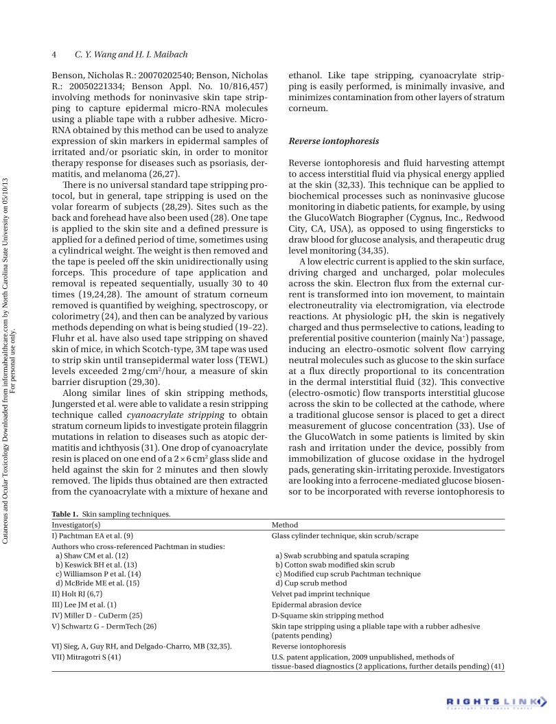
Table 1. Skin sampling techniques.Investigator(s) MethodI) Pachtman EA et al. (9) Glass cylinder technique, skin scrub/scrapeAuthors who cross-referenced Pachtman in studies: a) Shaw CM et al. (12) b) Keswick BH et al. (13) c) Williamson P et al. (14) d) McBride ME et al. (15)
a) Swab scrubbing and spatula scraping b) Cotton swab modified skin scrub c) Modified cup scrub Pachtman technique d) Cup scrub method
II) Holt RJ (6,7) Velvet pad imprint techniqueIII) Lee JM et al. (1) Epidermal abrasion deviceIV) Miller D – CuDerm (25) D-Squame skin stripping methodV) Schwartz G – DermTech (26) Skin tape stripping using a pliable tape with a rubber adhesive
(patents pending)VI) Sieg, A, Guy RH, and Delgado-Charro, MB (32,35). Reverse iontophoresisVII) Mitragotri S (41) U.S. patent application, 2009 unpublished, methods of
tissue-based diagnostics (2 applications, further details pending) (41)
Cut
aneo
us a
nd O
cula
r T
oxic
olog
y D
ownl
oade
d fr
om in
form
ahea
lthca
re.c
om b
y N
orth
Car
olin
a St
ate
Uni
vers
ity o
n 05
/10/
13Fo
r pe
rson
al u
se o
nly.
Minimally invasive skin sampling techniques 5
improve upon the existing technology for noninvasive glucose monitoring (36).
Other uses for reverse iontophoresis include use as a potential diagnostic tool for skin inflammatory reactions. Increased levels of prostaglandin E2 were extracted at the anode in response to transdermal delivery of irritant drugs. Reverse iontophoresis can also be used to aid in making decisions for dialysis in renal disease patients without repeated blood draws. Reverse iontophoresis has extracted urea by electro-osmosis to the cathode after a 5-minute application of current, and the levels of urea were reported to correlate well with serum levels. Pre- and postdialy-sis urea extracts were also respectively different (34). This technique can also be used to extract amino acids, such as phenylalanine, to aid in the diagno-sis of metabolic diseases such as phenylketonuria (37,38). Investigators have successfully extracted up to 13 amino acids from the stratum corneum via reverse iontophoresis (39,40) (Tables 1 and 2).
Conclusion
There are numerous noninvasive to minimally invasive skin sampling techniques available, many to the research arena, with the potential to be devel-oped for wider clinical practice. Biologic material obtained in said fashion can offer many windows into the inner workings of the human body, with minimal subject discomfort, side effects, risk, and time. Of the various methods available, none are easily interchangeable, nor are there direct labo-ratory comparisons of the techniques. This could be an area for future evaluation, and will help the dermatologic investigator make more rational meth-odologic choices. We suspect that these techniques
will offer many branches of science an efficient alternative to venipuncture. In addition, these tech-niques provide experimental access to many skin cell systems and mechanisms.
Declaration of interest
The authors report no conflicts of interest. No monetary funding was sought for this article.
References
1. Lee JM, Carson R, Arce C, Mahajan M, Lobst S. Development of a minimally invasive epidermal abrasion device for clinical skin sampling and its applications in molecular biology. Int J Cosmet Sci 2009;31(1):27–39.
2. Kruse R, Guttenbach M, Schartmann B, Schubert R, van der Ven, H, Schmid, M, et al. Genetic counseling in a patient with XXY/XXXY/XY mosaic Klinefelter’s syndrome: estimate of sex chromosome abberations in sperm before intracytoplasmic sperm injection. Fertil Steril 1998;69:482–485.
3. Rogers G, Koike K. Laser capture microscopy in a study of expression of structural proteins in the cuticle cells of human hair. Exp Dermatol 2009;18:541–547.
4. Matsuhashi S, Narisawa Y, Ozaki I, Mizuta T. Expression pat-terns of programmed cell death 4 protein in normal human skin and some representative skin lesions. Exp Dermatol 2007;16:179–184.
5. Tazón-Vega B, Ars E, Burset M, Santin S, Ruiz P, Fernandez-Llama P, et al. Genetic testing for X-linked Alport syndrome by direct sequencing of COL4A5 cDNA from hair root RNA samples. Am J Kidney Dis 2007 Aug;50(2):257 e1–e14.
6. Reinisch C, Aumaid U, Erovic BM, Pammer J. Expression of BMI-1 in normal skin and inflammatory and neoplastic skin lesions. J Cutan Pathol 2007;34:174–180.
7. Afridi HI, Kazi TG, Kandhro GA, Baig JA, Shah AQ, Jamali, MK, et al. Evaluation of toxic elements in scalp hair samples of myocardial infarction patients at different stages as related to controls. Biol Trace Elem Res 2010;134(1):1–12.
8. Sakuyama S, Hirabayahsi C, Hasegawa J, Yoshida S. Analysis of human face skin surface molecules in situ by
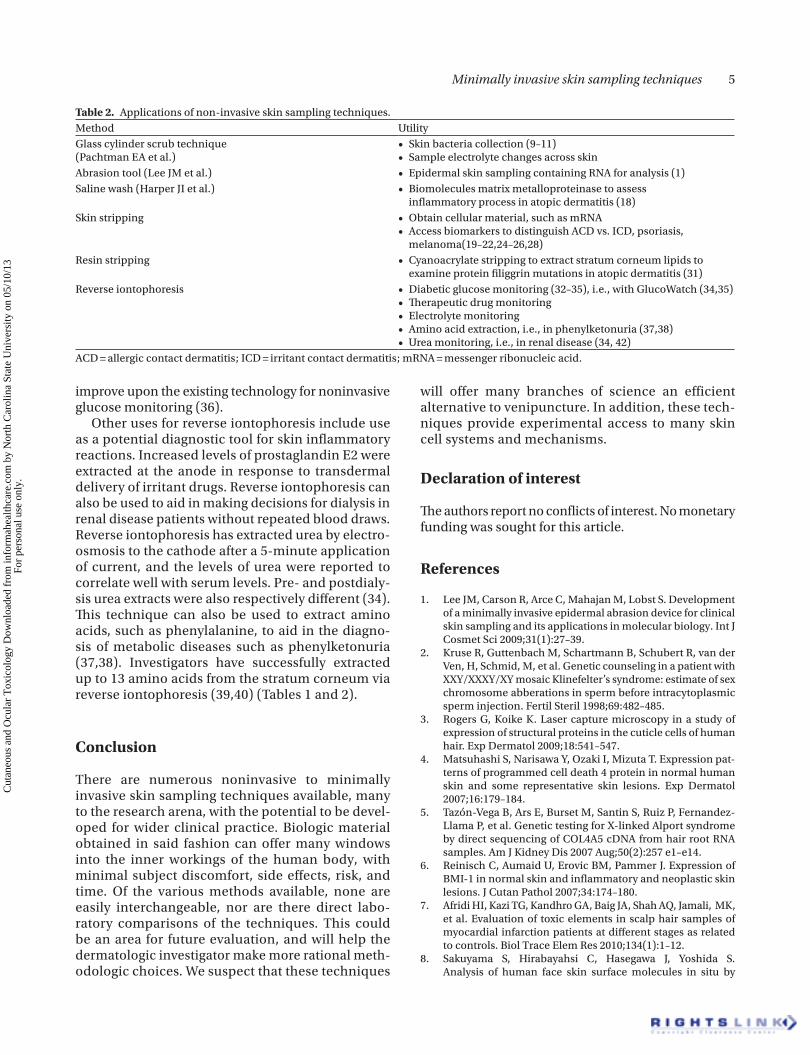
Table 2. Applications of non-invasive skin sampling techniques.Method UtilityGlass cylinder scrub technique (Pachtman EA et al.)
• Skin bacteria collection (9–11) • Sample electrolyte changes across skin
Abrasion tool (Lee JM et al.) • Epidermal skin sampling containing RNA for analysis (1)Saline wash (Harper JI et al.) • Biomolecules matrix metalloproteinase to assess
inflammatory process in atopic dermatitis (18)Skin stripping • Obtain cellular material, such as mRNA
• Access biomarkers to distinguish ACD vs. ICD, psoriasis, melanoma(19–22,24–26,28)
Resin stripping • Cyanoacrylate stripping to extract stratum corneum lipids to examine protein filiggrin mutations in atopic dermatitis (31)
Reverse iontophoresis • Diabetic glucose monitoring (32–35), i.e., with GlucoWatch (34,35) • Therapeutic drug monitoring • Electrolyte monitoring • Amino acid extraction, i.e., in phenylketonuria (37,38) • Urea monitoring, i.e., in renal disease (34, 42)
ACD = allergic contact dermatitis; ICD = irritant contact dermatitis; mRNA = messenger ribonucleic acid.
Cut
aneo
us a
nd O
cula
r T
oxic
olog
y D
ownl
oade
d fr
om in
form
ahea
lthca
re.c
om b
y N
orth
Car
olin
a St
ate
Uni
vers
ity o
n 05
/10/
13Fo
r pe
rson
al u
se o
nly.
6 C. Y. Wang and H. I. Maibach
Fourier-transform infrared spectroscopy. Skin Res Technol 2010;16:151–160.
9. Pachtman EA, Vicher EE, Brunner MJ. The bacterio-logic flora in seborrheic dermatitis. J Invest Dermatol 1954;22:389–396.
10. Aly R, Maibach HI, Shinefield HR, Strauss WG. Survival of pathogenic microorganisms on human skin. J Investig Dermatol 1972;58:205–210.
11. Lo JS, Oriba HA, Maibach H, Bailin PL. Transepidermal potassium ion, chloride ion, and water flux across delip-idized and cellophane tape-stripped skin. Dermatologica 1990;180(2):66–68.
12. Shaw CM, Smith JA, McBride ME, Duncan WC. An Evaluation of Techniques for sampling Skin Flora. J Invest Dermatol 1970;54:160–163.
13. Keswick BH, Frank D. Modified Scrub Technique for Sampling Infant Skin Microflora. J Clin Microbiol 1987;25: 2400–2401.
14. Williamson P, Kligman AM. A new method for the quantita-tive investigation of cutaneous bacteria. J Invest Dermatol 1965;45:498–503.
15. McBride ME, Duncan WC, Knox JM. Correlations between epithelial cells and bacterial populations in bacteriological skin samples. Br J Dermatol 1978;99:537–543.
16. Holt RJ. Pad culture studies on skin surfaces. J Appl Bacteriol 1966;29:625–630.
17. Holt RJ. Aerobic bacterial counts on human skin after bath-ing. J Med Microbiol 1971;4:319–327.
18. Harper JI, Godwin H, Green LE, Wilkes NJ, Holden M, Moffatt WO, et al. A study of matrix metalloproteinase expression and activity in atopic dermatitis using a novel skin wash sampling assay for functional biomarker analysis. Br J Dermatol 2010;162:397–403.
19. Escobar-Chavez JJ, Merino-Sanjuan V, Lopez-Cervantes M, Urban-Morlan Z, Pinon-Segundo E, Quintanar-Guerrero D, et al. The tape-stripping techniques as a method for drug quan-tification in skin. J Pharm Pharm Sci 2008;11(1):104–130.
20. Karan A, Alikhan A, Maibach HI. Toxicologic implications of cutaneous barriers: a molecular, cellular, and anatomical overview. J Appl Toxicol 2009;29:551–559.
21. Morhenn VB, Chang EY, Rheins LA. A noninvasive method for quantifying and distinguishing inflammatory skin reactions. J Am Acad Dermatol 1999;41(5, part 1):687–692.
22. Broccardo CJ, Mahaffey SF, Strand M, Reisdorph NA, Leung DYM. Peeling off the layers: Skin taping and a novel proteomics approach to study atopic dermatitis. J Allergy Clin Immunol 2009;124: 1113–1115.
23. Kezic S, Kammeyer A, Calkoen F, Fluhr JW, Bos JD. Natural moisturizing factor components in the stratum cor-neum as biomarkers of filaggrin genotype: evaluation of minimally invasive methods. Br J Dermatol 2009;161: 1098–1104.
24. Dreher F, Modjtahedi BS, Modjtahedi SP, Maibach HI. Quantification of stratum corneum removal by adhesive tape stripping by total protein assay in 96-well microplates Skin Res Technol 2005;11:97–101.
25. CuDerm Website. http://www.cuderm.com (accessed 30 Aug 2010).
26. DermTech International Website. http://www.dermtech.com (accessed 30 Aug 2010).
27. United States Patient and Trademark Office Website. http://www.uspto.gov/ (accessed 30 Aug 2010).
28. Löffler H, Dreher F, Maibach HI. Cutaneous biology. Stratum corneum adhesive tape stripping: influence of anatomi-cal site, application pressure, duration and removal. Br J Dermatol 2004;151:746–752.
29. Fluhr JW, Elsner P, Berardesca E, Maibach HI. Bioengineering of the Skin: Water and the Stratum Corneum. 2nd Ed; London, UK: Informa Healthcare. Aug.2004.
30. Fluhr JW, Mao-Qiang M, Brown BE, Wertz PW, Crumrine D, Sundberg JP, et al. Glycerol regulates stratum corneum hydra-tion in sebaceous gland deficient (asebia) mice. J Invest Dermatol 2003;120:728–737.
31. Jungersted JM, Hellgren LI, Drachmann T, Jemec GBE, Agner T. Validation of cyanoacrylate method for collection of stratum corneum in human skin for lipid analysis. Skin Pharmacol Physiol 2010;23(2):62–67.
32. Sieg A, Guy RH, Delgado-Charro MB. Noninvasive and mini-mally invasive methods for transdermal glucose monitoring. Diabetes Technol Ther 2005;7(1):174–197.
33. Tura A. Noninvasive glycaemia monitoring: back-ground, traditional findings, and novelties in the recent clinical trials. Curr Opin Clin Nutr Metab Care 2008;11: 607–612.
34. Leboulanger B, Guy RH, Delgado-Charro MB. Reverse ion-tophoresis for non-invasive transdermal monitoring. Physiol Meas 2004;25:R35–R50.
35. Sieg A, Guy RH, Delgado-Charro MB. Electroosomosis in transdermal iontophoresis: implications for noninva-sive and calibration-free glucose monitoring. Biophys J 2004;87:3344–3350.
36. Ching CT, Sun TP, Huang SH, Shieh HL, Chen CY. A mediated glucose biosensor incorporated with reverse iontophoresis function for noninvasive glucose monitoring. Ann Biomed Eng 2010;38:1548–55.
37. Sieg A, Jeanneret F, Fathi M, Hochstrasser D, Rudaz S, Veuthey JL, et al. Extraction of amino acids by reverse iontophoresis: simulation of therapeutic monitoring in vitro. Eur J Pharm Biopharm 2008;70:908–913.
38. Sieg A, Jeanneret F, Fathi M, Hochstrasser D, Rudaz S, Veuthey JL, et al. Extraction of amino acids by reverse ion-tophoresis in vivo. Eur J Pharm Biopharm 2009;72:226–231.
39. Bouissou CC, Sylvestre JP, Guy RH, Delgado-Charro MB. Reverse iontophoresis of amino acids: identification and separation of stratum corneum and subdermal source in vitro. Pharm Res 2009;26:2630–2638.
40. Delgado-Charro, Begona M, Sylvestre JP, Bouissou CC, Guy RH, Delgado-Charro MB. Extraction and quantifica-tion of amino acids in human stratum corneum in vivo. Br J Dermatol. 2010 Apr 15. [Epub ahead of print].
41. Mitragotri Lab Group Website. http://drugdelivery.engr.ucsb.edu/ (accessed 30 Aug 2010).
42. Wascotte V, Caspers P, de Sterke J, Jadoul M, Guy RH, Preat V. Assessment of the “skin reservoir” of urea by confocal Raman microspectroscopy and reverse iontophoresis in vivo. Pharm Res. 2007 Oct; 24(10): 1897–901.
Cut
aneo
us a
nd O
cula
r T
oxic
olog
y D
ownl
oade
d fr
om in
form
ahea
lthca
re.c
om b
y N
orth
Car
olin
a St
ate
Uni
vers
ity o
n 05
/10/
13Fo
r pe
rson
al u
se o
nly.