white paper - accueil - implanet€¦ · · 2015-07-23white paper . postoperative ... in order to...
TRANSCRIPT

Version 01, July 2015 – 12 pages
Postoperative infections after surgical correction
of Adolescent Idiopathic Scoliosis
using polyester sublaminar bands
Brice Ilharreborde MD, PhD
Department of Pediatric Orthopaedic Surgery,
Robert Debré Hospital, Paris Diderot University, France.
White paper

Postoperative infections after surgical correction of AIS using polyester sublaminar bands
2
Introduction
Surgical site infections (SSIs) are the most frequent adverse event requiring revision after spinal
fusion for pediatric scoliosis. The incidence of SSI after Adolescent Idiopathic Scoliosis (AIS) surgery
remains highly variable, currently ranging from 0.5 to 7% [1, 2, 3, 4]. Numerous risk factors have
been investigated, but most of them are patient-related and therefore non-modifiable, such as
previous surgery or underlying medical status. Recent literature has focused on deep wound
infections management and described prevention strategies to decrease their occurrence, but the
influence of the type of instrumentation used for surgical correction remains unknown [3]. All-
pedicle screws constructs are currently the most popular, but several limitations have been reported
lately, resuscitating interest for posteromedial translation techniques using hybrid instrumentation
[5,6,7,8]. While implant distribution remains variable in all-screw constructs, there is a tendency to
increase implant density in order to optimize the correction, and a trend towards intraoperative
navigation and/or imaging control, both increasing operative time and risk of SSI [8,9]. In opposition,
fewer implants are necessary when posteromedial translation is the chosen method for correction,
especially with sublaminar bands [9], and intraoperative imaging is not necessary. However, the main
concern to date corresponds to the risk of infection, and the ability of the pathogen to colonize the
polyester plait, which could make SSI eradication more difficult. In addition, very little literature is
available regarding SSI sequelae. As a matter of fact, bone graft is often partially removed during
surgical debridement, thus altering the conditions for optimal fusion. In addition, the multiple
procedures that can be necessary to eradicate the pathogen and the longer hospitalization stay
might lead to poorer satisfaction. The goal of this study was therefore to report SSI rate after AIS
surgery using sublaminar bands, describe their management, and examine the influence of deep
wound infections on radiological and functional outcomes.

Postoperative infections after surgical correction of AIS using polyester sublaminar bands
3
MATERIALS AND METHODS
Patients
After institutional review board approval, medical data of all patients operated for AIS in our
institution between June 2006 and June 2014 were retrospectively reviewed. Patients with previous
spinal surgery or medical conditions increasing the risk for infection were excluded. The cases of
infections were first identified, using the recommended criteria, in order to determine the incidence
rate of SSI associated with the sublaminar bands technique [10]. Patient-related and procedure-
related parameters from the SSI group were then compared to a control group, matched for age,
Lenke curve type and initial Cobb angle [11].
Preoperative care
On the day of surgery and on the day before, all patients took a shower with povidone-iodine scrub.
The operative team systematically checked the back skin at the time of admission, and the procedure
was postponed in case of significant acne. Skin preparation before incision included one surgical site
povidone-iodine scrub, followed by two 5% alcoholic povidone-iodine paints performed by a nurse.
The surgical field was recovered by an iodine-impregnated adherent plastic drape, and two plastic
bags were placed laterally in order to protect the bands from falling down during the procedure
(Figure 1).
Figure 1: Lateral view of the installation, showing the position of the plastic bags placed on each side of the patient, in order to receive the extra spinal part of the sublaminar bands intraoperatively.

Postoperative infections after surgical correction of AIS using polyester sublaminar bands
4
The same blood saving strategy was applied during the entire study period. Patients were
preoperatively treated by weekly injections of Erythropoietin (EPO), in order to reach a hemoglobin
rate of 15 mg/dl, and antifibrinolytic (tranexamic acid) was systematically used during the procedure.
As recommended, antibiotic prophylaxis relied on cefamandol, with 2 grams (g) injected within one
hour before incision, followed by a second injection of 1g 4 hours later, and antibiotics were stopped
after wound closure [12].
Operative procedures
All patients underwent posterior segmental spinal correction and fusion using hybrid constructs,
performed by one of the two senior surgeons of the department. Spinal cord monitoring was
systematically performed. Fusion levels were selected following the same criteria during the study
period [5]. In all cases, pedicle screws were placed at the distal extremity of the curve (from L4 to L1
or T12), while thoracic levels were instrumented with sublaminar bands (Jazz from Implanet,
Bordeaux, France or Universal Clamps from ZimmerSpine, Bordeaux, France) on the concave side and
at the apex on the convex side. The 2 upper thoracic levels, located at the proximal end of the
construct, were bilaterally instrumented with autostable hooks (ZimmerSpine, Bordeaux, France),
protected by 2 adjacent sublaminar bands (Figure 2).
Figure 2: Preoperative and postoperative frontal radiograph of a Lenke 1 AIS instrumented with a hybrid construct combining pedicle screws from L3 to L1, 7 sublaminar bands placed on the main thoracic curve, and 2 autostable claws placed on T3-T4.

Postoperative infections after surgical correction of AIS using polyester sublaminar bands
5
Correction was performed on both sides simultaneously, using 5.5 mm CoCr rods contoured
according to the desired sagittal alignment. Both rods were first connected to lumbar screws, and
derotation, compression/distraction and in situ contouring were applied. Once the lumbar curve was
corrected and the last instrumented vertebra leveled, the rods were connected to the stable
proximal anchor with the set screws left loose in order to allow distraction. The bands were then
connected to the rods and multilevel tension was applied on the concave side in order to obtain a
posteromedial translation of the scoliotic curve (Figure 3) [6]. After tensioning all bands and
setscrews were revisited and the residual length of the bands were cut and removed. After
decortication, autograft was used in combination with bioglass (Novabone, Novabone products,
Alachua, Florida, USA), as previously reported [13].
Figure 3: Intraoperative view of the patient, with the extraspinal part of the bands placed and protected in the lateral plastic bags, before being cut and removed after curve correction and setscrews revisit.
In addition, a convex thoracoplasty was performed when the patient and/or caregiver expressed
concern over the prominence of the rib hump deformity. Wound closure was performed over 2
drains after irrigation (1000mL of sterile saline solution) and muscle debridement if necessary.
Postoperative care and SSI management
Wound dressings were changed on day 2 and 5 postoperative, with drains being removed on day 2.
Patients started to stand on the second postoperative day with the physiotherapist, and worked in
erect position twice a day during hospitalization stay. Patients were discharged from the hospital and

Postoperative infections after surgical correction of AIS using polyester sublaminar bands
6
sent to a rehabilitation center (3 weeks period) between 6 and 7 days after surgery. When SSI was
diagnosed, patients were treated according to the same standardized protocol, associating surgical
debridement, wound closure without removal or change of the implants, and 15 days of intravenous
antibiotherapy administrated through a central catheter placed during the revision procedure,
followed by 2.5 months of oral antibiotics adapted to the pathogen. When necessary, serum dosage
of antibiotics was performed before discharge. In all cases, the antibiotherapy was discussed and
decided during a multidisciplinary meeting with the referent surgeon, a pediatrician and a
microbiologist.
Evaluation at follow-up
In order to determine the influence of SSI on fusion and/or instrumentation failure, radiological
analysis was performed preoperatively, postoperatively and at latest follow-up (minimum 1-year)
using low-dose biplanar stereoradiographs (EOS imaging, Paris, France). In addition, the final
functional outcomes were evaluated using SRS 30 and SF 12 questionnaires [14,15].
Statistical analysis
Demographic data, loss of correction and functional outcomes were compared between the SSI and
the control groups using unpaired t-tests (Statview, SAS Institute Inc, Cary, NC, USA). A p<0.05 was
considered significant.
Results
Patients
Among the 524 patients who underwent AIS corrections using sublaminar bands during the study
period, 28 (5.3%) were diagnosed with SSI. The average follow-up was 38 months (± 24). Early
infections (within 45 days postoperative) were reported in 26 of the 28 SSI patients (93%), after a
mean free interval of 18 days (± 8), while 2 late SSI occurred after 84 days and 674 days, respectively.
Demographic data of the SSI and control groups are reported in Table 1. No significant difference was
found, except for the number of levels fused, which was significantly higher in the non-infected
group.

Postoperative infections after surgical correction of AIS using polyester sublaminar bands
7
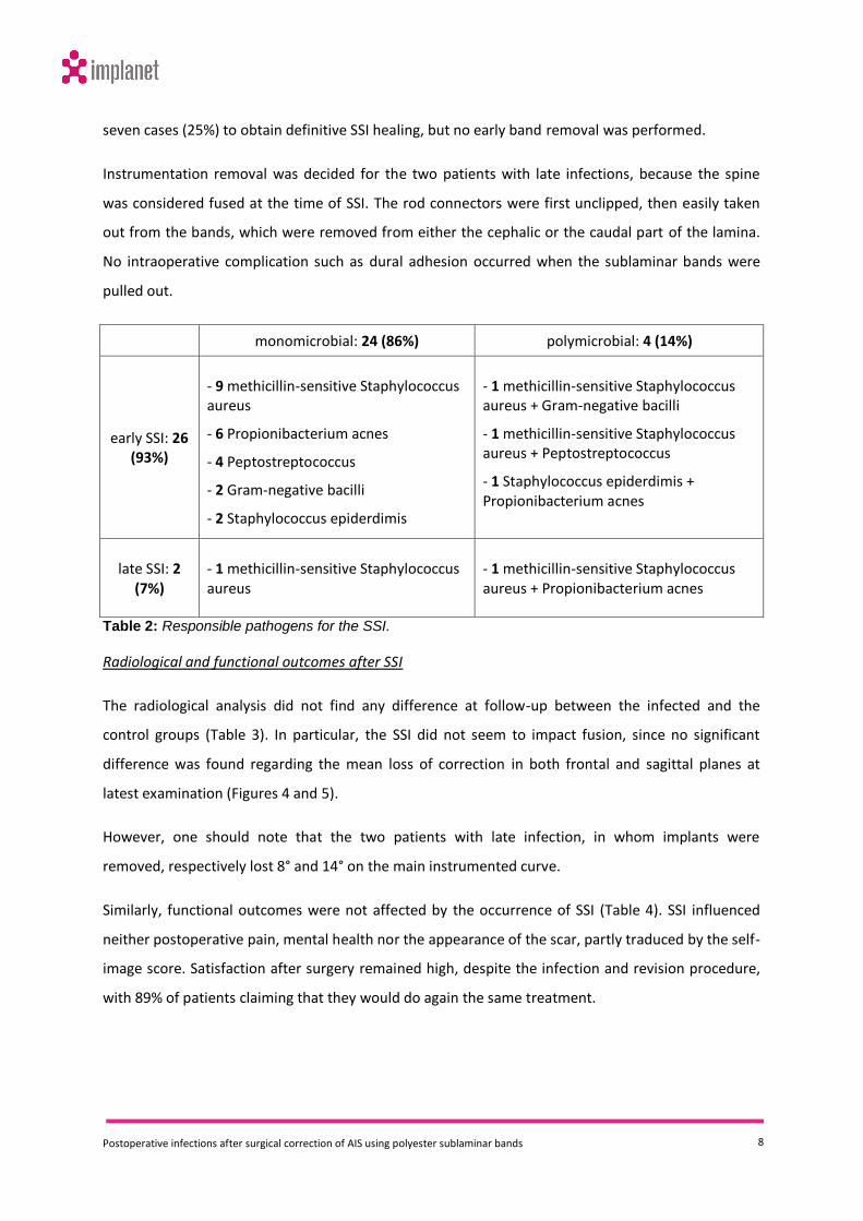
Pathogens
Pathogens were identified in all cases, using either direct examination and/or molecular biology
(table 2). SSI was monomicrobial in 24 cases (86%) and polymicrobial in 4 cases (14%). Responsible
bacteria are reported in Table 2. The most frequent pathogen was methicillin-sensitive
Staphylococcus aureus (13 cases, 46%), followed by Propionibacterium acnes (8 cases, 29%), all with
community profile. Skin germs (Staphylococcus (epiderdimis and aureus and Propionibacterium
acnes) were responsible for the SSI in 86% of cases.
Management
All patients with SSI underwent surgical debridement within 24 hours after diagnosis. Initial wound
closure was performed in all cases, over 2 or 3 drains that were removed after 5 days if the liquid
culture was negative and no other clinical or biological sign of infection was present. Patients were
initially treated by double probabilistic antibiotherapy, but an adapted oral antibiotherapy was
possible after 15 days in all cases. More than one surgical debridement (2 to 3) was necessary in
Infected group
(N = 28) Control Group
(N = 28) p
Age (years) 15.3 ± 2.1 14.8 ± 2.6 0.219
Sex ratio 26 girls / 2 boys 25 girls / 3 boys 0.646
Weight (Kg) 55.5 ± 8.0 54.1 ± 13.2 0.64
Height (cm) 166.3 ± 9 164.2 ± 9 0.441
BMI 20.1 ± 3.1 19.9 ± 4.0 0.799
Risser stage 3.8 ± 1 3.4 ± 1 0.48
Number of thoracoplasty 6 7 0.755
Number of levels fused 12.2 ± 1.3 13.7 ± 0.8 <0.001
Number of pedicle screws 5.8 ± 1.2 6.0 ± 0.9 0.692
Number of sublaminar bands
7.1 ± 1.8 6.9 ± 1.2 0.492
Table 1: Demographic data of the two study groups (mean ± standard deviation).
Postoperative infections after surgical correction of AIS using polyester sublaminar bands
8
seven cases (25%) to obtain definitive SSI healing, but no early band removal was performed.
Instrumentation removal was decided for the two patients with late infections, because the spine
was considered fused at the time of SSI. The rod connectors were first unclipped, then easily taken
out from the bands, which were removed from either the cephalic or the caudal part of the lamina.
No intraoperative complication such as dural adhesion occurred when the sublaminar bands were
pulled out.
monomicrobial: 24 (86%) polymicrobial: 4 (14%)
early SSI: 26 (93%)
- 9 methicillin-sensitive Staphylococcus aureus
- 6 Propionibacterium acnes
- 4 Peptostreptococcus
- 2 Gram-negative bacilli
- 2 Staphylococcus epiderdimis
- 1 methicillin-sensitive Staphylococcus aureus + Gram-negative bacilli
- 1 methicillin-sensitive Staphylococcus aureus + Peptostreptococcus
- 1 Staphylococcus epiderdimis + Propionibacterium acnes
late SSI: 2
(7%)
- 1 methicillin-sensitive Staphylococcus aureus
- 1 methicillin-sensitive Staphylococcus aureus + Propionibacterium acnes
Table 2: Responsible pathogens for the SSI.
Radiological and functional outcomes after SSI
The radiological analysis did not find any difference at follow-up between the infected and the
control groups (Table 3). In particular, the SSI did not seem to impact fusion, since no significant
difference was found regarding the mean loss of correction in both frontal and sagittal planes at
latest examination (Figures 4 and 5).
However, one should note that the two patients with late infection, in whom implants were
removed, respectively lost 8° and 14° on the main instrumented curve.
Similarly, functional outcomes were not affected by the occurrence of SSI (Table 4). SSI influenced
neither postoperative pain, mental health nor the appearance of the scar, partly traduced by the self-
image score. Satisfaction after surgery remained high, despite the infection and revision procedure,
with 89% of patients claiming that they would do again the same treatment.

Postoperative infections after surgical correction of AIS using polyester sublaminar bands
9
Infected group
(N=28) Control group
(N=28) p
SRS 30 total score (/150) 121.8 ± 7 123.5 ± 7 0.386
function/activity (/35) (post surgery (/10))
27.0 ±3 (5.7 ± 2)
27.7 ± 2 (6.2 ± 2)
0.372 0.487
pain (/30) (post surgery (/5))
26.1 ± 2 (4.2 ± 1)
26.9 ± 3 (4.5 ± 1)
0.381 0.423
self image (/45) (post surgery (/15))
35.6 ± 3 (11.5 ± 2)
35.6 ± 3 (11.8 ± 2)
0.926 0.603
mental health (/25) 20.4 ± 2 19.8 ± 2 0.352
satisfaction with management (/15)
(post surgery (/5))
12.9 ± 2 (4.2 ± 1)
13.6 ± 1 (4.3 ± 1)
0.071 0.516
positive answers (%) to the question: “would you do it again?”
89% 90% 0.709
SRS 30 post surgery score (/35) 25.6 ± 4 26.8 ± 5 0.308
SF 12 (physical score) 47.5 ± 6 49.5 ± 5 0.206
SF 12 (mental score) 50.5 ± 6 50.7 ± 4 0.919
Table 3: Functional outcomes (SRS 30 and SF 12 scores) at follow-up.
Figure 4: Preoperative (a), immediate postoperative (b) and 2-year follow-up (c) frontal microdose radiographs of a 14 year-old girl AIS, who developed and early SSI (2 weeks postoperative) due to Staphylococcus aureus after posterior T2L3 fusion.

Postoperative infections after surgical correction of AIS using polyester sublaminar bands
10
Figure 5: Preoperative (a), immediate postoperative (b) and 2-year follow-up (c) lateral microdose radiographs of a 14 year-old girl AIS, who developed and early SSI (2 weeks postoperative) due to Staphylococcus aureus after posterior T2L3 fusion.
Conclusion
Results of the current study demonstrate that the use of sublaminar bands in AIS did not increase SSI
rate. As a matter of fact, the 5.3% incidence found in this cohort is in accordance with literature,
even though it stands in the upper range [1,2,3,4,16,17]. However, results are consistent with a
previous study, performed by the same department, in which hooks were used in similar hybrid
constructs [7]. Sublaminar bands can therefore safely replace pedicle screws or hooks at thoracic
levels, in order to optimize sagittal correction, while reducing operative time, blood loss and
radiation exposure [18,19,20]. Nevertheless, the use of lateral plastic bags is recommended, since
sublaminar bands measure approximately 40cm long before being tensioned.
Results also show that a concern raised by some surgeons about a higher risk of implant colonization
in case of SSI, due to the band structure, was not justified, and the management of SSI remained
unchanged. As shown in vascular surgery literature about Dacron patches (similar constitution), the
use of polyester did not increase SSI rate, and all infections healed after debridement and
appropriate antibiotherapy, without necessitating band withdrawal [21,22]. On the other hand, in
cases of late infections occurring after spinal fusion in which instrumentation removal was decided to

Postoperative infections after surgical correction of AIS using polyester sublaminar bands
11
ease pathogen eradication, no adherence with the dura and no intraoperative complication was
observed when bands were pulled out, unlike what had been reported with Luque wires [23]. The
fact that 25% of the infected patients underwent multiple debridements (average 1.3 / patient) can
seem high, but it is comparable to recent published studies, in which the mean number of
debridement ranged from 1.6 to 2, in large series of pediatric deformities with multiple etiologies
[24,25].
In conclusion, posteromedial translation with sublaminar bands does not increase SSI rate in AIS. The
choice of implant material does not influence early morbidity and bands can be removed safely, even
in case of delayed infection. Results of this series also show that the occurrence of SSI does not alter
radiological or functional outcomes.
References
1. Ahmed R, Greenlee JD, Traynelis VC. Preservation of spinal instrumentation after development of postoperative bacterial infections in patients undergoing spinal arthrodesis. J Spinal Disord Tech. 2012;25(6):299-302
2. Li Y, Glotzbecker M, Hedequist D. Surgical site infection after pediatric spinal deformity surgery. Curr Rev Musculoskelet Med. 2012;5:111-119
3. WG. Mackenzie, H. Matsumoto, BA.Williams, J. Corona, C. Lee, SR. Cordy, L. CovingtonL. Saiman, JM.Flynn, DL. Skaggs, DP. Roye, MG. Vital. Surgical site infection following spinal instrumentation for scoliosis. A multicenter analysis of rates, risk factors, and pathogens. J Bone Joint Surg Am. 2013;95(9):800-6, S1-2
4. Coe JD, Smith JS, Berven S, Arlet V, Donaldson W, Hanson D, Mudiyam R, Perra J, Owen J, Marks MC, Shaffrey CI. Complications of spinal fusion for scheuermann kyphosis: a report of the scoliosis research society morbidity and mortality committee. Spine 2010;35(1):99-103
5. Ilharreborde B, Even J, Lefevre Y, Fitoussi F, Presedo A, Souchet P, Penneçot GF, Mazda K . How to determine the upper level of instrumentation in Lenke type 1 and type 2 adolescent idiopathic scoliosis? A prospective study of 132 patients. J Pediatr Orthop 2008;28:733-9
6. Mazda K, Ilharreborde B, Even J, Lefevre, Y Fitoussi F, and Pennecot GF. Efficacy and safety of posteromedial translation for correction of thoracic curves in adolescent idiopathic scoliosis using a new connection to the spine: the Universal Clamp. Eur Spine J 2009;18:158-69
7. Ilharreborde B, Even J, Lefevre Y, Fitoussi F, Presedo A, Pennecot GF, and Mazda K. Hybrid constructs for tridimensional correction of the thoracic spine in adolescent idiopathic scoliosis: a comparative analysis of Universal Clamps versus hooks. Spine 2010; 35:306-14
8. De Kleuver M, Lewis SJ, Germscheid NM, Kamper SJ, Alanay A, Berven SH, Cheung KM, Ito M, Lenke LG, Polly DW, Qiu Y, van Tulder M, Shaffrey C. Optimal surgical care for adolescent idiopathic scoliosis: an international consensus. Eur Spine J. 2014 Dec;23(12):2603-18
9. Le Navéaux F, Aubin CÉ, Larson AN, Polly DW Jr, Baghdadi YM, Labelle H. Implant distribution in surgically Instrumented Lenke 1 Adolescent Idiopathic Scoliosis: Does it Affect curve correction? Spine 2015;40:462-8.
10. Horan TC, Gaynes RP, Martone WJ, et al.: CDC definitions of nosocomial surgical site infections, 1992: a modification of CDC definitions of surgical wound infections. Infect Control Hosp Epidemiol 1992;13:606–608.
11. Lenke LG, Betz RR, Harms J, et al. Adolescent idiopathic scoliosis: a new classification to determine extent of spinal arthrodesis. J Bone Joint Surg Am 2001;83:1169 -81.
12. Milstone AM, Maragakis LL, Townsend T, et al. Timing of preoperative antibiotic prophylaxis: a modifiable risk factor for deep surgical site infections after pediatric spinal fusion. Pediatr Infect Dis J. 2008;27:704–8.
13. Ilharreborde B, Morel E, Fitoussi F, Presedo A, Souchet P, Penneçot GF, Mazda K. Bioactive glass as a bone substitute for spinal fusion in adolescent idiopathic scoliosis: a comparative study with iliac crest. J Pediatr Orthop. 2008;28(3):347-51.

Postoperative infections after surgical correction of AIS using polyester sublaminar bands
12
14. Lonjon G, Ilharreborde B, Odent T, Moreau S, Glorion C, Mazda K. Reliability and validity of the french canadian version of the scoliosis research scociety 22 questionnaire in France. Spine 2014;39:26-34.
15. Gandek B et al. Cross-validation of item sélection and scoring for the SF-12 health Survey in nine countries : results from the IQOLA project. International quality of life assessment. J Clin Epidemiol. 1998;51:1171-8
16. Bachy M , Bouyer B, Vialle R. Infection after spinal correction and fusion for spinal deformities in childhood and adolescence. Int Orthop. 2012;36(2):465-9
17. Rihn JA, Lee JY, Ward WT. Infection after surgical treatment of adolescent idiopathic scoliosis evaluation of the diagnosis, treatment, and impact on clinical outcomes. Spine 2008;33:289-94.
18. J.Sale de Gauzy, JL. Jouve, F. Accadbled, B. Blondel,G. Bollini. Use of the Universal Clamp in adolescent idiopathic scoliosis for deformity correction and as an adjunct to fusion: 2 years follow-up. J Child Orthop. 2011 Aug;5(4):273-82.
19. Hongo M, Ilharreborde B, Gay RE, Zhao C, Zhao KD, Berglund LJ, Zobitz M, An K. Biomechanical evaluation of a new fixation device for the thoracic spine. Eur Spine J 2009;18:1213–1219
20. E. Polirsztok, M. Gavaret, T. Gsell, I. Suprano, E. Choufani, G. Bollini, Jean-Luc Jouve. Sublaminar bands, are they safe? Eur Spine J 2015;24:1441-9
21. Knight BC, Tait WF. Dacron patch infection following carotid endarterectomy: a systematic review of the literature. Eur J Vasc Endovasc Surg. 2009;37(2):140-8.
22. Katz SG, Kohl RD. Does the choice of material influence early morbidity in patients undergoing carotid patch angioplasty? Surgery 1996;119(3):297-301.
23. Songer MN, Spencer DL, Meyer PR Jr, Jayaraman G. The use of sublaminar cables to replace Luque wires. Spine 1991;16(8 Suppl):S418-21.
24. Ho C, Skaggs DL, Weiss JM, Tolo VT. Management of infection after instrumented posterior spine fusion in pediatric scoliosis. Spine 2007;32(24):2739-44.
25. Cahill PJ, Warnick DE, Lee MJ, Gaughan J, Vogel LE, Hammerberg KW, Sturm PF. Infection after spinal fusion for pediatric spinal deformity: thirty years of experience at a single institution. Spine 2010;35(12):1211-7.