white blood cells disorders - med study group -...
TRANSCRIPT
White blood cells disorders
Non-neoplastic
Leukopenia
• Leukopenia: decrease in WBC count below average levels, results most commonly from a decrease in neutrophils
• Lymphopenia is much less common; it is associated with rare congenital immunodeficiency diseases, advanced human immunodeficiency virus (HIV) infection, and treatment with high doses of corticosteroids
Neutropenia
• ANC < 1500 cell/ microliter
• Severe neutropenia: <500, spontaneous infection
Causes of neutropenia
Decreased production • Part of pancytopenia: aplastic, myelophthisic,
megaloblastic anemias, myelodysplastic syndrome, chemotherapy
• Isolated neutropenia: Acquired: drugs (anti epileptic, anti psychotic, anti-
hyperthyroidism) Congenital: • Schwachman-Diamond syndrome: AR, SBDS gene mutation,
skeletal abnormalities, pancreatic exocrine deficiency • Chediak- Higashi syndrome: AR, LYST gene, abnormal
lysosomal aggregation and dysfunction, platelet dysfunction, albinism
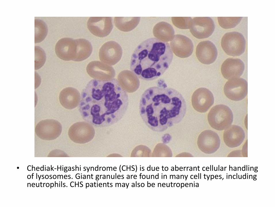
• Chediak-Higashi syndrome (CHS) is due to aberrant cellular handling of lysosomes. Giant granules are found in many cell types, including neutrophils. CHS patients may also be neutropenia
Causes
Increased destruction
• Special infection settings (severe sepsis, salmonella, brucella)
• Immune mediated
• Cyclic neutropenia (ELANE gene mutation, abnormal Elastase accumulation, apoptosis)
• Hypersplenism
• PNH
Reactive Leukocytosis
• An increase in the number of white cells in the blood is common in a variety of inflammatory states caused by microbial and nonmicrobial stimuli. Leukocytoses are relatively nonspecific and are classified according to the particular white cell series that is affected
• Leukemoid reaction: marked increase in WBC count with left-shifted granulpoiesis, mimiking chronic myelogenous leukemia. Occurs in severe stress, paraneoplastic syndrome
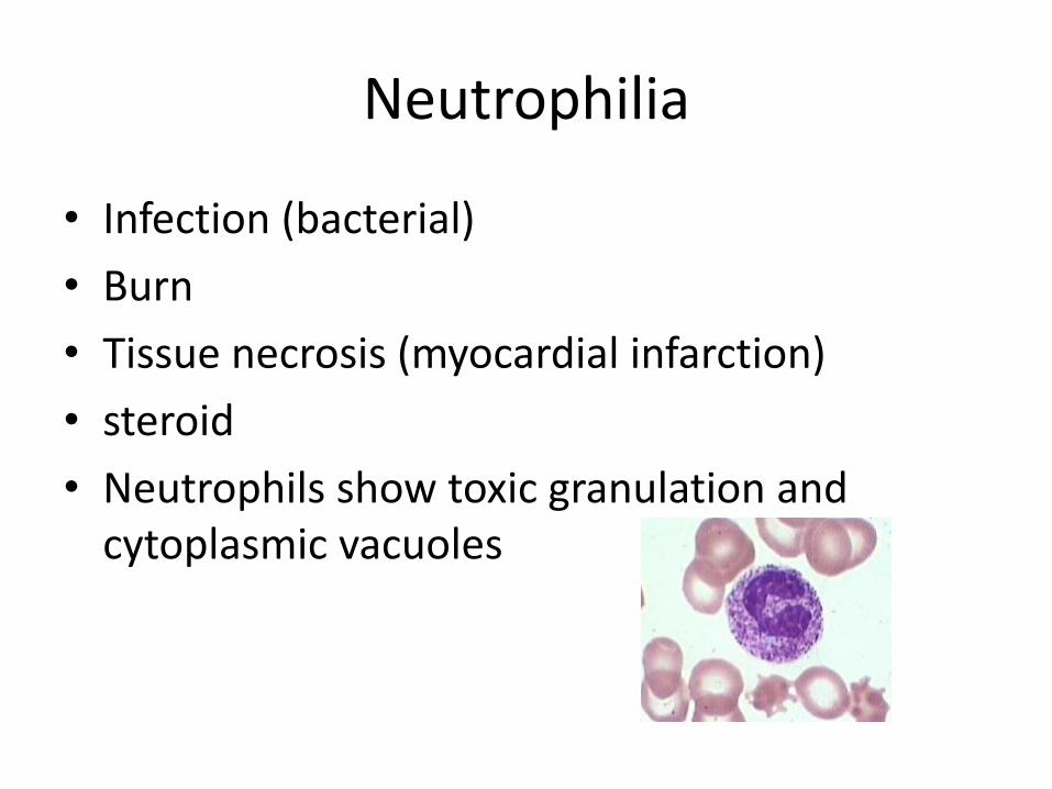
Neutrophilia
• Infection (bacterial)
• Burn
• Tissue necrosis (myocardial infarction)
• steroid
• Neutrophils show toxic granulation and cytoplasmic vacuoles
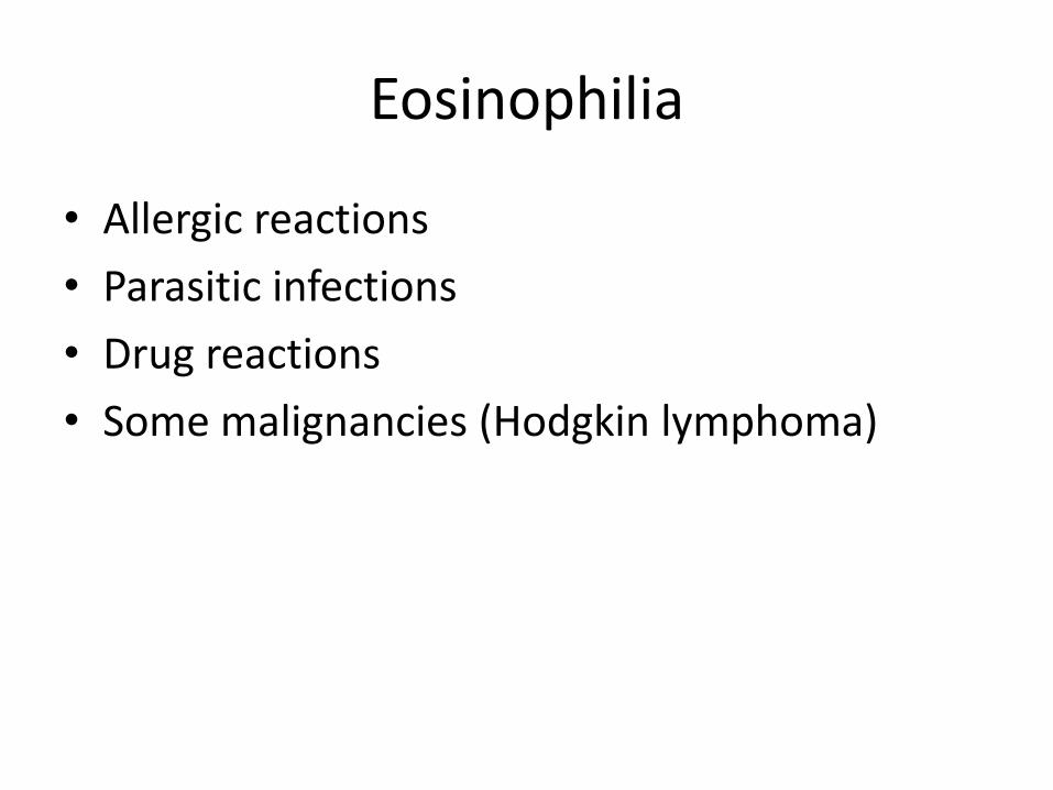
Eosinophilia
• Allergic reactions
• Parasitic infections
• Drug reactions
• Some malignancies (Hodgkin lymphoma)
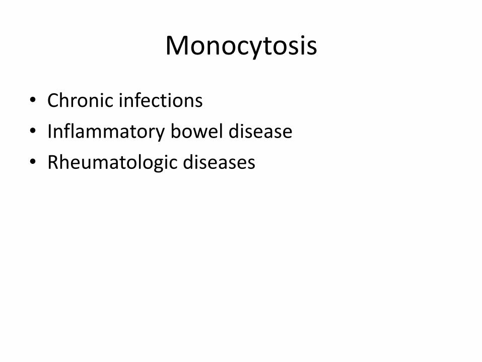
Monocytosis
• Chronic infections
• Inflammatory bowel disease
• Rheumatologic diseases
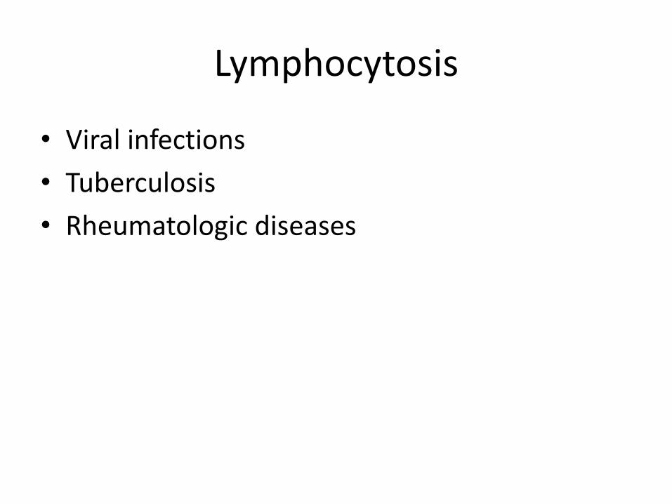
Lymphocytosis
• Viral infections
• Tuberculosis
• Rheumatologic diseases
Reactive Lymphadenitis
• Lymphocyte response to antigen stimulus in the body (Infections, autoimmune)
• Leads to lymph node enlargement (lymphadenopathy)
• Acute is commonly painful, follows bacterial or viral infections
Chronic Reactive Lymphadenitis
• Follicular hyperplasia: proliferation of germinal center B-cells resulting in enlarged follicles, occur in HIV, Toxoplasmosis, Rheumatologic diseases
• Paracortical (diffuse) hyperplasia: proliferation of T-cells in the interfollicular areas, caused by viral infection, drug reaction, post vaccination
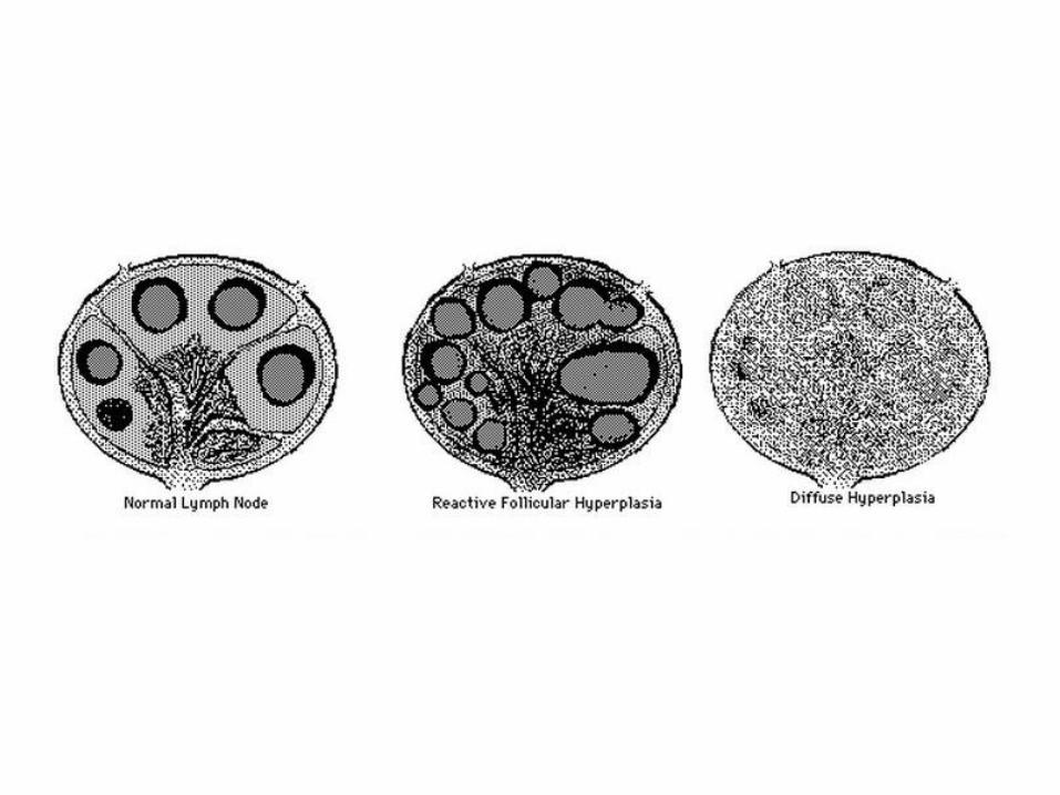
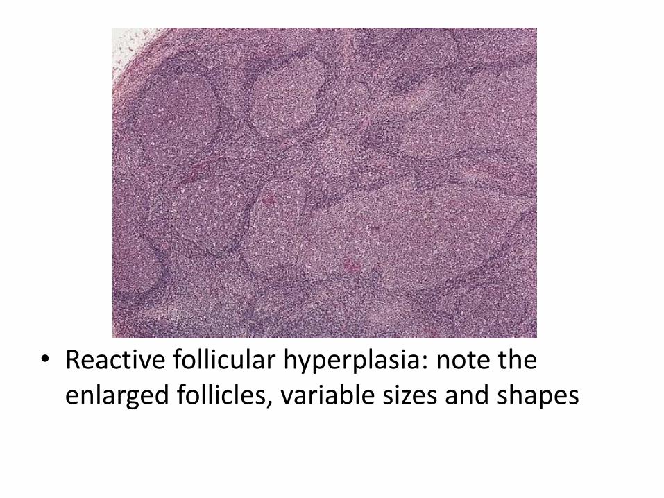
• Reactive follicular hyperplasia: note the enlarged follicles, variable sizes and shapes
Hematopoietic malignancies
• Myeloid
• Lymphoid
• Histiocytic
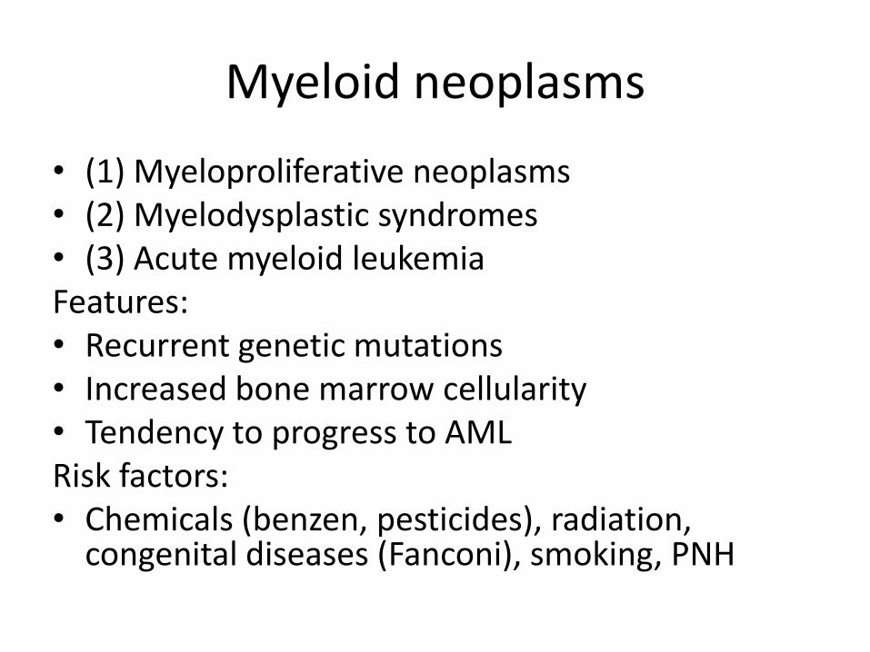
Myeloid neoplasms
• (1) Myeloproliferative neoplasms • (2) Myelodysplastic syndromes • (3) Acute myeloid leukemia Features: • Recurrent genetic mutations • Increased bone marrow cellularity • Tendency to progress to AML Risk factors: • Chemicals (benzen, pesticides), radiation,
congenital diseases (Fanconi), smoking, PNH
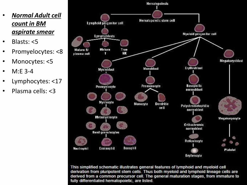
• Normal Adult cell count in BM aspirate smear
• Blasts: <5
• Promyelocytes: <8
• Monocytes: <5
• M:E 3-4
• Lymphocytes: <17
• Plasma cells: <3
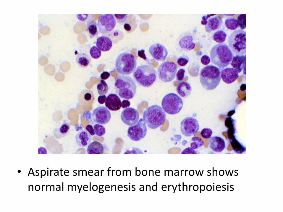
• Aspirate smear from bone marrow shows normal myelogenesis and erythropoiesis
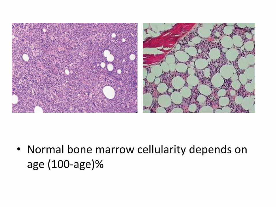
• Normal bone marrow cellularity depends on age (100-age)%
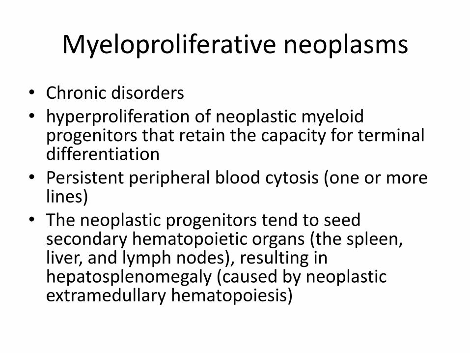
Myeloproliferative neoplasms
• Chronic disorders • hyperproliferation of neoplastic myeloid
progenitors that retain the capacity for terminal differentiation
• Persistent peripheral blood cytosis (one or more lines)
• The neoplastic progenitors tend to seed secondary hematopoietic organs (the spleen, liver, and lymph nodes), resulting in hepatosplenomegaly (caused by neoplastic extramedullary hematopoiesis)
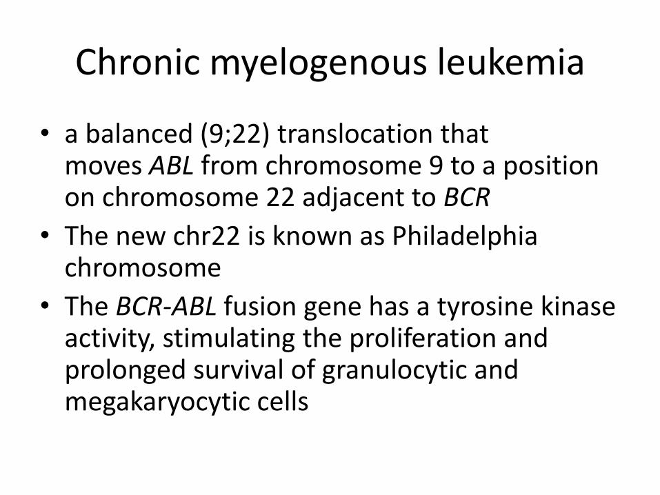
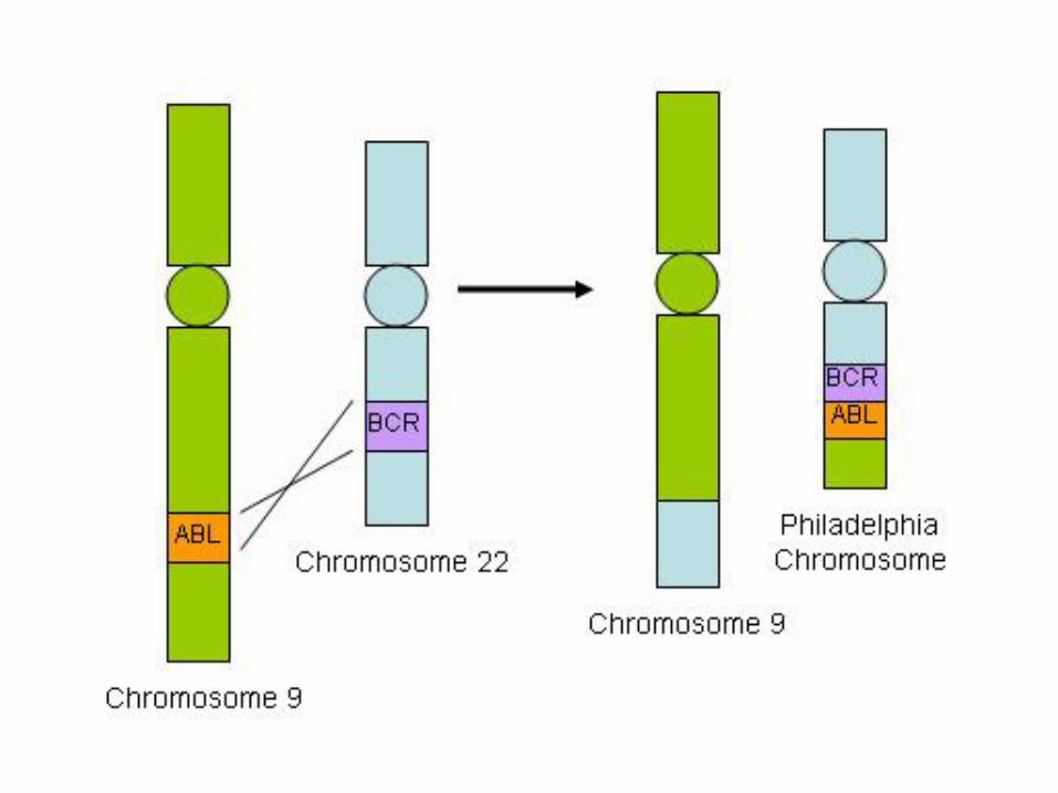
Chronic myelogenous leukemia
• a balanced (9;22) translocation that moves ABL from chromosome 9 to a position on chromosome 22 adjacent to BCR
• The new chr22 is known as Philadelphia chromosome
• The BCR-ABL fusion gene has a tyrosine kinase activity, stimulating the proliferation and prolonged survival of granulocytic and megakaryocytic cells
manifestations
• Peripheral blood shows markedly increased WBC count, sometimes exceeding 100,000 cell/uL
• Most of the cells are neutrophils, metamyelocytes and myelocytes
• Basophils and eosinophils are also increased
• Thrombocytosis and anemia are common
• The bone marrow is hypercellular owing to increased numbers of granulocytic and megakaryocytic precursors
• Spleen is enlarged with extramedullary hematopoiesis
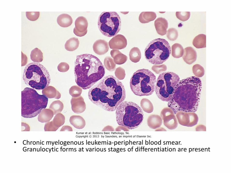
• Chronic myelogenous leukemia-peripheral blood smear. Granulocytic forms at various stages of differentiation are present
Polycythemia Vera
• Janus Kinase-2 (JAK-2) + other mutations
• Stem cell hypersensitive to erythropoietin and growth factors
• Characterized by marked erythropoiesis, also granulopoiesis and megakaryopoiesis (panmyelosis)
• Erythropoietin is low
• Splenomegaly
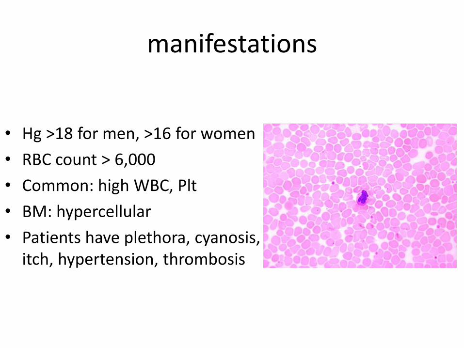
manifestations
• Hg >18 for men, >16 for women
• RBC count > 6,000
• Common: high WBC, Plt
• BM: hypercellular
• Patients have plethora, cyanosis, itch, hypertension, thrombosis
Secondary polycythemia
• Absolute: true increased RBC mass
• Relative: increased Hg concentration (low plasma)
• A prominent cause of hypoxia is present: smoking, lung or heart diseases, high altitude
• High erythropietin, reversible, no splenomegaly
• Also: renal carcinoma, surreptitious
• Alcohol: depresses respiration, prevents anti-diuretic hormone
Primary Myelofibrosis
• Brief period of granulopoiesis and megakaryopoiesis, rapidly followed my BM fibrosis and elimination of hematopoietic elements
• The fibroblast proliferation is stimulated by platelet-derived growth factor and transforming growth factor β released from neoplastic megakaryocytes
• Hematopoiesis takes place in spleen and liver • RBC’s escaping the fibotic stroma in BM are
deformed and take the shape of “tear-drops”
Manifestations
• BM is initially hypercellular with increased atypical megakaryocytes
• PB: leukocytosis, shift to left, thrombocytosis, anemia, nucleated RBCs, tear drop cells
• Later in disease, become fibrotic and hypocellular, pancytopenia
• Spleen shows marked extramedullary hematopoiesis
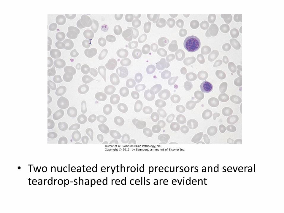
• Two nucleated erythroid precursors and several teardrop-shaped red cells are evident
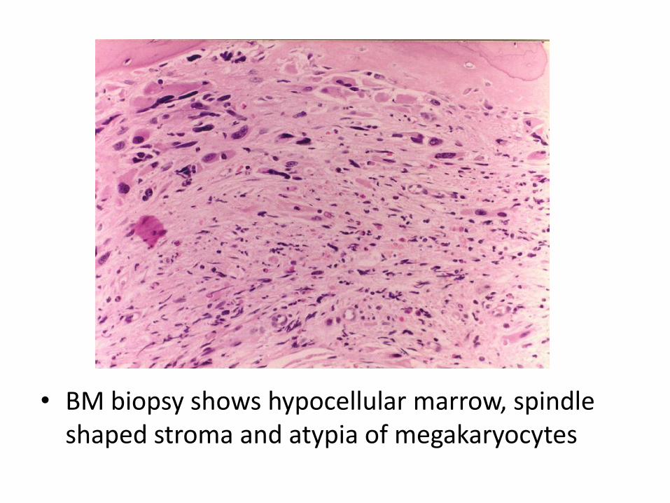
• BM biopsy shows hypocellular marrow, spindle shaped stroma and atypia of megakaryocytes
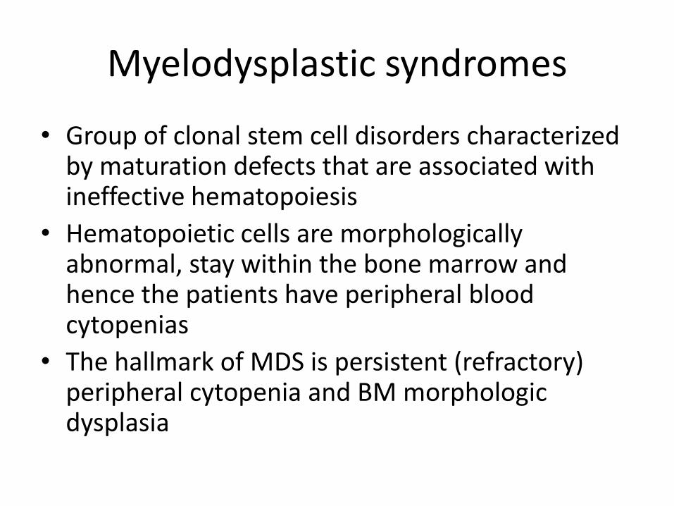
Myelodysplastic syndromes
• Group of clonal stem cell disorders characterized by maturation defects that are associated with ineffective hematopoiesis
• Hematopoietic cells are morphologically abnormal, stay within the bone marrow and hence the patients have peripheral blood cytopenias
• The hallmark of MDS is persistent (refractory) peripheral cytopenia and BM morphologic dysplasia
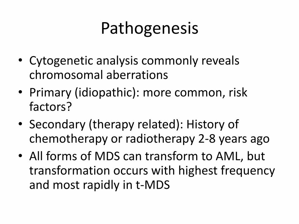
Pathogenesis
• Cytogenetic analysis commonly reveals chromosomal aberrations
• Primary (idiopathic): more common, risk factors?
• Secondary (therapy related): History of chemotherapy or radiotherapy 2-8 years ago
• All forms of MDS can transform to AML, but transformation occurs with highest frequency and most rapidly in t-MDS
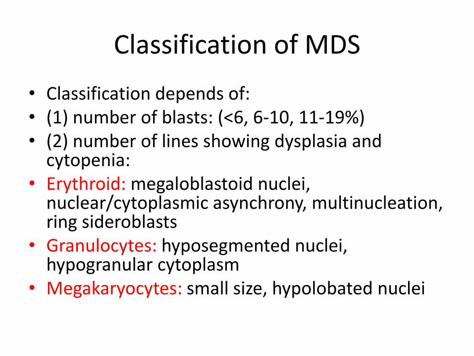
Classification of MDS
• Classification depends of: • (1) number of blasts: (<6, 6-10, 11-19%) • (2) number of lines showing dysplasia and
cytopenia: • Erythroid: megaloblastoid nuclei,
nuclear/cytoplasmic asynchrony, multinucleation, ring sideroblasts
• Granulocytes: hyposegmented nuclei, hypogranular cytoplasm
• Megakaryocytes: small size, hypolobated nuclei
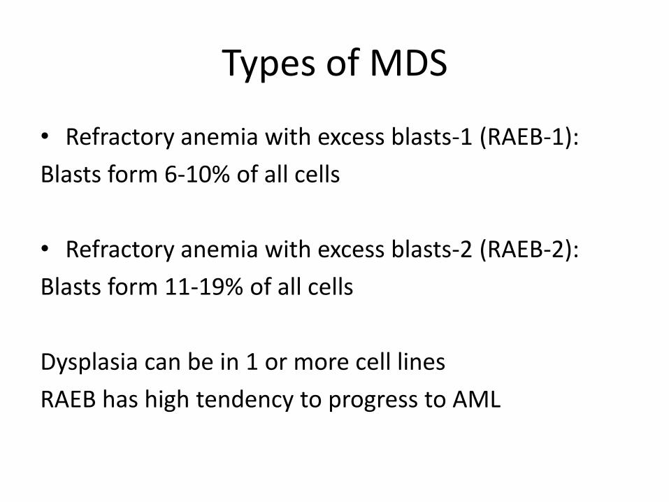
Types of MDS
• Refractory anemia with excess blasts-1 (RAEB-1):
Blasts form 6-10% of all cells
• Refractory anemia with excess blasts-2 (RAEB-2):
Blasts form 11-19% of all cells
Dysplasia can be in 1 or more cell lines
RAEB has high tendency to progress to AML
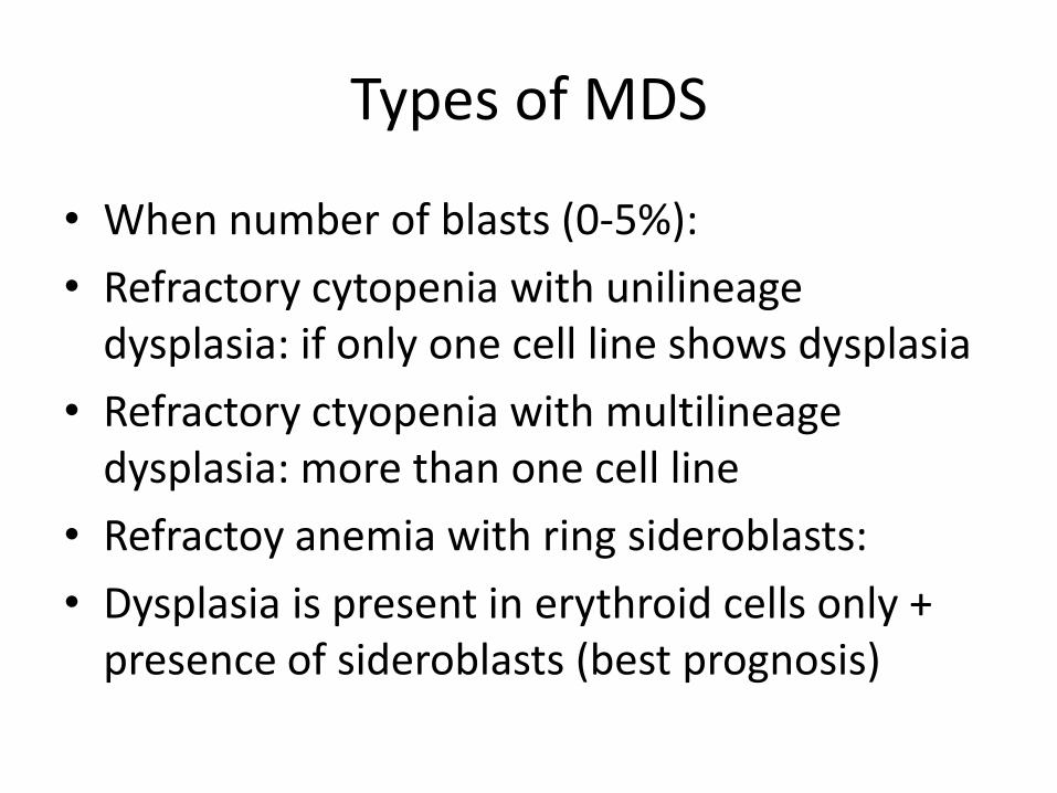
Types of MDS
• When number of blasts (0-5%):
• Refractory cytopenia with unilineage dysplasia: if only one cell line shows dysplasia
• Refractory ctyopenia with multilineage dysplasia: more than one cell line
• Refractoy anemia with ring sideroblasts:
• Dysplasia is present in erythroid cells only + presence of sideroblasts (best prognosis)
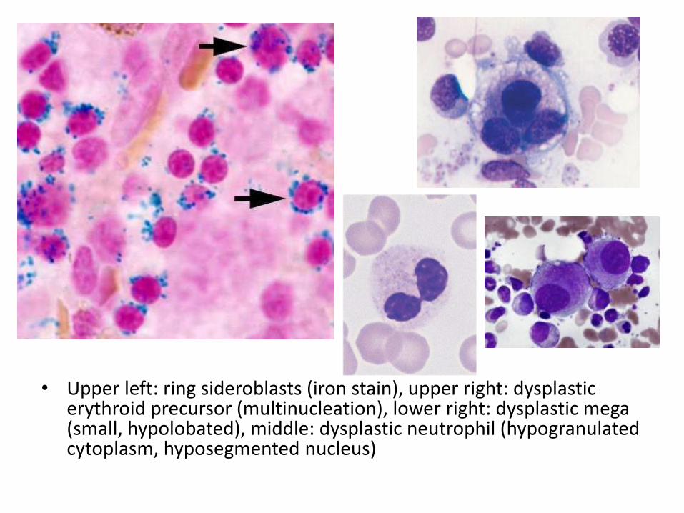
• Upper left: ring sideroblasts (iron stain), upper right: dysplastic erythroid precursor (multinucleation), lower right: dysplastic mega (small, hypolobated), middle: dysplastic neutrophil (hypogranulated cytoplasm, hyposegmented nucleus)
Acute myeloid leukemia
• mutations that impede myeloblast differentiation, and increases proliferation,
• Accumulated blasts leads to marrow failure (myelophthisic anemia)
• AML occurs at all ages, but the incidence rises throughout life
• Diagnosis of AML: blast count is ≥ 20% of bone marrow cells
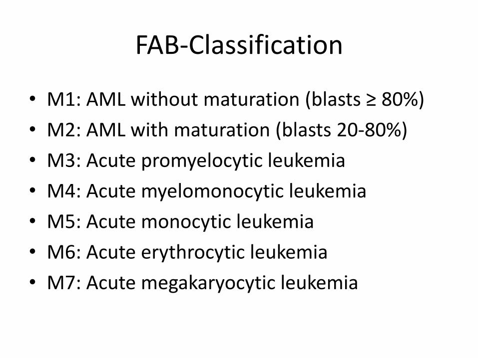
FAB-Classification
• M1: AML without maturation (blasts ≥ 80%)
• M2: AML with maturation (blasts 20-80%)
• M3: Acute promyelocytic leukemia
• M4: Acute myelomonocytic leukemia
• M5: Acute monocytic leukemia
• M6: Acute erythrocytic leukemia
• M7: Acute megakaryocytic leukemia
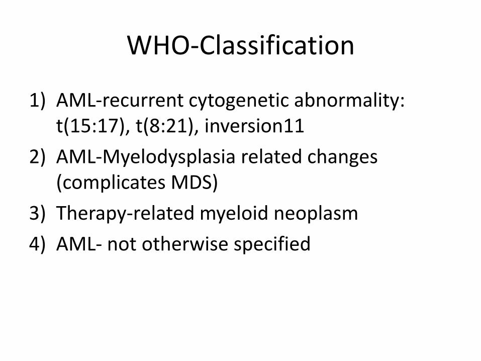
WHO-Classification
1) AML-recurrent cytogenetic abnormality: t(15:17), t(8:21), inversion11
2) AML-Myelodysplasia related changes (complicates MDS)
3) Therapy-related myeloid neoplasm
4) AML- not otherwise specified
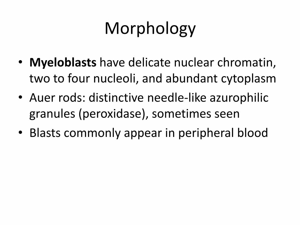
Morphology
• Myeloblasts have delicate nuclear chromatin, two to four nucleoli, and abundant cytoplasm
• Auer rods: distinctive needle-like azurophilic granules (peroxidase), sometimes seen
• Blasts commonly appear in peripheral blood
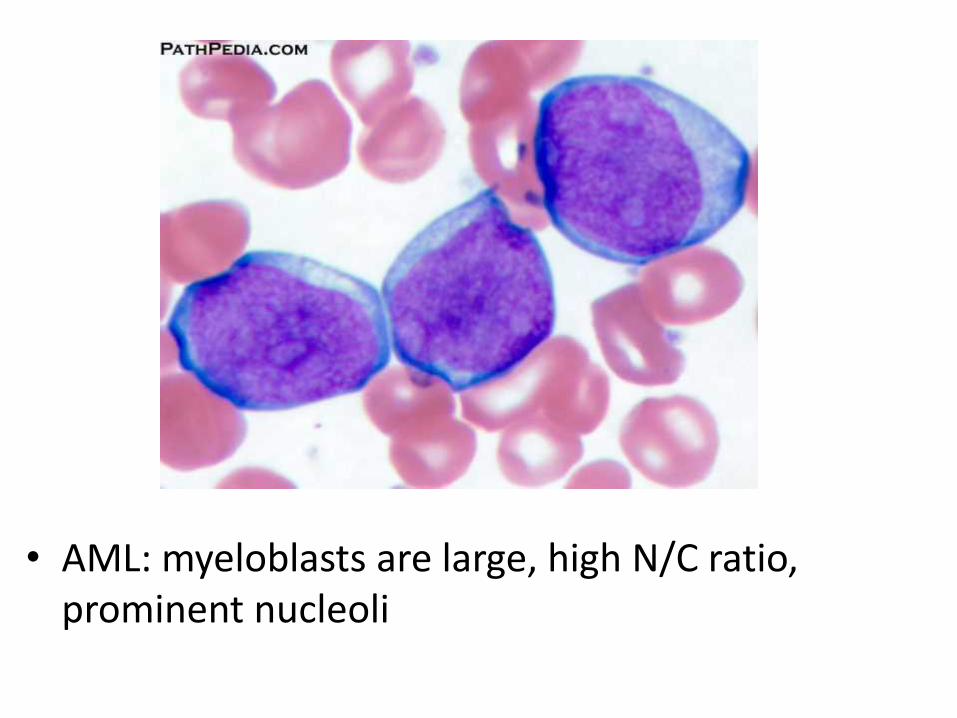
• AML: myeloblasts are large, high N/C ratio, prominent nucleoli
Acute promyelocytic leukemia
• FAB-M3 • WHO: AML-t(15:17), PML-RARA gene fusion • Promyelocytic leukemia gene – retinoic acid receptor
alpha • New protein binds cell DNA, blocking maturation
(reversed by vitamin A) • Cells are arrested at promyelocytic stage, showing
prominent cytoplasmic granules and Auer rods • Malignant promyelocytes secrete tissue factor,
activating thrombin, initiating coagulation cascade (disseminated intravascular coagulation-DIC)
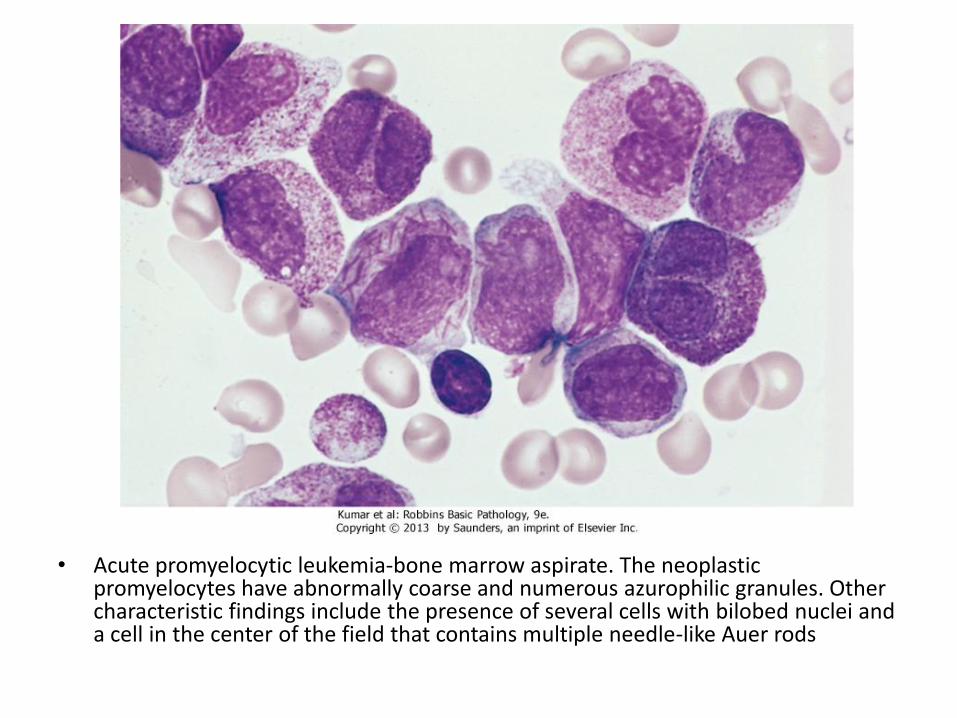
• Acute promyelocytic leukemia-bone marrow aspirate. The neoplastic promyelocytes have abnormally coarse and numerous azurophilic granules. Other characteristic findings include the presence of several cells with bilobed nuclei and a cell in the center of the field that contains multiple needle-like Auer rods
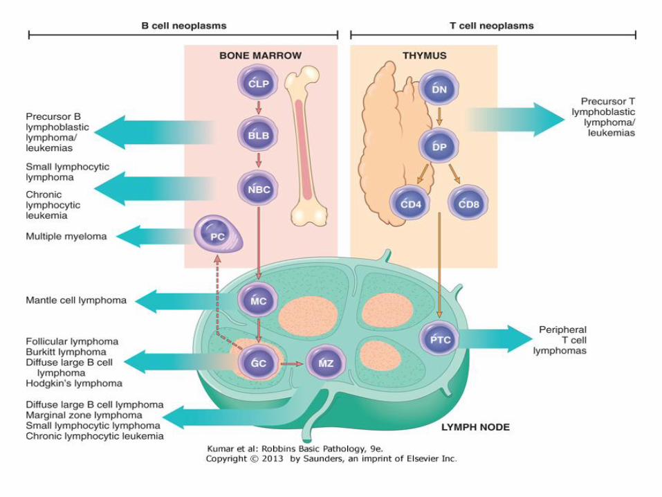
Lymphoid neoplasms
Lymphoma
• Neoplastic disorders originate from B or T lymphocytes
• Most commonly arise in lymph nodes
• If circulates peripheral blood or bone marrow, it is called lymphoid leukemia
• They vary widely in their clinical presentation and behavior, low or high-grade lymphomas
• Generally classified as Hodgkin and non-Hodgkin lymphomas
• Risk factors: immune suppression, chronic inflammation, EBV, HHV8
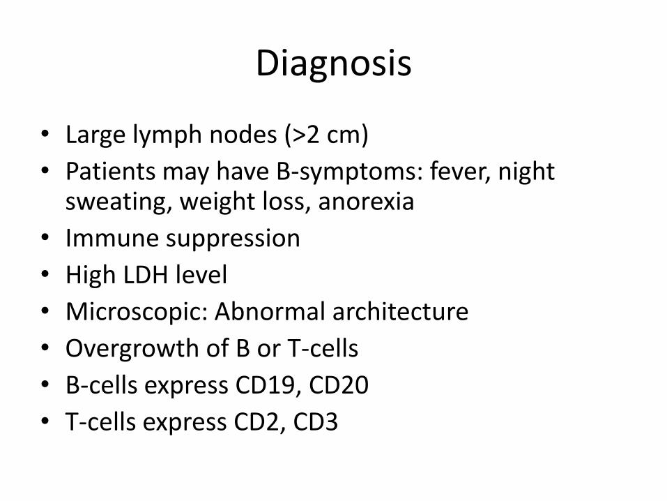
Diagnosis
• Large lymph nodes (>2 cm)
• Patients may have B-symptoms: fever, night sweating, weight loss, anorexia
• Immune suppression
• High LDH level
• Microscopic: Abnormal architecture
• Overgrowth of B or T-cells
• B-cells express CD19, CD20
• T-cells express CD2, CD3
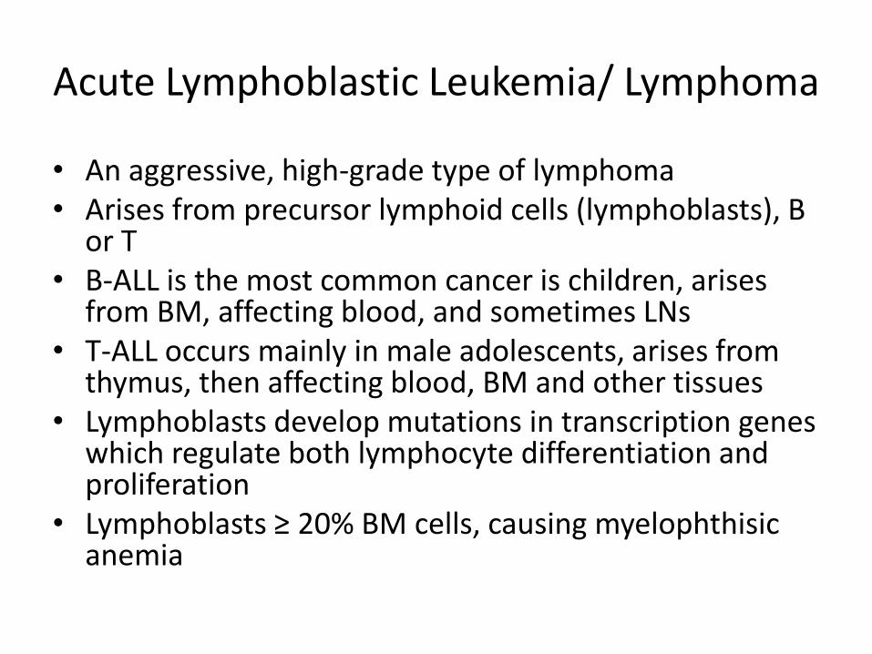
Acute Lymphoblastic Leukemia/ Lymphoma
• An aggressive, high-grade type of lymphoma • Arises from precursor lymphoid cells (lymphoblasts), B
or T • B-ALL is the most common cancer is children, arises
from BM, affecting blood, and sometimes LNs • T-ALL occurs mainly in male adolescents, arises from
thymus, then affecting blood, BM and other tissues • Lymphoblasts develop mutations in transcription genes
which regulate both lymphocyte differentiation and proliferation
• Lymphoblasts ≥ 20% BM cells, causing myelophthisic anemia
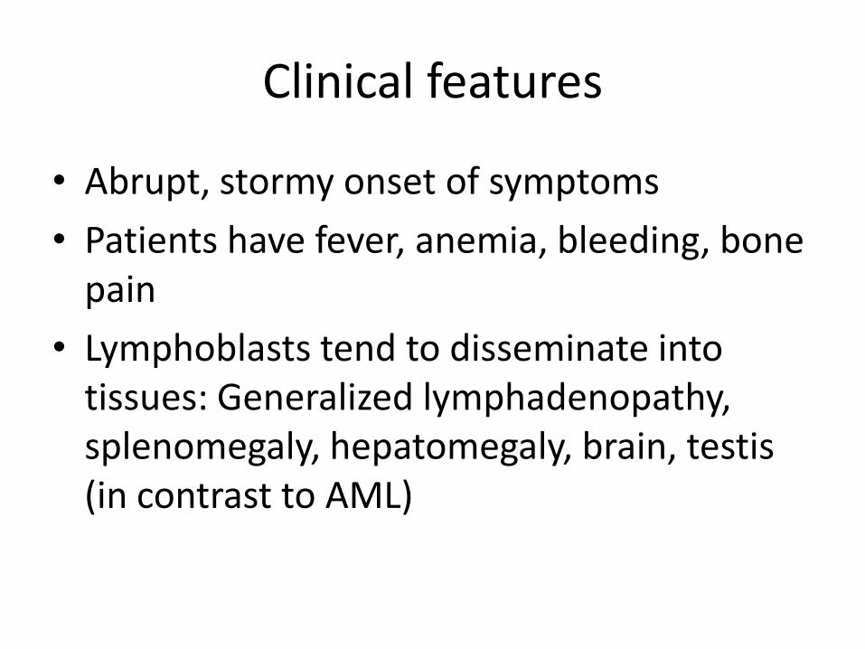
Clinical features
• Abrupt, stormy onset of symptoms
• Patients have fever, anemia, bleeding, bone pain
• Lymphoblasts tend to disseminate into tissues: Generalized lymphadenopathy, splenomegaly, hepatomegaly, brain, testis (in contrast to AML)
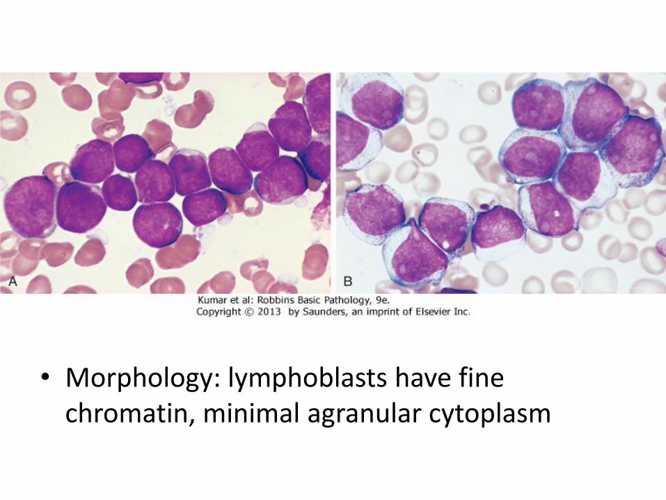
• Morphology: lymphoblasts have fine chromatin, minimal agranular cytoplasm
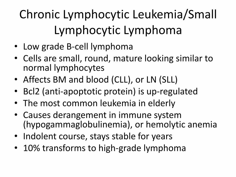
Chronic Lymphocytic Leukemia/Small Lymphocytic Lymphoma
• Low grade B-cell lymphoma • Cells are small, round, mature looking similar to
normal lymphocytes • Affects BM and blood (CLL), or LN (SLL) • Bcl2 (anti-apoptotic protein) is up-regulated • The most common leukemia in elderly • Causes derangement in immune system
(hypogammaglobulinemia), or hemolytic anemia • Indolent course, stays stable for years • 10% transforms to high-grade lymphoma
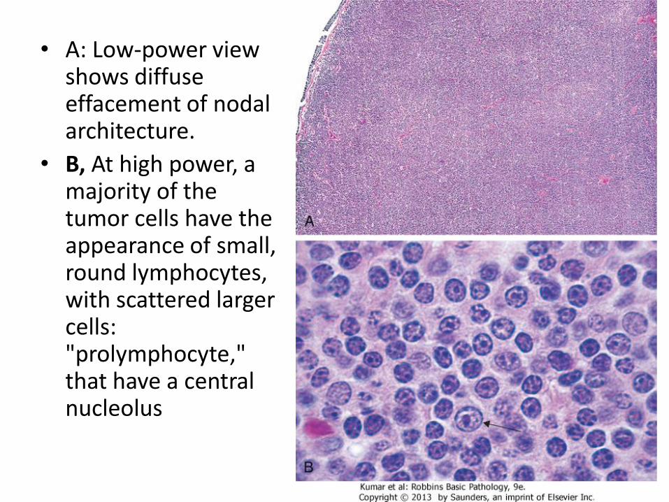
• A: Low-power view shows diffuse effacement of nodal architecture.
• B, At high power, a majority of the tumor cells have the appearance of small, round lymphocytes, with scattered larger cells: "prolymphocyte," that have a central nucleolus
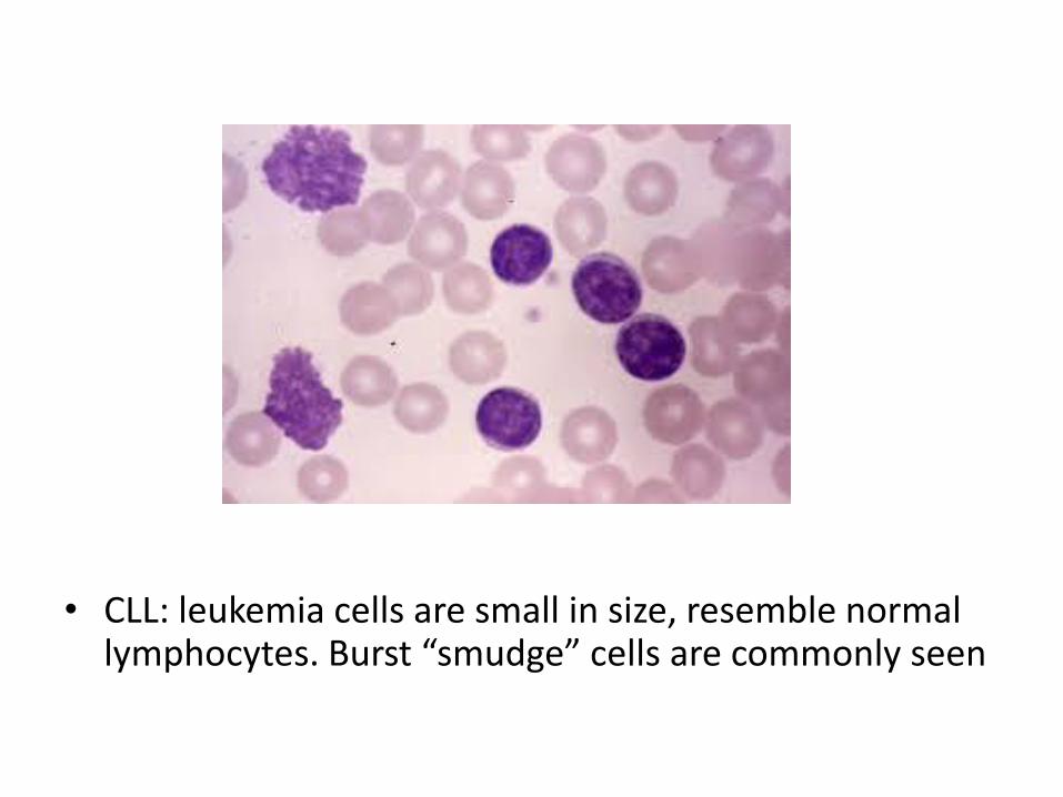
• CLL: leukemia cells are small in size, resemble normal lymphocytes. Burst “smudge” cells are commonly seen
Follicular Lymphoma
• Common (West), low-grade B-cell lymphoma • Affects elderly • Arises from germinal center B-cell • Lymphoma cells have specific translocation t(14:18), in
which Bcl2 gene on chr18 fuses with IgH gene on chr14, causing overexpression of Bcl2
• Patients has generalized lymphadenopathy • Lymphoma cells proliferate to form abnormal, large,
crowded follicles • Patients have indolent course, transforms into high
grade lymphoma in 40% of cases
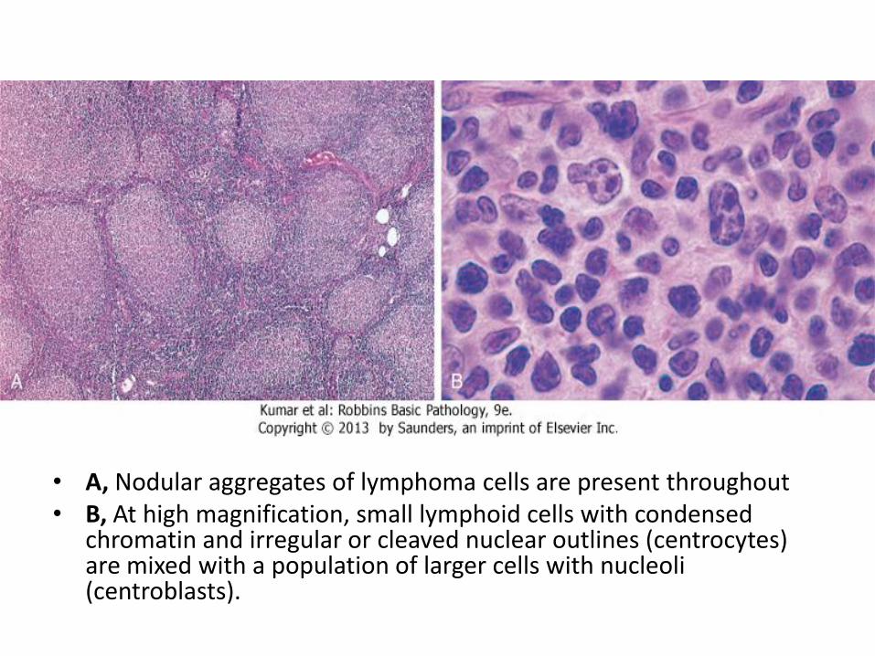
• A, Nodular aggregates of lymphoma cells are present throughout • B, At high magnification, small lymphoid cells with condensed
chromatin and irregular or cleaved nuclear outlines (centrocytes) are mixed with a population of larger cells with nucleoli (centroblasts).
Diffuse Large B Cell Lymphoma
• most common type of lymphoma in adults, accounting for approximately 50% of adult NHLs, also arises in children
• Arises de novo, as a transformation from low grade B-cell lymphoma, in the setting of chronic immune stimulation
• High-grade lymphoma, progressive and fatal if not treated

• Tumor cells have large nuclei with open chromatin and prominent nucleoli.
Burkitt lymphoma
• High-grade B-cell lymphoma
• Endemic in Africa, sporadic worldwide
• High association with EBV
• t(8:14), myc gene fuses with IgH gene, causing overexpression of myc, which activates other transcription factors and causes continuous cell proliferation
• Lymphoma commonly arises in extranodal sites (jaw, ileum)
• Lymphoma is rapidly growing and fatal if not treated
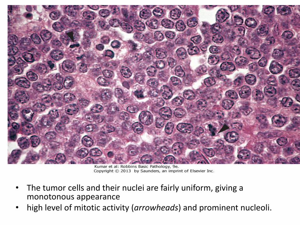
• The tumor cells and their nuclei are fairly uniform, giving a monotonous appearance
• high level of mitotic activity (arrowheads) and prominent nucleoli.
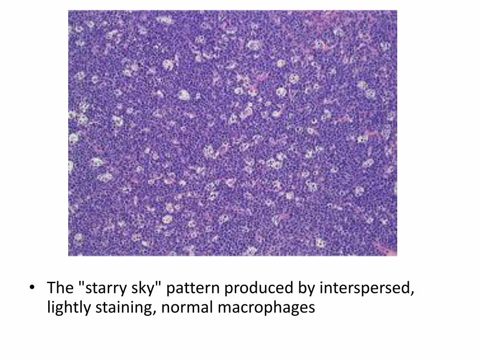
• The "starry sky" pattern produced by interspersed, lightly staining, normal macrophages
Hodgkin Lymphoma
• a group of lymphoid neoplasms that differ from NHL in several respects
• Localized to a single axial group of nodes, most commonly in cervical, axillary and mediastinal LNs
• Orderly spread by contiguity
• Extra-nodal presentation rare
Hodgkin Lymphoma
• Presence of neoplastic giant cells called Reed-Sternberg cells
• RS cells constitute only a minority of tumor size, the rest is composed of reactive lymphocytes, histiocytes and granulocytes
• neoplastic RS cells are derived from crippled, germinal center, B cells
• Immunophenotype is very different from normal B-cells (negative for CD3, CD20, positive for CD30)
• EBV plays a role in the evolution of disease
Clinical features
• Bimodal age distribution: children + old age
• Presents as painless lymphadenopathy
• Constitutional symptoms (B-symptoms), such as fever, night sweats, and weight loss are common
• Spread: nodal disease first, then splenic disease, hepatic disease, and finally involvement of the marrow and other tissues
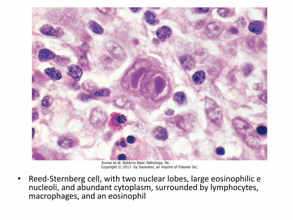
• Reed-Sternberg cell, with two nuclear lobes, large eosinophilic e nucleoli, and abundant cytoplasm, surrounded by lymphocytes, macrophages, and an eosinophil
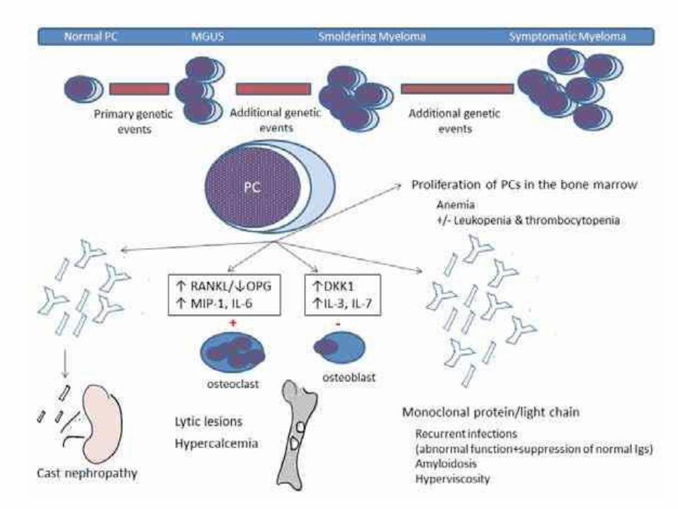
Plasma cell myeloma
• Neoplasm of plasma cells that secrets monoclonal Immunoglobulin (M-protein)
• 10% of BM tumors
• Arises from long-lived plasma cells in the BM
• Aggressive tumor, difficult to control
• Affects elderly
• Clinically known as multiple myeloma
Pathogenesis
• Risk factors: older age, male, blacks, radiation, family history, obesity?
• Accumulation of genetic mutations and chromosomal aberrations
• Transformed plasma cells proliferate modestly, interact with stromal cells in BM (resistant to chemotherapy)
• Secrete IgG (>other Igs)
• Plasma cell count ≥ 10%
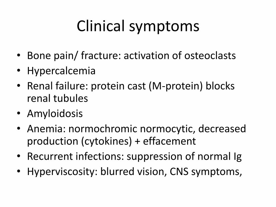
Clinical symptoms
• Bone pain/ fracture: activation of osteoclasts
• Hypercalcemia
• Renal failure: protein cast (M-protein) blocks renal tubules
• Amyloidosis
• Anemia: normochromic normocytic, decreased production (cytokines) + effacement
• Recurrent infections: suppression of normal Ig
• Hyperviscosity: blurred vision, CNS symptoms,
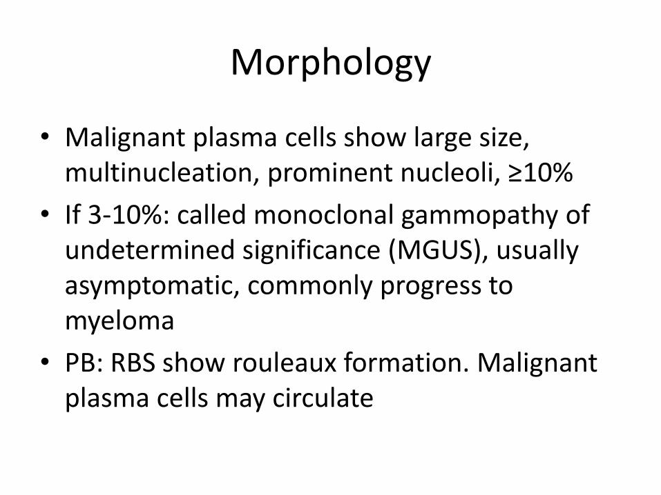
Morphology
• Malignant plasma cells show large size, multinucleation, prominent nucleoli, ≥10%
• If 3-10%: called monoclonal gammopathy of undetermined significance (MGUS), usually asymptomatic, commonly progress to myeloma
• PB: RBS show rouleaux formation. Malignant plasma cells may circulate
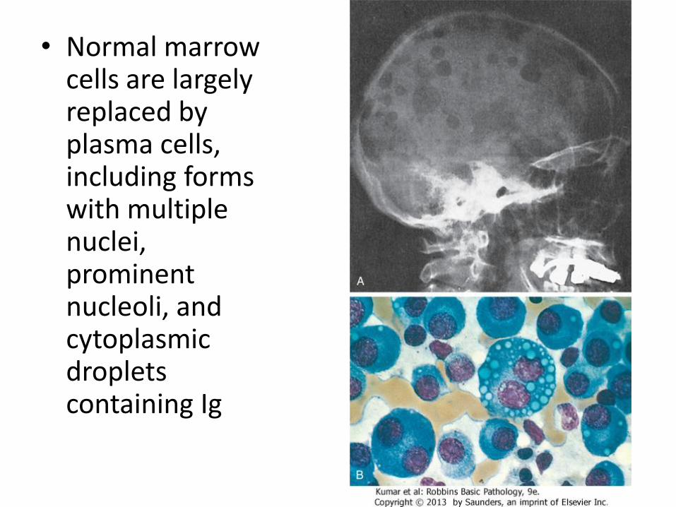
• Normal marrow cells are largely replaced by plasma cells, including forms with multiple nuclei, prominent nucleoli, and cytoplasmic droplets containing Ig
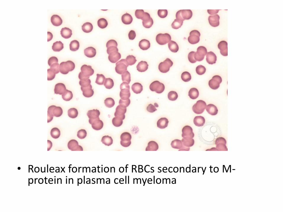
• Rouleax formation of RBCs secondary to M-protein in plasma cell myeloma
Bleeding disorders
Types of clotting factor deficiency
• Acquired (more common, multiple): a) Chronic liver disease b) Vitamin K deficiency (Warfarin) c) microangiopathic hemolytic anemias d) Autoimmune • Hereditary: Hemophilias A, B Von Willibrand factor deficiency
Hemophilia A
• Inherited factor VIII deficiency • Normally, factor VIII is a cofactor for IX, activating factor X
• X-linked
• 70% has family history, 30% new mutation
• Spontaneous bleeding occur at levels ≤ 1% normal activity
• Milder deficiencies may only become apparent when other predisposing conditions, such as trauma, are also present
• 90% have quantitative deficiency, 10% functional

Clinical features
• Easy bruising
• Early in life: circumcision, crawling
• Bleeding tends to occur in deeper sites (joints, muscles, gum, GI, GU, nosebleed, menorrhagea, post surgery)
• Joint deformity
• Skin bleeding (petechiae) is typically absent (capillary leak)
• Anemia of blood loss
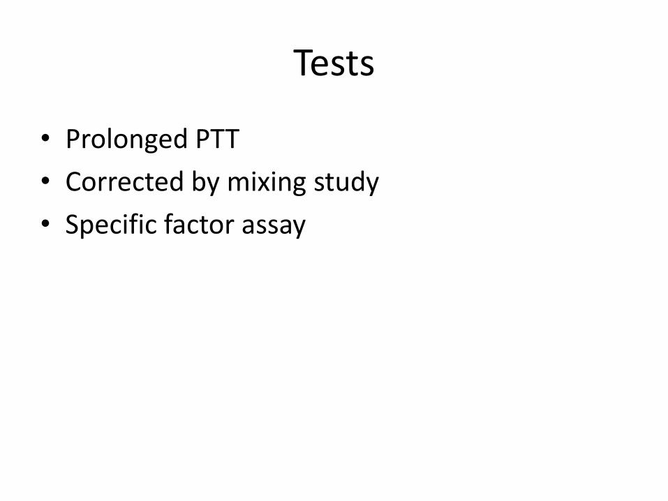
Tests
• Prolonged PTT
• Corrected by mixing study
• Specific factor assay
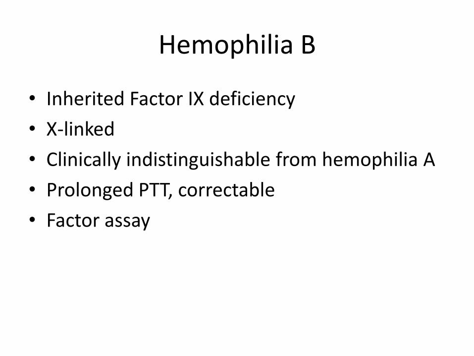
Hemophilia B
• Inherited Factor IX deficiency
• X-linked
• Clinically indistinguishable from hemophilia A
• Prolonged PTT, correctable
• Factor assay
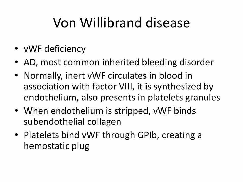
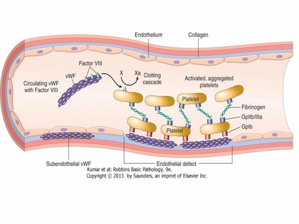
Von Willibrand disease
• vWF deficiency
• AD, most common inherited bleeding disorder
• Normally, inert vWF circulates in blood in association with factor VIII, it is synthesized by endothelium, also presents in platelets granules
• When endothelium is stripped, vWF binds subendothelial collagen
• Platelets bind vWF through GPIb, creating a hemostatic plug
vWF deficiency
• Type 1: Quantitative • Type 2: Qualitative, abnormal binding to platelets,
short half life (thrombocytopenia) • No platelets adhesion, bleeding • Factor VIII is not stabilized, secondary factor IIIV
deficiency • Bleeding from wounds, mucous membranes, deep
seated organs • Usually mild disease • PTT is prolonged • Bleeding time: can be prolonged
Platelet disorders
• Immune thrombocytopenic purpura (ITP)
• Microangiopathic hemolytic anemia:
• (a) Thrombotic thrombocytopenic purpura (TTP)
• (b) Disseminated intravascular coagulation (DIC)
ITP
• Acquired disease
• Auto antibodies (IgG) against platelets GP IIb/IIIa, synthesized in spleen
• Coated platelets are engulfed and destroyed in spleen
• Acute setting: children, follows viral infection, self limited
• Chronic: middle age, females, persistent, benefit of splenectomy
• PB: isolated thrombocytopenia, platelets are large
• BM: active proliferating megakaryocytes
• Patients present with petechiae, bruising and internal organ bleeding
TTP
• Pentad of fever, thrombocytopenia, microangiopathic hemolytic anemia, transient neurologic deficits, and renal failure
• A similar disease, Hemolytic Uremic Syndrome, has similar manifestations but is distinguished from TTP by the absence of neurologic symptoms, the dominance of acute renal failure, and frequent occurrence in children after infection with E.Coli
• In both, there is a widespread formation of platelet-rich thrombi in the microcirculation. The consumption of platelets leads to thrombocytopenia, and the narrowing of blood vessels by the platelet-rich thrombi results in a microangiopathic hemolytic anemia
Pathogenesis
• Deficiency in ADAMTS 13, a protease enzyme that degrades precursor of vWF (very-high-molecular-weight multimers)
• Abnormal large vWF multimers accumulate in plasma
• Giant vWF multimers promote the formation of platelet microaggregates throughout the circulation
• ADAMTS 13 deficiency can be inherited or acquired (auto antibodies, more common)
• No activation of coagulation cascade (normal PT, PTT)
DIC
• Systemic activation of coagulation and results in the formation of thrombi throughout the microcirculation
• As a consequence, platelets and coagulation factors are consumed and, secondarily, fibrinolysis is activated, results in secondary bleeding
• Thus, bleeding time, PT and PTT are prolonged
Pathogenesis
• Intense activation of the extrinsic or intrinsic pathways, or widespread endothelial injury
Examples of pathways activation:
• Release of tissue factor in APL, or by monocytes in sepsis (secondary to endotoxins)
• Release of mucin by carcinoma cells
• Release of placenta fragments in circulation
• Release of brain tissue in circulation after trauma
Pathogenesis
Examples of widespread endothelial injury:
• Snake venom
• burn
• heat
• Systemic lupus erythematosus (immune complex deposition)
Clinical features
• Microangiopathic hemolytic anemia is an emergency situation
• Patients may bleed to death if not treated