which new cartilage repair options are worth considering
TRANSCRIPT
Which New Cartilage Repair Options are Worth Considering for Our Patients?
Jack Farr, MDIndianapolis, IN
OrthoIndy Center for Knee Preservation, Cartilage Regeneration and OrthoBiologics

Financial Disclosures(AAOS disclosures up to date)
Royalties• Arthrex
Consulting• Arthrex• JRF• MedShape• Moximed• Norvartis• Organogenesis• Samumed
Books• 2018 Farr & Gomoll Editors of Cartilage Restoration: Practical
Clinical Applications (royalties donated)
Use of Product Brand Names is for Clarity OnlyThis is NOT an Endorsement or Product Promotion

What is Here Now?
• Autograft Osteochondral Plugs
• Fresh Osteochondral Allograft (multiple tissue banks)
• Marrow Stimulation and Augments
• Cell Therapy MACI/ACI
• Sterilized acellular OCA
• PJAC
• “OCA” with “minimal bone”
• Particulated Autograft Cartilage

Why Not More Options? Significant Barriers to the Market
Human cells, tissues, and cellular and tissue-based products
HCT/P 351, which requires FDA monitored RCT for Biologic License if:
• Manipulation of Cells/Tissue
• Culturing
• Addition of Growth Factors
• Non-Homologous use
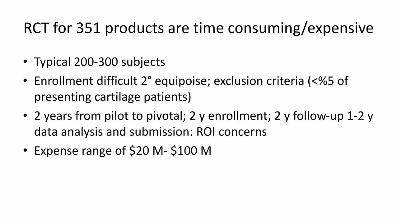
RCT for 351 products are time consuming/expensive
• Typical 200-300 subjects
• Enrollment difficult 2° equipoise; exclusion criteria (<%5 of presenting cartilage patients)
• 2 years from pilot to pivotal; 2 y enrollment; 2 y follow-up 1-2 y data analysis and submission: ROI concerns
• Expense range of $20 M- $100 M

Cancelled 2° ROI
DeNovo ET (Zimmer)
allogeneic chondrocyte implant
study cancelled in Phase 3 due to enrollment issues
CAIS (DePuy Mitek)
particulated cartilage autograft
study cancelled in Phase 3 due to enrollment issues
Chondrocelect (Tigenix)
ACI variant with optimized culture conditions
Company elected not to start study and close US cell
culture facility after discussions with FDA on design

Current HCT/P 351 FDA approved Trials
– Agili-C (Cartiheal)Coral based acellular plug
– Gelrin C (Regentis)PEG/Fibrinogen hydrogel cured in situ with UV
– Novocart 3D (Aesculap)autologous chondrocyte implant in phase 3
– Cartistem (Medipost)allogeneic umbilical cord blood stem cells in phase 2
– Adipose-derived stem cells (Stanford)autologous single-step ADSC RCT against MFx
– HyalofastTM (Anika Therapeutics)– Nonwoven hyaluronic acid (HA) with BMAC
– Neocart (Histogenics)failed superiority in phase 3

Option “around” HCT/P 351 required RCT
• HCT/P 361 allows allograft tissue use which is not FDA regulated IF:
Minimally Manipulated
Homologous Tissue Use
Not combined with other materials/drugs/GF

361 “Gray Areas”FDA announced Nov 2017 there may be stricter definitions in Nov 2020
• Is mincing/particulating/micronizing minimally manipulative?
• Is separation of blood products manipulative?
• Is use of bone marrow aspirates and blood products homologous to the joint?
• Is fat homologous to the joint?
• Are placenta and amnion homologous to the joint?

Does FDA approval or 361 allowed use mean insurance will pay? NO
• CMS medical policies
• Private Payer individual insurance policies
• Based on Medical Policy on Medical Policies
Adequately powered RCT
2 or more independent studies in the English peer-reviewed literature
Long-term outcomes

361 Products in Use (No RCT trials)
• Marrow stimulation augments, e.g., micronized cartilage
• PJAC
• Sterilized decellularized OCA
• Minimal bone cartilage disks
• Viable allograft cartilage strips/ECM
• Particulated autograft cartilage

How to add New Products into Your PracticeWhen there are no LOE 1 studies
• Evaluation of Novel Cartilage Treatment Options for Clinical Use, Farr and Sherman, SMAR 2018
• Develop a personal system to evaluate each novel product—as new is not always better.

One Approach to New Products
1. Read the pre-clinical literature; understand FDA regulatory pathway regarding loose or strict regulations for the particular products2. Read the industry produced literature (“white papers” or sponsored case series) with a critical eye 3. Seek out the design surgeons to learn the “best results” possible and current indications 4. Seek out “earlier adapters” for their experience 5. Attend podium presentations and/or read about early clinical series in the literature 6. Conduct your own pilot historical controlled trial of approximately 10-20 patients----and wait……………. 7. Evaluate your results and compare to historical control and other surgeons/literature

A cautionary tale: Decellularized OCA
• Applications similar to OCA• 361 classification allowed clinical implementation without human
trials: 2010 Equine trial then available for human implantation 2011+ Early Clinical Outcomes Associated with a Novel Osteochondral Allograft Transplantation System in the Knee, Long et al, Adv Orth Surg, Long et al 2016- High Failure Rate of a Decellularized Osteochondral Allograft for the Treatment of Cartilage Lesions, Farr et al, AJSM 2016- Acute Delamination of Commercially Available Decellularized Osteochondral Allograft Plugs, Degen et al, Cartilage 2016

Farr et al Failures
• Twenty-three of the 32 knees (72%) were considered failures
• 14 of the 32 knees (43%) had further surgery
• Implant survivorship was 19.6% at 2 years
Failure defined as structural damage of the graft diagnosed by: Arthroscopy or MRIAnd/or any reoperation resulting in removal of the allograft
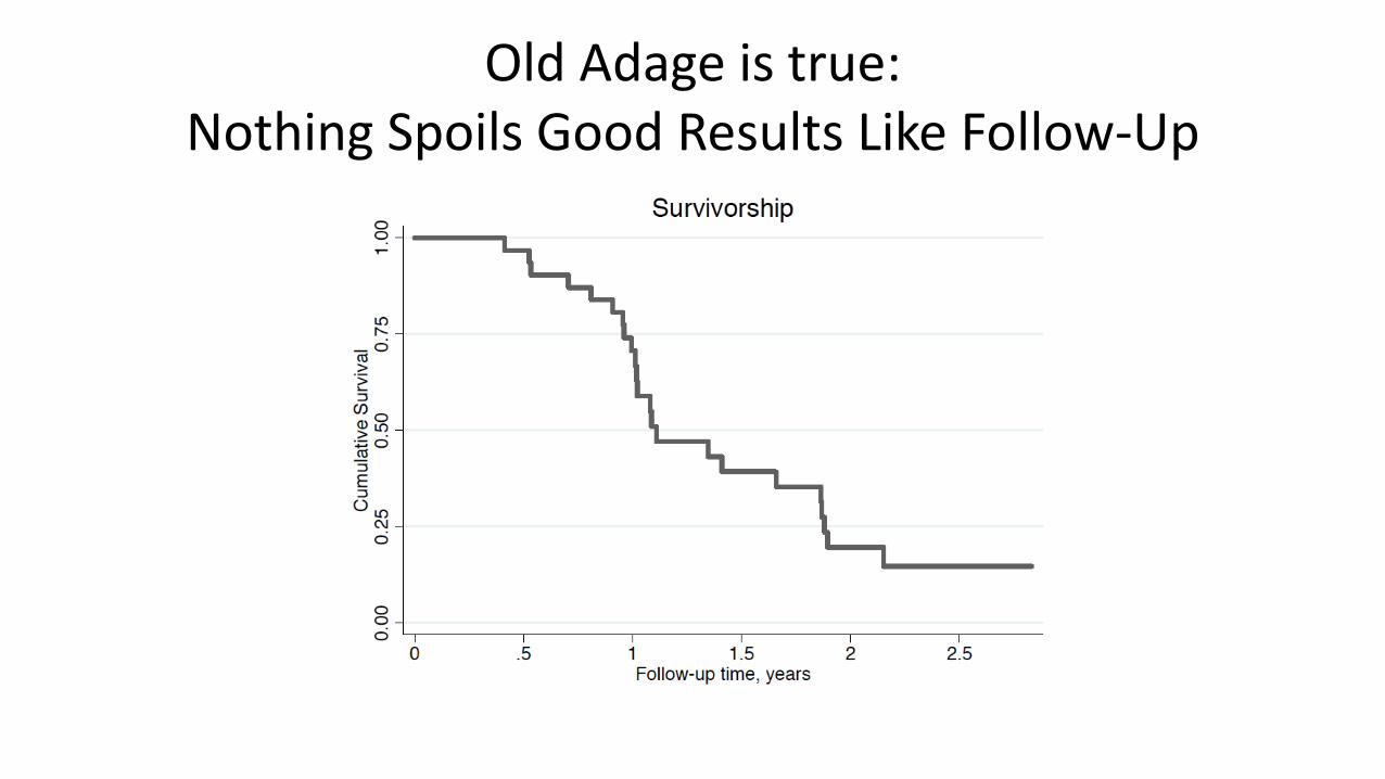
Old Adage is true:Nothing Spoils Good Results Like Follow-Up

Current Options

Particulated Juvenile Allograft Cartilage
• Fresh stored articular cartilage
• Juvenile - donors < 13 years old
• Higher (>10x) chondrocyte density
• Minced into 1 mm cubes
• Increases surface area
• Facilitates chondrocyte outgrowth
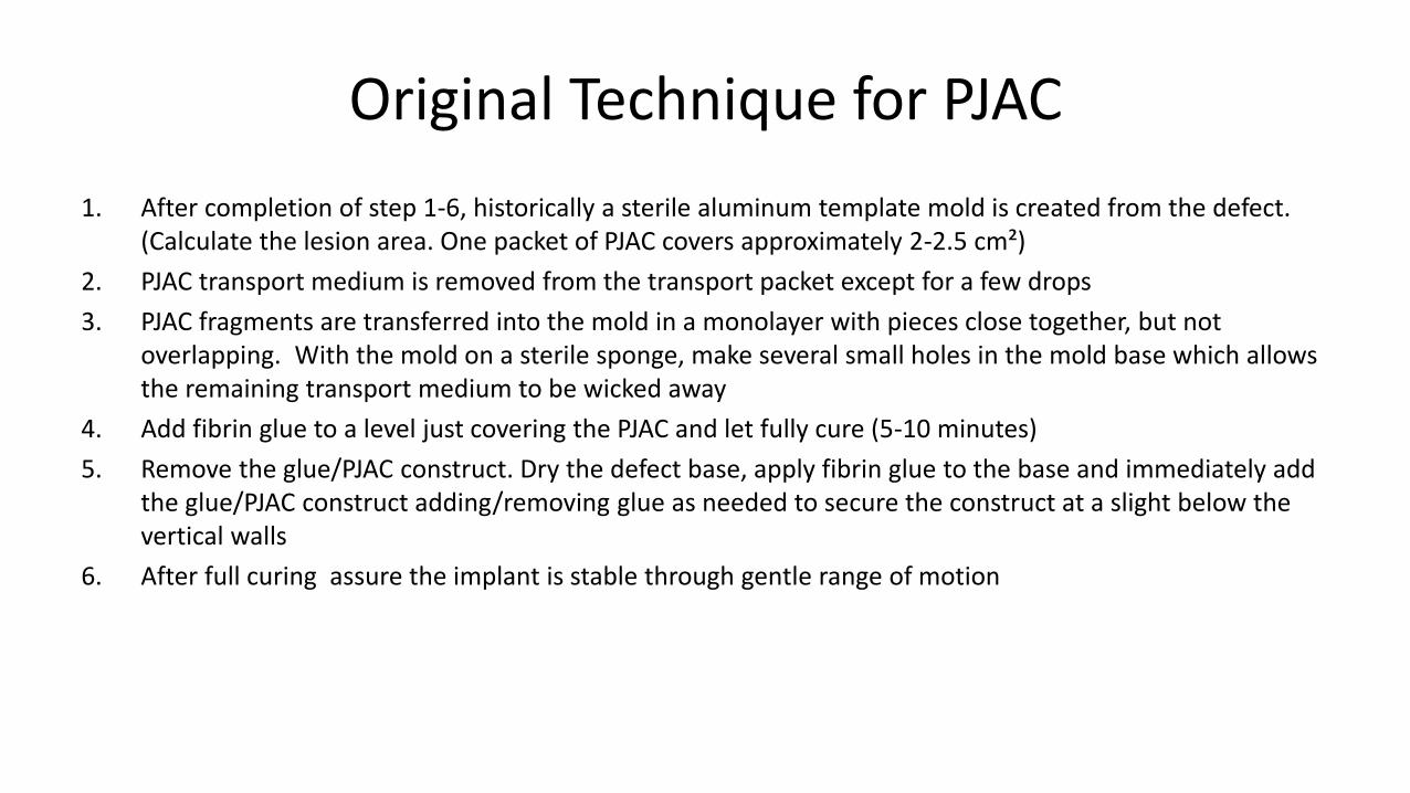
Original Technique for PJAC
1. After completion of step 1-6, historically a sterile aluminum template mold is created from the defect. (Calculate the lesion area. One packet of PJAC covers approximately 2-2.5 cm²)
2. PJAC transport medium is removed from the transport packet except for a few drops
3. PJAC fragments are transferred into the mold in a monolayer with pieces close together, but not overlapping. With the mold on a sterile sponge, make several small holes in the mold base which allows the remaining transport medium to be wicked away
4. Add fibrin glue to a level just covering the PJAC and let fully cure (5-10 minutes)
5. Remove the glue/PJAC construct. Dry the defect base, apply fibrin glue to the base and immediately add the glue/PJAC construct adding/removing glue as needed to secure the construct at a slight below the vertical walls
6. After full curing assure the implant is stable through gentle range of motion

Original Surgical Technique

Most Commonly used Method for PJAC
1. Steps 1 and 2 as above, but place the PJAC fragments directly into the defect noting the scant amount of transport medium allows the fragments to adhere even on non-horizontal surfaces (move the limb/table angle to avoid fully vertical or higher angles). Fragments are monolayer and touching or near touching.
2. Wick excess transport medium from the margin with a sponge
3. Apply fibrin glue and wait until fully cured before checking with gentle range of motion

Most Commonly Used Method for PJAC
Once glued in position, both techniques appear the same.

Alternative Method for PJACCollagen patch in place over PJAC in:
Monopolar lesion Bipolar lesion

Original lesion
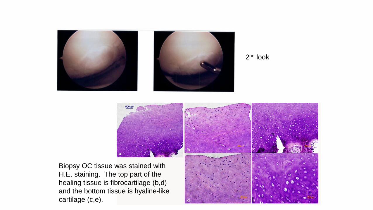
Biopsy OC tissue was stained with
H.E. staining. The top part of the
healing tissue is fibrocartilage (b,d)
and the bottom tissue is hyaline-like
cartilage (c,e).
2nd look

PJAC Pilot Knee StudyHistology of PJAC Graft at 2 yrs Post Op
Masson’s Trichrome Saf-O Col I Col II
37 yo Male, 3.4 cm2 MFC lesion, Prior MFX
Good integration
of repair tissue
with underlying
bone
High proteoglycan
content
Minimal collagen I High collagen II

PJACLong Term Observational Study in the Knee
• 15 Clinical centers, 197 lesions in 155 subjects (1/1/15)– 71@Preop, 100@12M, 58@18M, 73@24M, 60@36M, 36@48M, 17@60M
• Primary outcomes measures:– KOOS
– Graft survival
– Workdays lost pre- and post-op
• Demographics:– Mean age 32.8 +/- 9.1 (range 15-60)
– BMI 28.0 +/- 5.4 (range 17.6-46.2)
– 4.6 +/- 5.8 years from injury (range 0.0-25.2)
– 1.4 +/- 2.8 years from most recent symptom onset (range 0.0–20.7)

PJACLong Term Observational Study in the Knee
• Total Lesion Size 3.5cm2 per patient– Majority of lesions ≥ grade 3C
– 25% of subjects have >1 lesion
– 112 Femoral lesions (57%) mean 2.9cm2
– 83 Patella lesions (42%) mean 2.6cm2
– 2 Tibial lesions (1%) mean 1.8cm2
• 86 subjects (55.5%) ≥1 Concomitant Surgery:– 53 (34% of total) patellar realignment
– 22 (14% of total) meniscus
– 7 (5% of total) ligament

PJACLong Term Observational Study in the Knee
• 77% of subjects ≥ 1 prior surgery on Index Knee (mean 1.7, range 1-8)– 227 surgeries in 119 subjects
• 59% of subjects ≥1 cartilage repair on index knee– 125 surgeries in 91 subjects
– 44% Debridement
– 15% Microfracture
– 2% OC allograft, 2% ACI + periosteum
– 18 % other (fragment fixation, loose body removal…)

PJACKOOS Subdomain Scores
All post op times are statistically significant v baseline at p<0.05 except for Sports at 6M

PJAC ReoperationsLong Term Observational Study in the Knee
• 43 subjects (27% of total) had 55 reoperations:
– 20 (13%) chondromalacia
– 11 (7%) arthrofibrosis
– 14 (10%) painful hardware
– 6 (4%) DJD
– 16 (10%) total revisions at mean of 20.3 months
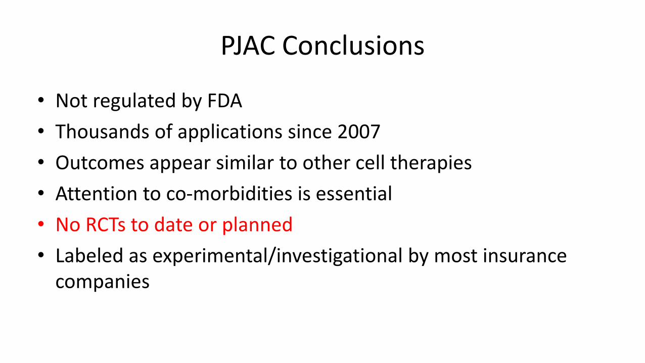
PJAC Conclusions
• Not regulated by FDA
• Thousands of applications since 2007
• Outcomes appear similar to other cell therapies
• Attention to co-morbidities is essential
• No RCTs to date or planned
• Labeled as experimental/investigational by most insurance companies
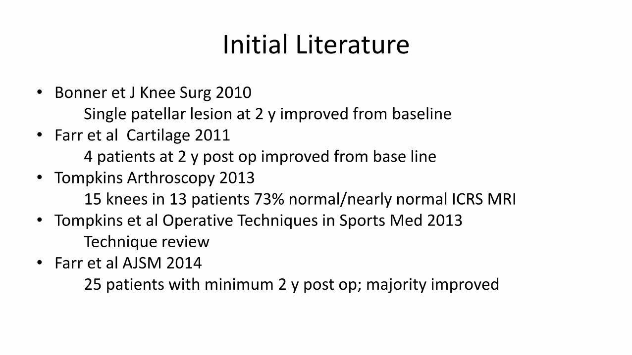
Initial Literature
• Bonner et J Knee Surg 2010 Single patellar lesion at 2 y improved from baseline
• Farr et al Cartilage 2011 4 patients at 2 y post op improved from base line
• Tompkins Arthroscopy 2013 15 knees in 13 patients 73% normal/nearly normal ICRS MRI
• Tompkins et al Operative Techniques in Sports Med 2013Technique review
• Farr et al AJSM 2014 25 patients with minimum 2 y post op; majority improved

Additional Literature
Cartilage Regeneration in Full-Thickness Patellar Chondral Defects Treated with Particulated Juvenile Articular Allograft Cartilage: An MRI Analysis Grawe et al Cartilage. 2017
Cartilage repair in the degenerative ageing knee: A narrative review and analysis Brittberg et al, Acta Orthop. 2016
Use of chondral fragments for one stage cartilage repair: A systematic review Bonasia et al World J Orthop. 2015
Use of Particulated Juvenile Articular Cartilage Allograft for Osteochondral Lesions of the Wrist Hess et al Hand (N Y) 2017
Particulated articular cartilage for symptomatic chondral defects of the knee Riboh et al Curr Rev MusculoskeletMed. 2015
Combined Particulated Juvenile Cartilage Allograft Transplantation and Medial Patellofemoral Ligament Reconstruction for Symptomatic Chondral Defects in the Setting of Recurrent Patellar InstabilityArshi et al Arthrosc Tech. 2016
Particulated Juvenile Articular Cartilage (PJAC) Allograft for Treatment of Lateral Femoral Condyle defect in High-Performance Athletes Bucci et alOrthop J Sports Med. 2018
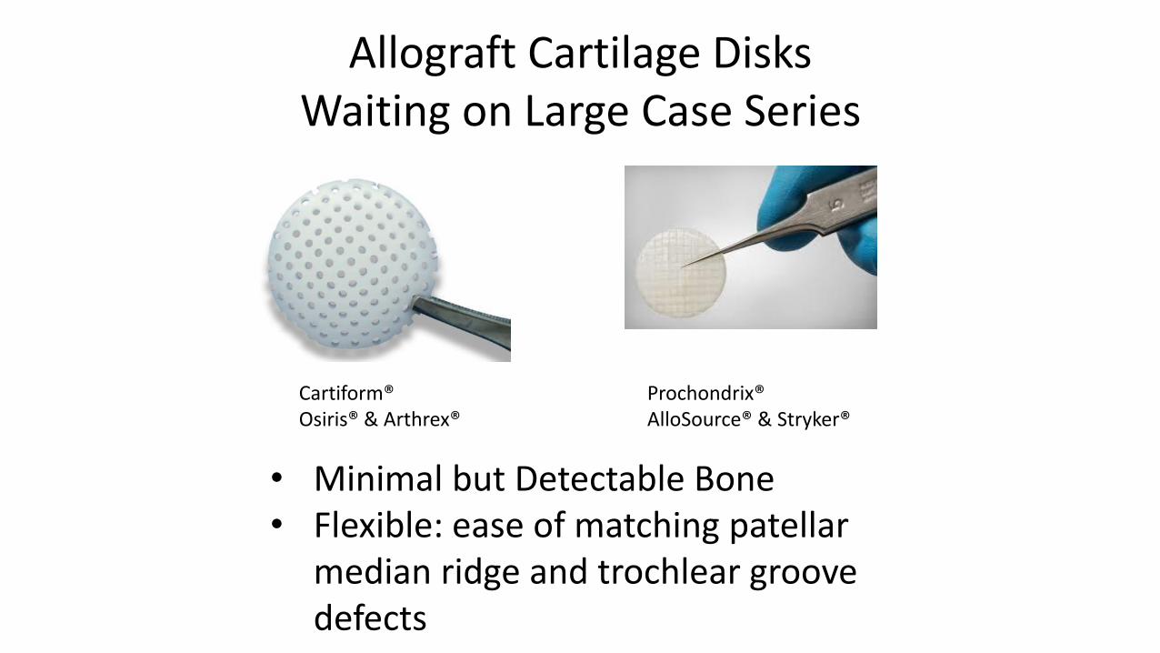
Allograft Cartilage DisksWaiting on Large Case Series
• Minimal but Detectable Bone• Flexible: ease of matching patellar
median ridge and trochlear groove defects
Cartiform®Osiris® & Arthrex®
Prochondrix®AlloSource® & Stryker®
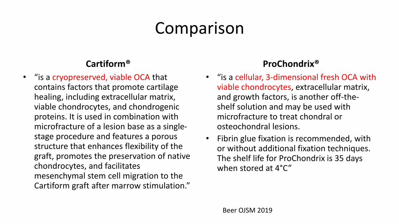
Comparison
Cartiform®
• “is a cryopreserved, viable OCA that contains factors that promote cartilage healing, including extracellular matrix, viable chondrocytes, and chondrogenic proteins. It is used in combination with microfracture of a lesion base as a single-stage procedure and features a porous structure that enhances flexibility of the graft, promotes the preservation of native chondrocytes, and facilitates mesenchymal stem cell migration to the Cartiform graft after marrow stimulation.”
ProChondrix®
• “is a cellular, 3-dimensional fresh OCA with viable chondrocytes, extracellular matrix, and growth factors, is another off-the-shelf solution and may be used with microfracture to treat chondral or osteochondral lesions.
• Fibrin glue fixation is recommended, with or without additional fixation techniques. The shelf life for ProChondrix is 35 days when stored at 4°C”
Beer OJSM 2019

Case Example 2013
Caton-Deschamps: 1.0TT-TG: 9mmTT-PCL: 24mm

Standard “Cell Therapy” Lesion Prep

Use of Cryopreserved Allograft Cartilage

Follow-Up Imaging
6 mon MRI:
Implant expansion of thickness with good basilar integration, incomplete marginal integration; small joint effusion
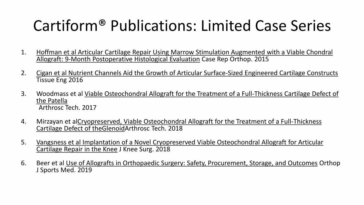
Cartiform® Publications: Limited Case Series
1. Hoffman et al Articular Cartilage Repair Using Marrow Stimulation Augmented with a Viable Chondral Allograft: 9-Month Postoperative Histological Evaluation Case Rep Orthop. 2015
2. Cigan et al Nutrient Channels Aid the Growth of Articular Surface-Sized Engineered Cartilage ConstructsTissue Eng 2016
3. Woodmass et al Viable Osteochondral Allograft for the Treatment of a Full-Thickness Cartilage Defect of the PatellaArthrosc Tech. 2017
4. Mirzayan et alCryopreserved, Viable Osteochondral Allograft for the Treatment of a Full-Thickness Cartilage Defect of theGlenoidArthrosc Tech. 2018
5. Vangsness et al Implantation of a Novel Cryopreserved Viable Osteochondral Allograft for Articular Cartilage Repair in the Knee J Knee Surg. 2018
6. Beer et al Use of Allografts in Orthopaedic Surgery: Safety, Procurement, Storage, and Outcomes Orthop J Sports Med. 2019

Viable allograft cartilage strips/ECM
• CartiMax® by MTF ® /Conmed ®
• “Cryopreserved, viable, cartilage fibers combined with cartilage allograft matrix to make a biologically-active scaffold with putty-like handling characteristics used to treat focal cartilage defects. This product offers the potential to facilitate cartilage healing and regeneration along with ease of use.”
• Prepare defect as per cell therapy; create the CartiMax® putty and fill defect; no scaffold necessary

Conmed® Literature Support of CartiMax®
1Data on File, MTF Biologics.2Albrecht F, Roessner A, Zimmermann E. Closure of osteochondral lesions using chondral fragments and brin adhesive. Archives of orthopaedic and traumatic surgery Archiv fur orthopadische und Unfall-Chirurgie. 1983;101:213–7. [PubMed].3Lu Y, Dhanaraj S, Wang Z, et al. Minced cartilage without cell culture serves as an effective intraoperative cell source for cartilage repair. Journal of orthopaedicresearch : official publication of the Orthopaedic Research Society. 2006;24:1261–70. [PubMed].4Frisbie DD, Lu Y, Kawcak CE, DiCarlo EF, Binette F, McIlwraith CW. In vivo evaluation of autologous cartilage fragment-loaded scaffolds implanted into equine articular defects and compared with autologous chondrocyte implantation. The American journal of sports medicine. 2009;37(Suppl 1):71S–80S.

CartiMax® Specific PublicationsNone found on PubMed & Google searches 4/15/2020
Per Conmed®
“These grafts have been studied in a small number of patients and preliminary outcomes (up to 6 months) are very promising with early indications of decrease in pain and increase in IKDC [International Knee Documentation Committee] and KOOS [Knee Injury and Osteoarthritis Outcome Score] scores, no significant bone edema or graft delamination, and early observation of complete fill and excellent incorporation. We continue to collect data and plan to publish clinical results in the coming months.”

Particulated Autograft
• First Animal Study (Rabbit) in German literature, Albrecht 1983
• Cartilage Autograft Implant System Cole, Farr, et al AJSM 2011
2 year post-op

KOOS change from baselineKOOS pain KOOS Symptoms/Stiffness KOOS ADL
KOOS Func/ Sports & Recreation KOOS Quality of Life
Cole, Farr et al AJSM 2012

CAIS cancelled for ROI concernsCurrent Particulated Autograft Cartilage Options
• Manual Chipped cartilage pursued in Denmark
Foldager Cartilage 2015 Clinical Case Series
Christensen 2016 Mini Pig
• Mechanical particulated autograft: Reveille™ from Exactech 2017
• Capture of shaver harvested autograft: GraftNet® from Arthrex 2019

Exactech Reveille™ CPAutograft Cartilage Processing System
Point-of-care cartilage autograftprocessor
Conveniently powered by surgical drill handset

Exactech Reveille™ CPAutograft Cartilage Processing System
Precision in size reduction and separation
Rapid increase in surface area and chondrocyteexposure

Exactech Reveille™ CPAutograft Cartilage Processing System
Chondrocyte outgrowth in culture following particulation
Chondrocytes per mL
Reveille CP ≈ 1.5x Hand minced
0.00E+00
1.00E+04
2.00E+04
3.00E+04
4.00E+04
5.00E+04
6.00E+04
Hand minced Reveille CP

Exactech Reveille™ CPAutograft Cartilage Processing System
Chondrocyte viability in culture following particulation
Percent chondrocyteviability
Chondrocyte viability maintained at >90%
0.00%
10.00%
20.00%
30.00%
40.00%
50.00%
60.00%
70.00%
80.00%
90.00%
100.00%
Biopsy (T=0) Hand minced Reveille CP

Reveille® PublicationsNone per Google and PubMed search 4/15/2020
Alternative Mincing in Gian Salzmann Lab:
Levinson et al Chondrocytes From Device-Minced Articular Cartilage Show Potent Outgrowth Into Fibrin and Collagen Hydrogels Orthop J Sports Med. 2019
Conclusion:
“The outgrowth potential, the viability after 28 days in culture, and the matrix deposition were not different between the mincing techniques and the tested biomaterials, yet device mincing is faster and results in significantly smaller cartilage particles."

GraftNet® Arthrex® Released 2019
• Collects arthroscopic shavings• Bone and Cartilage reported viable• May be used as with prior particulated autograft
techniques
Lavender et al Autograft Cartilage Transfer Augmented With Bone Marrow Concentrate and Allograft Cartilage Extracellular Matrix Arthroscopy Techniques January 2020
Mead et al A Technique Utilizing GraftNet to Fill Graft Donor Sites in Bone-Patellar-Bone Anterior Cruciate Ligament Reconstruction Arthroscopy TechniquesDecember 2019

All these options need careful study
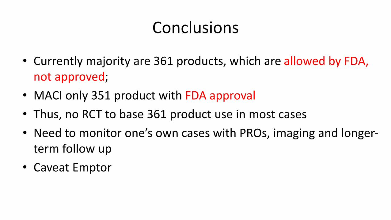
Conclusions
• Currently majority are 361 products, which are allowed by FDA, not approved;
• MACI only 351 product with FDA approval
• Thus, no RCT to base 361 product use in most cases
• Need to monitor one’s own cases with PROs, imaging and longer-term follow up
• Caveat Emptor

Thank You