whey proteins as microencapsulating agents
TRANSCRIPT

Food Structure Food Structure
Volume 12 Number 1 Article 4
1993
Whey Proteins as Microencapsulating Agents. Whey Proteins as Microencapsulating Agents.
Microencapsulation of Anhydrous Milkfat - Structure Evaluation Microencapsulation of Anhydrous Milkfat - Structure Evaluation
M. Rosenberg
S. L. Young
Follow this and additional works at: https://digitalcommons.usu.edu/foodmicrostructure
Part of the Food Science Commons
Recommended Citation Recommended Citation Rosenberg, M. and Young, S. L. (1993) "Whey Proteins as Microencapsulating Agents. Microencapsulation of Anhydrous Milkfat - Structure Evaluation," Food Structure: Vol. 12 : No. 1 , Article 4. Available at: https://digitalcommons.usu.edu/foodmicrostructure/vol12/iss1/4
This Article is brought to you for free and open access by the Western Dairy Center at DigitalCommons@USU. It has been accepted for inclusion in Food Structure by an authorized administrator of DigitalCommons@USU. For more information, please contact [email protected].

FOOD STRUCTURE, Vol. 12 (1993), pp. 31-41 I 046-705X/93$5. 00 +. 00 Scanning Microscopy International , Chicago (AMF O 'Hare) , IL 60666 USA
WHEY PROTEINS AS MICROENCAPSULATING AGENTS. MICROENCAPSULATION OF ANHYDROUS MILKFAT- STRUCTURE EVALUATION
M. Rosenberg , and S. L. Young
Department of Food Science and Technology University of California, Davis
Davis, CA 95616
Abstract
Microencapsulation of milkfat may open new fields of application for this milk constituent by transforming it into dry and stable powder. Research has been undertaken to study the microstructure of whey protein based, anhydrous milkfat-containing, spray-dried microcapsules. Whey protein concentrates of 50% and 75% protein and whey protein isolate were evaluated as microencapsulating agents (wall materials). The effects of wall composition, fat load , and drying conditions on the capsule's structure were studied by scanning electron microscopy (SEM). Spherical capsules with smooth , wrinkle- free surfaces were observed in all cases. Whey protein isolate-based microcapsules dried at a temperature higher than 105 °C were free of surface indentation and only a limited extent of indentation was observed when whey protein concentrate served as the wall material. Microcapsules prepared from emulsions containing more than 10 % wall solids and dried at I6o • c exhibited no cracks or holes. Comparing the microstructure of spray-dried microcapsules with and without milkfat (prepared under the same conditions) suggested that high milkfat load limits the extent of surface indentation. The milkfat was encapsulated as 50-600 nm particles that were uniformly distributed throughout the interior of the wall matrices . No visible pores or cracks exposing the core material to the environment were detected. Theresults suggest that whey proteins can be considered as microencapsulating agents for anhydrous milkfat.
Key Words: Anhydrous milkfat, inner structure, microencapsulation , microstructure, outer topography , whey protein concentrate, whey protein isolate.
Initial paper received December 21 , 1992 Manuscript received February 24, 1993 Direct inquiries to M. Rosenberg Telephone number: (916) 752 4682 Fax number: (916) 752 4759
31
Introduction
Microencapsulation is a "micro-packagi ng" technique that makes it possible to pack liquid droplets or solid particles by thin films (called wall) consisting of an encapsulating material. The wall protects the encapsulated material (the core) from deterioration. Microencapsulation is also used to transform liquids into dry, free-flowing powders, limit volatile losses, and control release of active material [2 , 3, 4, II, 19, 23 , 34]. Effective microencapsulation requires capsules of high physical integrity, i.e. , the core material should be completely surrounded and protected by the wall system. The functionality of any microencapsulated system is critically dependent on its structu ral features. In addition , both the flowability and the wettability of the final product (a powder) are affected by the outer topography of the powder particles [27] .
In the food industry , typical core materials are aroma and flavor compounds, oils, fats , essential oils , oleoresins, minerals , and vitamins [23, 35] . Spray-drying is a well -established technique in the food industry and at present is the most commonly used technique to microencapsulate food ingredients [28 , 37].
The variety of wall materials commonly used in food applications is relatively limited and includes natural gums, carbohydrates, waxes, gelatin , and some chemically modified natural polymers [2 , 3, 23]. A need for additional wall materials exists.
Any wall material used for microencapsulation by spray drying should have bland flavor, high solubility, and possess the necessary emulsification properties, film-forming and drying characteristics. In addition , its concentrated solution should have low viscosity [28].
The functional properties of whey proteins have been extensively studied and reported [22 , 26] and they exhibit many of the properties of a good wall material. The functionality of whey proteins in the microencapsulation of anhydrous milkfat has recently been reported [38].
The evaluation of a potential wall material is based (among other things) on thoroughly understanding the effect of compositional aspects and process parameters on the structural characteristics of the microcap-
M. Rosenberg and S.L. Young
sules. The microstructure of various spray-dried, milkderived , and other food-related powders has been studied [6-9 , 12-18 , 20,2 1,24, 25] ; however, the information on food -related , spray-dried microcapsules has been established chiefly for systems consisting of natural gums and carbohydrates [5 , 10, 30-33]. It has been demonstrated that wall com position, drying conditions, and storage conditions affect the structure of spray-dried microcapsules [30-33]. The retention of the core material during the process and during storage is affected by the structural features of the capsules. The microstructure of whey protei n-based microcapsules has not yet been addressed. The objective of this research was to determine the effects of wall composition , core load, and dryi ng conditions on the microstructure of whey proteinbased, anhydrous milkfat-containing, spray-dried microcapsules.
Materials and Methods
Materials
. Whey protein concentrates containing SO and 7S% protein (WPC 50 , WPC 75 , respectively) were obtained from Calpro Ingredients (Corona , CA); whey protein is~ta'te (WPI) was purchased from LeSueur Isolates (Le Sueur, MN) . Anhydrous milkfat (California Cooperative Creamery, Hugh son, CA) containing 99.8% milkfat was used as a model core material. The anhydrous milkfat (AMF) was stored at 0 °C until use.
Preparation of emulsions
WPI or WPC solution s containi ng 10-30 % (weight / weight , w/w) sol id s were prepared in deionized water. For simplic ity, these solutions are called "wall solutions" in the subsequent text. The wall solutions were de-aerated (at 3S oc and 13 kPa) to remove trapped air. AMF was em ul sified into the wall solutions at proportions of 50 and 75% (g AMF/g dry, non fat wall solids x 100). The emulsification was carr ied out in two stages. A coarse emulsion was prepared using an UltraTurrax T-2S (IKA Works, Cincinnati, OH) homogenizer operated at 13,500 rpm (position 3 on the instrument dial) for 30 seconds. The second stage consisted of four successive homogenization steps using a type 8.30-H Mini-Lab high pressure homogenizer (APV Rannie, St. Paul, MN) operated at SO MPa. The emulsion constituents were heated to 50°C prior to the emulsification and this temperature was maintained throughout the emulsification process. These homogenization conditions were selected based on the results of a different study [39] in which the influence of emulsion composition and homogenization conditions on the structural and rheological properties of the emulsion were studied. The emulsification conditions selected in the present study resulted in small mean particle size (Table I) and did not increase the viscosity of the emulsion.
Spray drying
Microencapsulation was carried out by spray drying emulsi.ons of AMF in wall solutions. Spray drying
32
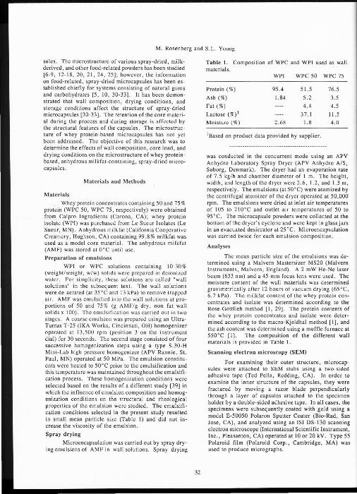
Table I. Composition of WPC and WPI used as wall materials.
WPI WPC 50 WPC 75
Protein( %) 95.4 51.5 76.5
Ash(%) 1.84 5.2 3.5 Fat(%) 4.4 4.5 Lactose (%) t 37.1 11.5 Moisture(%) 2.68 1.8 4.0
1Based on product data provided by supplier.
was conducted in the concurrent mode using an APV Anhydro Laboratory Spray Dryer (APV Anhydro A/S , Soborg, Denmark). The dryer had an evaporation rate of 7.S kg/hand chamber diameter of 1 m. The height , width, and length of the dryer were 2.6, 1.2 , and 1.3 m, respectively. The emulsions (at 50 °C) were atomized by the centrifugal atomizer of the dryer operated at 50,000 rpm. The emulsions were dried at inlet air temperatures of 105 to 210 oc and outlet air temperatures of 50 to 9S °C. The microcapsule powders were collected at the bottom of the dryer 's cyclpne and were kept in glass jars in an evacuated desiccator at 25°C . Microencapsulation was carried twice for each emulsion composition.
Analyses
The mean particle size of the emulsions was determined using a Malvern Mastersizer MS20 (Malvern Instruments, Malvern , England). A 2 mW He-Ne laser beam (633 nm) and a 45 mm focus lens were used. The moisture content of the wall materials was determined gravimetrically after 12 hours of vacuum drying (65°C, 6.7 kPa). The milkfat content of the whey protein concentrates and isolate was determined according to the Rose-Gottlieb method [I , 29]. The protein contents of the whey protein concentrates and isolate were determined according to the macro-Kjeldhal method [I] , and the ash content was determined using a muffle furnace at 550°C [1]. The composition of the different wall materials is provided in Table 1.
Scanning electron microscopy (SEM)
For examining their outer structure, microcapsules were attached to SEM stubs using a two-sided adhesive tape (Ted Pella, Redding, CA). In order to examine the inner structure of the capsules, they were fractured by moving a razor blade perpendicularly through a layer of capsules attached to the specimen holder by a double-s ided adhesive tape. In all cases, the specimens were subsequently coated with gold using a model E-50050 Polaron Sputter Coater (Bio-Rad, San Jose, CA) , and analyzed using an lSI DS-130 scanning electron microscope (International Scientific Instrument, Inc. , Pleasanton , CA) operated at 10 or 20 kV. Type 55 Polaroid film (Polaroid Corp., Cambridge, MA) was used to produce micrographs.

Microencapsulation by whey proteins
Table 2. Composition of emulsions.
System Wall Concentration of wall AMF Particle component component(%) w/ w1 load2 size3 (~m)
I WPI 20 75 0 .40 2 WPI 10 50 0.45 3 WPC 50 30 50 0.45 4 WPC 50 20 50 0.38 5 WPC 75 20 75 0.38 6 WPC 75 30 75 0.45
1 Concentration of WPC or WPI (% w/w) in the wall solution. 2 Concentration of anhydrous milk fat (g AMF/g dry , no-fat wall solids). 3 Mean particle size of AMF in the emulsion.
In a separate set of experiments, the possibility to fracture microcapsules embedded in Lowicryl HM-20 (Electron Microscopy Sciences, Fort Washington , PA) was evaluated by using the method described by Rosenberg eta/. [30-33]. Polymerization was carried out by exposure to ultraviolet (UV) radiation at 360 nm (30 W) at 10°C, at a bulb-to-sample distance of 35 em for I hour. The blocks were fractured using a model 820 Rotary Microtome (American Optical , Buffalo , NY), gold coated, and examined by scanning electron microscopy (SEM) under the conditions described above. In several cases , hardening of the microcapsules by suspending them for I hour in glutara ldeh yde (10 %, w/w) prior to embedding was evaluated . At the end of the hardening stage, the mi crocapsules were washed with ethanol, dried (55 °C, 7 kPa , for 12 hours), and embedded as described above.
Results and Discussion
Table 2 presents the composition of the emulsions and the mean AMF particle size in these emulsions prior to drying.
The outer topograph y of WPI -based , AMF-containing microcapsules is presented in Figures l -3. Spherical capsules (1-25 J.Lm) were observed in all cases. The relati vely wide size distribution is attributed to both , the properties of the atomizer and the effects of drying as described bellow. The surfaces of the capsules were smooth regardless of dryi11g tt:mperature or composition. Microcapsules that were dried at l05 °C (Figure I) exhibited shallow surface dents only to a very limited extent. At drying (inlet) air temperature of 160°C, almost no surface dents could be observed (Figures 2 and 3). The capsules were usual ly characterized by high a degree of integrity , i.e., no cracks or pores could be detected on the outer surfaces. However, a few of the large capsules that were dried at 160°C, particularly those prepared from emulsion that contained I 0% wall solids, exhibited holes. These features can be attributed to the results of excessive "ballooning .. and subsequent rupturing effects associated with spray-d ried microcap-
33
sules [II , 12, 30-33]. Microcapsules prepared at an inlet air temperature of l05 °C were free from such defects. Although the capsules presented in Figures 1-3 were prepared from emulsions that differed in their composition , no significant differences in outer topography could be detected.
Figures 4 and 5 present AMF-free microcapsules prepared by spray drying a 20% WPI solution at 105 and 160 °C, respectively. In both cases, particles with smooth surfaces were observed; however , some of the spheres had a slightly elongated shape. Figure 5 reveals the presence of many fragments of broken particles. Such features could not be detected in the AMF-contain ing capsule batches and may suggest a difference in mechanical propert ies between the particles of these two systems. Since the systems differed only in the AMF con tent , such differences may be linked to a effect that AMF (or other core) may have on the physical properti es of the capsules. The exact reasons for the differences have not yet been determined. However, no st ructural differences between systems containing 50 and 75% AMF could be detected.
Microcapsules in which WPC 50 served as wall material are presented in Figures 6-8. Particles of 2-35 J.Lm were observed. Surface dents were associated with the capsules dried at 105 and 160°C, and were manifested to a very limited extent in capsules that were dried at 210 °C. In all cases, the capsules were of spherical shape and exhibited smooth surfaces . Inlet air temperature of 21 0 °C was associated with few damaged capsules (as presented in Figure 8). Such defects could not be detected in batches dried at lower drying temperatures. The surface dents observed in these micrographs are shallower than those reported by Mistry eta/. [25] for WPC powder, and there was no evidence of wrinkled surfaces.
Representative micrographs of AMF-containing microcap_sules in which WPC 75 served as the wall material are presented in Figures 9-11. Under all drying conditions, capsules with smooth surfaces were observed. Capsules dried at 105 oc were characterized by

M. Rosenberg and S.L. Young
Figures l-3. Micrographs of WPI-based , AMP-containing microcapsules prepared from system I (Table 2) by spray-drying. Inlet and outlet air temperatures were lOS and so•c (Figure l) ; 160 and so•c (Figure 2) ; and 210 and go•c (Figure 3), respectively. Bars= 10 I'm (Fig. I) and 20 I'm (Figs. 2 and 3).
34
Figures 4-5. Micrographs of spray-dried WPJ particles prepared from a 20% WPI solution. Inlet and outlet air temperatures were 105 and so•c (Figure 4) ; and 210 and go•c (Figure 5) , respectively. Bars = 20 I'm (Fig. 4) and 10 I'm (Fig. 5).
Figure 6. Micrograph of spray-dried , WPC 50-based, AMF-containing microcapsules prepared from system 4 (Table 2). Inlet and outlet air temperatures were 105 and 50 °C, respectively. Bar = 20 p.m.
Figure 7. Micrograph of spray-dried, WPC 50-based, AMF-containing microcapsules prepared from system 3 (Table 2). Inlet and outlet air temperatures were 160 and 80 °C, respectively. Arrow: surface dent , bar = 20 I'm.
Figure 8. Micrograph of spray-dried , WPC 50-based , AMP-containing mic rocapsules prepared from system 4 (Table 2). Inlet and outlet air temperatures were 210 and 90 °C, respectively. Arrow: surface dent, D: damaged microcapsule, bar = 10 p.m.

Microencapsulation by whey proteins
Figures 9~11. Micrograph of spray-dried, WPC 75-based , AMP-containing microcapsules prepared from system 5 (Figs. 9, 11) and system 6 (Fig. 10); see Table 2. Inlet and outlet air temperatures were 105 and 50 'C (Fig. 9); 160 and 80 'C (Fig. 10); and 210 and 90'C (Fig. 11), respectively. Bars = 20 I'm.
35

M. Rosenberg and S.L. Young
deep surface dents (Figure 9). The proportion of capsules exhibiting su rface dents was significantly reduced when drying temperature was increased to 160°C, and the phenomenon was almost completely eliminated at inlet air temperature of 210 °C. At this temperature, few of the large capsules (Figure II) exhibited small holes. This may be linked to rapid parti cle expansion at high drying temperatures.
It is necessary to fracture the microcapsules in order to examine their inner structure. Embedding the microcapsules in Lowicryl HM-20 resulted in significant shape distortion (micrographs not provided here). This was attributed to softening of the capsule wall which can be linked to the relativel y high hydrophobici ty of the whey proteins that permitted the interaction with the apolar embedding material. ln addition , man y of the embedded capsules were cracked during the resin polymerization (under UV at 4 cc). This effect was attributed to the shrinkage of the resin during polymerization [32]. Treatment with glutaraldehyde prior to the embedding did not eliminate the adverse effects. Consequently , capsules were fractured by razor blade in this study, in order to allow examination of their inner structure.
Figure 12 reveals the inner st ructure of a WPIbased, AMF-free microcapsule. Figure 13 is a low-magnification micrograph presenting the typical inner structure of a WPJ, AMF-containing, spray-dried microcapsule. Comparing the structural details observed in these micrograph s indicates that small particles of the core material (AMF) were evenly distributed throughout the protein matr ices of the wall . These struc tural features are similar to those reported for spray-dried, gum arabic-based microcapsules [30-33] . The inner structure of AMF-containing , WPI-and-WPC 50 based microcapsules is presented in Figures 14 and 15, respectively. A central void was found in all cases , regard less of drying conditions or composition. Thi s is in contrast to the resu lts reported for gum arabic-based mic rocapsules. In the latter , low drying temperature resulted in limited formation of central voids (30-33]. In all cases, the AMF was distributed as small droplets (50-600 nm) embedded in the capsule wall. Similar structural details were reported by Burna (8] regarding the structure of spray-dried, whole milk powder. No effects of wall composition on the inner structure could be detected. The diameter range of the encapsulated AMF (Figures 14 and 15) was similar to that of the AMF droplets at the emulsion stage as was determined prior to drying (Table 2). This indicates that no coalescence of the AMF droplets occurred during the drying process. No visible cracks or channels exposing the encapsulated AMF droplets to the environment coul d be detected. The inner structure of spray-dried WPC 50 and WPC 75 solutions was examined (micrographs not presented here) in a way similar to that presented in Figure 12 for WPI. The fat content of the WPCs was dispersed in the particle wall in the form of small droplets having the size and shape of those found in the AMP-containing particles. The fat content of the WPCs could not be structurally
36
Figure 12. The inner structure of a spray-dried particle from the batch presented in Figure 4. The micrograph reveals the existence of a central void (CV) , some craters or holes on the surface of the central void (C), and vacuoles (V) embedded in the particle ' s wall (W). Bar =I ~m.
Figure 13. Micrograph revealing the typical inner structure of a WPI-based, AMP-containing microcapsule from the batch presented in Figure 2. CV: central void, W: wall in which the AMF droplets are embedded (arrows). The existence of holes or craters on the central void surface is evident. Dimples or protrusions (many of which exhibit a hole) can be seen on the surface of the central void. Bar = 5 J.Lffi.
Figure 14. A representative micrograph revealing the inner structure of WPI-based , AMP-containing microcapsule from the batch presented in Figure 3. CV: central void, W: wall. AMF droplets are embedded in the capsule's wall (arrows). Bar = I J.Lm.
Figure 15. A representat ive micrograph reveal ing the inner structure of WPC 50-based , AMP-containing microcapsule from the batch presented in Figure 7. CV: central void, W: wall. AMF droplets are embedded in the capsule's wall (arrows). Bar = 0.5 J.Lm.
Figure 16. Micrograph showing the outer topography of WPC 50 particl es prepared by spay drying a solution of 20% total solids. Inlet and outlet air temperatures were I 60 and 80 °C, respec tively. It is possible to observe particles exhibiting deep su rface dents , particles exhibiting "caps" within dents (small arrows) that probably represent early stages of expansion after shrinkage, particles with a "wavy" surface (big arrow), and particles with no dents at all. Bar = 5 ftm.
Figure 17. Micrograph presenting the outer topography of spray-dried WPC 50 particles prepared by spray drying a solution of 20 % total solids . Inlet and outlet air temperatures were 210 and 90 °C, respect ive ly. Many of the large capsules exhibit no surface dents while the small particles exhibit deep dents or intermediate stages of expansion (after shrinkage) in the form of "caps" within surface dents. Bar = 5 J.Lm.
distinguished from the added AMF. It should be mentioned that in all the WPC-based systems , the proportion of fat contributed by the wall material was very small in comparison to the added AMF.
Some voids (larger than the AMF droplets) found in the wall (Figures 13) seemed to be typical, and was also observed with AMF-free WP! capsules (Figure 12). This finding can be attributed either to air bubbles trapped in the wall solution (or emulsion) , or to small capsules trapped in the wall of large capsules. The latter has been reported as occurring with spray-dried, milkderived powders ((5], and was also observed in some specimens in this research (micrographs not presented here) .

Microencapsulation by whey proteins
37

M. Rosenberg and S.L. Young
The surfaces of the central void found in the microcapsules were characterized in some cases by "craters" or holes (Figure 13). Thi s could not be attributed to the AMF as it was also found in the AMF-free capsules (Figure 12) . It is unclear whether these features were part of the holes that were found in some cases to be associated with the spray-dried particles (especially with those dried at high temperature). The particles presented in Figures 12 and 13 were prepared at 160 and l05°C , respectively , so no conclusion regarding the effect of drying temperature in this regard could be reached.
Another feature observed on the surface of the central void presented in Figure 13 was the presence of many dimples or protrusions (Figure I 3). Many of these structural details exhibited very small holes (Figure 13). The nature of these features , their source, or their orientation is not clear. Similar structural details were observed while studying the structure of spray-dried capsules consisting of gum arabic and esters [30 , 33]. So far, such features have been detected only in systems containing core material (both in this study and previ ously reported ones). It is possible that these details represent the contours of core droplets that are very close to the surface of the central void; however, the phenomenon needs further study.
The morphology of spray-dried, milk-derived powder particles has been extensively studied , and the existence of surface dents in such particles (as well as in other spray dried food systems) is well known. The formation of wrinkled surfaces has also been addressed. There is conflicting information regarding the role of the different milk constituents in affecting particle structure.
The presence of surface dents reported for spraydried skim milk powder [6-8] has been attributed to the effects of drying on casein [6 , 7], and to the effects of atomization and drying conditions [6]. Surface dents (found regardless of chemical composition) and wrinkled surfaces (attributed to lactose content) were reported for particles of different spray-dried retentates prepared from ultrafiltered and diafiltered skim milk [25]. However, no surface dents or wrinkled surfaces were detected in spray-dried lactose particles [7 , 20]. Hayashi [14] attributed the formation of wrinkled surfaces in milk-derived powders to the effect of high dryi ng temperature. Surface dents were reported for particles of spray-dried milk protein concentrate [24] and for spray-dried ultrafiltered (UF) skim-milk r~t~ntate powders [18]. Surface dents and wrinkled surfaces were reported for particles of different whey-derived spray-dried powders [6 , 25]. Deep surface dents were reported to be characteristic of spray-dried particles of soy protein isolate [36].
Burna and Henstra (8] and Burna [6] studied the structure of spray-dried whole and skim milk powders. They suggested that the observed surface folds, pores , and cracks represented the effects of mechanical stresses induced by uneven drying at different parts of the drying droplets and by shrinkage of casein . Burna and Henstra (8] concluded that casein rather than lactose was proba-
38
bly responsible for surface dents. For whole milk, these authors [8] attributed the limited surface indentation to the presence of a large number of fat globules that may hinder the shrinkage of the casein.
The research reported here indicates that WPIbased , spray-dried microcapsules have a structure different from that reported for other milk-derived powders or soy protein isolate particles. Shallow surface dents found at low drying temperatures were eliminated -{WPI systems) or significantly limited (WPCs systems) by increasing the drying temperature. Regardless of the lactose or fat contents, surface wrinkles were not detected . These results are thus in conflict with the aforementioned hypothesized role of lactose in introducing wrinkles. To date , it has not been possible to correlate the extent of surface dents in WPCs capsules with the lactose content. However , the existence of dents in WPC particles and their absence in the case of WPI particles may be associated with the lactose content of WPC. The absence of surface dents in the WPI capsules was typical for AMP-containing as well as for AMF-free capsules and thus seems to be characteristic of WPI.
The structure of the WPJ microcapsules reported here differs from the reported structure of spray dried microcapsules in which gum arabic or dextrins served as wall material [5, 10 , 30-33]. It has been reported that the formation of deep surface dents in microencapsulation by spray drying could be eliminated by introducing a fast drying rate [30-33] . The results regarding WPI capsules did not indicate any significant influence of wall solids concentration (at the emulsion stage) on the morphological properties of the dry capsules. The results regarding the WPCs capsules are in agreement with the reported effect which the drying temperature has on the surface morphology of spray-d ried particles in general, and on spray-dried microcapsules in particular [12, 13 , 30-33]. Spray-dried, AMF-free , WPC 50 particles in which "caps" developed within dents are clearly visible in Figures 16 and 17. This phenomenon can be explained by some theories dealing with dent formation during spray-drying [12, 13, 30-33]. According to these theories , dent form ation is related to uneven shrinkage of the atomized droplets at the early stages of spray drying. The surface topography of the final product thus depends on the drying temperatures or drying rates. At high drying rates, the thermal expansion of either the air or water vapors inside the drying particle can "erase" the dents. In such cases, the expansion must take place after the dent formation, but at a stage when the wall system is still elastic enough to allow "ballooning" [30, 33]. The "caps" within dents that are visible in Figures 16 and I 7 can be attributed to cases where the solidification of the wall occurred prior to the completion of the expansion and thus intermediate stages associated with "dents erasing" by expansion are evident. In extreme cases, excessive expansion of the capsules during the late stages of drying may result in rupturing [30-33] . The small particles observed in Figures 16 and 17 show deep indentation . This can be linked to the very fast

Microencapsulation by whey proteins
drying (and hence early solidification) of these particles as a result of high surface area to volume ratio. In such cases dents could not be "erased" by the mechanism mentioned above. To date, "caps" within dents could be fou nd in neither AMP-containing WPC microcapsu les nor in WPI-based microcapsules.
An attempt to develop mathematical models correlating surface topography of spray-dried particles with the effects of compositional or process (drying) parameters has been reported by Keith [20]. It was established that the tendency to develop folds depends on the ratio of low-to high -molecular weight solutes in the feed [20]. A mechanism based on a surface-tension-driven, viscous flow of surface folds formed by uneven shrinkage of drying droplets was hypothesi zed [20]. Based on this theory, it was speculated that a critical viscosity for folding exists, below which the surface tension forces are sufficient to smooth out surface irregularities [20]. This mechanism calls for a critical role of the physicochemical properties of the wall material (and maybe of the core material as well) in determining the surface features of spray-dried capsules. The fact that the results reported here differ from those reported for other milkderived powders may be due to the noticeable differences in composition of the powders examined in this study and that of the reported milk powders. The high concentration of whey proteins and milkfat in the systems studied in this research may play a part in limiting surface folds. Thi s composition effect may be explained by the mechanisms hypothesized by Keith [20], or by significant differences in the viscoelastic properties of the various emulsions and solutions at extremely high solids concentration (associated with late stages in the drying process). The significan t difference in dent formation between the WPC 50 systems with AMF and those without AMF as well as the compositional and structural differences between the WPI- and WPC-based systems may support the suggested surface ten sion-driven theory. The results regarding the WPC 50-based (with and without AMF) systems suggest that AMF has a role in limiting dent formation and are in agreement with the conclusions of Burna (8] regarding the st ructure of whole milk powder.
Conc lusions
The results of the structural study indicate that whey proteins can be considered as effective microencapsulating agents for AMF and that preparing an "all milk-derived" powder with high fat load is feasible . These, in turn, may open new applications for whey proteins and AMF.
The inner and outer structural features of the spray-dried capsules indicated that good physical protection is provided to the AMF. However, this should be confirmed by a stability study. Structural analyses revealed that the whey protein-based microcapsules exhibited the typical structure of microcapsules prepared by spray-drying. The outer structure of WPI capsules was
39
essentiall y free of dents , and that of WPC-based capsules could be easily manipulated by adjusting the drying temperature to eliminate shallow surface dents. High fat loads were not associated with detectable adverse effects on the outer or inner structure of the capsules.
Comparing the structure of capsules prepared at different drying temperatures, it appears that a drying tempera ture of 160 oC and 20% solids in the wall system at the emulsion stage can be recommended.
The presence of surface dents adversely affects the f1owability (as well as the wettability) of powders because it leads to the formation of clumps or aggregates. This research ind icates that whey proteins in general and WPJ in particular may offer advantages in this regard.
The stability of encapsulated milkfat , as well as the structure of capsules in which core materials other than milkfat are encapsulated, are currently being evaluated by the authors.
References
I. Association of Official Analytical Chemists. (1990). Official methods of analysis. 15th ed., AOAC , Arlington, VA, p. 835.
2. Bakan JA . (1973). Microencapsulation of food and related products . Food Techno!. 27, 11 -44.
3. Bakan JA. (1978). Microencapsulation. In: Encyclopedia of Food Science. Peterson MS, Johnson AH (Eds.). A vi Publishing Co., Westport, CT. pp. 499-507.
4. Balassa LL, Fanger GP. (1971). Microencapsulation in the food indust ry. CRC Crit. Rev. Food Techno!. 2 , 245-265.
5. Beatus Y, Raziel A, Rosenberg M, Kopelman 11. (1985). Spray-drying mi croencapsulation of paprika oleoresin. Lebensm.-Wiss. u.- Techno!. 18 , 28-34.
6 . Burna TJ, Henstra S. (1971). Particle structure of spray-dried milk products as observed by scanning electron microscope. Neth. Milk Dairy J. 25 , 75-80.
7. Burna TJ, Henstra S. (1971). Particle structure of spray-dried caseinate and spray-dried lactose as observed by scanning electron microscope. Neth. Milk Dairy J. 25, 278-283.
8. Burna TJ. (1971). Free fat in spray-dried whole milk. 8. The relation between free fat content and particle porosity of spray-dried whole milk. Neth. Milk Dairy J. 25 , 123-140.
9. Carie M, Kalab M . (1987) . Effect of drying techniques on milk powders quality and microstructure: a review. Food Microstruc . 6 , 171-180.
10. Chang YI , Scire J, Jacobs B. (1988) . Effect of particle size and microstructure properties on encapsulated orange oil. In: Flavor Encapsulation . Reineccius GA, Risch SJ (eds.). American Chemical Society Symposium No. 370, Washington, DC . pp. 87-102.
II . Dziezak JD. (1988). Microencapsulation and encapsulated ingredients. Food Techno!. 42, 136-151.
12. Greenwald CG. (1980). Particle morphology

M. Rosenberg and S.L. Young
in the spray drying of foods. Ph.D. Thesis, University of California , Berkeley, U.S.A. (Microfilm# 8029410. Available from University Microfilm International , Ann Arbor, Ml).
13. Greenwald CG, King CJ. (1982). The mechanism of particle expansion in spray drying of foods. AIChE Symp. Ser. 218, 101-108.
14. Hayashi H. (1962). Studies on spray drying mechanism of milk powders. Rept. Res. Lab. Snow Brand Milk Prod. 66, 42-46 (available from Snow Brand Milk Products Company , 1-1-2 Minamidai , Kawagoe, Saitama 350, Japan).
15. Kalab M. (1979). Microstructure of dairy foods. 1. Milk products based on protein. J. Dairy Sci. 62, 1352-1364.
16. Kalab M. (1980). Possibilities of an electronmicroscopic detection of buttermilk made from sweet cream in adulterated skim milk. Scanning Electron Microsc. 1980:111 , 645-651.
17. Kalab M. (1981). Electron microscopy of milk products: a review of techniques. Scanning Electron Microsc. 1981:111 , 453-472.
18. Kalab M, Carie M, Zaher M, Harwalkar VR. (1989). Composition and some properties of spray-dried retentates obtained by ultrafiltration of milk. Food Microstruc. 8, 225-233.
19. Karel M, Langer R. (1988). Controlled release of food ingredients. In: Flavor Encapsulation. Reineccius GA, Risch SJ (eds .). American Chemical Society Symposium No. 370, Washington , DC. pp. 177-191.
20. Keith A. (1983) . Factors governing su rface morphology in the spray-drying of foods. Ph.D. thesis, University of California, Berkeley, U.S.A. (Microfi lm # 8413299. Available from University Microfilm International, Ann Arbor, MI).
21. Kerr TJ , Washam CJ , Evans AL, Rigsby WE. (1983). Structural characterization of spray-dried dairy products by scanning electron microscopy. De vel. lndustr. Microbiol. 24, 475-484.
22. Kinsella JE. (1984). Milk proteins: Physicochemical and functional properties. Crit. Rev. Food Sci. and Nutr. 21, 197-262.
23. Lauren SJ, Ken L. (1991). Microencapsulation and the food industry. Lebensm.-Wiss. u.-Technol. 24, 289-274.
24. Mistry VV, Hassan HN. (1991) . Delactosed, high milk protein powder. 2. Physical and functional properties. J. Dairy Sci. 74, 3716-3723.
25. Mistry VV, Hassan HN, Robinson DJ. (1992). Effect of lactose and protein on the microstructure of dried milk. Food Microstruc. 11, 73-82.
26. Morr CV, Foegeding EA . (1990). Composition and functionality of whey and milk protein concentrates and isolates: A status report. Food Techno!. 44 , 100-112.
27. Peleg M, Mannheim CH. (1973). Effect of conditioners on the flow properties of powdered sucrose. Powder Techno!. 35, 2352-2357.
40
28. Reineccius GA. (1988). Spray drying in food flavors. In: Flavor Encapsulation. Reineccius GA, Risch SJ (eds.). American Chemical Society Symposium No. 370, Washington , DC. pp. 55-66 .
29. Richardson GH (ed.) (1985). Standard Methods for the Examination of Dairy Products, 15th edition. American Public Health Association, Washington , DC. p. 358.
30. Rosenberg M, Talmon Y, Kopelman IJ. (1984). A new preparation technique for the study of microencapsulation by scanning electron microscopy. Proc. of the 8th European Congress on Electron Microscopy. 1984:1, 653-654 (copies available from M. Rosenberg, address on first page of this paper).
31. Rosenberg M. (1985). Structural characterization and volatiles retention in microencapsulation by spray drying. D.Sc. thesis, Technion, Israel Institute of Technology, Haifa, Israel.
32. Rosenberg M, Kopelman II , Talman Y. (1985). A scanning electron microscopy study of microencapsulation. J. Food Sci. 50 , 139-144.
33. Rosenberg M, Talmon Y, Kopelman IJ. (1988). The microstructure of spray-dried microcapsules. Food Microstruc. 7 , 15-23.
34. Taylor AH. (1983). Encapsulation systems and their applications in the flavor industry. Food Flavors, Ingredients , Packaging , and Processing. 5, 48-49 , 51 -52.
35. Todd RD. (1970). Microencapsulation and the flavo r industry. The Flavor Ind. I , 768-771.
36. Wolf WJ , Baker FL. (1980). Scanning electron microscopy of soybeans and soybean protein products. Scanning Electron Microsc. 1980:111, 621-634.
37. Young RA. ( 1986). Spray drying encapsulation- today's view. Food Flavors, Ingredients , Packaging , and Processing. 8 , 31-33.
38. Young SL , Sarda X, Shoemaker C, Rosenberg M. (1992). Microencapsulation of anhydrous milkfat by whey proteins. J. Dairy Sci., Suppl. I , 93 (abstract).
39. Young SL, Lee SY , Shoemaker C , and Rosenberg M. (1992). Rheological properties of anhydrous milkfat/whey protein emulsions. J. Dairy Sci. Suppl. 1, 93 (abstract).
Discussion with Reviewers
B.E Brooker: To what extent are the minor structural features you (and other authors) describe in spray-dried powders, e.g. , "craters" , the result of examining them at ambient temperature in the SEM? Are the same feature present when powders are examined by cold stage SEM? Authors: The structure of the microcapsules was studied by SEM at ambient temperature. We did not examine the structure by cold stage SEM. However, based on previous studies in our laboratory (unpublished data) in which irradiation damage could be detected in microcapsules, the structural features described here do not seem to be associated with examination conditions.

Microencapsulation by whey proteins
v~E. Colombo: Please, could you specify possible causes of deterioration which may be involved in unprotected material? Authors: One of the main objectives for microencapsulation is to isolate sensit ive ingredient or active material from factors that may o therwise cause its deterioration. Generally, the wall sys tem should be design to provide protection agai nst mass transport phenomena between the inner part of the microcapsule and the environment. Such transport phenomena can be permeation of gases to inner parts of the capsule through the wall matrix, or, the migration of core material (th rough the wall matrix) to the surface of the capsules. Deterioration of food ingredients that are microencapsulated in an improper way includes oxidation, interaction with moisture, change in aroma composition (due to losses of some of its constituents), etc . Wall systems of high porosity, improper polarity, or those with structural defects may lead to the deterioration of the core under various conditions.
41
V .E. Colombo: Could you list some chemicall y modified natural polymers which would according to your opinion be su itable as wall material? Authors: Different modified carbohydrates have been evaluated as wall materials. Chemical or enzymatic modifications of starch to change its li pophilic/hydrophi li c balance are the main approaches in this regard. CapsuJill and N-Lok® (National Starch and Chemical Corporation, Bridgewater , NJ) are examples of such materials.
V.E. Colombo: What is the flow through in the spray drying apparatus you were using for your experiments? Authors: The drying capacity of the dryer is 7.5 kg/ h. The dimensions of the apparatus are provided in the Materials and Methods section.
V .E. Colombo: Could you explain , in more details, the method by Rose-Gottlieb for measuring the mi lk fat? Authors: The Rose-Gottlieb method for fat determination is an IDF-ISO-AOAC Method and can be found as method 932.06 in text reference I. One gram of sample is dispe rsed in 10 ml of water. About I ml of NH40H is then added and the system is heated for 15 minutes in a water ba th at 60-70 °C. The mixture is cooled and the fat is ex tracted by ether/alcohol. The solvents are evaporated and the fat is determined grav imetrically .