when an unexpected diagnosis occurs: a vaginal ... · cancro vaginal sarcoma vaginal radioterapia...
TRANSCRIPT

When an Unexpected Diagnosis Occurs:a Vaginal Premenopausal Sarcoma
Quando um diagnóstico inesperado ocorre: um sarcomavaginal na pré-menopausa
Pedro Marcos-Figueiredo1 Diana de Sousa da Costa Moreira2 Manuel Gonçalves Morim3
Júlia Leite Pereira3 Lurdes Silva Salgado2
1Serviço de Ginecologia/Obstetrícia, Hospital Senhora da OliveiraGuimarães, Guimarães, Portugal
2Serviço de Radiologia, Instituto Português Oncologia do PortoFrancisco Gentil, Porto, Portugal
3Serviço de Ginecologia/Obstetrícia, Centro Hospitalar da Póvoa deVarzim/Vila do Conde, Póvoa de Varzim, Portugal
Rev Bras Ginecol Obstet 2018;40:47–52.
Address for correspondence Pedro Marcos Figueiredo, MD, HospitalSenhora da Oliveira Guimarães, Rua dos Cutileiros 114, Guimarães,Portugal (e-mail: [email protected]).
Keywords
► vaginal cancer► vaginal sarcoma► pelvic radiotherapy► postcoital bleeding► gynecologic
hemorrhage
Abstract Vaginal cancer is a rare entity. The evidence on its management resides mostly inclinical cases or small case series. Of the histological types, the most frequent is thesquamous cell carcinoma, followed by adenocarcinoma. But what to do whenidentifying an even more infrequent sarcoma in a premenopausal woman? In thisstudy, we describe the case of a 53-year-old woman presenting with metrorrhagia fortwo months, who was evaluated after an intense episode. A necrotic and ulcerativevaginal swelling was documented and then submitted to biopsy, which revealed avaginal sarcoma. The patient was referred to radiation therapy with 50 Gy (aiming tocontrol the symptoms and to cause tumor reduction for posterior pelvic exenterationwith intraoperative radiotherapy) and developed an extra-pelvic metastization at theend of the treatment, which caused a fast negative outcome. Despite the initial poorprognosis, a chemo-irradiation or primary surgery regimen might have achieved(although with greater side effects) a better survival. This case-report entails adiscussion about the strategies to manage vaginal sarcoma in advanced stage andin premenopausal women.
Palabras Clave
► cancro vaginal► sarcoma vaginal► radioterapia pélvica► coitorragia► hemorragia
ginecológica
Resumo O cancro vaginal é uma doença rara. A evidência para a sua abordagem residefundamentalmente em casos clínicos ou pequenas séries de casos. Dentre os tiposde cancro histológicos, o mais frequente é o carcinoma espinocelular, seguido doadenocarcinoma. Mas o que fazer em presença de um sarcoma ainda mais raro numamulher pré-menopáusica? No presente estudo, descrevemos o caso de uma mulher de53 anos apresentando metrorragia por dois meses, avaliada após um episódio intenso.Foi então documentada uma tumefacção vaginal necrótica e ulcerativa, submetida abiópsia, que revelou um sarcoma vaginal. A paciente foi encaminhada para radioterapia
receivedJuly 20, 2017acceptedNovember 11, 2017
DOI https://doi.org/10.1055/s-0037-1615293.ISSN 0100-7203.
Copyright © 2018 by Thieme RevinterPublicações Ltda, Rio de Janeiro, Brazil
THIEME
Case Report 47
Introduction
The primary vaginal cancer comprises approximatelyonly 3%of all malignant neoplasms of the female genital tract.1 Thistype of cancer is rare, and a metastatic disease or localextension from adjacent gynecologic structures to the vaginais not uncommon.2 Consequently, it must be excluded beforeassuming a primary neoplasm.
The most common clinical presentation of vaginal canceris vaginal bleeding, typically postcoital or postmenopaus-al.3,4 Many women are asymptomatic. A vaginal mass mayalso be noted by the patient.3,4Other potential symptoms arerelated to local extension of the disease. The posterior wall ofthe upper one-third of the vagina is the most common site ofprimary vaginal carcinoma.4 The lesion may appear as amass, a plaque or an ulcer.
Leiomyosarcomas, endometrial stromal sarcomas, malig-nant mixed Müllerian tumors and rhabdomyosarcomas arethe major types of primary vaginal sarcomas.4 They repre-sent less than 3% of all vaginal cancers. Less than 50 cases ofvaginal leiomyosarcoma are reported in the literature. Stillrare, the most common type of leiomyosarcoma is theembryonal rhabdomyosarcoma.5
Vaginal tumors may invade locally and disseminate byseveral routes: direct extension to pelvic soft tissue struc-tures, lymphatic spread and hematogenous dissemination,depending on the stage and histological type. The mostimportant variable affecting the prognosis is the stage atthe time of presentation. The global poor survival rates (evenlower in sarcomas) may reflect the higher proportion ofvaginal tumors initially diagnosed at an advanced stage, andthe potential for treatment complications that preventsaggressive therapy.4,6
Case Description
A 53-year-old premenopausal woman presented to the ur-gent care department with severe vaginal bleeding and ahistory of postcoital and intermenstrual bleeding over theprevious twomonths. In her gynecological/obstetrical histo-ry, she had two pregnancies — one vaginal delivery and oneectopic, for which she underwent a salpingectomy at lapa-rotomy. She was known to have no relevant medical historyor concerns. Her father and mother died of lung and breastcancer, respectively.
At the physical examination, the patient weighed 66 Kg,appeared pale and lethargic, but otherwise well. The abdom-inal examination was normal. Upon speculum examination,an ulcerative and necrotic lesionwas noted in the upper twothirds of the vagina, raising suspicion of neoplasia and,consequently, a biopsy was performed. The bimanual exami-nation was painful, and the lesion was noted to be irregular,but it was not obliterating the fornices or reaching the cervix;the corpus and cervixof the uterusweremobile, not enlargedand no adnexal masses, inguinal or parametrial lymphade-nopathies were palpable. Digital rectal examination found anormotonic sphincter and free rectal ampulla.
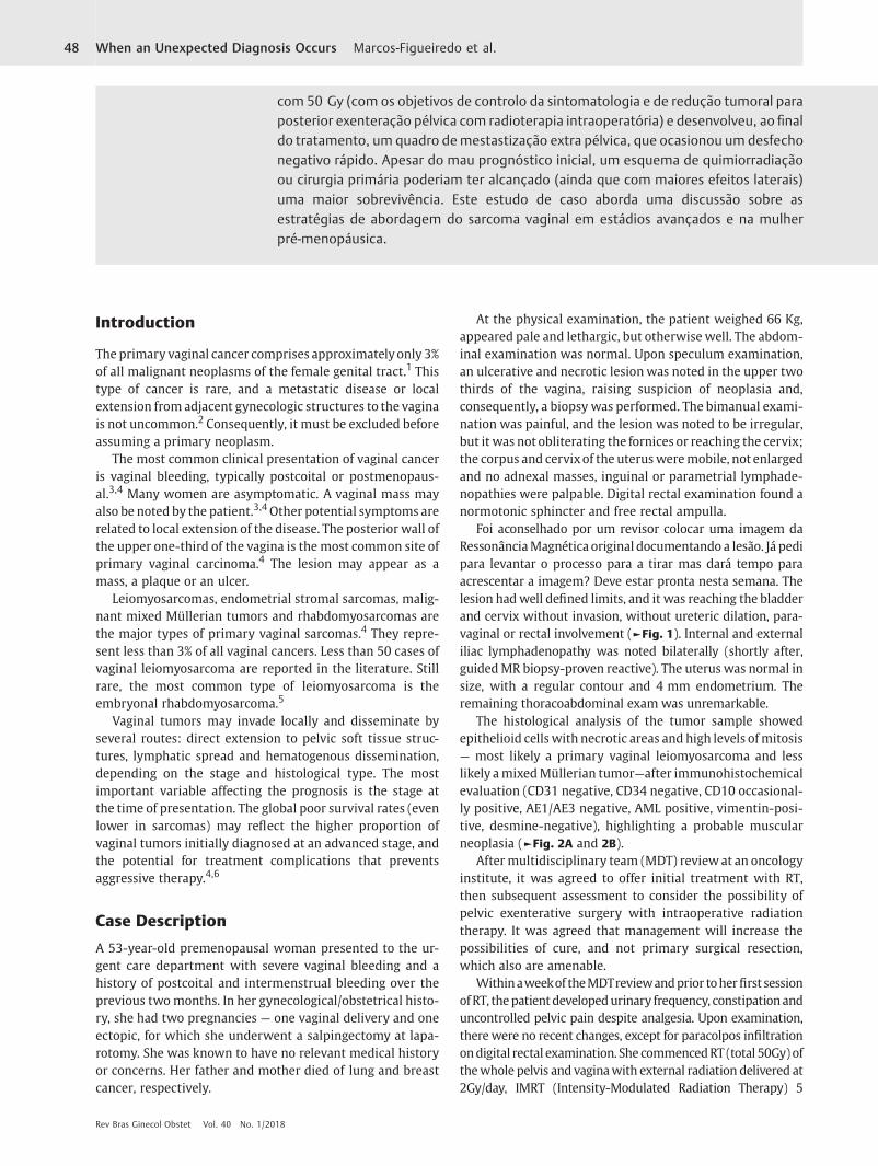
Foi aconselhado por um revisor colocar uma imagem daRessonânciaMagnética original documentando a lesão. Já pedipara levantar o processo para a tirar mas dará tempo paraacrescentar a imagem? Deve estar pronta nesta semana. Thelesion hadwell defined limits, and it was reaching the bladderand cervix without invasion, without ureteric dilation, para-vaginal or rectal involvement (►Fig. 1). Internal and externaliliac lymphadenopathy was noted bilaterally (shortly after,guidedMR biopsy-proven reactive). The uterus was normal insize, with a regular contour and 4 mm endometrium. Theremaining thoracoabdominal exam was unremarkable.
The histological analysis of the tumor sample showedepithelioid cells with necrotic areas and high levels ofmitosis— most likely a primary vaginal leiomyosarcoma and lesslikely amixedMüllerian tumor—after immunohistochemicalevaluation (CD31 negative, CD34 negative, CD10 occasional-ly positive, AE1/AE3 negative, AML positive, vimentin-posi-tive, desmine-negative), highlighting a probable muscularneoplasia (►Fig. 2A and 2B).
After multidisciplinary team (MDT) reviewat an oncologyinstitute, it was agreed to offer initial treatment with RT,then subsequent assessment to consider the possibility ofpelvic exenterative surgery with intraoperative radiationtherapy. It was agreed that management will increase thepossibilities of cure, and not primary surgical resection,which also are amenable.
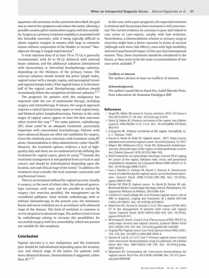
Withinaweekof theMDTreviewandprior toherfirst sessionofRT, thepatientdevelopedurinary frequency, constipationanduncontrolled pelvic pain despite analgesia. Upon examination,therewere no recent changes, except for paracolpos infiltrationondigital rectal examination. She commencedRT (total 50Gy)ofthewhole pelvis and vaginawith external radiation delivered at2Gy/day, IMRT (Intensity-Modulated Radiation Therapy) 5
com 50 Gy (com os objetivos de controlo da sintomatologia e de redução tumoral paraposterior exenteração pélvica com radioterapia intraoperatória) e desenvolveu, ao finaldo tratamento, um quadro demestastização extra pélvica, que ocasionou um desfechonegativo rápido. Apesar do mau prognóstico inicial, um esquema de quimiorradiaçãoou cirurgia primária poderiam ter alcançado (ainda que com maiores efeitos laterais)uma maior sobrevivência. Este estudo de caso aborda uma discussão sobre asestratégias de abordagem do sarcoma vaginal em estádios avançados e na mulherpré-menopáusica.
Rev Bras Ginecol Obstet Vol. 40 No. 1/2018
When an Unexpected Diagnosis Occurs Marcos-Figueiredo et al.48

Fig. 2 (A and B) Histology - Epithelioid cells with necrotic areas and high mitotic rate, most likely a leiomyosarcoma.
Fig. 1 Lesion limits, reaching the bladder and cervix.
Rev Bras Ginecol Obstet Vol. 40 No. 1/2018
When an Unexpected Diagnosis Occurs Marcos-Figueiredo et al. 49
times/week with diary portal vision during the first week, andafter then, once per week (►Fig. 3). Seven days later, the pelvicpain and constipation were controlled.
After two weeks of RT, 20 Gy had been administered andthe patient reported an improvement in her appetite, leth-argy and intermenstrual bleeding, despite the diagnosis ofmicrocytic anemia of 8,1 g/dL, then managed with transfu-sion. The gynecological examination revealed large tumoralnecrosis, and the patient felt able to manage on less pain-killers and was commenced on oral doxycycline.
During the last week of RT, the patient developed lethargyand anorexia. No changes were found during the examina-tion at this time, other than her weight had dropped by 5 kgto 61 Kg. The serumbiochemistry revealed hypomagnesemiaand a borderline potassium deficiency, which was treatedwith oral supplements. Finally, she completed 50 Gy of RTwith average global state; maintaining weight (61 Kg) andexamination findings.
Two days later, she complained of epigastric/right hypo-chondrium pain and bowel subocclusion semiology, withworsening anorexia. She had slightly painful deep palpationof the right hypochondrium and epigastrium (suspectedhepatomegaly) without signs of peritoneal irritation. Shewas tachycardic (130 bpm), but the remaining vitals werenormal. In the thoracic X-ray, pulmonary metastasis wasrevealed (micro and macronodules of irregular limits) andan abdominal X-ray confirmed subocclusion. The progres-sion of the disease was also documented in the serumsamples, which revealed thrombocytopenia of 47,000/uL,hyponatremia, hypomagnesemia and elevated liver param-eters (aspartate transaminase [AST]/alanine transaminase[ALT] double the upper normal limit (UNL), total bilirubin1.5 x UNL, alkaline phosphatase 10 x UNL, lactate dehydro-genase 6.8 x UNL), raising concern of probable hepatic/peritoneal metastasis.
The patient was admitted for supportive care, under anil-by-mouth regimen, and with fluid resuscitation. Duringthe subsequent MDT review, a decision was made to referher care to palliative care. The patient passed away on thefollowing day.
Discussion
Vaginal sarcoma is a very rare oncology malignancy, withinthe already infrequent vaginal cancer category.
The staging is clinical, based upon findings from physicalexamination cystoscopy, proctoscopy, and chest and skeletalradiography.6,8 An MRI can assist in determining the primaryvaginal tumor size and local extent. Vaginal tumors aregenerally best seen on T2 imaging.9 Despite the fact that themajority of available evidence is for squamous cell carcinomaand adenocarcinoma, the positron emission tomography–computed tomography (PET-CT) value appears to be usefulto address metastization, mainly in advanced stages.10,11 Theguided biopsy performed may have a role in cancer dissemi-nation, but this appear to bevery unlikely. In thefirst place, thelymphadenopathies were reactive (even in pathologic revi-sion). Furthermore, we only found studies justifying thisconcern in uterine cancers, when morcelation is used. TheInternational Federation of Gynecology and Obstetrics (FIGO)and the Tumor, Nodes, Metastasis (TNM) classification recom-mend a clinical staging system for vaginal cancer.6
With regard to global vaginal cancer, 37% of the patients arediagnosed at stage II, and 37% at stages III or IV.8 In the case ofsarcomas, theoutcome isevenworse.4,6,8 In thiscase,dueto thealreadyadvancedstageof thedisease, thepatients areoftennotcandidates for surgery.Given the relativelypooroutcomeswithtreatment using RT alone, chemoradiation is often adminis-tered, rather than RT. This is largely based on an extrapolationof the improved outcomes with chemoradiation for the treat-ment of locally advanced cervical cancer, but studies in vaginalcancer show that locoregional control rates are high afterchemoradiotherapy, and radiation-related long-term sideeffects do not seem worse compared with RT alone.12–14
However, given the lack of high-quality data to inform thebenefits of chemoradiation, RT is a reasonable alternative,particularly for patients who are not candidates for cisplatin-basedchemotherapy, forwhatever reason.4Thedatato supportthis approach specifically for vaginal cancer is low-quality andlargely limited to small retrospective series.12–16 The leiomyo-sarcoma treatment (more rare) is often extrapolated for
Fig. 3 (A and B) Dosimetric plan and dose-volume histogram.
Rev Bras Ginecol Obstet Vol. 40 No. 1/2018
When an Unexpected Diagnosis Occurs Marcos-Figueiredo et al.50
squamous cell carcinoma. In the caseherein described, the goalwas to control the symptoms and reduce the tumor, allowing apossible curative pelvic exenterative surgerywith lessmorbid-ity. Surgery as a primary treatmentmodality is associatedwithless favorable outcomes, with it being typically difficult toobtain negative margins in women with large or extensivelesions without compromise of the bladder or rectum.4 Neo-adjuvant therapy is largely experimental.4
A total radiation dose of at least 70 to 75 Gy is generallyrecommended, with 45 to 50 Gy delivered with externalbeam radiation, and the additional radiation administeredwith intracavitary or interstitial brachytherapy radiation,depending on the thickness of the primary tumor. Theexternal radiation should include the pelvic lymph nodes,vaginal tumor with amargin, vagina, and paravaginal tissuesand inguinal lymph nodes, if the vaginal tumor is in the lowerhalf of the vaginal canal. Brachytherapy radiation shouldimmediately follow the completion of external radiation.4,17
The prognosis for patients with this malignancy hasimproved with the use of multimodal therapy, includingsurgery and chemotherapy. If chosen, the surgical approachrequires a radical hysterectomy, upper or total vaginectomy,and bilateral pelvic lymphadenectomy. Patients in the earlystages of vaginal cancer appear to have the best outcomeswhen treated this way.2–4 For some patients, radiotherapy(RT) alone could be an adequate treatment, particularlyimportant with concomitant brachytherapy. Patients withmore advanced disease are often not candidates for surgery.Given the relatively poor outcomes with treatment using RTalone, chemoradiation is often administered, rather than RT.However, the treatment options evidence a lack of high-quality data and there are no randomized trials defining thetreatment for vaginal cancer, given its rarity.4,6 Instead, thetreatment management is extrapolated from cervical or analcancers and should be individualized depending upon thelocation, size and clinical stage of the tumor.7 In addition, thetreatment must consider the local anatomic constraints andpsychosexual issues.
There is no treatment defined for vaginal sarcoma. Usuallyis surgery, as the most of others sites. For advanced gyneco-logic sarcomas, until now, and not possible to control bysurgery (not resection possible), the management can beconsidered palliative, what include radiotherapy with orwithout chemotherapy. In the present case, the metastasisfound and worse evolution are in accordance with advancedstage of the disease. This kind of evolution is common incervix neoplasia in advanced stage. The authors tried to treatby radiotherapy aiming to increase the possibilities forsuccessfull surgery with less comorbidity, which was provennot suitable for this neoplasia.
Conclusion
Vaginal sarcoma is a rare malignancy and the treatmentplan should be individualized depending upon the location,size, and clinical stage of the tumor. For patients withmore advanced disease, chemoradiation is suggested, ratherthan RT.4,12
In this case, with a poor prognosis, the expected evolutionis minimal and the primary best treatment is still controver-sial. The current evidence on sarcomas is poor and related tocase series or case-reports, usually with bad evolution.Nevertheless, a chemoradiation scheme or primary surgicalresection might have a better outcome in terms of survival(although with more side effects), even with high morbidityandworst psychosocial impact, in the case of premenopausalwomen. Thus, these treatments should be considered in thefuture, as they seem to be the main recommendation of lastcase series available.18
Conflicts to InterestThe authors declare to have no conflicts of interest.
AcknowledgmentsThe authors would like to thank Dra. Isabel Macedo Pinto,from Laboratório de Anatomia Patológica IMP.
References1 Siegel RL, Miller KD, Jemal A. Cancer statistics, 2015. CA Cancer J
Clin 2015;65(01):5–29. Doi: 10.3322/caac.212542 Dunn LJ, Napier JG. Primary carcinoma of the vagina. Am J Obstet
Gynecol 1966;96(08):1112–1116. Doi: 10.1016/0002-9378(66)90519-9
3 Livingston RC. Primary carcinoma of the vagina. Springfield, IL:C. C. Thomas; 1950
4 Karam A, Berek JS, Kidd EA. Vaginal cancer. 2017. https://www.uptodate.com/contents/vaginal-cancer. Accessed January 12, 2017
5 Hilgers RD, Malkasian GD Jr, Soule EH. Embryonal rhabdomyo-sarcoma (botryoid type) of the vagina. A clinicopathologic review.Am J Obstet Gynecol 1970;107(03):484–502
6 FIGO Committee on Gynecologic Oncology. Current FIGO stagingfor cancer of the vagina, fallopian tube, ovary, and gestationaltrophoblastic neoplasia. Int J Gynaecol Obstet 2009;105(01):3–4.Doi: 10.1016/j.ijgo.2008.12.015
7 Lian J, Dundas G, Carlone M, Ghosh S, Pearcey R. Twenty-yearreviewof radiotherapy for vaginal cancer: an institutional experi-ence. Gynecol Oncol 2008;111(02):298–306. Doi: 10.1016/j.ygyno.2008.07.007
8 Hacker NF, Eifel PJ. Vaginal cancer. In: Berek JS, Hacker NF, eds.Berek and Hacker’s Gynecologic Oncology. 6th ed. Philadelphia, PA:Lippincott Williams & Wilkins; 2015:608–624
9 Gardner CS, Sunil J, Klopp AH, et al. Primary vaginal cancer: role ofMRI in diagnosis, staging and treatment. Br J Radiol 2015;88(1052):20150033. Doi: 10.1259/bjr.20150033
10 Robertson NL, Hricak H, Sonoda Y, et al. The impact of FDG-PET/CT in the management of patients with vulvar and vaginalcancer. Gynecol Oncol 2016;140(03):420–424. Doi: 10.1016/j.ygyno.2016.01.011
11 Bentivegna E, UzanC, Gouy S, et al. [The accuracyof FDG-PET/CT inearly-stage cervical and vaginal cancers]. Gynecol Obstet Fertil2011;39(04):193–197. Doi: 10.1016/j.gyobfe.2011.02.008
12 Grigsby PW. Vaginal cancer. Curr Treat Options Oncol 2002;3(02):125–130. Doi: 10.1007/s11864-002-0058-4
13 Samant R, Lau B, e C, Le T, Tam T. Primary vaginal cancer treatedwith concurrent chemoradiation using Cis-platinum. Int J RadiatOncol Biol Phys 2007;69(03):746–750. Doi: 10.1016/j.ijrobp.2007.04.015
14 Miyamoto DT, Viswanathan AN. Concurrent chemoradiation forvaginal cancer. PLoS One 2013;8(06):e65048. Doi: 10.1371/jour-nal.pone.0065048
Rev Bras Ginecol Obstet Vol. 40 No. 1/2018
When an Unexpected Diagnosis Occurs Marcos-Figueiredo et al. 51

15 Frank SJ, Jhingran A, Levenback C, Eifel PJ. Definitive radiationtherapy for squamous cell carcinomaof the vagina. Int J Radiat OncolBiol Phys 2005;62(01):138–147. Doi: 10.1016/j.ijrobp.2004.09.032
16 Dalrymple JL, Russell AH, Lee SW, et al. Chemoradiation forprimary invasive squamous carcinoma of the vagina. Int J GynecolCancer 2004;14(01):110–117
17 Orton A, Boothe D, Williams N, et al. Brachytherapy improvessurvival in primary vaginal cancer. Gynecol Oncol 2016;141(03):501–506. Doi: 10.1016/j.ygyno.2016.03.011
18 Li L, Zhang R,Wu LY, et al. [Primary leiomyosarcoma of the vagina:a clinical analysis of 9 cases]. Zhonghua Fu ChanKeZa Zhi 2012;47(10):747–750
Rev Bras Ginecol Obstet Vol. 40 No. 1/2018
When an Unexpected Diagnosis Occurs Marcos-Figueiredo et al.52