what you need to know about osteoporosis · what you need to know about osteoporosis. ... risk...
TRANSCRIPT

Medical guide
2nd Edition 05/14
What you need to know about Osteoporosis

Copyright © Osteoporosis Australia 2014. Except as provided by the Copyright Act 1968, no part of this publication may be reproduced, stored in a retrieval system or transmitted in any form or by any means without the prior written permission of the publisher.
IntroductionHealth professionals play a central role in identifying people at high risk of osteoporosis and osteoporotic fractures.
Many patients may already be suffering from the ‘silent disease’ without knowing, as osteoporosis can progress without any signs or symptoms. In Australia, there is a real opportunity to improve the investigation of patients and combat the progression of the disease. Recognising risk factors and discussing bone health with patients is vital.
An estimated 1.2 million Australians have osteoporosis and another 6.3 million are affected by osteopenia (low bone density), a potential precursor to osteoporosis.1
1
Contents
Bone loss and fragility 2
Fracture incidence 3
The fracture cascade 3
Risk factors for osteoporosis and fractures 4
Assessment of patient bone health 4
Diagnosis of osteoporosis 5
Bone densitometry 5
Other forms of testing 5
Management of bone health and treatment of osteoporosis 6
Calcium 6
Vitamin D 6
Exercise 6
Medicines 7
Re-fracture prevention 8
Patient care in general practice post fracture 8
Patient care post hip fracture 9
Falls prevention 10
Falls risk screening tools 10
Assessment tools and interventions 10
Bone loss and fragilityTwo types of tissue form bones –
cancellous (or trabecular) bone and
cortical bone. Trabecular bone, which
forms an interconnecting latticework, is
more metabolically active and turns over
at a rate of approximately 25% per year.2
Less active and denser cortical bone
surrounds the trabecular bone, forming
the bone’s outer surface. The proportion
of trabecular to cortical bone differs
depending on the site. For example, spinal
vertebrae are largely trabecular while the
femur is predominately cortical.
It is estimated that the entire skeleton
replaces itself over a period of 10 years.
The destruction of old bone and its
replacement is largely regulated by the
activity of osteoblasts (cells that lay down
new bone matrix) and osteoclasts (cells
that resorb bone). Strong contributors to
the activity of these cells are the hormones
testosterone and oestrogen, which have
osteogenic qualities, and parathyroid
hormone, which is osteolytic. Stresses and
strains placed on the bone also have an
osteogenic effect, hence the importance
of exercise in building and maintaining
bone strength and quality. Bone quality is
dependent on both its bone mineral density
(BMD) and its structural properties, such as
architecture and geometry. As such, small
bones are not necessarily poor quality and
large bones are not invariably strong.
The bones are the major storage organ
for calcium. 99% of the body’s calcium
is stored in the bones as calcium
hydroxyapatite. The bones are responsible
for the homeostasis of calcium and release
it into the bloodstream as required. The
presence of vitamin D has a significant
role in the mineralisation of the bone.
The relationship between osteoblasts and
osteoclasts plays a role in determining the
speed of bone turnover and whether there
is a net loss or increase in bone volume.
Menopause, with its associated decline
in oestrogen production, results in an
increase in bone turnover. This is exhibited
by an accelerated bone loss, averaging
2% per year.3 Both cortical thinning and the
thinning and breakage of the latticework
forming the trabecular bone can occur.
Accelerated bone loss is greatest in the
first three to six years after menopause
and then gradually resumes the rate of
premenopausal bone loss. After the age
of 70, bone loss begins to accelerate again,
reaching 1-2% per year in women older
than 80 years.4
A 10% loss in bone mass in the vertebrae
can double the risk of vertebral fractures
and a 10% loss of bone mass in the hip
can result in a 2.5 times greater risk of
hip fracture.5
Men experience only a gradual decline in
testosterone, so that the structural integrity
of trabecular bone is maintained for longer.
In addition, men have a greater bone volume
with a faster cortical bone deposition than
women, hence are able to better offset
any inner bone loss. However, by the age
of 70 years, sufficient bone has usually
been resorbed to warrant investigation,
particularly where other osteoporotic risk
factors are present.
Apart from hormone levels there are a range
of other factors that have an impact on bone
health, including genetics, diet (particularly
calcium intake), vitamin D levels, exercise
and certain diseases and medicines.
When less than optimal, these factors may
compromise bone strength and increase the
risk of fracture.
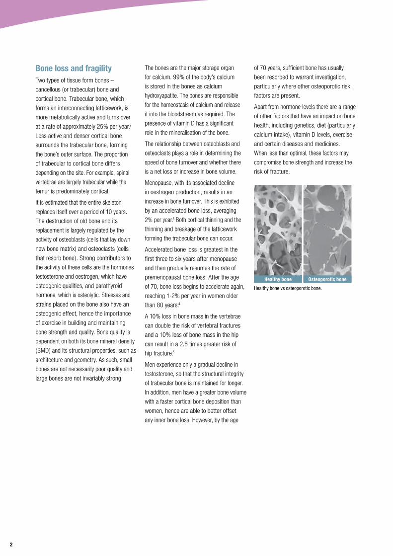
Healthy bone vs osteoporotic bone.
Healthy bone Osteoporotic bone
2

Fracture incidenceOsteoporosis remains largely undiagnosed and untreated. Only around 20% of people with osteoporotic fractures who come to medical attention are treated to prevent further bone loss and fractures.6 7 The healthcare system works to repair the fracture but does not investigate the underlying cause.
Any bone can be affected by osteoporosis. The most common fracture sites are the spine, hip and wrist. Other sites affected include the humerus, ribs, forearm and pelvis. Minimal trauma fractures (also known as fragility fractures) are defined as fractures resulting from trauma equal to (or less than) a fall from standing height.
Osteoporotic fractures in Australia
In 2012, 140,822 fractures occurred in Australia as a result of osteoporosis and osteopenia. By 2022, it is expected that there will be a 30% increase in the annual number of fractures, if action is not taken now to improve diagnosis and management of osteoporosis. The total costs of poor bone health and associated fractures is estimated to reach $33.6 billion over the 10 years from 2013-2022. Hospital costs currently account for 73%
of direct fracture costs.8
The fracture cascade
It has been well documented that following
a low trauma fracture at any site, there is a
2-4 fold increase in the risk of a subsequent
fracture.9 10 Patients with a history of prior
fracture at any site should be evaluated for
osteoporosis and fracture risk.5
Subsequent fracture is a particular problem
for the 50-75% of vertebral fractures that
do not come to medical attention. For
example, women who sustain a vertebral
fracture are 4 times more likely to sustain
another vertebral fracture. Vertebral
fractures may be asymptomatic, or signs
and symptoms such as back pain, height
loss or kyphosis can be misinterpreted
and not properly investigated.
Undiagnosed and untreated fractures
increase the chance of more severe
fractures, resulting in pain, disability
and even premature mortality.10 11
Osteoporosis care of fracture patients has been characterised as a Bermuda Triangle, comprised of orthopaedic surgeons, primary care physicians and osteoporosis experts, into which the fracture patient disappears.12 Everyone assumes that someone else is managing the patient’s fractures when in reality, often no one does.
Only 20% of people who come to medical attention with osteporotic fractures are treated to prevent further bone loss and fractures.6 7 Minimal trauma fractures are a clear signal that a patient’s bone health should be investigated.
Hip fractures
Most hip fractures occur following a fall and 90% occur in people over 50.9
Half of all patients who have had a hip fracture will be unable to regain their previous independence and will require some form of additional walking aid or assistance with mobility.13
Source: Osteoporosis costing all Australians. A new burden of disease analysis – 2012 to 2022. Osteoporosis Australia.
Osteoporotic fractures
Source: AIHW National Hospital Morbidity Database.**
Discharge to residential care: 11%
Discharge to usual residence: 30%
Transfer to other health service: 53%
Death: 6%
Results after a hip fracture
3
0 25,000 50,000 75,000 100,000 125,000 150,000 175,000 200,000
2012 140,8822022 183,105
1.6 million estimated fractures over 10 years
Assessment of patient bone health
● Review personal fracture history and any family fracture history.
Note: Minimal trauma fractures should be investigated.
● Assess calcium intake and vitamin D status.
● Review conditions or medications that can impact bone health (see risk factors).
● Check menstrual history in women, testosterone history in men.
● Determine smoking, alcohol and exercise status.
● Measure height and ask about back pain (any height loss 3 cm or more should be investigated).
Risk factors for osteoporosis and fractures
● Genetics.* ● Family history of osteoporosis
and minimal trauma fractures.
● Calcium and vitamin D status.** ● Inadequate calcium intake. ● Low/deficient vitamin D levels.
● Fracture history and age.* ● Previous spinal or minimal
trauma fracture. ● Increasing age (over 70).
● Body weight. ● Low body weight.* ● Obesity (new research suggests
impact on bone).
● Height loss.* ● Height loss (3cm or more). ● Kyphosis.
● Negative lifestyle factors.** ● Lack of physical activity. ● Excessive alcohol. ● Smoking.
● Recurrent falls.*
● Medical conditions (secondary
osteoporosis).* ● Inflammatory arthritis. ● Malabsorption (eg: coeliac disease). ● Premature menopause in women. ● Hypogonadism in men. ● Hyperparathyroidism/Hyperthyroidism
or thyroxine excess. ● Chronic kidney or liver disease. ● Multiple myeloma/monoclonal
gammopathy. ● Organ or bone marrow transplant. ● Inflammatory bowel disease. ● HIV and its treatment. ● Type 1 and type 2 diabetes mellitis.
● Medicines.* ● Glucocorticoids (longer than
3 months). ● Aromatase inhibitors. ● Gonadotropin-releasing agonists (commonly used to treat prostate and breast cancer).
● Antiepileptic drugs. ● Antidepressants (SSRI’s).
4
*Patients over 50 with these risk factors should be investigated with a bone mineral density scan (DXA).
**These factors may contribute to a patient’s overall risk of osteoporosis and fracture.
Diagnosis of osteoporosisThe diagnosis of osteoporosis is based on the measurement of bone mineral density (BMD). A presumptive clinical diagnosis can also be made following a minimal trauma fracture in middle aged or elderly patients. Checking BMD in patients over 50 with risk factors is recommended. BMD is measured in g/cm2 and the results expressed as T-Score [the number of standard deviations (SD) above or below the young normal mean] and Z-Score [the number of standard deviations (SD) above or below age matched controls].
Bone densitometry
Dual-energy X-ray Absorptiometry (DXA) to measure bone density of the hip and spine are the standard tests for diagnosing osteoporosis and monitoring response to treatment. The hip (femoral neck or total proximal femur sites) and lumbar spine (usually L1-L4 or L2-L4) are used unless there are abnormalities in these regions which may affect bone density. Proximal femur BMD appears to be the best overall predictor of fracture risk, particularly as it is less affected by osteoarthritis. Osteoarthritic changes may elevate spine BMD.
Note: The various DXA machine brands will report different absolute units (g/cm2) for BMD, and the results are not directly comparable. However, the widespread use of standardised reference ranges largely overcomes this problem.
The results of a BMD scan can show bone density in the range of normal, osteopenia or osteoporosis.
Suspected osteoporotic fracture in the spine should be confirmed by plain radiograph. Peripheral DXA and spinal quantitative computed tomography are sometimes used as alternatives for osteoporosis diagnosis but have not been evaluated for guiding therapeutic interventions.
Note: Quantitative ultrasound of the heel (often available in pharmacy) is not recommended as an appropriate standard test for BMD.
Investigate patients
Any patient over 50 with risk factors for
osteoporosis warrants investigation with
a DXA scan.
Medicare rebates are available for BMD
testing in high risk categories, including
all patients over 70 and for monitoring
treatment and low bone density. A rebate
is also available to confirm low bone density
in a patient with presumed osteoporosis
presenting with one (or more) minimal
trauma fractures.
Note: A BMD in the osteopenic range
does not invalidate a clinical diagnosis
of osteoporosis when a minimal trauma
fracture has occurred.
Other forms of testing ● Plain x-rays should be ordered if height
loss (3 cm or more) or kyphosis is
documented, to confirm the presence
of spinal fractures.
● Blood tests may be indicated to exclude
other causes of bone mineral loss
such as primary hyperparathyroidism,
malabsorption, thyroid disease or
vitamin D deficiency.
● Bone markers are biochemical markers
of bone turnover which can be measured
in serum and urine. They are not routinely
indicated for osteoporosis assessment.
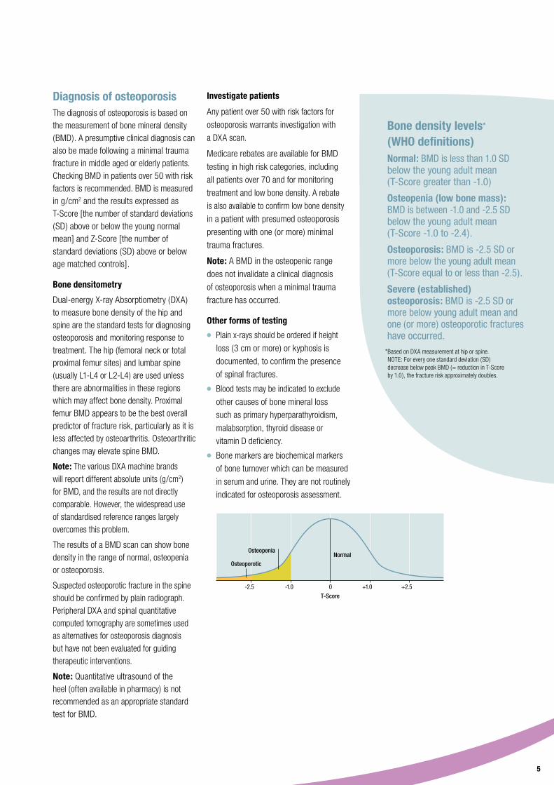
*Based on DXA measurement at hip or spine. NOTE: For every one standard deviation (SD) decrease below peak BMD (= reduction in T-Score by 1.0), the fracture risk approximately doubles.
Bone density levels* (WHO definitions)Normal: BMD is less than 1.0 SD below the young adult mean (T-Score greater than -1.0)
Osteopenia (low bone mass): BMD is between -1.0 and -2.5 SD below the young adult mean (T-Score -1.0 to -2.4).
Osteoporosis: BMD is -2.5 SD or more below the young adult mean (T-Score equal to or less than -2.5).
Severe (established) osteoporosis: BMD is -2.5 SD or more below young adult mean and one (or more) osteoporotic fractures have occurred.
-2.5 +2.5-1.0 +1.00
Osteoporotic
T-Score
OsteopeniaNormal
5

Vitamin D
Low vitamin D levels and vitamin D
deficiency are common in Australia,
but this can be easily rectified.
Adequate vitamin D levels should be
at least 50 nmol/L at the end of winter.
Levels should be 10-20 nmol/L higher
during summer to allow for seasonal
decrease in the winter months.
The main source of vitamin D is from
sunlight through the action of UVB light
on skin. Optimal exposure times will vary
based on season, skin type (people with
dark skin require more exposure than
fair skinned people) and location (lower
latitudes require longer exposure times).
For people who consistently do not achieve
the recommended levels of sun exposure
or have risk factors for vitamin D deficiency,
testing is recommended. Supplementation
is advised for people with mild, moderate
or severe deficiency.
Refer to the Vitamin D fact sheet provided with this booklet or on the Osteoporosis Australia website www.osteoporosis.org.au for background information and recommendations.
Exercise
Exercise has a range of benefits for bone
health, including increased bone density,
improved co-ordination, balance and
strength (to help prevent falls) and assisting
rehabilitation after a fracture.
For healthy adults, a combination of weight
bearing and progressive resistance training
is recommended to improve or maintain
bone density, muscle mass, strength and
functional capacity (balance and gait).
For older adults and people with osteopenia
or osteoporosis, exercise should be
multi-modal and supervised and include
weight bearing activities, progressive
resistance training and high challenge
balance activities each week. The aim of
exercise is to slow (or reverse) bone loss,
maintain bone strength, increase muscle
mass, strength and function and to improve
gait and mobility to help prevent falls.
Refer to the Exercise fact sheet provided with this booklet or on the Osteoporosis Australia website www.osteoporosis.org.au for background information and recommendations.
Management of bone health and treatment of osteoporosisCalcium, vitamin D and exercise all play an
important role in optimising bone mass and
preserving bone density in healthy adults
as well as slowing bone loss in patients
diagnosed with ostepenia and osteoporosis.
Calcium
More than half of Australian adults do not
get their recommended intake of calcium.
Daily calcium recommendations are
1,000 mg per day for adults, increasing
to 1,300 mg per day for women over 50
and men over 70 years. It is recommended
that these levels are achieved through diet.
Since calcium content in food varies, this
requires regular intake of calcium rich food.
For people with inadequate dietary
calcium intake, calcium supplements of
500-600 mg per day are recommended
and are equally effective.
Refer to the Calcium fact sheet provided with this booklet or on the Osteoporosis Australia website www.osteoporosis.org.au for background information and recommendations.
6
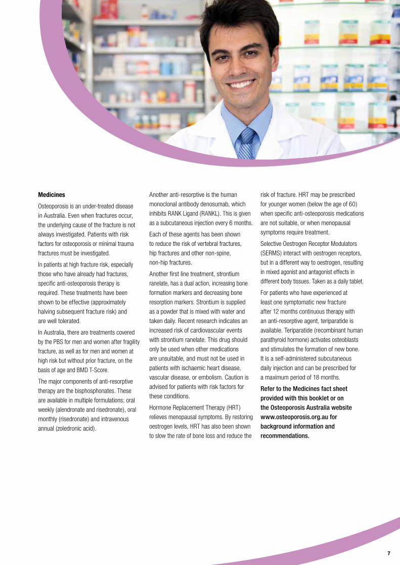
Medicines
Osteoporosis is an under-treated disease
in Australia. Even when fractures occur,
the underlying cause of the fracture is not
always investigated. Patients with risk
factors for osteoporosis or minimal trauma
fractures must be investigated.
In patients at high fracture risk, especially
those who have already had fractures,
specific anti-osteoporosis therapy is
required. These treatments have been
shown to be effective (approximately
halving subsequent fracture risk) and
are well tolerated.
In Australia, there are treatments covered
by the PBS for men and women after fragility
fracture, as well as for men and women at
high risk but without prior fracture, on the
basis of age and BMD T-Score.
The major components of anti-resorptive
therapy are the bisphosphonates. These
are available in multiple formulations; oral
weekly (alendronate and risedronate), oral
monthly (risedronate) and intravenous
annual (zoledronic acid).
Another anti-resorptive is the human
monoclonal antibody denosumab, which
inhibits RANK Ligand (RANKL). This is given
as a subcutaneous injection every 6 months.
Each of these agents has been shown
to reduce the risk of vertebral fractures,
hip fractures and other non-spine,
non-hip fractures.
Another first line treatment, strontium
ranelate, has a dual action, increasing bone
formation markers and decreasing bone
resorption markers. Strontium is supplied
as a powder that is mixed with water and
taken daily. Recent research indicates an
increased risk of cardiovascular events
with strontium ranelate. This drug should
only be used when other medications
are unsuitable, and must not be used in
patients with ischaemic heart disease,
vascular disease, or embolism. Caution is
advised for patients with risk factors for
these conditions.
Hormone Replacement Therapy (HRT)
relieves menopausal symptoms. By restoring
oestrogen levels, HRT has also been shown
to slow the rate of bone loss and reduce the
risk of fracture. HRT may be prescribed
for younger women (below the age of 60)
when specific anti-osteoporosis medications
are not suitable, or when menopausal
symptoms require treatment.
Selective Oestrogen Receptor Modulators
(SERMS) interact with oestrogen receptors,
but in a different way to oestrogen, resulting
in mixed agonist and antagonist effects in
different body tissues. Taken as a daily tablet.
For patients who have experienced at
least one symptomatic new fracture
after 12 months continuous therapy with
an anti-resorptive agent, teriparatide is
available. Teriparatide (recombinant human
parathyroid hormone) activates osteoblasts
and stimulates the formation of new bone.
It is a self-administered subcutaneous
daily injection and can be prescribed for
a maximum period of 18 months.
Refer to the Medicines fact sheet provided with this booklet or on the Osteoporosis Australia website www.osteoporosis.org.au for background information and recommendations.
7
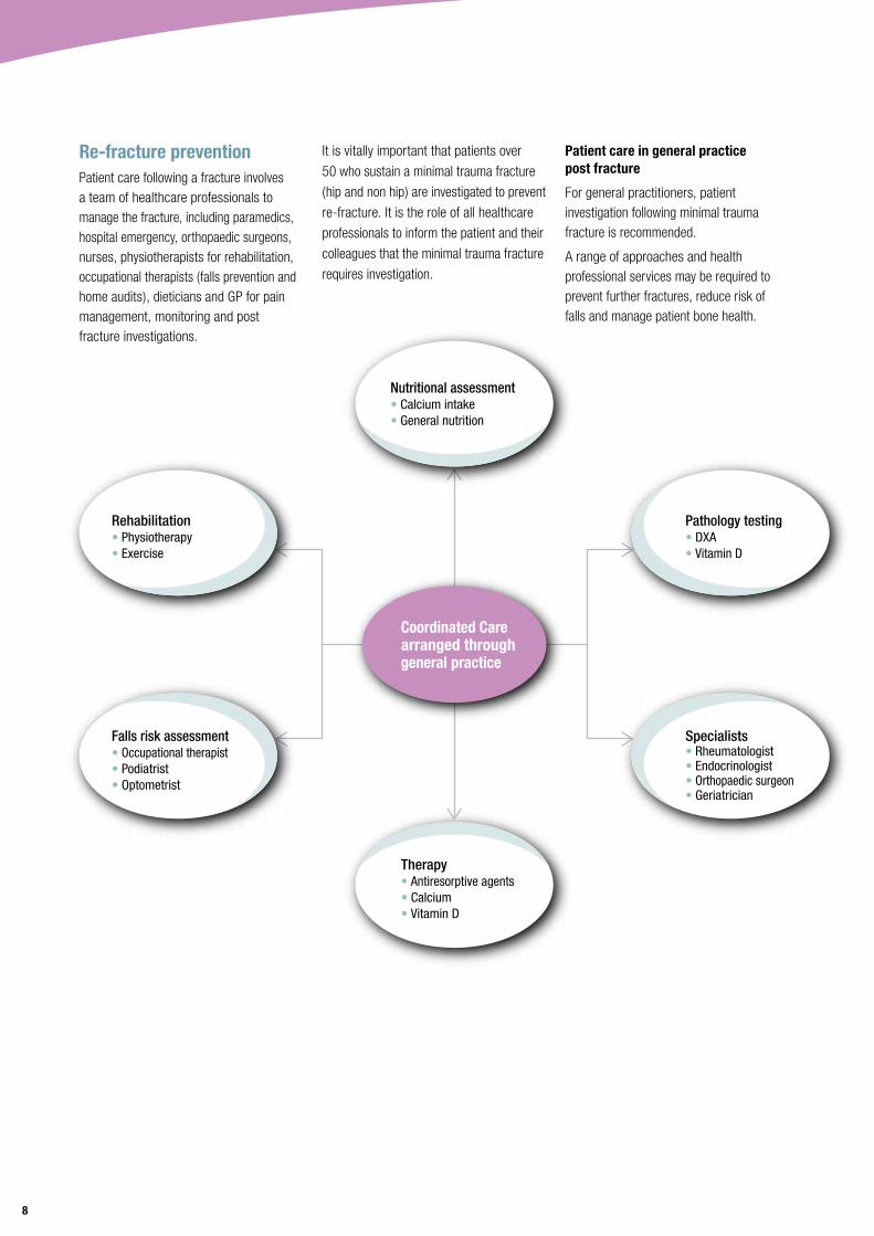
Re-fracture preventionPatient care following a fracture involves a team of healthcare professionals to manage the fracture, including paramedics, hospital emergency, orthopaedic surgeons, nurses, physiotherapists for rehabilitation, occupational therapists (falls prevention and home audits), dieticians and GP for pain management, monitoring and post fracture investigations.
Nutritional assessment• Calcium intake• General nutrition
Pathology testing• DXA• Vitamin D
Specialists• Rheumatologist• Endocrinologist• Orthopaedic surgeon• Geriatrician
Therapy• Antiresorptive agents• Calcium• Vitamin D
Falls risk assessment• Occupational therapist• Podiatrist• Optometrist
Rehabilitation• Physiotherapy• Exercise
Coordinated Carearranged throughgeneral practice
It is vitally important that patients over
50 who sustain a minimal trauma fracture
(hip and non hip) are investigated to prevent
re-fracture. It is the role of all healthcare
professionals to inform the patient and their
colleagues that the minimal trauma fracture
requires investigation.
8
Patient care in general practice post fracture
For general practitioners, patient investigation following minimal trauma fracture is recommended.
A range of approaches and health professional services may be required to prevent further fractures, reduce risk of falls and manage patient bone health.
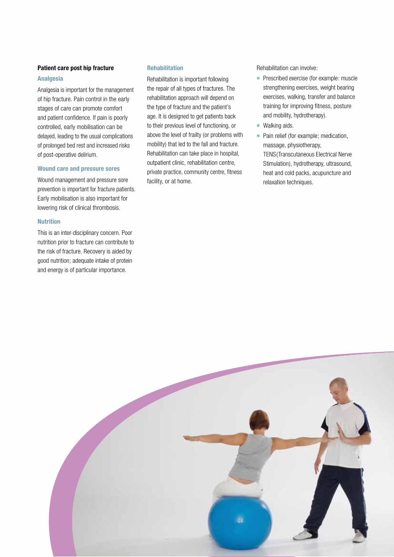
Rehabilitation
Rehabilitation is important following
the repair of all types of fractures. The
rehabilitation approach will depend on
the type of fracture and the patient’s
age. It is designed to get patients back
to their previous level of functioning, or
above the level of frailty (or problems with
mobility) that led to the fall and fracture.
Rehabilitation can take place in hospital,
outpatient clinic, rehabilitation centre,
private practice, community centre, fitness
facility, or at home.
Patient care post hip fracture
Analgesia
Analgesia is important for the management
of hip fracture. Pain control in the early
stages of care can promote comfort
and patient confidence. If pain is poorly
controlled, early mobilisation can be
delayed, leading to the usual complications
of prolonged bed rest and increased risks
of post-operative delirium.
Wound care and pressure sores
Wound management and pressure sore
prevention is important for fracture patients.
Early mobilisation is also important for
lowering risk of clinical thrombosis.
Nutrition
This is an inter-disciplinary concern. Poor
nutrition prior to fracture can contribute to
the risk of fracture. Recovery is aided by
good nutrition; adequate intake of protein
and energy is of particular importance.
Rehabilitation can involve:
● Prescribed exercise (for example: muscle
strengthening exercises, weight bearing
exercises, walking, transfer and balance
training for improving fitness, posture
and mobility, hydrotherapy).
● Walking aids.
● Pain relief (for example; medication,
massage, physiotherapy,
TENS(Transcutaneous Electrical Nerve
Stimulation), hydrotherapy, ultrasound,
heat and cold packs, acupuncture and
relaxation techniques.Nutritional assessment• Calcium intake• General nutrition
Pathology testing• DXA• Vitamin D
Specialists• Rheumatologist• Endocrinologist• Orthopaedic surgeon• Geriatrician
Therapy• Antiresorptive agents• Calcium• Vitamin D
Falls risk assessment• Occupational therapist• Podiatrist• Optometrist
Rehabilitation• Physiotherapy• Exercise
Coordinated Carearranged throughgeneral practice

Falls prevention33% of people over 65 experience a fall
each year.14 Falls are the leading cause of
injury-related hospitalisation in people over
65, accounting for 17% of emergency
admissions15 and 4% of all hospital
admissions in this age group.16
In older people, nearly half of falls
occur in the home and immediate home
surroundings.17 18 The remaining falls occur
in public places or in other people’s homes.
Falls can result in minor injuries including
abrasions, bruises and sprains, or cause
serious injury and fractures. Falls are
responsible for 90% of hip fractures.19
It is estimated that up to 6% of falls result
in a fracture.17
Falls risk screening tools
Screening patients
A simple screen: ask older patients about their history of falls in the past 12 months
and assess balance and mobility. People with a history of falls (one or more) in the last
year and who perform badly on a simple test of gait or balance should be assessed further.
Note: the following tools are for patients in a community setting. There are different screening and assessment tools for other settings (eg: Hospital sub-acute, Hospital acute, residential aged care facilities).
Falls risk screening tool
a Multifactorial fall risk assessment tools
Setting Assessment tool
Community QuickScreen© is a risk assessment tool designed specifically for general practice and assesses previous falls, medication usage, vision, peripheral sensation, lower limb strength, balance and co-ordination.21
Setting Assessment tool
Community The Timed Up and Go Test (TUGT) measures the time taken to rise from a chair, walk three meters (with usual assistive device), turn, return to the chair and sit down. A time of 12 seconds or more indicates increased risk of falls.20
b Individual functional mobility assessments
Risk factor Assessment tool
Balance and gait Tinetti performance-oriented mobility assessment tool22
Cognitive impairment Mini mental state examination23
Incontinence Urinary and fecal assessment
Feet and footwear Foot pain, safe-shoe checklist
Syncope/dizziness Tilt-table test 24
Medications Medication review
Vision Snellen eye chart
Environment Westmead home safety assessment 25
Assessment tools
Assessment tools provide detailed information about the underlying issues contributing to
overall falls risk and should be linked to tailored interventions.
Assessing falls risk usually involves either:
a Multifactorial assessment tools (that cover a wide range of falls risk factors) or;
b Individual functional mobility assessments, focused on postural stability (eg: vision,
strength, coordination, balance, gait).
10
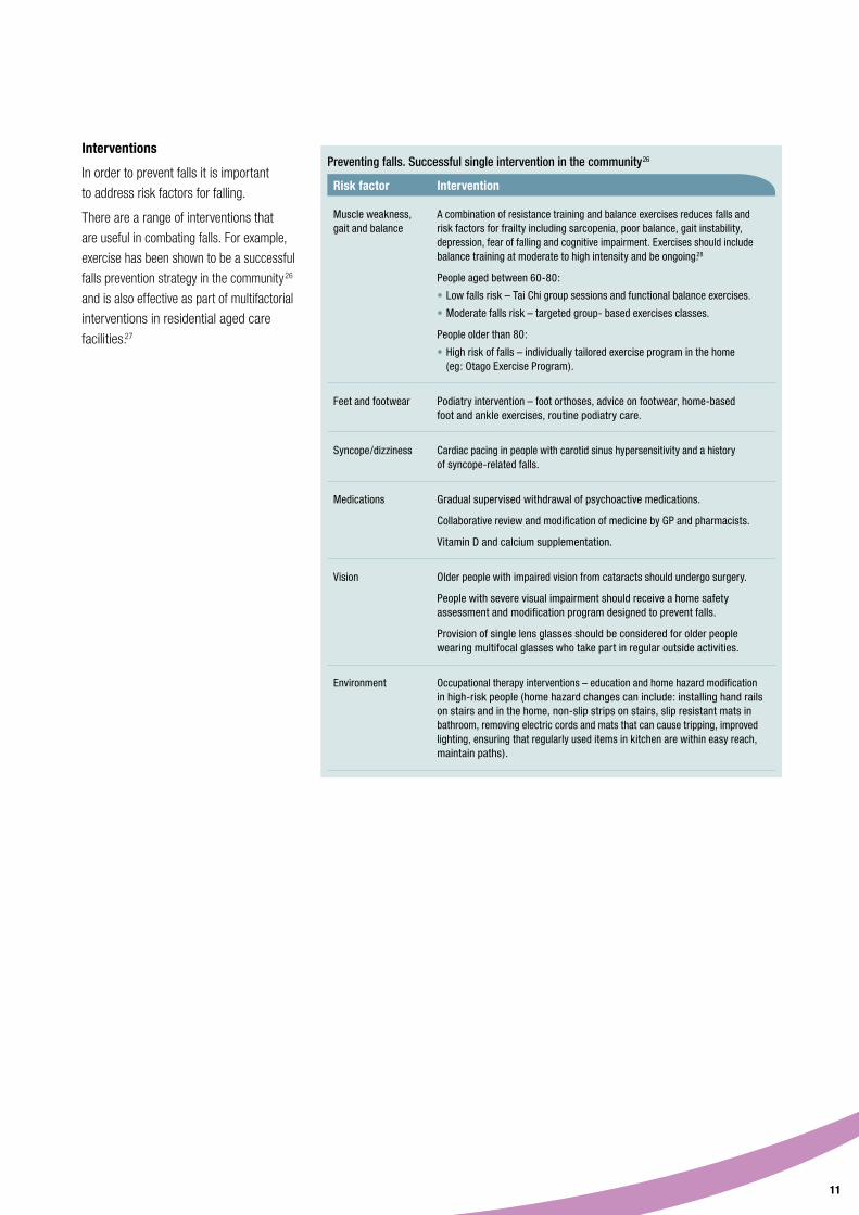
Interventions
In order to prevent falls it is important
to address risk factors for falling.
There are a range of interventions that
are useful in combating falls. For example,
exercise has been shown to be a successful
falls prevention strategy in the community26
and is also effective as part of multifactorial
interventions in residential aged care
facilities.27
Preventing falls. Successful single intervention in the community26
Risk factor Intervention
Muscle weakness, gait and balance
A combination of resistance training and balance exercises reduces falls and risk factors for frailty including sarcopenia, poor balance, gait instability, depression, fear of falling and cognitive impairment. Exercises should include balance training at moderate to high intensity and be ongoing.28
People aged between 60-80:
• Low falls risk – Tai Chi group sessions and functional balance exercises.
• Moderate falls risk – targeted group- based exercises classes.
People older than 80:
• High risk of falls – individually tailored exercise program in the home (eg: Otago Exercise Program).
Feet and footwear Podiatry intervention – foot orthoses, advice on footwear, home-based foot and ankle exercises, routine podiatry care.
Syncope/dizziness Cardiac pacing in people with carotid sinus hypersensitivity and a history of syncope-related falls.
Medications Gradual supervised withdrawal of psychoactive medications.
Collaborative review and modification of medicine by GP and pharmacists.
Vitamin D and calcium supplementation.
Vision Older people with impaired vision from cataracts should undergo surgery.
People with severe visual impairment should receive a home safety assessment and modification program designed to prevent falls.
Provision of single lens glasses should be considered for older people wearing multifocal glasses who take part in regular outside activities.
Environment Occupational therapy interventions – education and home hazard modification in high-risk people (home hazard changes can include: installing hand rails on stairs and in the home, non-slip strips on stairs, slip resistant mats in bathroom, removing electric cords and mats that can cause tripping, improved lighting, ensuring that regularly used items in kitchen are within easy reach, maintain paths).
11

Tables and graphs
* Risk Factors based on: The Royal Australian College of General Practitioners (2010) Clinical guideline for the prevention and treatment of osteoporosis in postmenopausal women and older men, viewed 28th April 2014, www.racgp.org.au/guidelines/musculoskeletaldiseases/osteoporosis, and Osteoporosis Australia. Is your patient (man or woman) over 50? Think osteoporosis. 1st Ed,4/2013, viewed 28th April 2014, www.osteoporosis.org.au/health-professionals/general-practitioners.
** Hip Fracture Graph – Outcomes of hospitalisation for osteoporotic hip fracture, 2006-07 AIHW (Soure:AIHW National Hospital Morbidity Database) The Problem of Osteoporotic Hip Fracture in Australia, AIHW, Bulletin 76, March 2010.
References
1 Henry M, Pasco JA, Nicholson GC, et al. Prevalence of osteoporosis in Australian men and women: Geelong Osteoporosis Study. Med J Australia 2011;195:321-322.
2 Manolagas SC, Jilka RL. Bone Marrow, cytokines and bone remodelling – emerging insights into the pathophysiology of osteoporosis. New Engl J Med 1995;332:305-311.
3 Ahlborg HG, Johnell O, Turner CH, et al. Bone Loss and Bone Size after Menopause. N Engl J Med 2003;349:327-334.
4 Cummings S 2002, ‘Bone biology, epidemiology and general principles’, in SR Cummings, F Cosman, SA Jamal (eds), Osteoporosis: an evidence based guide to prevention and management, American College of Physicians – American Society of Internal Medicine, Philadelphia, pp. 3-28.
5 Klotzbuecher CM, Ross PD, Landsman PB, et al. Patients with prior fractures have an increased risk of future fractures: a summary of the literature and statistical synthesis. J Bone Miner Res 2000;15:721.
6 Elliot-Gibson V, Bogoch ER, Jamal SA, et al. Practice patterns in the diagnosis and treatment of osteoporosis after a fragility fracture: a systematic review. Osteoporosis Int 2004;15:767-778.
7 Eisman J, Clapman S, Kehow L. Osteoporosis prevalence and levels of treatment in primary care: The Australian BoneCare Study. J Bone Miner Res 2004;19:1969-75.
8 Watts JJ, Abimanyi-Ochom J, Sander KM. Osteoporosis costing all Australians. A new burden of disease analysis – 2012 to 2022. 2013, Osteoporosis Australia.
9 The Royal Australian College of General Practitioners (2010) Clinical guideline for the prevention and treatment of osteoporosis in postmenopausal women and older men. Retrieved from: http://www.racgp.org.au/guidelines/musculoskeletaldiseases/osteoporosis/
10 Center JR, Bliuc D, Nguyen TV, et al. Risk of subsequent fracture after low trauma fracture in men and women. J Amer Med Assoc 2007;297:37-394.
11 Vestergaard P, Rejnmark L, Mosekilde L. Increased mortality in patients with a hip fracture-effect of pre-morbid conditions and post-fracture complications. Osteoporos Int 2007;18:1583-1593.
12 Harrington JT. Dilemmas in providing osteoporosis care for fragility fracture patients. US Musculoskeletal Review 2006;2:64-65.
13 Johansen A, Parker M 2006, ‘Hip fracture and orthogeriatrics’, in MSJ Pathy, AJ Sinclair, JE Morley (eds), Principles and practice of geriatric medicine, vol 2, John Wiley and sons, Ltd, Hoboken, pp.1329-1345.
14 Lord SR, Sherrington C, Menz H, Close JCT 2007, Falls in older people: risk factors and strategies for prevention, Cambridge University Press, Cambridge.
15 Close JC, Lord SR, Antonova EJ, et al. Older people presenting to the emergency department after a fall: a population with substantial recurrent healthcare use. Emerg Med J. 2012;29:742-747.
16 Bradley C, 2012. Hospitalisations due to falls by older people, Australia 2007-08. Injury research and statistics series: Canberra: Australian Institute of Health and Welfare (AIHW cat no. INJCAT 137).
17 Cripps R, Carman J, 2001. Falls by the elderly in Australia: Trends and data for 1998. Injury research and statistics series: Adelaide: Australian Institute of Health and Welfare (AIHW cat no. INJCAT 35).
18 Campbell AJ, Borrie MJ, Spears GF, et al. Circumstances and consequences of falls experienced by a community population 70 years and over during a prospective study. Age Ageing 1990;19:136-141.
19 Costa AG, Wyman A, Siris ES, et al. When, where and how osteoporosis-associated fractures occur: an analysis from the global longitudinal study of osteoporosis in women (GLOW). PLoS ONE 2013: 8. DOI: 10.1371/journal.pone.0083306.
20 Podsiadlo D, Richardson S. The timed “Up & Go”: a test of basic functional mobility for frail elderly persons. J Am Geriatr Soc. 1991;392:142-148.
21 Tiedemann A, Lord SR, Sherrington C. The development and validation of a brief performance-based fall risk assessment tool for use in primary care. J Gerontol A-Biol 2010;65 A:896-903.
22 Tinetti ME. Performance-oriented assessment of mobility problems in elderly patients. J Am Geriatri Soc 1986;34:119-26.
23 Folstein MF, Folstein SE, McHugh PR. Mini-mental state: A practical method for grading the cognitive state of patients for the clinician. J Psychiatr Res 1975;12:189-98.
24 Natale A, Akhtar M, Jazayeri M, et al. Provocation of hypotension during head-up tilt testing in subjects with no history of syncope or presyncope. Circulation. 1995;92:54-8.
25 Clemson L, Fitzgerald MH, Heard R. Content validity of an assessment tool to identify home fall hazards: The Westmead Home Safety Assessment.Br J Occup Ther 1999;62:171-9.
26 Gillespie LD, Robertson MC, Gillespie WJ, et al. Interventions for preventing falls in older people in the community. Cochrane DB Syst Rev 2008;Issue 2. Art No.:CD007146. http://summaries.cochrane.org/CD007146/interventions-for-preventing-falls-in-older-people-living-in-the-community/
27 Cameron ID, Murray GR, Gillespie LD, et al. Interventions for preventing falls in older people in nursing care facilities and hospitals. Cochrane DB of Syst Rev 2010; Issue 1. Art No.: CD005465. http://summaries.cochrane.org/CD005465/interventions-for-preventing-falls-in-older-people-in-nursing-care-facilities-and-hospitals/
28 Sherrington C, Whitney JC, Lord SR, et al. Effective exercise for the prevention of falls: A systematic review and meta-analysis. J Am Geriatr Soc 2008;56:2234-43.
12

Osteoporosis AustraliaOsteoporosis Australia is a national,
not-for-profit organisation committed to
improving awareness and understanding
of osteoporosis. Our goal is to reduce the
incidence of osteoporosis and osteoporotic
fractures in the Australian community.
Our services include:
● Educational materials for consumers
and health professionals.
● ‘Osteoblast’ magazine for consumers
and medical professionals.
● Osteoporosis Prevention and
Self-management Program for consumers.
● Community education seminars.
Our activities include: ● World Osteoporosis Day (October 20).
● Support for medical research.
● Advocacy to improve patient care.
Contact usOsteoporosis AustraliaFor more information call our national toll-free number: 1800 242 141 Visit our website: www.osteoporosis.org.au
Affiliated state organisations
Osteoporosis New South Walesp: (02) 9857 3300
Osteoporosis Australian Capital Territoryp: (02) 6288 4244
Osteoporosis Victoriap: (03) 8531 8000
Osteoporosis Tasmaniap: (03) 6228 4824
Osteoporosis South Australiap: (08) 8379 5711
Arthritis & Osteoporosis Western Australianp: (08) 9388 2199
Osteoporosis Northern Territoryp: (08) 8948 5232
Osteoporosis Queenslandp: (07) 3857 4200

The Australian Government has provided funding to support this publication; however views in this document are those of the authors and do not necessarily represent the views of the Australian Government.